IMAGING FINDINGS OF GAUCHER DISEASE IN ALL OVER THE BODY
|
|
|
- Ashley Manning
- 6 years ago
- Views:
Transcription
1 IMAGING FINDINGS OF GAUCHER DISEASE IN ALL OVER THE BODY V. Katsaros 1, P. Lampropoulou 2, M. Mitropoulou 1, A. Nikolaou 1, C. Drossos 2 Departments of CT and MRI 1 IKA Oncology Hospital and 2 Athens General Hospital, Greece
2 PURPOSE The review of imaging findings especially of Computerized Tomography (CT) and Magnetic Resonance Imaging (MRI) in Gaucher s disease all over the body. The presentation of volumetric imaging and evaluation of the spleen by multi slice CT, as a diagnostic tool in the follow up of Gaucher s disease after therapeutic interventions.
3 INTRODUCTION 1) Gaucher s disease is a lysosomal storage disease of glucosylceramidase (glucocerebroside), due to inherited defect of acid betaglucosidase activity, which is necessary for decomposition of this substance. 2) It emerges with three different phaenotypic types: a) Type 1 (classic Gaucher s disease) b) Type 2 (infantile form with bad prognosis) c) Type 3 (intermediate form)
4 INTRODUCTION 3) It is peculiar prevalent in the race of Ashkenazi Jews (1/2500 births) and less prevalent in the rest of population (0,005% of births). 4) The life expectancy of the disease is variable, depending on the disease s type and visceral involvement (liver spleen lungs heart). 5) Treatment is either symptomatic or etiologic (enzyme replacement therapy or bone marrow transplantation).
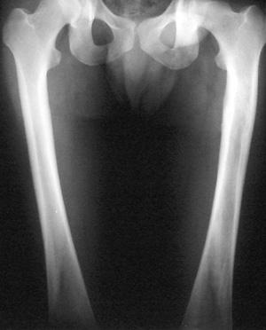
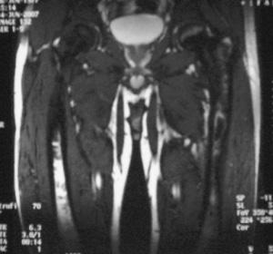
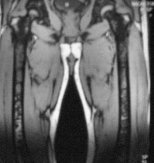
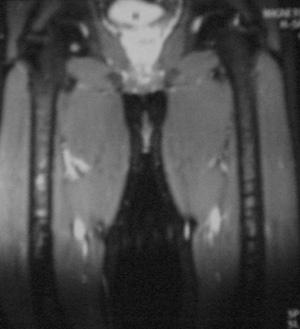
5 CLINICAL SIGNS AND SYMPTOMS Skeletal : Acute bone pain, pathological fractures, fever, regional lymphadenopathy, which are conjugated by imaging findings of osteoporosis, widening of tubular bones, regional osteolytic lesions, aseptic necrosis and osteoarthritic type epiphyseal lesions.
6
7
8 CLINICAL SIGNS AND SYMPTOMS Visceral : Hepatomegaly in the order of % of the normal liver size (it has been described in extraordinary cases proliferation to 10 times of the liver volume). Splenomegaly with subsequent anemia and thrombopenia Cardiac features as cardiomyopathy Pulmonary features as pulmonary hypertension and Pericarditis

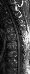
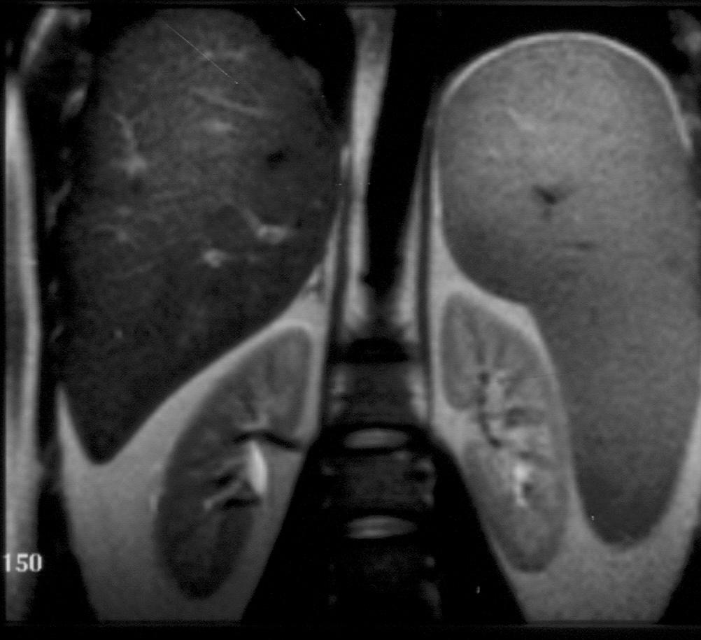
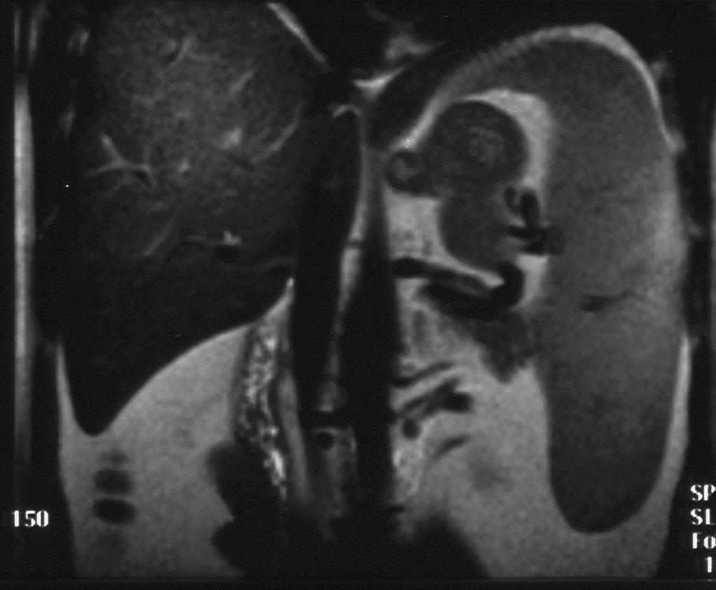
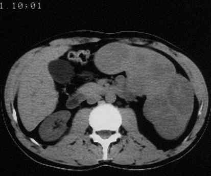
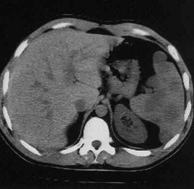
9 Hepatomegaly
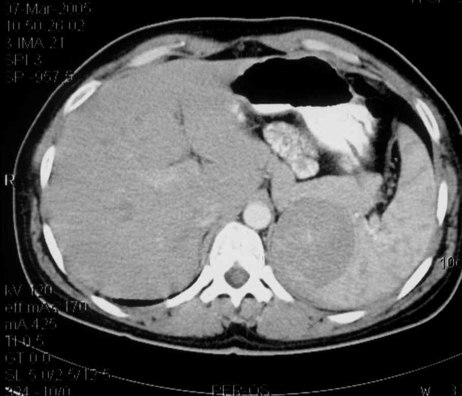
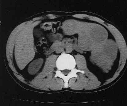
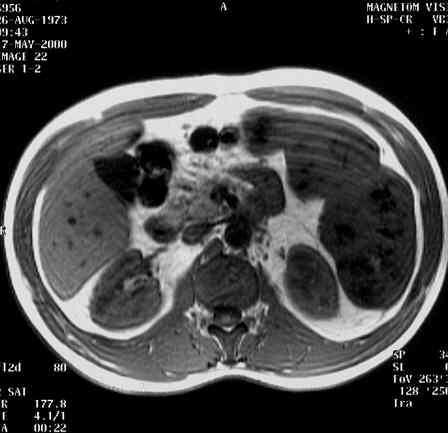
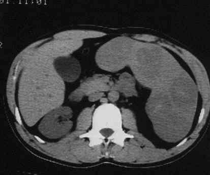
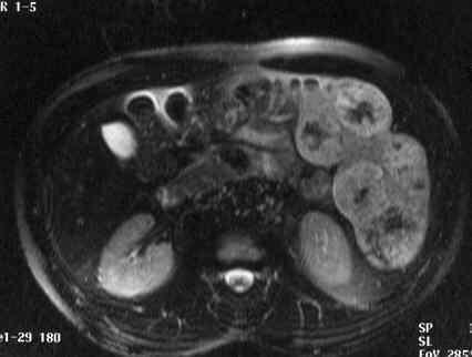
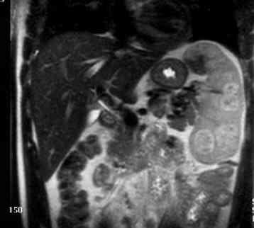
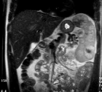
10 Splenomegaly
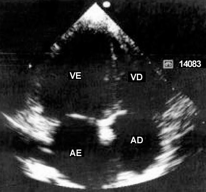
11 Cardiomyopathy

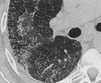
12 Reticulonodular Pattern throughout the lung fields

13 Diffuse, coarse, reticulonodular pattern throughout the lung fields
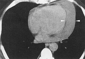
14 Pericarditis
15 CLINICAL SIGNS AND SYMPTOMS Dermatologic features as melanchrosis and ichthyosis. Increased occurrence of neoplasms as multiple myeloma, chronic lympogenic leukemia, Hodgkin and non-hodgkin lymphoma. CNS features, as thickening of the meninges The clinical picture of the disease is mainly due to accumulation of many enlarged macrophages, in which the undestructed material of sphingolipids is deposited (Gaucher s cells) in the liver, the spleen, the bone marrow and the skeleton of the patients.


16 Multiple Myeloma STIR T1-W T1-W Fs +Gd


17 Thickening of the left side of the tentorium Normal white matter myelination for 6-month old
18 SPLEEN AND GAUCHER S S DISEASE Splenomegaly is a constant finding of Gaucher s disease and the degree of it os proportional of the degree of splenic hyperfunction. The enlargement of the organ is commonly 20 times the normal splenic size (and can in extreme case reach 75 times of the normal splenic volume) Frequently splenomegaly is associated by hypocapsular infarcts, fibrotic areas in the splenic parenchyma, as well as regions of accumulated Gaucher s cells.
19 PATIENTS AND METHODS The last 10 years in our institutions were examined 35 patients with Gaucher s disease either by CT, MRI, or both methods. The routine protocol of abdomen MRI includes transverse Τ1-weighted, Τ2-weighted HASTE, T1- weighted fat-saturation, T2* weighted fat-saturation, coronal Τ2-HASTE images, as well as contrast enhanced Τ1-weighted sequences (Gd- 0.2 ml/kg body weight). The multi-slice CT protocol includes axial slices of 5 mm thickness, followed by 3-D reconstruction and volumetric evaluation of the spleen.
20 CT AND MRI FINDINGS CΤ : a) Splenomegaly of various degree b) Multiple hypoattenuating, nodular lesions in the splenic parenchyma, which do not enhance after intravenous administration of contrast agent. MRI: a) Multiple nodular lesions in the splenic parenchyma or in the periphery of the organ hypointense in Τ1 weighted sequences and of various pathologic intensity in Τ2 weighted sequences b) They do not enhance after intravenous administration of gadolinium chelates.
21 IMAGING FINDINGS OF SPLENIC LESIONS IN Τ2 WEIGHTED IMAGES Normally hypointense with small central regions (accumulation of Gaucher s cells). A small number of lesions show hyperintensity (enlarged colpoids). Frequently lesions show low signal intensity centrally surrounded by a hyperintense ring (accumulation of Gaucher s cells in combination with enlarged colpoids and fibrotic regions)
22
23

24
25
 Appearance of")


26 VOLUMETRIC EVALUATION OF THE SPLEEN a) Appearance of workstation b) The images of one patient are uploaded (constant parameters: slice thickness,gantry height, Bed height, Magnification level and Reconstruction center).

 At last we proceed to")

27 VOLUMETRIC EVALUATION OF THE SPLEEN c) The system is reconstructing in coronal, sagittal and axial plane, MPR/MIP images are also available d) At last we proceed to the Planning, either free-hand or elliptical, of the contour of the region or volume of interest


28 e) Choosing Start Evaluation button the system calculates the volume showing us the table of results.
29 The volumetric evaluation of the spleen in Gaucher s is an important diagnostic tool in the follow-up of patients treated by enzyme replacement therapy, as the splenic volume is proportional to the severity of the disease
30 CONCLUSIONS -Gaucher s disease can be imaged in all over the body. -Splenomegaly and focal abnormalities of the spleen are due to Gaucher cells accumulation in combination with infarcts, enlarged colpoids and fibrotic regions. - Imaging findings of Gaucher s disease are characteristic, especially in MRI. - The volumetric evaluation of the spleen is considered as necessary in every imaging control of patients with Gaucher s disease, as there is a proportional relationship between the volume of the organ and the disease severity.
Gaucher Disease: a multiorgan rare disease in Internal Medicine. M.Domenica Cappellini Fondazione Policlinico IRCCS University of Milan
 Gaucher Disease: a multiorgan rare disease in Internal Medicine M.Domenica Cappellini Fondazione Policlinico IRCCS University of Milan XXXI Congreso Nacional de la Sociedad Espanola de Medicina Interna
MR Evaluation of Bone Marrow Disorders. Nisha Patel, MD
 MR Evaluation of Bone Marrow Disorders Nisha Patel, MD 1 Introduction Nearly all imaging modalities evaluate the marrow, which is a site of significant pathology Radiography Nuclear Medicine CT MR 2 Topics
Hematologic Malignancies of the Liver : Spectrum of Disease. Zhou Jian
 Hematologic Malignancies of the Liver : Spectrum of Disease Zhou Jian 2015-7-8 Hematologic malignancies include a wide spectrum of lymphoproliferative and myeloproliferative disorders with nodal and extranodal
FOR CMS (MEDICARE) MEMBERS ONLY NATIONAL COVERAGE DETERMINATION (NCD) FOR MAGNETIC RESONANCE IMAGING:
 MEMBERS ONLY NATIONAL COVERAGE DETERMINATION (NCD) FOR MAGNETIC RESONANCE IMAGING:") National Imaging Associates, Inc. Clinical guidelines BONE MARROW MRI Original Date: July 2008 Page 1 of 5 CPT Codes: 77084 Last Review Date: September 2014 NCD 220.2 MRI Last Effective Date: July 2011
National Imaging Associates, Inc. Clinical guidelines BONE MARROW MRI Original Date: July 2008 Page 1 of 5 CPT Codes: 77084 Last Review Date: September 2014 NCD 220.2 MRI Last Effective Date: July 2011
Revised Dec Spine MR Protocols
 Spine MR Protocols Sp 1: Cervical spine MRI without contrast Sp 2: Pre- and post-contrast cervical spine MRI Sp 3: Pre- and post-contrast cervical spine MRI (multiple sclerosis protocol) Sp 4: Thoracic
Anatomical and Functional MRI of the Pancreas
 Anatomical and Functional MRI of the Pancreas MA Bali, MD, T Metens, PhD Erasme Hospital Free University of Brussels Belgium mbali@ulb.ac.be Introduction The use of MRI to investigate the pancreas has
Cardiac MRI: Clinical Application to Disease
 Cardiac MRI: Clinical Application to Disease Jessi Smith, MD Cardiothoracic imaging, Indiana University Slides courtesy of Stacy Rissing, MD Outline Imaging planes Disease findings Pulse sequences used
Extraosseous myeloma: imaging features
 Extraosseous myeloma: imaging features C. Santos Montón, R. Corrales, J. M. Bastida Bermejo, M. Villanueva Delgado, R. E. Correa Soto, J. M. Alonso Sánchez; Salamanca/ES Learning objectives -To review
Screening for and Assessment of Osteonecrosis in Oncology Patients. Sue C. Kaste, DO SPR Postgraduate Course 2015
 Screening for and Assessment of Osteonecrosis in Oncology Patients Sue C. Kaste, DO SPR Postgraduate Course 2015 The author declares no potential conflicts of interest or financial disclosures Osteonecrosis
Cardiac MRI: Clinical Application to Disease
 Cardiac MRI: Clinical Application to Disease Stacy Rissing, MD! Cardiothoracic imaging, Indiana University! Outline Imaging planes Disease findings Pulse sequences used for each indication Pathophysiology
Whole body MR in patients with multiple myeloma
 Whole body MR in patients with multiple myeloma Alina Piekarek, Piotr Sosnowski, Adam Nowicki, Mieczysław Komarnicki Received: 11.05.2009 Accepted: 13.07.2009 Subject: original article Clinical Radiology
Topics. Musculoskeletal Infection Extremities. Detection of Infection. Role of Imaging in Extremity Infection. Detection of Infection
 Topics Musculoskeletal Infection Extremities Nuttaya Pattamapaspong M.D. Department of Radiology, Faculty of Medicine, Chiang Mai University, Chiang Mai, Thailand Role of imaging in extremity infection
Why Cardiac MRI? Presented by:
 Why Cardiac MRI? Presented by: Lisa G. Carkner, MD, FACC 1 Disclosures I have no financial disclosures Objectives Review basic principles of Cardiac MRI. What patient characteristics do I need to consider
MRI XR, CT, NM. Principal Modality (2): Case Report # 2. Date accepted: 15 March 2013
: Case Report # 2. Date accepted: 15 March 2013") Radiological Category: Musculoskeletal Principal Modality (1): Principal Modality (2): MRI XR, CT, NM Case Report # 2 Submitted by: Hannah Safia Elamir, D.O. Faculty reviewer: Naga R. Chinapuvvula, M.D.
Radiological Category: Musculoskeletal Principal Modality (1): Principal Modality (2): MRI XR, CT, NM Case Report # 2 Submitted by: Hannah Safia Elamir, D.O. Faculty reviewer: Naga R. Chinapuvvula, M.D.
Jeffrey C. Weinreb, MD, FACR Yale School of Medicine Yale-New Haven Hospital
 Jeffrey C. Weinreb, MD, FACR Yale School of Medicine Yale-New Haven Hospital jeffrey.weinreb@yale.edu 1991 1997 Whole body MRI: multistation approach x z Isocenter: Table Move: Multiple Steps Whole body
Pathology of Hematopoietic and Lymphoid tissue
 CONTENTS Pathology of Hematopoietic and Lymphoid tissue White blood cells and lymph nodes Quantitative disorder of white blood cells Reactive lymphadenopathies Infectious lymphadenitis Tumor metastasis
Hodgkin's Lymphoma. Symptoms. Types
 Hodgkin's lymphoma (Hodgkin's disease) usually develops in the lymphatic system, a part of the body's immune system. This system carries disease-fighting white blood cells throughout the body. Lymph tissue
Essentials of Clinical MR, 2 nd edition. 73. Urinary Bladder and Male Pelvis
 73. Urinary Bladder and Male Pelvis Urinary bladder carcinoma is best locally staged with MRI. It is important however to note that a thickened wall (> 5 mm) is a non-specific finding seen in an underfilled
Pathology of Hematopoietic and Lymphoid tissue
 Pathology of Hematopoietic and Lymphoid tissue Peerayut Sitthichaiyakul, M.D. Department of Pathology and Forensic Medicine Faculty of Medicine, Naresuan University CONTENTS White blood cells and lymph
Case 9551 Primary ovarian Burkitt lymphoma
 Case 9551 Primary ovarian Burkitt lymphoma Monteiro V, Cunha TM, Saldanha T Section: Genital (Female) Imaging Published: 2011, Nov. 20 Patient: 23 year(s), female Authors' Institution V Monteiro 1, TM
Lymphatic System Disorders
 Lymphatic System Disorders Lymphomas Malignant neoplasms involving lymphocyte proliferation in lymph nodes Specific causes not identified // Higher risk in adults who received radiation during childhood
TYPE 1 GAUCHER DISEASE PRESENTING AS PERSISTENT THROMBOCYTOPENIA, ASSOCIATED FACTOR XI DEFICIENCY & EMERGENT MYELOMA
 TYPE 1 GAUCHER DISEASE PRESENTING AS PERSISTENT THROMBOCYTOPENIA, ASSOCIATED FACTOR XI DEFICIENCY & EMERGENT MYELOMA Trish Hyland, Medical Scientist, Department of Haematology, Cork University Hospital
MSK Tumors and Marrow Evaluation. Bone Marrow
 MSK Tumors and Marrow Evaluation Bone Marrow Thomas M. Link, MD Department of Radiology and Biomedical Imaging University of California, San Francisco (i) Introduction Bone marrow consists of trabecular
What s New in Newborn Screening?
 What s New in Newborn Screening? Funded by: Illinois Department of Public Health Information on Newborn Screening Newborn screening in Illinois is administered by the Illinois Department of Public Health.
Essentials of Clinical MR, 2 nd edition. 65. Benign Hepatic Masses
 65. Benign Hepatic Masses Pulse sequences acquired for abdominal MRI typically consist of fast acquisition schemes such as single-shot turbo spin echo (i.e. HASTE) and gradient echo schemes such as FLASH
General Imaging. Imaging modalities. Incremental CT. Multislice CT Multislice CT [ MDCT ]
![General Imaging. Imaging modalities. Incremental CT. Multislice CT Multislice CT [ MDCT ]](/thumbs/76/74079340.jpg "General Imaging. Imaging modalities. Incremental CT. Multislice CT Multislice CT [ MDCT ]") General Imaging Imaging modalities Conventional X-rays Ultrasonography [ US ] Computed tomography [ CT ] Radionuclide imaging Magnetic resonance imaging [ MRI ] Angiography conventional, CT,MRI Interventional
General Imaging Imaging modalities Conventional X-rays Ultrasonography [ US ] Computed tomography [ CT ] Radionuclide imaging Magnetic resonance imaging [ MRI ] Angiography conventional, CT,MRI Interventional
The musculoskeletal manifestations of Gaucher's disease
 The musculoskeletal manifestations of Gaucher's disease Poster No.: C-2179 Congress: ECR 2010 Type: Educational Exhibit Topic: Musculoskeletal Authors: S. M. M. McDonald, M. A. Hopper, P. W. P. Bearcroft;
Clinical Applications
 C H A P T E R 16 Clinical Applications In selecting pulse sequences and measurement parameters for a specific application, MRI allows the user tremendous flexibility to produce variations in contrast between
Primary B-cell lymphoma of the pelvic bone in a young patient: Imaging features of a rare case
 Case Report Primary B-cell lymphoma of the pelvic bone in a young patient: Imaging features of a rare case Nghi C. Nguyen 1*, Mujahid Khan 1, Muhammad Shah 1 1 Department of Radiology, University of Pittsburgh
Cardiovascular manifestations of HIV
 Cardiovascular manifestations of HIV Prabhakar Rajiah, MBBS, MD, FRCR Associate Professor of Radiology Associate Director, Cardiac CT and MRI University of Texas Southwestern Medical Center, Dallas, USA
CLINICO-PATHOLOGICAL CONFERENCE CLASS OF 2007/2012 PHASE IIIB SESSION 2010/2012
 CLINICO-PATHOLOGICAL CONFERENCE CLASS OF 2007/2012 PHASE IIIB SESSION 2010/2012 PRESENTATION OF CASE A 62-year-old woman was seen in the outpatient cancer center of this hospital because of anemia and
Radiology of hepatobiliary diseases
 GI cycle - Lecture 14 436 Teams Radiology of hepatobiliary diseases Objectives 1. To Interpret plan x-ray radiograph of abdomen with common pathologies. 2. To know the common pathologies presentation.
Multiple Myeloma: diagnosis and prognostic factors. N Meuleman May 2015
 Multiple Myeloma: diagnosis and prognostic factors N Meuleman May 2015 Diagnosis Diagnostic assessment of myeloma: what should we know? Is it really a myeloma? Is there a need for treatment? What is the
Case Report: Knee MR Imaging of Haemarthrosis in a Case of Haemophilia A
 Clinical > Pediatric Imaging Case Report: Knee MR Imaging of Haemarthrosis in a Case of Haemophilia A M. A. Weber, J. K. Kloth University Hospital Heidelberg, Department of Diagnostic and Interventional
TREATMENTS FOR GAUCHER DISEASE
 TREATMENTS FOR GAUCHER DISEASE Non-Discrimination Statement and Multi-Language Interpreter Services information are located at the end of this document. Coverage for services, procedures, medical devices
What s New in Newborn Screening?
 What s New in Newborn Screening? Funded by: Illinois Department of Public Health Information on Newborn Screening Newborn screening in Illinois is mandated and administered by the Illinois Department of
What is a hematological malignancy? Hematology and Hematologic Malignancies. Etiology of hematological malignancies. Leukemias
 Hematology and Hematologic Malignancies Cancer of the formed elements of the blood What is a hematological malignancy? A hematologic malignancy is a malignancy (or cancer) of any of the formed elements
Disseminated Primary Non-Hodgkin s Lymphoma of Bone : A Case Re p o r t 1
 Disseminated Primary Non-Hodgkin s Lymphoma of Bone : A Case Re p o r t 1 Hee-Jin Park, M.D., Sung-Moon Lee, M.D., Hee-Jung Lee, M.D., Jung-Sik Kim, M.D., Hong Kim, M.D. Primary lymphoma of bone is uncommon
Alice Fung, MD Oregon Health and Science University
 Alice Fung, MD Oregon Health and Science University Disclosure Comments The speaker Alice Fung, MD Has relevant financial relationships to disclose. Received honorarium from (Guerbet). This individual
Why Talk About Technique? MRI of the Knee:
 Why Talk About Technique? MRI of the Knee: Part 1 - Imaging Techniques Mark Anderson, M.D. University of Virginia Health Sciences Center Charlottesville, Virginia Always had an interest teach our fellows
Non-Hodgkin lymphomas (NHLs) Hodgkin lymphoma )HL)
 Hodgkin lymphoma )HL)") Non-Hodgkin lymphomas (NHLs) Hodgkin lymphoma )HL) Lymphoid Neoplasms: 1- non-hodgkin lymphomas (NHLs) 2- Hodgkin lymphoma 3- plasma cell neoplasms Non-Hodgkin lymphomas (NHLs) Acute Lymphoblastic Leukemia/Lymphoma
Non-Hodgkin lymphomas (NHLs) Hodgkin lymphoma )HL) Lymphoid Neoplasms: 1- non-hodgkin lymphomas (NHLs) 2- Hodgkin lymphoma 3- plasma cell neoplasms Non-Hodgkin lymphomas (NHLs) Acute Lymphoblastic Leukemia/Lymphoma
Pediatric Hepatobiliary, Pancreatic & Splenic US
 Pediatric Hepatobiliary, Pancreatic & Splenic US Susan J. Back, MD Department of Radiology, The Children s Hospital of Philadelphia No Disclosures Objectives Normal Abnormal: cases and US advances Objectives
Prof. Dr. NAGUI M. ABDELWAHAB,M.D.; MARYSE Y. AWADALLAH, M.D. AYA M. BASSAM, Ms.C.
 Role of Whole-body Diffusion MR in Detection of Metastatic lesions Prof. Dr. NAGUI M. ABDELWAHAB,M.D.; MARYSE Y. AWADALLAH, M.D. AYA M. BASSAM, Ms.C. Cancer is a potentially life-threatening disease,
DISEASES WITH ABNORMAL MATRIX
 DISEASES WITH ABNORMAL MATRIX MSK-1 FOR 2 ND YEAR MEDICAL STUDENTS Dr. Nisreen Abu Shahin CONGENITAL DISEASES WITH ABNORMAL MATRIX OSTEOGENESIS IMPERFECTA (OI): also known as "brittle bone disease" a group
Sickle Cell Disease. Edward Malters, MD
 Sickle Cell Disease Edward Malters, MD Introduction Vaso-occlusive phenomena and hemolysis are the clinical hallmarks of Sickle Cell Disease (SCD) Inherited disorder due to homozygosity for the abnormal
The Lymphomas. An overview..
 The Lymphomas An overview.. Peter Anglin MD, FRCPC, MBA Stronach Regional Cancer Centre Newmarket, ON The lymphomas are an important part of the history of medicine 1666 Magpighi publishes first recorded
This presentation is the intellectual property of the author. Contact them for permission to reprint and/or distribute.
 MRI of the Knee Jennifer Swart, M.D. Musculoskeletal Radiology South Texas Radiology Group Outline Coils, Patient Positioning Acquisition Parameters, Planes and Pulse Sequences Knee Arthrography Normal
After the Chest X-Ray:
 After the Chest X-Ray: What To Do Next Alan S. Brody Professor of Radiology and Pediatrics Chief of Thoracic Imaging Cincinnati Children s Hospital Cincinnati, Ohio USA What Should We Do Next? CT scan?
Case Dysbaric osteonecrosis of the humerus
 Case 14398 Dysbaric osteonecrosis of the humerus Magdalena Posadzy 1, Nicolas De Vos 2, 3, Filip Vanhoenacker2, 3, 4 1. W. Dega Orthopaedic and Rehabilitation University Hospital, Karol Marcinkowski University
This presentation is the intellectual property of the author. Contact them at for permission to reprint and/or distribute.
 MRI of the Knee Jennifer Swart, M.D. Musculoskeletal Radiology South Texas Radiology Group Financial Disclosure Dr. Jennifer Swart has no relevant financial relationships with commercial interests to disclose.
Semiotics in Radiology
 Adelino Santos Health Technology College Coimbra, Portugal Collaboration of António Agudo Student of Radiology College of Health Technology Coimbra, Portugal What are the most important points to evaluate
RADIOLOGY TEACHING CONFERENCE
 RADIOLOGY TEACHING CONFERENCE John Athas, MD Monica Tadros, MD Columbia University, College of Physicians & Surgeons Department of Otolaryngology- Head & Neck Surgery September 27, 2007 CT SCAN IMAGING
CT 101 :Pancreas and Spleen
 CT 101 :Pancreas and Spleen Shikha Khullar,, MD, MPH Division of Radiology University of South Alabama The Pancreas Normal Pancreas 3 Phase Pancreatic CT Non contrast Arterial phase : 30-35 35 second
41 year old female with headache. Elena G. Violari MD and Leo Wolansky MD
 41 year old female with headache Elena G. Violari MD and Leo Wolansky MD ? Dural Venous Sinus Thrombosis with Hemorrhagic Venous Infarct Acute intraparenchymal hematoma measuring ~3 cm in diameter centered
Mantle-Cell Leukemia: Lessons in Life and Death
 Mantle-Cell Leukemia: Lessons in Life and Death James J. Stark, MD, FACP Medical Director, Cancer Program and Palliative Care Maryview Medical Center Professor of Medicine, EVMS Case Presentation 60 y.o.
Pediatric metabolic bone diseases
 Pediatric metabolic bone diseases Classification and overview of clinical and radiological findings M. Mearadji International Foundation for Pediatric Imaging Aid www.ifpia.com Introduction Metabolic bone
Χριστίνα Χρυσοχόου Α Καρδιολογική Κλινική Πανεπιστηίου Αθηνών
 Ιωάννης Ανδρέου Χριστίνα Χρυσοχόου Α Καρδιολογική Κλινική Πανεπιστηίου Αθηνών Ιπποκράτειο Γ.Ν.Α Splenectomy at the age of 7yrs Episodes of persistent atrial fibrillation Hypothyroidism Osteoporosis Noncompliant
The Acute Calcific Prevertebral Tendinitis : Report of Two Cases
 The Acute Calcific Prevertebral Tendinitis : Report of Two Cases Asian Spine Journal Vol. 4, No. 2, pp 123~127, 2010 doi:10.4184/asj.2010.4.2.123 Dong-Eun Shin 1, Chang-Soo Ahn 2, Jung-Pil Choi 1 1 Department
Various presentations of multiple myeloma MRI review: A Case series
 Mary Hazarika Bhuyan/ International Journal of Biomedical Research 2015; 6(07): 531-536. 531 International Journal of Biomedical Research ISSN: 0976-9633 (Online); 2455-0566 (Print) Journal DOI: 10.7439/ijbr
DNA Day Illinois 2013 Webinar: Newborn Screening and Family Health History. Tuesday, April 16, 2013
 DNA Day Illinois 2013 Webinar: Newborn Screening and Family Health History Tuesday, April 16, 2013 Objectives Recognize the importance & impact of newborn screening Describe the process of newborn screening
Vascular Imaging in the Pediatric Abdomen. Jonathan Swanson, MD
 Vascular Imaging in the Pediatric Abdomen Jonathan Swanson, MD Goals and Objectives To understand the imaging approach, appearance, and clinical manifestations of the common pediatric abdominal vascular
Late effects, health status and quality of life after hemopoietic stem cell
 Late effects, health status and quality of life after hemopoietic stem cell transplantation (HSCT) THE 13th ESH-EBMT TRAINING COURSE ON BLOOD AND MARROW TRANSPLANTATION EBMT Slide template Barcelona 7
Medical Policy An independent licensee of the Blue Cross Blue Shield Association
 Substrate Reduction Therapy Page 1 of 7 Medical Policy An independent licensee of the Blue Cross Blue Shield Association Title: Substrate Reduction Therapy! Prime Therapeutics will review Prior Authorization
Imaging in gastric cancer
 Imaging in gastric cancer Gastric cancer remains a deadly disease because of late diagnosis. Adenocarcinoma represents 90% of malignant tumors. Diagnosis is based on endoscopic examination with biopsies.
Sex: 女 Age: 51 Occupation: 無 Admission date:92/07/22
 Sex: 女 Age: 51 Occupation: 無 Admission date:92/07/22 Chief complaint Unknown fever for one month Hand tremor and left huge renal tumor was noted Present illness Suffered from fever for one month, hand
Laura Tormoehlen, M.D. Neurology and EM-Toxicology Indiana University
 Laura Tormoehlen, M.D. Neurology and EM-Toxicology Indiana University Disclosures! No conflicts of interest to disclose Neuroimaging 101! Plain films! Computed tomography " Angiography " Perfusion! Magnetic
The role of MRI in the assessment of bone marrow
 The role of MRI in the assessment of bone marrow Poster No.: C-2180 Congress: ECR 2010 Type: Topic: Educational Exhibit Musculoskeletal Authors: J. Acosta Batlle, S. Hernandez Muñiz, B. Palomino Aguado,
Structural and functional imaging for the characterization of CNS lymphomas
 Structural and functional imaging for the characterization of CNS lymphomas Cristina Besada Introduction A few decades ago, Primary Central Nervous System Lymphoma (PCNSL) was considered as an extremely
FOR YOUR EYES ONLY: A Guide to Accurate Detection of Diffuse Infiltrators in the Liver Eric C. Ehman, MD 1
 FOR YOUR EYES ONLY: A Guide to Accurate Detection of Diffuse Infiltrators in the Liver Eric C. Ehman, MD 1 INFILTRATOR Brian T. Welch, MD 1 Naoki Takahashi, MD 1 Christine O. Menias, MD 2 Ajit H. Goenka,
CNS Imaging. Dr Amir Monir, MD. Lecturer of radiodiagnosis.
 CNS Imaging Dr Amir Monir, MD Lecturer of radiodiagnosis www.dramir.net Types of radiological examinations you know Plain X ray X ray with contrast GIT : barium (swallow, meal, follow through, enema) ERCP
FOR CMS (MEDICARE) MEMBERS ONLY NATIONAL COVERAGE DETERMINATION (NCD) FOR MAGNETIC RESONANCE IMAGING:
 MEMBERS ONLY NATIONAL COVERAGE DETERMINATION (NCD) FOR MAGNETIC RESONANCE IMAGING:") National Imaging Associates, Inc. Clinical guidelines SINUS MRI Original Date: November 2007 Page 1 of 5 CPT Codes: 70540, 70542, 70543 Last Review Date: July 2014 NCD 220.2 MRI Last Effective Date: July
National Imaging Associates, Inc. Clinical guidelines SINUS MRI Original Date: November 2007 Page 1 of 5 CPT Codes: 70540, 70542, 70543 Last Review Date: July 2014 NCD 220.2 MRI Last Effective Date: July
ACUTE PANCREATITIS: NEW CLASSIFICATION OF AN OLD FOE. T Barrow, A Nasrullah, S Liong, V Rudralingam, S A Sukumar
 ACUTE PANCREATITIS: NEW CLASSIFICATION OF AN OLD FOE T Barrow, A Nasrullah, S Liong, V Rudralingam, S A Sukumar LEARNING OBJECTIVES q Through a series of cases illustrate the updated Atlanta symposium
The follow-up of uterine fibroids treated with HIFU: role of DWI and Dynamic contrast-study MRI
 The follow-up of uterine fibroids treated with HIFU: role of DWI and Dynamic contrast-study MRI Poster No.: C-1137 Congress: ECR 2011 Type: Authors: Keywords: DOI: Scientific Exhibit V. Zampa, V. Vallini,
Many Faces of Infective Endocarditis- Radiological Features of Extracardiac Complications.
 Many Faces of Infective Endocarditis- Radiological Features of Extracardiac Complications. Poster No.: C-0923 Congress: ECR 2014 Type: Educational Exhibit Authors: M. Elsayed, M. Chiphang; Wigan/UK Keywords:
The Radiologic Features of Xanthogranulomatous Cholecystitis: An Important Mimic of Gallbladder Carcinoma
 The Radiologic Features of Xanthogranulomatous Cholecystitis: An Important Mimic of Gallbladder Carcinoma Poster No.: C-0691 Congress: ECR 2014 Type: Authors: Keywords: DOI: Educational Exhibit H. L. khosa
Case Fibrothecoma of the ovary
 Case 10646 Fibrothecoma of the ovary Elisa Melo Abreu, Teresa Margarida Cunha Section: Genital (Female) Imaging Published: 2015, Jan. 2 Patient: 70 year(s), female Authors' Institution Department of Radiology,
Granulocyte-Stimulating Factor-Induced Bone Marrow Reconversion Simulating Neuroblastoma Metastases on MRI: Case Report and Literature Review
 Radiology Case Reports Volume II, Issue 1, 2007 Granulocyte-Stimulating Factor-Induced Bone Marrow Reconversion Simulating Neuroblastoma Metastases on MRI: Case Report and Literature Review Jason C. Naples,
Malignant Cardiac Tumors Rad-Path Correlation
 Malignant Cardiac Tumors Rad-Path Correlation Vincent B. Ho, M.D., M.B.A. 1 Jean Jeudy, M.D. 2 Aletta Ann Frazier, M.D. 2 1 Uniformed Services University of the Health Sciences 2 University of Maryland
Imaging of cardio-pulmonary treatment related damage. Radiotheraphy and Lung
 Imaging of cardio-pulmonary treatment related damage Dr. Andrea Borghesi Dr. Emanuele Gavazzi Department of Radiology 2 University of Brescia Radiotheraphy and Lung The goal of radiation therapy (RT) is
Diagnosis, monitoring and treatment of adult Gaucher patients
 Diagnosis, monitoring and treatment of adult Gaucher patients Stephan vom Dahl, M.D., Professor of Medicine St. Franziskus Hospital, Köln, Germany Podčetrtek, Slovenia, April 22, 2006 Strokovni Sestanek
ARTICLE. The Clinical and Demographic Characteristics of Nonneuronopathic Gaucher Disease in 887 Children at Diagnosis
 ARTICLE The Clinical and Demographic Characteristics of Nonneuronopathic Gaucher Disease in 887 Children at Diagnosis Paige Kaplan, MBBCh; Hans C. Andersson, MD; Katherine A. Kacena, PhD; John D. Yee,
Report of Four Children with Gaucher Disease and Review of Literature
 http:// ijp.mums.ac.ir Case Report (Pages: 2287-2293) Report of Four Children with Gaucher Disease and Review of Literature Wajiha Maan 1, *Manoochehr Karjoo 1, Mirza Beg 112 1 Department of Pediatric
Infections and nonmicrobial inflammatory stimuli can cause leukocytosis (as seen in Lab 1) as well as lymph node enlargement (lymphadenopathy).
 as well as lymph node enlargement (lymphadenopathy).") LAB 5: LYMPHOID TISSUE AND SKIN The focus of this week s lab will be pathology of the lymphoid tissue and skin. The lymphoid organs include the thymus, spleen, and lymph nodes. Abnormalities in the lymph
LAB 5: LYMPHOID TISSUE AND SKIN The focus of this week s lab will be pathology of the lymphoid tissue and skin. The lymphoid organs include the thymus, spleen, and lymph nodes. Abnormalities in the lymph
Magnetic Resonance Imaging. Basics of MRI in practice. Generation of MR signal. Generation of MR signal. Spin echo imaging. Generation of MR signal
 Magnetic Resonance Imaging Protons aligned with B0 magnetic filed Longitudinal magnetization - T1 relaxation Transverse magnetization - T2 relaxation Signal measured in the transverse plane Basics of MRI
Uroradiology For Medical Students
 Uroradiology For Medical Students Lesson 8 Computerized Tomography 2 American Urological Association Objectives In this lesson you will: Gain more experience reading CT images Learn how computer generated
RADIATION PROTECTION OF THE PATIENT IN PAEDIATRIC RADIOLOGY. Bahnarel Ion, Dimov Nicolae, Coretchi Liuba, Cujba Natalia
 RADIATION PROTECTION OF THE PATIENT IN PAEDIATRIC RADIOLOGY Bahnarel Ion, Dimov Nicolae, Coretchi Liuba, Cujba Natalia Medical Diagnostic Centre Magnific Chisinau, Republic of Moldova, e-mail: Ndimov@mail.ru
 INTRAUTERINE DEVICE = IUD INTRAUTERINE DEVICE = IUD CONGENITAL DISORDERS Pyometra = pyometrea is a uterine infection, it is accumulation of purulent material in the uterine cavity. Ultrasound is usually
MRI findings in childhood neurohypophyseal germinomas
 MRI findings in childhood neurohypophyseal germinomas Poster No.: C-1587 Congress: ECR 2015 Type: Scientific Exhibit Authors: C. Laganâ, S. I. Sirvent, M. A. Lopez-Pino, G. Albi, I. Solis Muniz, E. García
Interesting Cases from Liver Tumor Board. Jeffrey C. Weinreb, M.D.,FACR Yale University School of Medicine
 Interesting Cases from Liver Tumor Board Jeffrey C. Weinreb, M.D.,FACR Yale University School of Medicine jeffrey.weinreb@yale.edu Common Liver Diseases Hemangioma Cyst FNH Focal Fat/Sparing THID Non-Cirrhotic
Soft Tissue Tumour & Sarcoma Imaging Guidelines 2012
 Soft Tissue Tumour & Sarcoma Imaging Guidelines 2012 Version Control This is a controlled document please destroy all previous versions on receipt of a new version. Date Approved: March 2011 reissued April
The role of multimodality imaging in Multiple Myeloma: Past, Present and Future
 The role of multimodality imaging in Multiple Myeloma: Past, Present and Future Poster No.: C-1661 Congress: ECR 2015 Type: Educational Exhibit Authors: J. Niza, R. Gil, P. Pereira, C. Oliveira ; Setúbal/PT,
Case Report Traumatic Haemorrhagic Cervical Lymphadenopathy with Underlying Infectious Mononucleosis
 Hindawi Case Reports in Radiology Volume 2017, Article ID 3097414, 4 pages https://doi.org/10.1155/2017/3097414 Case Report Traumatic Haemorrhagic Cervical Lymphadenopathy with Underlying Infectious Mononucleosis
CT abdomen and pelvis
 CT abdomen and pelvis General indications: Assessment of vague abdominal symptoms (pain, colics,distenstion,...) Varifecation of a lesion discovered by other diagnostic modalities as US, barium,ivp, Staging
Emergency Abdominal MRI Protocols
 2018 SPR Annual Meeting & Postgraduate Course May 15-19, 2018 Nashville, Tennessee Emergency Abdominal MRI Protocols Unni Udayasankar MD Associate Professor Department of Medical Imaging University of
Bilateral Chest X-Ray Shadowing and Bilateral leg lesions - A case of Pulmonary Kaposi Sarcoma
 Article ID: WMC005047 ISSN 2046-1690 Bilateral Chest X-Ray Shadowing and Bilateral leg lesions - A case of Pulmonary Kaposi Sarcoma Peer review status: No Corresponding Author: Dr. Mohammad Fawad Khattak,
Role of MRI Diffusion in Assessment of Mediastinal Lymphadenopathy
 Med. J. Cairo Univ., Vol. 85, No. 3, June: 925-931, 2017 www.medicaljournalofcairouniversity.net Role of MRI Diffusion in Assessment of Mediastinal Lymphadenopathy YOUSSRIAH Y. SABRI, M.D.*; MARIAN FAYEK,
CHAPTER:4 LEUKEMIA. BY Mrs. K.SHAILAJA., M. PHARM., LECTURER DEPT OF PHARMACY PRACTICE, SRM COLLEGE OF PHARMACY 8/12/2009
 LEUKEMIA CHAPTER:4 1 BY Mrs. K.SHAILAJA., M. PHARM., LECTURER DEPT OF PHARMACY PRACTICE, SRM COLLEGE OF PHARMACY Leukemia A group of malignant disorders affecting the blood and blood-forming tissues of
Significance of MRI in diagnostics, outcome prognosis and definition the therapeutic tactics for cases of aseptic necrosis of the femoral head
 Significance of MRI in diagnostics, outcome prognosis and definition the therapeutic tactics for cases of aseptic necrosis of the femoral head Abstract: 539 Congress: ESMRMB 2013 Type: Scientific Poster
Musculoskeletal Sarcomas
 Musculoskeletal Sarcomas Robert C. Orth, M.D., Ph.D. Edward B. Singleton Department of Pediatric Radiology Texas Children s Hospital Page 0 xxx00.#####.ppt 9/23/2012 9:01:18 AM No disclosures Page 1 xxx00.#####.ppt
Review Course «Musculoskeletal Oncology» October 6, 2011 UNIKLINIK BALGRIST. Imaging of Bone and Soft Tissue. Tumors
 Imaging of Bone and Soft Tissue Tumors Approach from a radiologist s point of view Florian Buck Radiology Radio- Radio- Oncologist Oncologist Orthopedist Orthopedist Patient Management Oncologist Oncologist