PRELIMINARY DATA ON THE UPPER CRETACEOUS EUTHERIAN MAMMALS FROM BAYN DZAK, GOBI DESERT
|
|
|
- Anthony Knight
- 5 years ago
- Views:
Transcription
1 ZOFIA KIELAN-JAWOROWSKA PRELIMINARY DATA ON THE UPPER CRETACEOUS EUTHERIAN MAMMALS FROM BAYN DZAK, GOBI DESERT (WST~PNE DANE 0 G6RNO-KREDOWYCH SSAKACH LOZYSKOWYCH Z BAJN DZAK NA PUSTYNI GOB!) (Plates XXII-XXVII) Ab stract, - An account is given of the remains of eutherian mammals, collected by the Polish-Mongolian Palaeontological Expeditions from the Upper Cretaceous sandstone (Djadokhta Formation) of Bayn Dzak in the Gobi Desert. The monotypic genus Kennalestes nov., assigned tentatively to the superfamily Leptictoidea, uncertain family, is erected and Kennalestes gobiensis n, sp. is described and figured. Zalambdalestes GREGORY & SIMPSON, 1926 is re-diagnosed on the base of new material of Z. grangeri. The dentition of Z. grangeri and a fragmentary lower jaw identified as Zalambdalestes sp. are described and figured. The presence of two more eutherian new genera in the collection under investigation is noted. INTRODUCTION The discovery of eutherian (placental) mammals in the Cretaceous sandstone of Bayn Dzak (Shabarakh Usu), in the Gobi Desert, by the Central Asiatic Expeditions of the American Museum of Natural History in New York, in 1923 and 1925, caused a sensation in palaeontology. These were the first placental mammals to be recognized in beds older than the Tertiary and they seemed to confirm Professor OSBORN'S hypothesis which held Central Asia to be most probably the centre of development of placental mammals before the Tertiary. When the Cretaceous eutherian mammal skulls were found at Bayn Dzak again in 1964 and 1965, by the Polish-Mongolian Palaeontological Expeditions (see KIELAN-JAWOROWSKA & DOVCHlN, 1968), the Cretaceous eutherians were not the rarity they had been some 40 years before. They have recently been found in Upper Cretaceous beds in various places in North America (SIMPSON, 1951; CLEMENS, 1961, 1963a; CLEMENS & L. S. RUSSELL, 1965; SLOAN & VAN VALEN, 1965; VAN VALEN& SLOAN, 1965), one single tooth was found in Europe (LEDOUX et al., 1966) and a fragmentary lowerjaw with two teeth in South America (GRAMBAST et al., 1967). Moreover, a fragmentary therian lower jaw with M1-M a was foundin the Lower Cretaceous referred to as Upper Jurassic (SHIKAMA, 1947) of South Manchuria, therian teeth were discovered in the Lower Cretaceous (Albian) of Texas (PATTERSON, 1956; SLAUGHTER, 1965) and in the Wealden of Great Britain (CLEMENS, 1963b). The Cretaceous mammals from the Gobi, by virtue of their good state of preservation, differ from all those known' from other continents. Whereas all the North American materials obtained by washing the sediment and screening the residue consist of isolated teeth or, at
2 172 ZOFIA KIELAN-JAWOROWSKA best, fragmentary upper and lower jaws, the Gobi material embraces entire or partial skulls, often with lower jaws in occlusion, well preserved and showing the details of the skull structure. The Central Asiatic Expeditions assembled at Bayn Dzak in the red, poorly cemented sandstone of Djadokhta Formation (SIMPSON, 1928a, p. 2) a collection of I1 individuals (partial skull s and fragments of postcranial skeletons), assigned by SIMPSON (1925, 1928a) a nd GRE GORY & SIMPSON (1926) to Multituberculata and Insectivora (presently Multituberculata, Insectivora and Deltatheridia). One multi tuberculate species (Djadochtatherium matthewi SIMPSON), and five insectivore species (Deltatheridium pretrituberculare GREGORY & SIMPSON, Deltatheroides cretacicus GREGORY & SIMPSON, Hyotheridium dobsoni GREGORY & SIMPSON, Zalambdalestes lechei GREGORY & SIMPSON, and Zalambdalestes grangeri SIMPSON) were described. The Polish-Mongolian Palaeontological Expeditions assembled a collection of 25 individuals of Cretaceous mammals in 1964 and 1965 at the same locality. Five further specimens were later revealed in the laboratory, during the preparation of sandstone nodules collected at Bayn Dzak. In 1967, a five person field party from the Polish Academy of Sciences and the Academy of Sciences of the Mongolian People's Republic spent one week in Bayn Dzak. This group collected 6 further specimen s of Cretaceous mammals. As a result, the collection of Cretaceous mammals from Bayn Dzak, housed (see also KlELAN-JAWOROWSKA & DOVCHIN, 1968, p. 12) in the Institute of Palaeozoology in Warsaw, consi sts of 36 individuals, 9 of which were collected in 1964, 21 in 1965 and 6 in Thirteen specimens of this collection belong to the eutherian mammals and 23 to the multituberculates. All the mammals (except one skull), collected at Bayn Dzak by both the American and Polish-Mongolian Palaeontological Expeditions, come from one stratigraphical horizon, designated by GRADZINSKI et al. (1968, p. 71 and Fig. 30) by the numeral 2. This is a 10 metre thick layer of orange to moderate reddish-brown sand or poorly cemented sandstone with sandstone nodules. One multituberculate skull (Z. Pal. No. MgM-IjI7) was found in 1965, some 13 metres higher than the sandstone with nodules, near the top of the Flaming Cliffs, in a horizon designated by the numeral 4. The poorly cemented sandstone, in which mammal remains were found (horizon No. 2), crops out in Bayn Dzak in three fields. The first, designated as "The Main Mammal Field" (GRADZINSKI et al., 1968), lies at the base of the Flaming Cliffs. All the mammal specimens collected by the American Expeditions and by the Polish-Mongolian Palaeontological Expedition in 1964 come from this field. During the 1965 Polish-Mongolian Expedition and in the course of field-work in 1967 mammals remains were collected, in addition to the Main Field, in two other fields, named "The Ruins", situated 2500 metres NW of the Flaming Cliffs and "The Volcano", which lies at the foot of the volcano-shaped hill, 2000 metres NW of the Ruins. The majority ofspecimens were found in sandstone nodules, the only exception being a fragmentary multituberculate skull (Z. Pal. No. MgM-Ij12) found during 1965 in the Main Field in situ, in a sandstone yielding Protoceratops andrewsi. The paper of NOVOZHlLOV (1954) cast some doubts as to the Cretaceous age of the mammals from Bayn Dzak (see VAN VALEN, 1966; MAC INTYRE, 1966, and others). However, the geological observations carried out during the Polish-Mongolian Palaeontological Expeditions (LEFELD, 1965; GRADZINSKI et al., 1968), and in particular the finding of a mammal skull in situ, in a sandstone containing Protoceratops andrewsi, proves without doubt that the mammals from Bayn Dzak are indeed of Cretaceous age and together with Proto ceratops andrewsi, Pinacosaurus grangeri, dinosaurs eggs and lizards, characterize the sandstone of the Djadokhta Formation.
3 UPPER CRETACEOUS EUTHERIAN MAMMALS 173 It is difficult to establish the exact age of the Djadokhta Formation, before all the fauna collected in the sandstone from Bayn Dzak has been elaborated, the more so since all the species of dinosaurs, lizards and ma mmals described from this form ation are endemic species. The Djad okhta Format ion is usually referred to as an Upper Cretaceous Formation, without further qu alification. Preliminary examination of th e eutherian mammals from Bayn Dzak, elaborated by the present writer, and their degree of an atomical differentiation seem to indicate that the fauna from Bayn Dzak is older than tha t of the Hell Creek Formation of Montana and the Lance Formatio n of Wyoming, while younger than the fauna of the Forestburg "Trinity Sandstone" (Albi an) of Texas. This agree s with the conclusion of Professor R. E. SLOAN who, after examining some photographs of multituberculate s from Bayn Dzak collected by the Polish-Mongolian Palaeontological Expeditions, wrote to the pre sent writer (letter of March 14, 1966): "The mul tituberculates from Bayn Dzak are almost exactly intermediate between those from the Trinity Sandstone of the Albian of Texas and those of the Judith River fauna (same age as Belly River of Alberta) of the Mid-Campanian of Montana". Should thi s be the case, the Djadokhta Formation would correspond to the lower part of the Upper Cretaceous (Coniacian- Santonian). In 1966, the pre sent author began elaboration of the eutherian mammals from Bayn Dzak. The collection of multituberculates will be elaborated at a later date. At present, all the eutherian mammals from Bayn Dzak collected in 1964 and 1965 ha ve been prepared, while those from 1967 are in the process of preparation. The cement of sandstone nodules, yielding the mammal skulls is calcareous, however the skulls could not be prepa red by chemic al methods, as all the bones found in the sandstone of Djadokhta Formation are calcified and acetic acid affects the bone. The surrounding sandstone has been sepa rate d from the.skulls by mea ns of the finest entomological steel needle s und er a binocular microscope. Preliminary determination of the eutherian ma mmals from Bayn Dzak gives the following species : 1. Zalambdalestes grangeri SIMPSON, represented by two skulls : Z. Pal. No. MgM-I/14 and Z. Pal. No. MgM-I/16, dentition of which is described in the pres ent paper. 2. Z alambdalestes lechei GREGORY & SIMPSON, represented by two specimens: Z. Pal. No. MgM-I/13, badl y damaged partial skull with lower jaws in occlusion ; Z. Pal. No. MgM-I/32, fragm ent of the right lower jaw with damaged MI-Ms. 3. Z alambdalestes sp., represented by a single individual: Z. Pal. No. MgM-I/4, described in the present paper. 4. Kennalestes gobiensis n. gen., n. sp., represented by three specimens: Z. Pal. No. MgM-I/3, type specimen ; Z. Pal. No. MgM-I/2 and Z. Pal. No. MgM-I/5, described in the present pa per. 5. New genu s and species, so far undescrib ed, represented by a single invidual: Z. Pal. No. MgM-I/I, an almost complete skull of a young individual, with lower jaws in occlusion. 6. New genus and species so far undescribed, represented by a single individual : Z. Pal. No. MgM-I/29, a partial left lower jaw with PI-Ms. 7. Unidentified specimen Z. Pal. No. MgM-I/30, strongly damaged anterior fragment of a skull, with fragmentary lower jaws in occlu sion, which might belong to Deltatheridium pretrituberculare GREGORY & SIMPSON. 8. Unidentified specimen Z. Pal. No. MgM-I/31, partial left lower ja w with strongly damaged M 2-Ms.
4 174 ZOFIA KIELAN-JAWOROWSKA 9. Unidentified specimen Z. Pal. No. MgM-lj15 - partial left lower jaw with strongly damaged M1-M a The representatives of Deltatheridium pretrituberculare GREGORY & SIMPSON (except uncertain Z. Pal. No. MgM-Ij30), of Deltatheroides cretacicus GREGORY & SIMPSON and of Hyotheridium dobsoni GREGORY & SIMPSON, described from the same locality by GREGORY and SIMPSON (1926), have not been found by our expeditions. Thus the collections of eutherian mammals from Bayn Dzak, found by both the Central Asiatic Expeditions of the American Museum of Natural History and the Polish-Mongolian Palaeontological Expeditions, embraces about 9 species of eutherian mammals belonging to 6 or 7 genera. From the results of the American Expeditions, which found only one species of multituberculates (Djadochtatherium matthewi) in Bayn Dzak, one could assume that eutherian mammals were more differentiated in the Djadokhta Formation than multituberculates. The collection at the disposal of the present author, however, shows that this is not the case. Before all the species of multituberculates have been prepared, it is difficult to give even an approximate number of species and genera. However a cursory glance at the prepared collection of multituberculates shows that, in the Djadokhta Formation. : this group was differentiated to more or less the same extent as were the eutherian mammals. As the elaboration of the whole collection of eutherian mammals from Bayn Dzak will take some time, it is thought desirable to publish separately preliminary results from the studied material. These contain a diagnosis of Kennalestes n. gen., with a description of Kennalestes gobiensis n. sp., an emended diagnosis of Zalambdalestes GREGORY & SIMPSON, a description of the dentition of Z. grangeri SIMPSON and a description of Zalambdalestes sp. A more detailed description of the mentioned species, particularly the description of the skull of Zalambdalestes, including studies of the ear region, a description of the petrosal of Kennalestes and a discussion on the relationship and systematic position of the studied forms, will be published later, together with a description of the remaining eutherian mammals from Bayn Dzak.. Dental nomenclature employed in this paper is adopted in part from VAN VALEN, The following abbreviations are used: Z. Pal. - Palaeozoological Institute of the Polish Academy of Sciences, Warsaw. A. M. N. H. - American Museum of Natural History, New York. ACKNOWLEDGEMENTS The author wishes to express her deep gratitude to Prof. M. C. McKENNA (American Museum of Natural History, New York), for plaster casts of mammals collected in Bayn Dzak by the Central Asiatic Expeditions, for his great interest in the author's work, and for his reading of the manuscript and valuable criticism. Thanks are due to Dr. W. A. CLEMENS (Department of Palaeontology, University of California) who kindly sent drawings of eutherian mammals from the Lance Formation of Wyoming and discussed several points with the writer, and to Prof. R. E. SWAN (Department of Geology, University of Minnesota) for his comments as to the tentative age of the multituberculates from the Djadokhta Formation. The writer is indebted to the members of the Polish-Mongolian Palaeontological Expeditions of 1964 and 1965 and to the members of the 1967 field party, who took part in the laborious work of collecting the mammals in Bayn Dzak. Special thanks are due to Mr. M. Ku CZYNSKI for his skilful preparation of the studied mammals. The drawings here published were made by Mrs. E. GADOMSKA and photographs taken by Miss M. CZARNOCKA.
5 UPPER CRETACE OUS EUTHERIAN MAMMALS 175 Superfamily? LEPTICTOIDEA Uncertain Family Genus KENNALESTES novo Type species: Kennalestes gobiensis n. sp. Derivation of the name: Named in honour of Professor MALCOLM C. McKENNA (American Museum of Natural History in New York); the second part of the name - lestes - alludes to the similarities between the new genus and Zalambdalestes and Cimolestes. Diagnosis. - Naslas expanded posteriorly, in contact with lacrimals. Infraorbital foramen deep, above P", lacrimal foramen large. Postpalatine torus present. Transverse palatine suture opposite P4_MI embrasure, posterior palatine foramina lacking, developed as small notches on the posterior palatine border. Angular process of the lowe'r jaw inflected. Mental foramina beneath PI and posterior half of P4. Upper canine two-rooted, p4 molariform, but without true metacone (incipient metacone present). Upper molars transverse, with wide embrasures, postcingulum wider than precingulum. Conules small, stylar shelf wide in parastylar area, with prominent parastyle. Lower canine two-rooted. P4 not molariform. Lower molars with very tall cusps, protoconid the tallest, paraconid the shortest, connate at base with metaconid, talonid cusps poorly developed. Stratigraphical and geographical range. - Known only from the Upper Cretaceous sandstone of the Djadokhta Formation at Bayn Dzak, Gobi Desert. Discussion. - The monotypic genus Kennalestes novo cannot be assigned to any known insectivorous or deltatheridian family. It is tentatively assigned to the superfamily Leptictoidea, and a new family will be eventually erected for it later, when all the details of skull structure have been described. Kennalestes differs from the representatives of the Leptictidae(see MATIHEW, 1909, 1918, 1937; SCOrf & JEPSEN, 1936; SIMPSON, 1937; BUTLER, 1956, and D. A. RUSSELL, 1964) in having P 4 not molariform and p4 without the true metacone. Further differences concern the skull structure, in particular the nasals, which in the new genus are expanded posteriorly, in contact with the lacrimals, which is not the case in the Leptictidae. This latter feature seems, however, to be characteristic of all the eutherian mammals from the Djadokhta Formation. From Procerberus (see SLOAN & VAN.vALEN, 1965), Kennalestes differs in having upper molars much more transverse, with longer embrasures between them, wider stylar shelves and a different structure of p1, which in Procerberus are molariform (not in Kennalestes). Of the Leptictoidea families, Kennalestes n. gen. recalls also certain features of the Zalambdalestidae. Zalambdalestes, which is the only representative of this family, is highly specialized, particularly in the structure of the anterior part of the snout (strongly enlarged 12, long diastema between 13 and C, semi-procumbent lower incisors and peg-like, one-rooted lower canine). In the structure of the anterior part of the snout, Kennalestes differs strongly from Zalambdalestes. The similarities oftwo genera concern the skull structure, the shape ofthe nasals, the lacrimals and the maxillary recess. Further simila~ities concern the structure of the upper canine, P', p2 and P", which are similarly shaped, particularly so in lateral view. Kennalestes differs, however, from Zalambdalestes in having P" less elongated transversely and in having upper molars provided with precingulum and postcingulum. The degree of the molarization of p4 is more or less similar in both genera, the incipie nt metacone being developed in a similar manner in both cases on the posterior edge of the paracone V. In the structure of the lower
6 176 ZOFIA KIELAN-JAWOROWSKA molars Zalambdalestes is more similar to the Leptictidae than Kennalestes is. In Zalambdalestes P 4 is almost completely molariform, with a well developed trigonid, which is not the case in Kennalestes. In the structure of P' and in the shape of the upper molars, Kennalestes is similar to the Pantolestidae (see MATTHEW, 1909, 1918, 1937; SIMPSON, 1936, 1937). It differs from the representatives of the Pantolestidae in the structure of P 4, which in pantolestids is submolariform, having a distinct paraconid and an incipient talonid, which is not so in the new genus. In the details of skull structure, however, Kennalestes differs as considerably from the Pantolestidae as it does from the Leptictidae. P' without more than an incipient metacone, a feature characteristic for Kennalestes, Zalambdalestes and the Pantolestidae, occurs also in other groups of primitive insectivores, e.g. in the Geolabidinae (see McKENNA, 1960), in Apternodontidae (see SCHLAIKJER, 1935, and SCOTT & JEPSEN, 1936) and others. Kennalestes may be compared with the North American Upper Cretaceous genera: Gypsonictops SIMPSON and Cimolestes MATTHEW. Gypsonictops was assigned recently by ROMER (I966) to the Adapisoricidae. A discussion on the systematic position of Gypsonictops lies, however, beyond the scope of the present paper. Kennalestes resembles Gypsonictops (see SIMPSON, 1951) in the general pattern of the upper and lower molars, differing in having the upper molars with wider stylar shelves and a different structure of Pt, which in Gypsonictops are molarized. On the other hand, Kennalestes shows con siderable similarities to the North American Upper Cretaceous genus Cimolestes MARSH, assigned (see CLEMENS & L. S. RUSSELL, 1965) to the Palaeoryctidae, M 1 and M 2 of Kennalestes gobiensis are very similar to the lower molar being the type of C. incisus MARSH (see MARSH, 1889', PI. 4, figs 12-15, and SLAUGHTER, 1965, fig. 6), differing however in having the trigonid relatively shorter with regard to the talonid, anterior cingulum less conspicuous and no fourth cusp (mesoconid) on the talonid. The lower jaw of Cimolestes magnus CLEMENS & RUSSELL figured by these authors (/. c., Fig. 5) shows that P 4 in Cimolestes magnus was, as in Kennalestes gobiensis, not molariform, however in C. magnus P4 is provided with a small anterior basal cusp and a talonid-like basin, which are lacking in the new species. The new material of Gypsonictops and Cimolestes from the Lance Formation of Wyoming, presently under investigation by Dr. CLEMENS, shows that the similarities of Kennalestes to the two genera in question concern many other characters of the dentition and skull structure. However, this can be discussed in detail only when the material from the Lance Formation is described by Dr. CLEMENS. It should be also mentioned that Kennalestes shows certain similarities in the structure of the skull and lower jaw to the primitive marsupials, which will be discussed by the present writer in detail when all the eutherian mammals from Bayn Dzak are described. At the end ofthis discussion, the author would like to point out that the similarities between the new genus and Endotherium SHIKAMA, known from the Lower Cretaceous of South Manchuria (SHIKAMA, 1947), as well as between the new genus and the Lower Cretaceous therians of Texas (PATTERSON, 1956; SLAUGHTER, 1965) are not close enough to warrant a detailed comparison. This also applies to the isolated lower molar, described from the Upper Cretaceous of France (LEOOUX et ai., 1966). As regards the two lower molars (M 1-M2 ), described from the Upper Cretaceous of South America as Perutherium altiplanense THALER (in GRAMBAST et al., 1967), they do not seem to the present author to be of the Cretaceous pattern of the tooth structure.
7 UPPER CR ETAC EOUS EUT HERIAN MAMMALS 177 Kennalestes gobiensis n. sp. (Pis. XXII-XXV ; Text-figs. 1-3) Type specimen: Z. Pal. No. MgM-I /3 - almost complete, somewhat distorted skull, without the posterior portion of the cranium, with right and left lower jaws in occlusion. Right root of P, broken off P, root of P and C-Ms; left 13-Ms. Right lower jaw almost complete, the condyle lacking, with J,-P S, dp.', M,-Ms. In left lower jaw: root of 1 10 broken off J. and C-Ms preserved, most anterior portion of the jaw as well as posterior one (coronoid process, condyle and angular process) lacking. One isolated upper incisor (possibly P) has been found lying by the palate of the skull. Type horizon and locality: Upper Cretaceous (Djadokhta Formation), Bayn Dzak (Main Field), Gobi Desert. Derivation of the name: gobiensis - occurring in the Gobi Desert. Diagnosis. - As for the genus. Material. - Three individuals, all found at the Main Field in Bayn Dzak, Gobi Desert. Z. Pal. No. MgM-I/3 - type specimen describe above ; Z. Pal. No. MgM-I/2 - partial skull, without lower jaw; most anterior portion of face and posterior portion of cranium lacking. Right side: alveoli for C-P1 and P2_M3; left side: alveoli for C_P2 and P3_M3. All teeth are in an excellent state of preservation. In the same piece of sandstone, about 1 cm behind the skull, was found an incomplete mould of the most posterior portion of the cranium, preserving parietal and interparietal bones and close to it an isolated, well preserved petrosal bone. There is no doubt that all these bones belong to the same individual. Z. Pal. No. MgM-I/5 is an incomplete, somewhat distorted skull, with incomplete right and left lower jaws in occlusion. The anterior portion of the face and posterior portion of the cranium are lacking. P3_M3are preserved on the right side, broken off p2and P3_M3 on the left. t eft mandible with a partial coronoid process, angular process and masseteric fossa, root of P3, incomplete dp 4, M-1M3, incomplete right mandible with P3, dp 40 M1-M2, and a trigonid of M 3. Description. - Dental formula: ?214.3 Measurements - see Table 1. Upper dentition. Incomplete upper incisors are preserved only in Z. Pal. No. MgM-I/3. On the right side of the specimen there are traces ofthree incisors. Incisor row antero-posterior rather than transverse. The root of P is well seen on the right side of Z. Pal. No. MgM-I/3, both in lateral and occlusal views. It is broken off at the level of the palatal part of premaxilla. It shows a suboval (almost rounded) section perforated in the middle by a canal. The isolated, broken off tooth, found lying at the palatal part of marilla of the same specimen, is regarded tentatively as P. It is a simple tooth, which was directed downwards, somewhat bent posteriorly in the distal part. There is a short diastema between F I and , preserved on the right side of the same specimen, has its most distal part broken off. It is a peg-like tooth, directed downwards, somewhat posteriorly. On the right side of the same specimen, only a part of the root of 13 is preserved, poorly seen in lateral view. This tooth is well preserved on the left side of the same specimen, but, because the anterior part of the skull is distorted, the left J3 is situated more anteriorly than the right 13, opposite right F' I Left J3 is a peg-like tooth, directed downward s, somewhat anteriorly, A reconstruction of the anterior part of the skull (Text-fig. 3), based on left and right sides of Z. Pal. No. MgM-I/3, shows that there is a very short diastema between the roots of 12 and 13, however, as 12 is directed slightly posteriorly and J3 anteriorly, they converge distally somewhat. As all the described incisors are somewhat distorted or broken off, and the same concerns the Palaeont ologia Polonica No

8 178 ZOFIA KI ELA N JAWOROWSKA T able Kennalestes gobiensis n. sp. (measurements in mm) I Z. Pal. Mu s. cat. No. MgM-I/3 MgM-I/2 MgM-I/5 Cops ant-post., ext.. Ml_Ms am-post., ext. M' tr.. M' ant.-post., ext. MS tr MS ant.-post., ext. CoPs ant-post., ext. M1-M s ant.-post., ext. M 1 tr.. M 1 anr-post., ext. M, tr. M, ant.-post., ext. Depth of anterior opening of infraorbital canal. Length of infraorbital canal. Depth of lower jaw below p. The same below Ms ea ea ca. la ea ea ca. 2.5 ca premaxilla, it is possible that the presented reconstruction is not entirely correct, and that in fact the incisors were arranged in somewhat different directions. The upper canine is situated after the short diastema behind the premaxillo-maxillary suture. It is a two-rooted, very strong tooth, directed downwards. pi is a compressed, very small tooth with one main cusp, and uncertain posterior basal cusp, two-rooted, subtriangular, situated after a short dia stema behind C. It is preserved only on both sides of Z. PaJ. No. MgM-Ij3. p 2 is very slightly larger than pi and differs from pi in being provided, in addition to the main cusp, with two basal cusps, the posterior larger than the anterior one. The shape ofthis tooth is very well seen on the rightside ofz. PaJ. No. MgM-Ij2 (Plate XXIV, fig. I a). A very short diastema is present between p 2 and P", which is a three-rooted tooth, subtriangular in lateral view. p3 is the highest of all the cheek teeth and only slightly lower than the canine. There is no cingulum or stylar shelf. The lingual root is arranged somewhat asymmetrically, in the posterior part of the tooth. In addition to the main cusp, which is very high, there are two additional basal cusps, the anterior low and the posterior more prominent and higher, well seen on the right side of Z. PaJ. No. MgM-Ij2, though in lateral view the posterior cusp is obscured by P4. P' (possibly dp4) is similar in structure to the molars, however without a well-developed metacone. It is a comparatively small tooth, shorter (both long. and Ir.) than MI. In lateral view, its height is a little more than a half that of P3. The stylar shelf is comparatively wide laterally, narrowing in the middle. There is a very prominent parastyle and behind it an incipient, almost unrecognizable stylocone seen easily only in lateral view. In the posterior part of the stylar shelf which is narrow, an almost undiscernible style (?metastyle) may be recognized. The paracone is situated in the middle of the labial part of the tooth. In lateral view, on the posterior crest of the paracone V there is a basal cusp which, judging from its position and size, can be recognized as an incipient metacone. In lateral view, the incipient metacone appears poorly separated from the stylar shelf and might be mistakenly regarded as one of the styles. The protocone is much lower than the paracone,


9 UPPER CR ETACEOUS EUTHERIAN MAMMALS 179 but prominent and lingually situated, the conules hardly discernible. The protofossa is hollowed out by wear, directed obliquely downwards towards 'the posterior, postprotocrista being situated lower than the preprotocrista. The precingulum is very indistinct, the postcingulum more prominent. I M' and M2 are similarly developed and are described here together. M2 is wider and more robust than Mt. The ectoflexus is very deep, particularly so in M2. The stylar shelf is o Fig. I Kennalestes gobiensis n. sp. A-C Anterior part of the skull of Z. Pal. No. MgM-I /3 in lateral and occlusal views, showing a root of 1', broken off", fragmentary root of P, C and P' on the right side, and 1', C, P' and p' (the latter only in occlusal view), on the left side. D Upper incisor, possibly 1', found lying by the palate of the same specimen. (Figs. A-D in the same scale). E Fragment of the posterior part of the skull of Z. Pal. No. MgM-I/2, somewhat compressed laterally, found in the same piece of rock as the skull, figured on Plate XXIV, lying 1 cm behind the anterior part of the skull. 12*

 as a large, rounded lobe, with a prominent parastyle.")

10 180 ZOFJA KI ELAN-JAWOROWSKA ~l Fig. 2 Kennalestes gobiensis n. sp. A-B Upper canine and cheek dent ition in occlusal and labial views. C Lower canine and cheek dent ition in occlusal view. D- E Lower dentition in labial and lingual views. comparatively wide at the corners of the tooth, strongly narrowing in the middle. The parastylar area is developed (in occlusal view) as a large, rounded lobe, with a prominent parastyle. In labial view behind the parastyle, there is a stylocone, closely adhering to the parastyle and lower than it. On the metastylar area, there is a small, sharp stylar cusp, situated in front of the metastyle, very well seen in later al view. The paracone is larger and higher than the metacone. The conules are distinct, the metaconule situated more labially than the paraconule. The


11 U PP ER CRETACEOU S EUT HERIAN MAMMA LS J81 protocone is promi nent, placed asymmetrically at the anteri or par t of the tooth oppo site th e paracone. Preprotocrista and postprotocrista are developed as distinct edges, protofossa hollowed out by wear, directed obliquely, but less so than in P4. The anterior face of protocone is more vertical th an the posterior one; the precingulum is distinct but small, and much less prominent than the long and wide postcingulum. M3 is smaller than M2, asymmetrical, with very large parasty lar area, small ectoflexus, the metastylar area not developed. In occlusal view, the outer edge of the tooth is directed strongly obliquely, the parastylar area developed as a large, suboval lobe, stretchi ng somewhat out behind the outer margin of M2. On the parastylar area there are two styles, adheri ng to each other, para style and stylocone, well seen in lateral view. The protocone is larger and higher than the metacone, which is situated at the postero-labial corner of the tooth. The metaconule is placed somewhat more labially than the paraconule. The protocone tip is situated asy rnmetr i- Fig. 3 Kennalestes gobiensis n. sp., schematic reconstruction of the anterior part of the skull in lateral view; x 6. cally in the anterior part of the tooth; the postprotocrista placed lower than the preprotocri sta. The postcingulum is larger than the precingulum. Lower dentition. The lower incisors are preserved only in Z. Pal. No. MgM-I j3, parti ally broken 1 and 12in the right lower jaw, and the root of 1 and broken off 12in the left. The structure of the anterior part of the lower jaws of Z. Pal. No. MgM-I j3 shows that there is no room in front of the preserved incisors for one tooth more. 1 1 is situated in the prolongation of the lower edge of the jaw, being strongly procumbent. 12 is somewhat less procumbent. There is a short diastema between 12 and the canine. There are no traces of 1 3 or its alveolus; since, however, both lower jaws with lower incisors are strongly damaged, the presence of thr ee incisors cannot be excluded. All lower teeth behind 12 includin g the canine are two-rooted. The canine is high, subtriangular, trench ant, slightly procumbent. There is a very sho rt diastema between the canine and Ph which is subtriangular, compressed, with a main cusp and very small, posterior basal cusp. There is also a short diastema between PI and P 2 P 2 is similar in shape to PI' but distinctly larger and somewha t less procum-
12 182 ZOFIA KIELAN-JAWOROWSKA bent, provided with a main anterior cusp and a prominent posterior basal cusp. The distema between P 2 and P3 is very short. P3 is somewhat higher than the canine, directed upwards, pro vided with an anterior main cusp and a low posterior basal cusp. P 4 is preserved only on the right side of Z. Pal. No. MgM-I/3. On the left side of the same specimen, and in the right and left lower jaws of Z. Pal. MgM-I /5, dp 4 is present. DP 4 is a strongly compressed tooth, slightly lower than P3, with a centrally disposed main cusp, very low anterior basal cusp, and comparatively high posterior cusp, placed somewhat lingually with regard to the longitudinal axis of the tooth. P 4 is somewhat higher than dp 4 It is not molariform and consi sts of a main cusp in the anterior part of th e tooth, and an unbasined heel with a single cusp. Three lower molars are similarly shaped and will be described together. The trigonid is compressed anteroposteriorly, being in M 1 slightly less than one half of the total length of tooth. The talonid increases in length towards the posterior, and in M 3, the trigonid is equal to 0.40 of the tot al tooth length, M3 being the longe st of the molars. The protoconid is the largest cusp. The metaconid is larger than the paraconid, which is situated more centrally on the anterior face of the tooth. Since in the studied collection there are no isolated teeth, the anterior face of the molars is difficult to examine. However, in the left lower jaw of Z. Pal. No. MgM-I/3, which bear s the best preserved molars, one can recognize on the anterior face of the trigonid of M 2 and M3 a very weak anterior cingulum, originating near the base of the crown at the buccal edge and projecting diagonally upwards. The talonid is very low in comparison with the high trigonid. Of the three talonid cusps on M3, the hypoconulid is the highe st, entoconid lower, hypoconid the lowest. There is a very steep ridge, connecting the entoconid to the base of th e trigonid on the lingual side, running steeply downwards, due to which the large part of the postfossid is seen in lingual view. The crista obliqua connects the hypoconid to the po sterior face of the trigonid, almost opposite (slightly labial) the notch between the paraconid and the metaconid. In all the examined M 1 and M 2, the talonid cusp s are somewhat worn ; it appears, howe ver, th at they were developed in a similar manner as in M 3. The talonid basin (postfossid) is comparatively large, rounded, hollowed out somewhat obliquely by wear in all the molars, forming a sort of furrow which originates at the lingu al side of the trigonid and run s diagonally upwards towards the hypoconid, the entoconid and hypoconulid forming the high edge of thi s furrow. In M3, the crown behind the hypoconulid is somewhat swollen posterio rly, which cau ses the talonid of M3 to be longer than that of M 2 and M 1 Skull. The snout is elongate and tubular, strongly narrowing anteriorly, more sharply so in front of the anterior opening of the infraorbital foramen. The bones of the cranium are broken and incompletely preserved. The anterior part of frontals is preserved in all the three skulls. Since the bones of the cranial roof are in all the specimens broken, one cannot recognize with certainty the suture between the nasal and the frontal. Evaluation of its course - opposite the anterior margin of the orbit and directed subtra nsversely (Text-fig. 3), mu st be regarded as tentative. The antero-lateral corners of the frontals contact the lacrimals. The suture between the frontal and maxilla (or palatinum) in the pterygo-palatine fossa is indeterminate. The postorbital process is lacking. In all the specimens, the frontals are either flat, or directed somewhat obliquely downwards towards the median suture. The latter position, however, seems to be cau sed by the state of preservation. The preserved part of the bra in-case (covered by the frontals) is very narrow, tubular. The contact of the front als with parietals is not pre served. The fragmentary pari etal s are preserved only in a fragment of the po sterior part of the brain case of Z. Pal. No. MgM-I/2 (see Text-fig. I e, and Plate XXIV, figs. If, Ig), preserved in the same piece of sandstone as the anterior part of the skull, 1 cm behind it. Thi s specimen
13 UPPER CRETACEOUS EUTHERIA N MAMMALS 183 is somewhat compre ssed laterally; in the anterior part, the bones of the cranial roof are not preserved, showing the part of the endocranial mould. In the middl e of the posterior part of the brain case, fragmentary parietals are preserved. A large part of the interparietal bone, separated from the parietals by a discernible suture, is preserved in the same specimen. The interparietal is comparatively extensive, wedged between the parietals. The sagittal crest is very low at the posterior part, becomes more prominent in the middle and disappears again in the anterior one third of the interparietal. The posterior edge of interparietal is not preserved. In the piece of rock yielding the skull designated as Z. Pal. No. MgM-I /2, an isolated, well-preserved petrosal, regarded as belonging to the same specimen, was found lying between the two fragments of the skull. The petrosal will be described in a later paper, together with the description of the ear region of other eutherian mammals from Bayn Dzak. The bones of the face are much more completely preserved than those ofthe cranium. The fragmentary premaxilla is preserved only in Z. Pal. No. MgM-I/3, its body and nasal process being almost entirely damaged. The palatine process of the premaxilla is somewhat concave. The posterior extremity of the palatine process is convex posteriorly, the suture between it and palatine proces s of maxilla being directed from the. middle antero-iaterally. The maxilla is elongate and comparatively low in a vertical sense. It is highest at the margin of the orbits, and thence its height diminishe s slightly anteriorly and strongly posteriorly. The premaxillo-maxillary suture is not preserved, it has been placed (in Z. Pal. No. MgM-I/3) about I mm in front of the canine, which lies entirely within the maxilla. The anterior extremity of the maxilla, in front of the infraorbital foramen is somewhat concave. The alveolar border is slightly sigmoid. The infraorbital fora men is deep, directed somewhat diagonally, having a thickened posterior edge, situated above the middle of P3. It appears from the preserved parts of the maxilla that the premaxillo-maxillary suture was directed more or less vertically (not very diagonally), the nasal process of the premaxilla not being very well developed. There is a very long maxillo-nasal contact. It appears from the structure of Z. Pal. No. MgM-I/5 (Plate XXV, fig. I a), that the nasal is bevelled for articulation with maxilla, which covers it dorsally. At the anterior edge of the orbital cavity placed opposite M', the maxilla contacts the lacrimal. The zygomatic process of the maxilla is comparatively long, strongly narrowing posteriorly, being over a large area covered dorsally and laterally by the malar bone, which however is preserved only on the right side of Z. Pal. No. MgM-I/5 (see Plate XXV, fig. I b). On the left side of the same specimen and in all other specimens, the malar bone is lacking and the wide contact of the zygomatic process with the malar is well exposed. The recessus maxillaris is very large, almost flat, subtriangular, occupying the whole base of the orbit. It is perforated by numerous foramina alveolaria posteriora, in which the tips of the molar roots are visible. In the anterior corner of the recess, the maxillary foramen, leading to the infaorbital canal, is clearly visible, The opening of the posterior palatine foramen is not discernible. Also the maxillo-palatine suture in this part is not possible to trace, and one cannot be certain whether maxillo-frontalcontact existed. Between the horizontal part of the maxillary recess and the vertical (proximal) wall of the orbits, there is a longitudinal groove, with an uncertain opening at its end, opposite M3, which might correspond to the sphenopalatine foramen. On the left side of Z. Pal. No. MgM-I/5, in the upper part of the vertical wall of the orbit, there is a longitudinal line, which could be interpreted as a suture between the expanded maxilla and the frontal. In such a case, the perpendicular wall of the palatine would be crowded out from the orbit by the expanded maxilla. As, however, all these elements are poorly preserved,
14 184 ZOFIA KIELAN-JAWOROWSKA the above consideration concerning the structure of the orbit must be regarded as entirely tentative. The transverse palatine suture is preserved only in Z. Pal. No. MgM-Ij2. As however, this part of the palatine is somewhat cracked, the evaluation of its course must be regarded as tentative. It is situated opposite P 4-MI embrasure, directed from the middle somewhat postero-medially. The anterior palatine foramina are large, suboval, placed entirely within the palate. The palatine groove is shallow and distinct, well seen only on the left side of Z. Pal. No. MgM-Ij2, proceeding anteriorly from the palatine foramen to a point opposite posterior root of P2. The palatine bones are not well preserved except for the hard palate. The posterior palatine foramina are apparently lacking, judging from Z. Pal. No. MgM-Ij2 (right side), it may be presumed that there was a notch in the lateral part of palatine bone, at the rear of palate, for the minor palatine artery. The palatines form a transversely directed torus, at the rear of palate, best preserved on Z. Pal. No. MgM-Ij2. The nasals are strongly elongate, very narrow anteriorly, expanded posteriorly in contact with the lacrimals. Anterior margin of nasals is not preserved. The lacrimal is in contact with maxilla, nasal, frontal and malar. Its facial wing is large, crescent-like, while the orbital wing seems to be smaller; its boundaries are, however, not well recognized. The lacrimal foramen is large, situated in the margin of the orbit. The lacrimal foramen is best seen on the left side of Z. Pal. No. MgM-Ij2 (see Plate XXIV, fig. 1c), on this specimen, however, the lacrimal bone is incomplete, its dorsal part lacking. It appears that an almost complete lacrimal bone is preserved on the right side of Z. Pal. No. MgM-Ij3, where the lacrimal foramen is somewhat compressed, but the superior part of the lacrimal bone is more completely preserved (Plate XXIII, fig. I a). The malar bone is partially preserved on the right side of Z. Pal. No. MgM-Ij5, covering the maxilla both dorsally and laterally, the orbital part of the malar very narrow. The preserved part narrows anteriorly, towards the lacrimal. The posterior extremity is broken off, suggesting the presence of a well developed zygomatic arch. On the right side of Z. Pal. No. MgM-Ij3 the most anterior part of the malar (contacting with lacrimal) is preserved, while posteriorly one can recognize on the maxilla a furrow, indicating the lower edge of the malar (see Plate XXIII, fig. I a). The lower jaw is almost completely preserved on the right side of Z. Pal. No. MgM-Ij3, and only the condyle and posterior part of coronoid process are lacking. On the left side of the same specimen it is less complete, while in Z. Pal. No. MgM-Ij5 only small fragments of the lower jaws are preserved. In Z. Pal. No. MgM-Ij3 right and left lower jaws were preserved separately due to distortion (see Plate XXII, fig. 1a). The lower jaw is slender, tapering strongly anteriorly, with lower margin sigmoid, convex opposite the alveolar border and concave opposite the masseteric fossa. The foramina mentalia are situated beneath PI and the posterior root of P 4 The coronoid process ascends steeply upwards, the masseteric fossa is sharply limited by the masseteric crest, but not limited by a ridge below. The angular process in outer view is subquadrangular, directed slightly downwards, in occlusal view it may be seen (Plate XXIII, fig. 1c) to be inflected inwards. On the inner side, a longitudinal ridge may be recognized, which runs subparallel to the alveolar border, beneath the second incisor and canine. The symphyseal surface is limited to the area below this ridge and reaches back as far as PI' The mandibular foramen is well discernible in right Z. Pal. MgM-Ij3, situated far backwards, opposite the posterior broken edge of the coronoid process (Plate XXIII, fig. I,D. There is a distinct, comparatively deep furrow, running
15 UPPER CRETACEOUS EUTHERIAN MAMMALS 185 from the mandibular foramen posteriorly. A more detailed description of this region and its interpretation will be given in the forthcoming paper. Discussion. - See p Superfamily LEPTICTOIDEA Family ZALAMBDALESTIDAE GREGORY & SIMPSON, 1926 Genus ZALAMBDALESTES GREGORY & SIMPSON, 1926 Revised diagnosis. - Comparatively large insectivores; skull varying about 5 cm in length, strongly constricted in front of P"; snout long, tubular, bent downwards; brain-case small; zygomata strongly expanded; postorbital constriction present. Nasals expanded posteriorly, in contact with lacrimals. Sagittal and lambdoid crests moderately developed. Infraorbital foramen above p3_.p4 embrasure. Anterior palatine foramen very small, opposite P3. Choanae very narrow, opposite M3, posterior palatine foramina very large, suboval, with anterior border situated in one line with choanae. P unknown (?lacking), J2 enlarged, caniniform, J3 small. Long diastema between J3 and C, which is two-rooted. Short diastema between C and P'. P' very small, p2somewhat larger, both compressed, two-rooted. p3in lateral view the largest ofall the teeth, with prominent paracone and spur-like protocone. p4 similar to P", without true metacone, but with protocone developed as in molars. Molars strongly elongated transversely, with well developed stylar shelf, paracone larger than metacone, conules incipient, precingulum and postcingulum lacking. M3 reduced. 1 procumbent, enlarged. 12, 1 3 and C with decreasing procumbency, styliform, C one-rooted. PI and P2 trenchant, procumbent, PI larger than P 2. P a with large main cusp and small unbasined heel, P4 semi-molariform with three-cusped trigonid and unbasined talonid. M, and M2 with trigonids narrower than talonids, paraconid and metaconid connate at bases, paraconid very small, protoconid the highest, talonids strongly basined. M3narrower than M 2 with strongly basined talonid, provided with three cusps.? Dental formula: ' Stratigraphical and geographical range. - Upper Cretaceous (Djadokhta Formation), Bayn Dzak, Gobi Desert. Discussion. - Zalambdalestes was erected by GREGORY and SIMPSON in 1926 to include Z. lechei, a species based on three specimens (two skulls associated with lower jaws and one lower jaw). SIMPSON (1928a) described Zalambdalestes grangeri, based on a partial skull, fragmentary pelvis and femur. The lower jaw of Z. grangeri was not known up to now. It cannot be excluded but that the differences between Z. lechei, Z. grangeri and Z. sp. (here described) are connected with individual and age variations and are not of specific rank. Should this be the case, Z. grangeri and Z. sp. would be younger synonyms of Z. lechei. This question could be decided only after examination of all the specimens of Zalambdalestes, housed both in the Palaeozoological Institute of the Polish Academy of Sciences in Warsaw and in the American Museum ofnatural History in New York. UntiI this is possible, the present author accepts, for the time being, the validity ofzalambdalestes species erected by GREGORY and SIMPSON (1926), and SIMPSON (1928a). In the collection of Cr~taceous mammals from Bayn Dzak, collected by the Polish-
16 186 ZOFIA KI ELAN-JAWOROWSKA Mongolian Palaeontological Expeditions, there are five specimens of Zalambdalestes, two of which are assigned provisionally to Z. grangeri, two to Z. lechei and one (incomplete lower jaw) is described here as Zalambdalestes sp. The new details of the skull structure, including the interpretation of the ear region and description of the endocranial mould of Zalambdalestes, and a discussion on its affinities will be given later. In the present paper, the author gives only a description of the dentition of Z. grangeri, which so far has been poorl y known, and a description of Zalambdalestes sp. Zalambdalestes grangeri SIMPSO N, 1928 (PIs. XXVI-XXVII ; Text-fig. 4) Zalambdalestes grangeri n. sp. : G. G. SIMPSO N. Further notes... p, 2, Figs a. Zalambdalestes grangeri SIMPSON ; G. G. SIMPSON. Affinities... Fig. l A Zalambdalestes grangeri SIMPSON; G. VANDERBROEK. The evolution PI. loa Zalambdalestes grangeri SIMPSON ; L. V AN VA LEN, A possible origin, Fig. 2 (partim). Diagnosis. - pi asymmetrical, shaped as a scalene triangle, directed downwards ; upper molars moderately elongated transversely, MI width/length ratio varying around Depth of the lower jaw along P I-M3 not increasing posteriorly; diastema between P1-C wider than the base of C, PI subtriangular with a wide base, slightly procumbent; diastema between P1-P2 lacking; P 3 directed upwards, provided with a prominent anterior basal cusp; lower molars comparatively wide; C-Pa/P4-MI ratio (ext. length) about 1 4. Material. - - Two individuals from the Upper Cretaceous sandstone (Djadokhta Formation) of Volcano, Bayn Dzak (Gobi Desert). Z. Pal. No. MgM-I/14 - well preserved partial skull with right and left mandibles in occlusion. Posterior portion of cranium and anterior portion of face (in front of the upper canine) lacking. Lower and upper canines and cheek teeth very well preserved. Z. Pal. No. MgM-I/16 - nearly complete, well preserved skull (without mandibles), showing a somewhat damaged endocranial mould. The bones of the cranial roof and of the anterior portion of face lacking. Description. - Measurements are given in Table 2. Dentition: P unknown, 1 2 enlarged, caniniform, directed downwards, 13 small, situated after a short diastema behind 1 2. Long diastema between J3 and C, equal to ext. P'-P' length. Canine two-rooted, diastema between C and pi short, somewhat longer than the exterior length of Pi. pi small, two-rooted, slightly compressed, asymmetrical, with a single cusp situated in the prolongation of anterior root. No cingulum. In lateral view, the tooth (excluding roots), has an appearance of a scalene triangle, with anterior crest directed downwards, posterior crest longer, directed diagonally. A very short diastema between pi and P2. p2is well preserved only on right side of Z. Pal. No. MgM-I/14; on right side of Z. Pal. No. MgM-I/16 it is also preserved, but has the tip of the cusp broken off. It is somewhat larger than P', two-rooted, compressed laterally, with a prominent main cusp and a minute anterior basal cusp. A lingual cingulum extends from the anterior cusp to a point opposite the middle of the main cusp, and is absent from the posterior half of the tooth. p3 is subtriangular in occlusal view, with a large main ' cusp (paracone) situated close the external border and a spur-like protocone. On the posterior crest of the paracone V (in lateral view), there is a very small cuspule, which is identified here as an incipient metacone. At the posterior end of this crest, in the prolongation of the paracone and metacone cusps, there is a large, prominent meta style. The stylar shelf


17 UPPE R CRETACEOUS EU T HE R lan MAMMA LS 187 A B ~~~~~ C Fig. 4 Z alambdalestes grangeri SIMPSON. A-B Up per can ine and cheek dent ition in occ lusa l and lab ial views. C- Lower ca nine and cheek dentition in occlusal, lab ial and lingual views. is not actually develop ed, however one can recogni ze in the po sterior part of the tooth, in addition to th e metastyle, two minute styles hardly discernible. In front of these minute styles, the out er part of the tooth is smooth ; in the most anterior part a parastyle is present. The

18 188 ZOFIA KIELAN-JAWOROWSKA - - Table 2 Measurements (in mm) of Z alambdalestes species * Species Z. lechei GREGORY & SIMPSON I z. grangeri SIMPSON I Z. sp, _. Mus. cat. No. Z. Pal.!A. M.N.H. Z. Pal. I Z. Pal. IA. M.N.H. Z. Pal. MgM-I /l MgM-I/14 Mg M-I/ MgM- I/4 J2-C ant. post., ext ca. l Diastema J3-C ext ca Dia stema C-pl ext ca Pl-P" ant.-post., ext p'_pol ant.-post., ext MI-M' an t.-post., int M'-M3 ant.-post., ext. 5 I P'-M' an t.-post., ext. 9.4 ea, MI ant.-post., ext MI tr M' ant.-post., ext.. - ca I.I M' tr. - ea CoP, an t.-po st., ext P,-M I ant.post., ext MI-M, ant.-post., ext MI tr MI ant.-post., ext M, tr M, ant. -post., ext.. 2.\ Depth of lower jaw (ext.) below PI The same below M, , * Measurement s of A. M. N. H. specimens - after SIMPSON, I928a. spur-like protocone is asymmetrical, directed obliquely ante ro-media lly, much lower than the paracone. P' is similar in structure to P", but has a protocone developed as in true molars. In lateral view, it is smaller than P3. On the posterior crest of paracone V, there is a small cuspule identified as an incipient metacone, similar to that in P", a prominent metastyle being situated in its posterior prolongation. The stylar shelf is developed though narrower in the middle than in the molars. In addition to the prominent parastyle and metastyle, there are three hardl y discernible, minu te styles, two posterior ones situated close to the meta style, anterior - close to the parastyle. The protocone is situated lower than the par acone, direct ed nearly transversely, with a slight anterior deviation. The precingulum and postcingulum are lacking. The surface between the protocone and paracone is hollowed out by wear, retaining a very cha racteristic transverse ridge in the middle, ab sent from other teeth. M' and M2 are similarly developed and are described here together, M' being somewhat wider transversely than M2. The externa l margin is strongly incurved in the middle. The paracone and metacon e are situated stro ngly labially, the paracon e higher th an the metacone. The parastyle and meta style are promi nent, the stylar shelf well developed (particularly at the ends). On the stylar shelf, in addition to the parastyle and metastyle, there are two or
19 UPPER CRETACEOUS EUTHERIAN MAMM ALS 189 three minute accessory styles, hardly discernible, the mesostyle not being developed. The protocone is situated lower than the paracone and metacone. Incipient conules are very indi stinct. Precingulum and postcingulum are lacking. The surface between the protocone and paraconemetacone W is strongly concave, hollowed out by wear. MS is a small tooth. The metacone is distinctly lower than the paracone. The parastyle is distinct, the stylar shelf is present only in the anterior half of the tooth, while the metastyle is lacking. The protocone is situated lower than the paracone-metacone. On the postprotocrista, near the metacone base, is a curvature, convex outwards. The surface of the crown is hollowed out by wear, the conules not discernible. Lower teeth. Lower incisors are unknown. The canine, pre served only on the left side of Z. Pal. No. MgM-I/14 is styliform, one-rooted, semiprocumbent. Between C and P, there is a short diastema. All the lower teeth po sterior to canine are two-rooted. P, is trenchant, very slightly procumbent, with a prominent main cusp and a minute accessory cusp situated posteriorly. P 2 is situated after a very short diastema behind Pt. It is very small, insignificantly procumbent, with anterior main cusp and a minute posterior one. P, is larger than P, and P 2, consisting of a main cusp, with a minute accessory cusp in front of it, and small, unbasined heel with one low cusp (entoconid). The entoconid is situated lingually, the labial part of the heel sloping steeply downward s. P 4 is submolariforrn, highest of all the teeth, with three-cusped trigonid and an unba sined talonid. The protoconid is highest, somewhat connate (in posterior view) at the base with the lower metaconid. The paraconid is much lower and smaller than the protoconid and metacon id, situated at the anterior edge of the tooth. The talonid is much shorter than that of Mj, with a single cusp situated linguall y and the labial part steeply sloping downward s. Mt and M 2 are similarly shaped. M 2 is lower and smaller than Mt. The trigonid in M, is sma ller than in P 4, the talonid larger. The paraconid and metaconid are connate at their bases, the former being distinctly smaller and lower than the latter. The protoconid is somewhat higher than the metaconid. The talo nid is strongly basined, with poorly defined cusps. One can recognize the higher entoconi d an d lower hypoconid which slightly projects laterally. M, is the smallest of the molars, narrower and shorter than M 2, with very small trigonid. The paraconid is vestigial, strongly connate with the metaconid, the protoconid is lower than the metaconid. The talonid is comparatively large, strongly ba sined, with three cusps. The entoconid is the highest, the hypoconulid situa ted close to it and insignificantly lower, the hypoconid the lowest. Zalambdalestes sp. (PI. XXVI, fig. 2) Material. - Incomplete right lower jaw, with alveoli of It-C, broken off Pt and Pt-Mt (Z. Pal. No. MgM-Ij4), from the Upper Cretaceous, Djadokhta Formation, Bayn Dzak (Main Field), Gobi De sert. Description. - The teeth of the lower jaw tightly adhere to each other, without any diastemas. The partial root of It> preserved in the broken alveolus, shows that It was entirely procumbent, lying in the prolongation of the lower edge of the jaw. Well preserved alveoli of 1 2, I, and C show that these teeth were prob ably styliform, with decreasing procumbency. All the teeth posterior to C are two-rooted. The base of PI' which is pre served, shows that P, was slightly procumbent. P, is an extremely small tooth, with tiny roots directed almost upwards, asymmetrical, with prominent anterior cusp and a trace of the po sterior one. P,
20 190 ZOF IA KI ELAN-JAWOR OWSK A (partly broken off) is a large, subtriangula r tooth, directed upward s, without an anterior basal cusp. It is provided with short, unbasined heel, with a single cusp (entoconid), situa ted lingually. The labial part of th e heel slopes steeply down wards. P~ is submolariform, with a comparat ively large trigonid and sh orter talonid. The met aconid is higher than the pro toconid. The paraconid is the lowest, overhan ging somew ha t above the heel of P a. The unbasined ta lonid is provided with a single cusp (entoco nid), situated lingually, the labial pa rt of the ta lonid sloping downwards. M 1 consists of a comparatively short tr igon id and a large, stro ngly ba sined talonid. The metaconid is somewhat higher th an th e protoconid. T he paraconid, placed very clo se to the metaconid, is very small. On the talonid, the entoconid and hypoconid may be recognized, the latt er slightly projecting la terally. The hypoconulid is hardly di scernible. T he lower jaw is comparatively slender, its depth do es not cha nge below M1-P a, tapering anteriorly below P a -I 2 The lower edge (in labial view) is somewhat inc ur ved below P a. Small mental foramina are situated below PI and anterior part of P a. On the lingual side there is a longitudina l arch-like ridge, convex upwards, extending below 1 2-P2 The symphyseal area exte nd s belo w this rid ge, reaching back to P2' Discussion. - The lower jaw described her e as Zalambdalestes sp. is more slender than those of Z. grangeri and Z. lechei. It differ s from the previou sly described species in having no diastemas between the lower teeth, a nd con sequently the anterior part of the jaw is shorter in proportion to the posterior one than in Z. grangeri and Z. lechei. The exterior length C- P a! P ~ -MI ratio is in Z. minor 1.08, while in Z. grangeri and Z. lechei, which are provided with diastemas between C-P h and a short diastema between P CP2, th is ratio is la and 1.6 respectively. F urther differences concern the sha pe of P 2, which in Z. minor is styliform, while in Z. grangeri and Z. lechei it is subtria ngular. From Z. grangeri, it differs also in the sha pe of P a, which in Z. grangeri is provided with anterior cusp, while in Z. minor and Z. lechei the anterior cusps are absent. It ma y be pre sumed that the above difference s are not of specific ran k, but are connected with the yo ung age of th e ind ividua l. Palaeozoological Institute of the Polish Academy of Sciences Warszawa, November 1967 REFERENCES BUTLER, P. M The skull of Ictops and the class ification of the Insect ivora. - Proc. Zool, Soc. London, 126, 3, , London. CLEM ENS, W. A A late Cretaceo us mammal from Dragon Ca nyo n, Utah. - J. Paleont., 35, 3, , Menasha. 1963a. Fossil mammals of the type Lance Fo rmation Wyom ing: Part I. Int roduction and Mult ituberculata. Univ, Calif. Publ. Geol. Sci., 48, 1-105, Berkeley. 1963b. Wealden mammalian fossils. - Palaeontology, 6, I, 55 69, London. & RussELL, L. S Mamm alian fossils from the Upper Edmonton Formation: I ll : Vertebrate Paleontology in Alberta, 32-40, Alberta. GRADZINSKI, R., KAZMIERCZAK, J. & LEFELD, J Geographical and geological data of the Polish-M ongolian Palaeontological Expeditions. Results of the Polish-M ong olian Paleon tological Expeditions, I. - Palaeont. Pol., 19, 33-82, Warszawa. GRAMBAST, L., MARTINEZ, M., MATTAu ER, M. & THALER, L Perutherium altiplanense, novo gen., novosp., prem ier Mamrnifere mesozolque d'amerique du Sud. - C. R. Acad. Sci., 264, Ser. D, 5, , Par is. GREGORY, W. K. & StMPSON, G. G Cretaceous mammal skulls from Mong olia. - Amer. Mus. Novit., 225, 1-20, New York.
21 UPPER CRETACEOUS EUTHERIAN MAMMALS 191 KIELAN-JAWOROWSKA, Z. & DOVCHIN, N Narrative of the Polish-Mongolian Palaeontological Expeditions. Result s of the Polish-Mongolian Palaeontological Expeditions, I. - Palaeont, Pol., 19, 7-30, Warszawa. LEDOUX, J. C., HARTENBERGER, J. L., MICHAUX, J., SUDRE, J. & THALER, L Decouverte d'un Marnrnife re dan s le Cretace superieur a Dinosaures de Champ-Garimond pres de Fon s (Gard). - C. R. Acad. Sci., Ser. D, 262, 18, , Paris.. LEFELD, J The age of mammal-containing beds at Bain-Dzak, Northern Gobi Desert. - Bull. Acad. Pol. Sci., Ser. geol, geogr., 13, I, 81-83, Warszawa. MAc INTYRE, G. T The Miacidae (Mammalia, Carnivora). Part 1. The systematics of Ictidopappus and Proticti s. Bull. Am er. Mus. Nat, Hist., 131, 2, , New York. McKENNA, M. C The Geolabidinae: a new subfamily of earl y Ceno zoic crinaceoi d insectivores. - Univ, Calif. Publ Geol. S ci., 37, 2, , Berkeley, MARSH, O. C Discovery of Cretaceous Mammalia. Part I. - Amer. J. Sci., 38, 81-92, New Haven. MATTHEW, W. D The Carnivora and Insectivora of the Bridger Basin, Middle Eocene. - Mem. Amer. Mus. Nat. Hist., 9, , New York A revision of the Lower Eocene Wasatch and Wind River fauna. Part V. Insectivora (continued), Glire s, Edentata. - Bull. Amer, Mus. Nat. Hist., 34, , New York Paleocene faunas of the San Juan Basin, New Mexico. - Trans. Amer. Phi!. Soc., 30, 1-5\0, Philadelphia. NOVOZHILOV, N. I. - see HOBOmHJIOB, H. H. PATTERSON, B Early Cretaceous mammals and the evolution of mammalian molar teeth. - Fieldiana, 13, I, 1-105, Chicago. ROMER, A. S Vertebrate Paleontology. Un iv. Chicago Press, 3rd Edition , Chicago-London. RUSSELL, D. E Les Marnrniferes paleocenes d'europe. - Mem. Mus. Nat, Hist, Nat., N. ser., C, 13, 1-324, Paris. SCHLAIKJER, E. M A detailed study of the structure and relationships of a new zalambdodont insectivore from the Middl e Oligocene. - Bull. Mus. Compar. Zool., 76, 1-27, Cambridge. SCOTT, W. B. & JEPSEN, G. L The mammalian fauna of the White River Oligocene. Par t I. Insectivora and Car nivora. - Trans. Amer. Phil. Soc., 28, 1-153, Philadelphia. SHlKAM A, T Teilhardosaurus and Endotherium, new Jurassic Reptili a and Mamm alia from the Husin Coal Field, South Man churia. - Proc. Japan. Acad., 23, 7, 76-84, Tokyo. SIMPSON, G. G A Mesozoic mammal skull from Mongolia. - Amer. Mus. Novit., 201, 1-11, New York. 1928a. Further note s on Mongolian Cretaceous mammals. - Ibidem, 329, b. Affinities of the Mongolian Cretaceous insectivores. - Ibidem, 330, A new fauna from the Fort Union of Montana. - Ibidem, 873, The Fort Un ion of the Crazy Mountain Field, Montana, and its mammalian faunas. - V. S. Mus, Bull., 169, 1-287, Washington American Cretaceous insectivores. - Amer. Mus. Novit., 1541, 1-19, New York. SLAUGHTER, B. H A therian from the Lower Cretaceous (Alb ian) of rexas. - Postilla, Peabody Mus. Nat. Hist., Yale Vniv., 93, 1-18, New Haven. SLOAN, R. E. & VAN VALEN, L Cretaceous mammals from Montana. - Science, 148, , Washington. VAN VALEN, L A possible orig in for rabbits. - Evolution, 18, , Lancaster Deltatheridia, a new order of mammals. - Bull, Amer, Mus. Nat, Hist., 132, 1, 1-126, New York. - & SLOAN, R. E The earliest Primates. - Science, 150, 3697, , Washington. YANDERBROEK, G The comparative anatomy of the teeth of lower and non specialized mammals. Int, Colloq. Evol. Lower and Non Special. Mamm als. Kon. VI. Acad. Wetensch. Lett. Sch. Kunsten Belgie, Parts I, 11, , Pis. 1-44, Brussels. HOBOH<UJIOB, H. H MCCToHaxomJ\cHUlI MJICKOmITaIOIl.\UX HHH<HCrO 301tCHa B nepxuero naneoue ua MOHrOJIHH. - Tp. MOHl. KOAl., AH CCCP, 59, 33-46, Mocx na.
22
23 PLATES
24
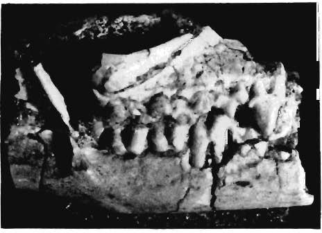
25 Palaeotuologia Polonica, No. 19, 1968 Z. Kl ELAN-JAWOROWSKA: UPPER CR ETACEOUS EUTHERIAN MAMMALS PLATE XXII Kennalestes gobiensis n. sp. (see also Plates XIII-XXV) Page 177 Uppe r Cretaceous, Djadokhta Formation, Bayn Dzak (Main Field), Gobi Deser t (Z. Pal. No. MgM-I/3) Fig. 1a. Type specimen, partial skull with lower jaw in occlusion, slightly prepared, in right lateral view. Po sterior portion of the right lower jaw preserve d in the counterpart, not seen in the photograph ; X 4. Fig. I b. The same in left lateral view; X 6. Fig. 1c. The same in dorsal view; X 4. Fig. 1d. Stereo-photograph of the same specimen, after the separati on of the lower jaws, in occlusal view, left I"-M3, right root of P, broken off 12, fragment ary root of 1", C-M3; x4. Phot o: AI. Czarnock a


26 Palaeontologia Polonica, No. 19, 196~ PI. XXII Id le z. KIELAN-]AWOROWSKA : UPPER CRETACEOUS EUTHERlAN MAMMALS
27 Palaeontologia Polonica, No. 19, 1968 Z. KIELAN-JAWO ROWSKA : UPPER CRETACEOUS EUTHERIAN MAMMALS PLAT E XXIII Kennalestes gobiensis n. sp. (see also Plates XXII and XXV) Page 177 Upper Cretaceous, Djadokhta Formation, Bayn Dzak (Main Field), Gobi Desert Fig. I a. Type specimen (Z. Pal. No. MgM-I/3), stereo-photograph of the skull after the separation of the lower jaws, in the right lateral view, showing right root of It, broken off P, fragmentary roo t of J8and C-M3. Fig. 1b. Anterior portion of the skull of the same specimen with left J3-M", in left lateral view. Fig. I c. Stereo-photograph of the separated lower jaws of the same specimen, in occlusal view, with 1 2-M3 in the left ja w. Figs. Id, e. Left lower jaw of the same specimen in labial and lingual views. Figs. If, g. Right lower jaw of the same specimen in lingual and labial views. Figs. 2a, b. Isolated left lower jaw of the specimen Z. Pal. No. MgM-I /5, with P 3, dp., M" M 2 and trigonid of M 3, in labial and lingual views. Figs. 2c, d. Isolated fragmentary right lower jaw of the same specimen, with fragments of P 3, dp h M t-m3, in lingual and labial views. All the specimens x 4 Photo: M. Czarnock a


28 Palaeontologla Polonica, No. 19, 1968 PI. XXII/ 2a 2b le ]a 2e Id le If 19 2d Ib Z. KIELAN -JAWOROWSK A : UPPER CRET ACroUS EUTHERI AN MAMMALS
29 Palaeontologia Po1011ica, No. 19, 1968 Z. KIELAN-JAWO RO WSKA: UPPER CRETACEOUS EUTHE R IAN MAMMALS PLAT E XXIV Kennalestes gobiensis n. sp.. (see also Plates XXII, XXIII, XXV) Page 177 Upper Cretaceous, Djadokhta Formation, Bayn Dzak (Main Field), Go bi Desert (Z. Pal. No. MgM -Ij2) Fig. 1a. Partial skull, slightly prepared, with right P'_Ms and alveoli for C and P', in right lateral view; X 6. Fig. I b. Stereo-photograph of the same specimen, fina lly prepared, in occlusal view. Right P2_M " and alveoli for C-P', left ps_ms and alveoli for C_P2; X 4. Fig. 1c. Stereo -photograph of the same, in left lateral view; X 4. Fig. 1d. Stereo-photograph of the same, in right lateral view; X4. Fig. 1e. Stereo-photograph of the same, in dorsal view; X 4. Fig. If Mo nta ge-photograph of two parts of the same specimen, put together, originally preserved in a distance of 1 cm, one behind the other, in the same piece of roek ; X 4. Fig. 1g. Poste rior part of the same specimen, in right lateral view ; X 4. Photo: M. Czarno cka Remark: The tips of the right and left ps were broken and lost during preparation. Righ t ps is originally preserved only in Fig. I a. In Figs I b, I c and I d, the tips of the right and left p. are reconstructed out of plastic.


30 Palaeontologia Polonica, No. 19, /968 PI. XXIV 1a 1f 1e 19' Z. KmLAN-]AWOROWSKA: UPPER CRETACEOUS EUTIIERlAN MAMMALS
31 Palaeontologia Polonica, No. 19, 1968 Z. KIELAN-J AWOROWSKA : UP PER CR ETACEOUS EUTHE R ran MAM MALS PLAT E XXV Kennalestes gobiensis n. Sp. (see also Plates XXH-XXIV) Page 177 Upper Cretaceo us, Djadokhta Formation, Bayn Dzak (Mai n Field), Go bi Desert (Z. Pal. No. MgM-r/5) Figs. 1a, b. c. Fragmentary skull with lower jaws in occlusion, in dorsal, right lateral and left lateral views. Fig. la - X 4; Figs. I b, l e - X 6. Fig. 1d. Stereo-pho tograph of isolated right and left lower jaws of the same specimen, in occlusal view, with Pa-Mo and a tr igonid of M, in the left jaw and Ps-M, in the right jaw; X 4. Fig. I e. Stereo-photograph of the same specimen, after the separation of the lower jaws, in occlusal view, with right po_m' and left po_m' ; x 4. Photo : AI. Cz arnocka


32 Palaeontologia Polonica, No. 19, 1968 PI. xxv Id la le lb le z. K!:ELAN JAWOROWSKA : UPPER CRETACEOUS EUTHERIAN MAMMALS
33 Palaeontologia Polonica, No. 19, 1968 Z. KIELAN-JAWOROWSKA: UPPER CRETACEOUS EUTHERIAN MAMMALS PLATE XXVI Zaiambdalestes grangeri S IM P SO~ (see also Plate XXVII ) Page ]86 Upper Cretaceous, Djadokhta Formation, Bayn Dza k (Volcano), Go bi Desert Figs. I a, b, c. Partial skull with lower jaws in occlusion, slightly prep ared, in right lateral, ventral and left lateral views (Z. Pal. No. MgM-I/14). Figs. la, lb - x 4 ; Fig. Ic - x 6. Fig. 1d. Stereo-photograph of separated lower jaws of the same specimen, in occlusal view, with C, Plo alveol us for P 2, P. M. in left jaw and P,-M. in right jaw ; X 4. Z alambdalestes sp Upper Cretaceous, Djadokhta Formation, Bayn Dzak (Mai n Field), Gobi Desert Figs. 2a, b. Fragmentary right lower jaw with alveoli for l,-c, broken off P,; Ps, p., p. and M" in labial and lingual views (Z. Pal. N o. MgM-I/4); X 4. Fig. 2e. Stereo-pho tograph of the same specimen, in occlusal view; X 4. Photo:,;,\1. Czarno cka


34 Palaeontologia Polonica, No. 19, 1968 PI. XXVI Z. KIELAN-JAWOROWSKA: UPPER CRETACEOUS EUTHElUAN MAMMALS
35 Palaeontologia Polonica, No. 19, 1968 Z. KIELA -JAWOROWSKA : U PPE R CR ETACEOUS EUTHE R IAN MA MM ALS PLATE XXVII Zalambdalestes grangeri SIMPSON (see also Plate XXVI) Page 186 Fig. 1a. Stereo-photograph of the skull of Z. Pal. No. MgM-1 j14, in occlu sal view, after the separat ion of the lower jaws, with C-M ' on the right side, and C, alveoli for Pl-P", and partially damaged P'- M' on the left side ; x 4. Fig. lb. The same specimen, in dorsal view; x 3.5. Figs. 1c, d. Separated right lower jaw of the same specimen with Pt-M " in lingua l and labial views ; x 4. Figs. 1e, [. Separated left lower jaw of the same specimen with C. PI' alveolus for p. and P,-M" in lingu al and labial views ; x 4. Fig. 2. Stereo-photograph of nearly complete skull (Z. Pal. No. MgM-IjI6), in occlu sal view. In the anterior part of the snout, the bones were lacking, right and left l' and left P have been preserved on their places in sandstone, as well as the partial moulds of right P and right and left canines. Plastic casts ha ve been made fro m them and plastic roof connecting the incisors with the middle part of the snout. On the right side : 1', plastic I', plastic C and PI_M', on the left P-P, plastic C and P3_Ms; x 2. Photo : A-l. Crarnoc k a


36 Palaeontologia Polonica, No. 19, 1968 PI. XXVll Z. KIELAN-JAWOROWSKA: UPPER CRETACEOUS EUTHERIAN MAMMALS
P3. (Figs. 3A-F, J-K, 4O-Q, 9C) P3 of P. gaoi bears three roots, the mesial and lingual
 P3 of P. gaoi bears three roots, the mesial and lingual") Boyer, Scott, and Fox, 2011: Supplementary File 2 Description of dentition of Pronothodectes gaoi P3. (Figs. 3A-F, J-K, 4O-Q, 9C) P3 of P. gaoi bears three roots, the mesial and lingual of which are subequal
Boyer, Scott, and Fox, 2011: Supplementary File 2 Description of dentition of Pronothodectes gaoi P3. (Figs. 3A-F, J-K, 4O-Q, 9C) P3 of P. gaoi bears three roots, the mesial and lingual of which are subequal
Fig. 12 Trigonias?osborni Lucas, SMNH PI637.2, left maxillary fragment with pi to MI; occlusal view, x I. DESCRIPTION
 Fig. 12 Trigonias?osborni Lucas, SMNH PI637.2, left maxillary fragment with pi to MI; occlusal view, x I. DESCRIPTION SMNH P1637.1 has the teeth somewhat cracked but otherwise well preserved. p 2 is very
EVOLUTION OF THE THERIAN MAMMALS IN THE LATE CRETACEOUS OF ASIA PART I. DELTATHERIDIIDAE
 ZOFIA KIELAN-JAWOROWSKA EVOLUTION OF THE THERIAN MAMMALS IN THE LATE CRETACEOUS OF ASIA PART I. DELTATHERIDIIDAE (plates XXVIII-XXXV) Abstract. - A Late Cretaceous mammalian family of metatherian-eutherian
Characters.-M1 more like that in Procynodictis than like that of Uintacyon. Prodaphcenus (?) robustus in known features of jaw and lower dentition,
 robustus in known features of jaw and lower dentition,") VOL. 20, 1934 GEOLOGY: C. STOCK NEW CREODONTA FROM THE SESPE UPPER EOCENE, CALIFORNIA By CHESTER STOCK BALCH GRADUATE SCHOOL OF THE GEOLOGICAL SCIENCES, CALIFORNIA INSTITUTE OF TECHNOLOGY Communicated
VOL. 20, 1934 GEOLOGY: C. STOCK NEW CREODONTA FROM THE SESPE UPPER EOCENE, CALIFORNIA By CHESTER STOCK BALCH GRADUATE SCHOOL OF THE GEOLOGICAL SCIENCES, CALIFORNIA INSTITUTE OF TECHNOLOGY Communicated
PAL A EON T 0 LOG I C A POLONICA PERCY M. BUTLER PLACENTALS
 ACT A Vol. 22 PAL A EON T 0 LOG I C A POLONICA ------------- 1977 Nr.3 PERCY M. BUTLER EVOLUTIONARY RADIATION OF THE CHEEK TEETH OF CRETACEOUS PLACENTALS Abstract. - Included is a comparative study of
PERISSODACTYLA OF THE SESPE EOCENE, CALIFORNIA
 260 PALEONTOLOGY: C. STOCK PROC. N. A. S. PERISSODACTYLA OF THE SESPE EOCENE, CALIFORNIA BY CHESTER STOCK BALCH GRADUATE SCHOOL OF THE GEOLOGICAL SCIENCES, CALIFORNIA INSTITUTE OF TECHNOLOGY Communicated
3. The Jaw and Related Structures
 Overview and objectives of this dissection 3. The Jaw and Related Structures The goal of this dissection is to observe the muscles of jaw raising. You will also have the opportunity to observe several
Bones of the skull & face
 Bones of the skull & face Cranium= brain case or helmet Copyright The McGraw-Hill Companies, Inc. Permission required for reproduction or display. The cranium is composed of eight bones : frontal Occipital
AMERICAN MUSEUM NOVITATES
 AMERICAN MUSEUM NOVITATES Number 511 Published by TNe AmzuacAwsMumoIJNATU2AL HISTorCir Dec. 15, 1931 56.9,31 (119:729.1) GENERA AND NEW SPECIES OF GROUND SLOTHS FROM THE PLEISTOCENE OF CUBA BY W. D. MATTHEW1
Identification of Mammal Skulls
 Identification of Mammal Skulls Mammalian teeth are heterodont. That is, they are different as you move from front to rear in the tooth row. This contrasts with the homodont condition of most toothed vertebrates.
THE SNOUT OF PAULOCNUS PETRIFACTUS (MAMMALIA, EDENTATA)
") THE SNOUT OF PAULOCNUS PETRIFACTUS (MAMMALIA, EDENTATA) by D. A. HOOIJER Rijksmuseum van Natuurlijke Historie, Leiden A specimen of the ground sloth discovered by Mr. P. Stuiver in Curasao, Paulocnus petrifactus
THE SNOUT OF PAULOCNUS PETRIFACTUS (MAMMALIA, EDENTATA) by D. A. HOOIJER Rijksmuseum van Natuurlijke Historie, Leiden A specimen of the ground sloth discovered by Mr. P. Stuiver in Curasao, Paulocnus petrifactus
Anatomy and Physiology. Bones, Sutures, Teeth, Processes and Foramina of the Human Skull
 Anatomy and Physiology Chapter 6 DRO Bones, Sutures, Teeth, Processes and Foramina of the Human Skull Name: Period: Bones of the Human Skull Bones of the Cranium: Frontal bone: forms the forehead and the
Infratemporal fossa: Tikrit University college of Dentistry Dr.Ban I.S. head & neck Anatomy 2 nd y.
 Infratemporal fossa: This is a space lying beneath the base of the skull between the lateral wall of the pharynx and the ramus of the mandible. It is also referred to as the parapharyngeal or lateral pharyngeal
Arrangement of the artificial teeth:
 Lecture Prosthodontic Dr. Osama Arrangement of the artificial teeth: It s the placement of the teeth on a denture with definite objective in mind or it s the setting of teeth on temporary bases. Rules
NOVITATES AMEIRIICAN MUSEUM NOTES ON AUSTRALIAN MARSUPIALS RARE OR LITTLE-KNOWN IN THE UNITED STATES BY G. H. H. TATE
 AMEIRIICAN MUSEUM NOVITATES PUBLISHED BY THE AMERICAN MUSEUM OF NATURAL HISTORY CITY OF NEW YORK JULY 17, 1951 NUMBER 1528 NOTES ON AUSTRALIAN MARSUPIALS RARE OR LITTLE-KNOWN IN THE UNITED STATES BY G.
Bones Ethmoid bone Inferior nasal concha Lacrimal bone Maxilla Nasal bone Palatine bone Vomer Zygomatic bone Mandible
 splanchnocranium - Consists of part of skull that is derived from branchial arches - The facial bones are the bones of the anterior and lower human skull Bones Ethmoid bone Inferior nasal concha Lacrimal
Figure (2-6): Labial frenum and labial notch.
: Labial frenum and labial notch.") The anatomy of the edentulous ridge in the maxilla and mandible is very important for the design of a complete denture. The consistency of the mucosa and architecture of the underlying bone is different
The anatomy of the edentulous ridge in the maxilla and mandible is very important for the design of a complete denture. The consistency of the mucosa and architecture of the underlying bone is different
Key points for starting off
 Key points for starting off First off, the five questions to ask yourself about a loose tooth before identifying it are: 1. 2. 3. 4. 5. Category (incisor, canine, premolar or molar)? Permanent or deciduous?
Upper arch. 1Prosthodontics. Dr.Bassam Ali Al-Turaihi. Basic anatomy & & landmark of denture & mouth
 1Prosthodontics Lecture 2 Dr.Bassam Ali Al-Turaihi Basic anatomy & & landmark of denture & mouth Upper arch Palatine process of maxilla: it form the anterior three quarter of the hard palate. Horizontal
Primary Teeth Chapter 18. Dental Anatomy 2016
 Primary Teeth Chapter 18 Dental Anatomy 2016 Primary Teeth - Introduction Synonyms deciduous teeth, baby teeth, temporary teeth, milk teeth. There are 20 primary teeth, designated as A thru T in the Universal
Morphology of an Anatomic Crown. By: Assistant Professor Dr. Baydaa Ali Al - Rawi
 Morphology of an Anatomic Crown By: Assistant Professor Dr. Baydaa Ali Al - Rawi October 4, 2009 Elevated landmarks Depressed landmarks A) Elevated landmarks : 1. Dental lobe : is one of the primary centers
Chapter 7. Skeletal System
 Chapter 7 Skeletal System 1 Skull A. The skull is made up of 22 bones: 8 cranial bones, 13 facial bones, and the mandible. B. The Cranium encloses and protects the brain, provides attachments for muscles,
DOUBLE TEETH IN THE SPERM WHALE (PHYSETER MACROCEPHALUS L.)
") DOUBLE TEETH IN THE SPERM WHALE (PHYSETER MACROCEPHALUS L.) by H. BOSCHMA Recently a fairly large number of teeth of the sperm whale were acquired for the collections of the Rijksmuseum van Natuurlijke
DOUBLE TEETH IN THE SPERM WHALE (PHYSETER MACROCEPHALUS L.) by H. BOSCHMA Recently a fairly large number of teeth of the sperm whale were acquired for the collections of the Rijksmuseum van Natuurlijke
Cranium Facial bones. Sternum Rib
 Figure 7.1 The human skeleton. Skull Thoracic cage (ribs and sternum) Cranium Facial bones Sternum Rib Bones of pectoral girdle Vertebral column Sacrum Vertebra Bones of pelvic girdle (a) Anterior view
Skull-2. Norma Basalis Interna Norma Basalis Externa. Dr. Heba Kalbouneh Associate Professor of Anatomy and Histology
 Skull-2 Norma Basalis Interna Norma Basalis Externa Dr. Heba Kalbouneh Associate Professor of Anatomy and Histology Norma basalis interna Base of the skull- superior view The interior of the base of the
Institute of Vertebrate Paleontology and Paleoanthropology, Chinese Academy of Sciences,
 A new symmetrodont mammal (Trechnotheria: Zhangheotheriidae) from the Early Cretaceous of China and trechnotherian character evolution Shundong Bi 1,2 *, Xiaoting Zheng 3,4, Jin Meng 5,2, Xiaoli Wang 3,4,
Fundamental & Preventive Curvatures of Teeth and Tooth Development. Lecture Three Chapter 15 Continued; Chapter 6 (parts) Dr. Margaret L.
 Dr. Margaret L.") Fundamental & Preventive Curvatures of Teeth and Tooth Development Lecture Three Chapter 15 Continued; Chapter 6 (parts) Dr. Margaret L. Dennis Proximal contact areas Contact areas are on the mesial and
Fundamental & Preventive Curvatures of Teeth and Tooth Development Lecture Three Chapter 15 Continued; Chapter 6 (parts) Dr. Margaret L. Dennis Proximal contact areas Contact areas are on the mesial and
GENERAL SCOPE AND USES OF PHYSICAL/BIOLOGICAL ANTHROPOLOGY. Paper No. & Title: B.A./B.Sc. (Honours) 2 dn semester. (Practical)
 2 dn semester. (Practical)") GENERAL SCOPE AND USES OF PHYSICAL/BIOLOGICAL ANTHROPOLOGY Course name: Physical Anthropology Paper No. & Title: B.A./B.Sc. (Honours) 2 dn semester (Practical) Topic No. & Title: 5/12 (Part-I) Drawing
GENERAL SCOPE AND USES OF PHYSICAL/BIOLOGICAL ANTHROPOLOGY Course name: Physical Anthropology Paper No. & Title: B.A./B.Sc. (Honours) 2 dn semester (Practical) Topic No. & Title: 5/12 (Part-I) Drawing
Phylogenetics Lab: Character Descriptions
 Phylogenetics Lab: Character Descriptions 1) Osseous Auditory Canal. 0= absent, 1= present. Does the organism have a bony auditory canal? This will look like a hole or opening in the skull behind the jaw
AXIAL SKELETON SKULL
 AXIAL SKELETON SKULL CRANIAL BONES (8 total flat bones w/ 2 paired) 1. Frontal forms forehead & upper portion of eyesocket (orbital) 2. Parietal paired bones; form superior & lateral walls of cranium 3.
Dental Anatomy High Yield Notes. **Atleast 35 questions comes from these areas of old lectures**
 Dental Anatomy High Yield Notes **Atleast 35 questions comes from these areas of old lectures** This review notes compiled and prepared by my sister for her own study, as a last day review session for
Temporal region. temporal & infratemporal fossae. Zhou Hong Ying Dept. of Anatomy
 Temporal region temporal & infratemporal fossae Zhou Hong Ying Dept. of Anatomy Temporal region is divided by zygomatic arch into temporal & infratemporal fossae. Temporal Fossa Infratemporal fossa Temporal
TOOTHED CETACEAN FROM
 SMITHSONIAN MISCELLANEOUS COLLECTIONS VOLUME 76, NUMBER 7 DESCRIPTION OF AN APPARENTLY NEW TOOTHED CETACEAN FROM, SOUTH CAROLINA (With Two Plates) BY REMINGTON KELLOGG Bureau of Biological Survey, U. S.
Anatomic Relations Summary. Done by: Sohayyla Yasin Dababseh
 Anatomic Relations Summary Done by: Sohayyla Yasin Dababseh Anatomic Relations Lecture 1 Part-1 - The medial wall of the nose is the septum. - The vestibule lies directly inside the nostrils (Nares). -
Structure Location Function
 Frontal Bone Cranium forms the forehead and roof of the orbits Occipital Bone Cranium forms posterior and inferior portions of the cranium Temporal Bone Cranium inferior to the parietal bone forms the
Dr. Sami Zaqout, IUG Medical School
 The skull The skull is composed of several separate bones united at immobile joints called sutures. Exceptions? Frontal bone Occipital bone Vault Cranium Sphenoid bone Zygomatic bones Base Ethmoid bone
Chapter 7 Part A The Skeleton
 Chapter 7 Part A The Skeleton Why This Matters Understanding the anatomy of the skeleton enables you to anticipate problems such as pelvic dimensions that may affect labor and delivery The Skeleton The
1. What is the highest and sharpest cusp on the lower first deciduous molar? 2. Which of the following is NOT the correct location of an embrasure?
 1 1. What is the highest and sharpest cusp on the lower first deciduous molar? a. mesiobuccal b. distobuccal c. distolingual d.mesiolingual 2. Which of the following is NOT the correct location of an embrasure?
Human Male Asian Skeleton, Robust
 Human Male Asian Skeleton, Robust Product Number: Specimen Evaluated: Skeletal Inventory: SC-287 Original Specimen Near-complete human skeleton with 28 teeth. Osteological Observations: This is a clean,
Tikrit University collage of dentistry Dr.Ban I.S. head & neck anatomy 2 nd y. Lec [5] / Temporal fossa :
![Tikrit University collage of dentistry Dr.Ban I.S. head & neck anatomy 2 nd y. Lec [5] / Temporal fossa :](/thumbs/88/115294566.jpg "Tikrit University collage of dentistry Dr.Ban I.S. head & neck anatomy 2 nd y. Lec [5] / Temporal fossa :") Lec [5] / Temporal fossa : Borders of the Temporal Fossa: Superior: Superior temporal line. Inferior: gap between zygomatic arch and infratemporal crest of sphenoid bone. Anterior: Frontal process of the
Lec [5] / Temporal fossa : Borders of the Temporal Fossa: Superior: Superior temporal line. Inferior: gap between zygomatic arch and infratemporal crest of sphenoid bone. Anterior: Frontal process of the
Superior View of the Skull (Norma Verticalis) Anteriorly the frontal bone articulates with the two parietal bones AT THE CORONAL SUTURE
 Anteriorly the frontal bone articulates with the two parietal bones AT THE CORONAL SUTURE") Superior View of the Skull (Norma Verticalis) Anteriorly the frontal bone articulates with the two parietal bones AT THE CORONAL SUTURE 1 The two parietal bones articulate in the midline AT THE SAGITTAL
Superior View of the Skull (Norma Verticalis) Anteriorly the frontal bone articulates with the two parietal bones AT THE CORONAL SUTURE 1 The two parietal bones articulate in the midline AT THE SAGITTAL
European Veterinary Dental College
 European Veterinary Dental College EVDC Training Support Document Preparation of Radiograph Sets (Cat and Dog) Document version : evdc-tsd-radiograph_positioning_(dog_and_cat)-20120121.docx page 1 of 13
The cribriform plate. ethmoid bone. Ethmoid bone consists from: 1) A horizontal cribriform plate. 2) A perpendicular plate. 3) Two lateral labyrinths.
 A horizontal cribriform plate. 2) A perpendicular plate. 3) Two lateral labyrinths.") ethmoid bone Ethmoid bone consists from: 1) A horizontal cribriform plate. 2) A perpendicular plate. 3) Two lateral labyrinths. The cribriform plate 1) Connect the two labyrinths to the perpendicular plate.
ethmoid bone Ethmoid bone consists from: 1) A horizontal cribriform plate. 2) A perpendicular plate. 3) Two lateral labyrinths. The cribriform plate 1) Connect the two labyrinths to the perpendicular plate.
Dr.Sepideh Falah-kooshki
 Dr.Sepideh Falah-kooshki MAXILLA Premaxillary/median palatal suture (radiolucent). Incisive fossa and foramen (radiolucent). Nasal passages (radiolucent). Nasal septum (radiopaque). Anterior nasal spine
GOMPHOTHERIUM ELEGANS, FROM THE PLEISTO-
 DESCRIPTION OF A NEW SPECIES OF MASTODON, GOMPHOTHERIUM ELEGANS, FROM THE PLEISTO- CENE OF KANSAS. By Oliver P. Hay, Research As.iu.iiitc of the Carnegie In-stituti'in of Wn-:]iingto)i. The specimen here
Introduction to Occlusion and Mechanics of Mandibular Movement
 Introduction to Occlusion and Mechanics of Mandibular Movement Dr. Pauline Hayes Garrett Department of Endodontics, Prosthodontics, and Operative Dentistry University of Maryland, Baltimore Assigned reading
Oligocene squalodont (Cetacea: Mammalia) from the Ashiya Group, Japan
 from the Ashiya Group, Japan") Bull. Kitakyushu Mus. Nat. Hist., 8: 75-80. December 27, 1988 Oligocene squalodont (Cetacea: Mammalia) from the Ashiya Group, Japan Yoshihiko Okazaki Kitakyushu Museum of Natural History, Kitakyushu, 805,
Bull. Kitakyushu Mus. Nat. Hist., 8: 75-80. December 27, 1988 Oligocene squalodont (Cetacea: Mammalia) from the Ashiya Group, Japan Yoshihiko Okazaki Kitakyushu Museum of Natural History, Kitakyushu, 805,
TRAUMA TO THE FACE AND MOUTH
 Dr.Yahya A. Ali 3/10/2012 F.I.C.M.S TRAUMA TO THE FACE AND MOUTH Bailey & Love s 25 th edition Injuries to the orofacial region are common, but the majority are relatively minor in nature. A few are major
Parotid Gland. Parotid Gland. Largest of 3 paired salivary glands (submandibular; sublingual) Ramus of Mandible. Medial pterygoid.
 Ramus of Mandible. Medial pterygoid.") Parotid region Parotid Gland Largest of 3 paired salivary glands (submandibular; sublingual) Ramus of Mandible Medial pterygoid Cross section of mandible Masseter D S SCM Parotid Gland Mastoid Process
Parotid region Parotid Gland Largest of 3 paired salivary glands (submandibular; sublingual) Ramus of Mandible Medial pterygoid Cross section of mandible Masseter D S SCM Parotid Gland Mastoid Process
Attachment G. Orthodontic Criteria Index Form Comprehensive D8080. ABBREVIATIONS CRITERIA for Permanent Dentition YES NO
 First Review IL HFS Dental Program Models Second Review Ortho cad Attachment G Orthodontic Criteria Index Form Comprehensive D8080 Ceph Film X-Rays Photos Narrative Patient Name: DOB: ABBREVIATIONS CRITERIA
Skeletal system. Prof. Abdulameer Al-Nuaimi. E. mail:
 Skeletal system Prof. Abdulameer Al-Nuaimi E-mail: a.al-nuaimi@sheffield.ac.uk E. mail: abdulameerh@yahoo.com Functions of Bone and The Skeletal System Support: The skeleton serves as the structural framework
Dr.Ban I.S. head & neck anatomy 2 nd y جامعة تكريت كلية طب االسنان مادة التشريح املرحلة الثانية أ.م.د. بان امساعيل صديق 6102/6102
 جامعة تكريت كلية طب االسنان مادة التشريح املرحلة الثانية أ.م.د. بان امساعيل صديق 6102/6102 Pterygopalatine fossa: The pterygopalatine fossa is a cone-shaped depression, It is located between the maxilla,
Techniques of local anesthesia in the mandible
 Techniques of local anesthesia in the mandible The technique of choice for anesthesia of the mandible is the block injection and this is attributed to the absence of the advantages which are present in
The Premolars. Chapter 17 Permanent Posterior Teeth (p )
") The Premolars Chapter 17 Permanent Posterior Teeth (p. 230-244) General Information Function: u Hold and grind food u Work with molars in mastication. u Even without molars one may be able to chew well
The Premolars Chapter 17 Permanent Posterior Teeth (p. 230-244) General Information Function: u Hold and grind food u Work with molars in mastication. u Even without molars one may be able to chew well
Central Incisor DR.Ahmed Al-Jobory B.D.S.,M.Sc. Conservative Department
 Dental Anatomy Lecture 3 Central Incisor DR.Ahmed Al-Jobory B.D.S.,M.Sc. Conservative Department The permanent maxillary Incisors Maxillary incisor are four in number. The maxillary central incisor is
A NEW DINOSAUR FROM THE LANCE
 SMITHSONIAN MISCELLANEOUS COLLECTIONS VOLUME 61. NUMBER 5 A NEW DINOSAUR FROM THE LANCE FORMATION OF WYOMING BY CHARLES W. GILMORE (Publication 2184) CITY OF WASHINGTON PUBLISHED BY THE SMITHSONIAN INSTITUTION
ANATOMY OF JUNGLE CAT SKULL (FELIS CHAUS, SCHREBER, 1777)
") CASE REPORT ZOOS' PRINT JOURNAL (): 0-0 ANATOMY OF JUNGLE CAT SKULL (FELIS CHAUS, SCHREBER, ) P.O. Nameer, P.O. Naseer, M.O. Ipe and P.A. Ommer Assistant Professor, Department of Wildlife Sciences, College
CASE REPORT ZOOS' PRINT JOURNAL (): 0-0 ANATOMY OF JUNGLE CAT SKULL (FELIS CHAUS, SCHREBER, ) P.O. Nameer, P.O. Naseer, M.O. Ipe and P.A. Ommer Assistant Professor, Department of Wildlife Sciences, College
Medical NBDE-II. Dental Board Exams Part I.
 Medical NBDE-II Dental Board Exams Part I http://killexams.com/exam-detail/nbde-ii Question: 149 Anatomically, the term "clinical root" can be defined as which of the following: A. The space in the tooth
PTERYGOPALATINE FOSSA
 PTERYGOPALATINE FOSSA Outline Anatomical Structure and Boundaries Foramina and Communications with other spaces and cavities Contents Pterygopalatine Ganglion Especial emphasis on certain arteries and
Chapter 7: Skeletal System: Gross Anatomy
 Chapter 7: Skeletal System: Gross Anatomy I. General Considerations A. How many bones in an average adult skeleton? B. Anatomic features of bones are based on II. Axial Skeleton A. Skull 1. Functionally
Mohammad Hisham Al-Mohtaseb. Lina Mansour. Reyad Jabiri. 0 P a g e
 2 Mohammad Hisham Al-Mohtaseb Lina Mansour Reyad Jabiri 0 P a g e This is only correction for the last year sheet according to our record. If you already studied this sheet just read the yellow notes which
Arrangement of posterior artificial teeth Standardized parameters Curve of Wilson Curve of Spee
 . Arrangement of posterior artificial teeth Posterior teeth are set up in tight centric occlusion. The mandibular teeth are set in the wax occlusion rim over the residual ridge in their ideal buccolingual
Skull basic structures. Neurocranium
 Assoc. Prof. Květuše Lovásová, M.V.D., PhD. Skull basic structures Skull consists of two groups of bones: neurocranium (bones forming the brain box) splanchnocranium (bones forming the facial skeleton)
SKULL AS A WHOLE + ANTERIOR CRANIAL FOSSA
 SKULL AS A WHOLE + ANTERIOR CRANIAL FOSSA LEARNING OBJECTIVES At the end of this lecture, the student should be able to know: Parts of skeleton (axial and appendicular) Parts of skull Sutures of skull
Dr.Noor Hashem Mohammad Lecture (5)
") Dr.Noor Hashem Mohammad Lecture (5) 2016-2017 If the mandible is discarded, the anterior part of this aspect of the skull is seen to be formed by the hard palate. The palatal processes of the maxillae
Dr.Noor Hashem Mohammad Lecture (5) 2016-2017 If the mandible is discarded, the anterior part of this aspect of the skull is seen to be formed by the hard palate. The palatal processes of the maxillae
6610 NE 181st Street, Suite #1, Kenmore, WA
 660 NE 8st Street, Suite #, Kenmore, WA 9808 www.northshoredentalacademy.com.08.900 READ CHAPTER The Professional Dental Assistant (p.-9) No Key Terms Recall Questions:,,,, and 6 CLASS SYLLABUS DAY READ
Introduction to Local Anesthesia and Review of Anatomy
 5-Sep Introduction and Anatomy Review 12-Sep Neurophysiology and Pain 19-Sep Physiology and Pharmacology part 1 26-Sep Physiology and Pharmacology part 2 Introduction to Local Anesthesia and Review of
Temporal fossa Infratemporal fossa Pterygopalatine fossa Terminal branches of external carotid artery Pterygoid venous plexus
 Outline of content Temporal fossa Infratemporal fossa Pterygopalatine fossa Terminal branches of external carotid artery Pterygoid venous plexus Boundary Content Communication Mandibular division of trigeminal
Maxilla, ORBIT and infratemporal fossa. Neophytos C Demetriades MD, DDS, MSc Associate professor European University of Cyprus School of Medicine
 Maxilla, ORBIT and infratemporal fossa Neophytos C Demetriades MD, DDS, MSc Associate professor European University of Cyprus School of Medicine MAXILLA Superior, middle, and inferior meatus Frontal sinus
Exercise 10. The Axial Skeleton
 Exercise 10 The Axial Skeleton The Axial Skeleton Consists of the skeletal structures found along the midline of the body. Includes the skull, hyoid, vertebrae, ribs, sternum, and sacrum. The cartilages
Skeletal System -Axial System. Chapter 7 Part A
 Skeletal System -Axial System Chapter 7 Part A Skeleton Learn: Names of the s. Identify specific landmarks that allow: Bones to fit into each other, Organs to fit into the cavities, Muscles to attach,
DEVELOPING ANALOGUE/SUBTITUTE FOR THE MANDIBULAR DENTURE BEARING AREA. Dr Muhammad Rizwan Memon FCPS Assistant Professor
 DEVELOPING ANALOGUE/SUBTITUTE FOR THE MANDIBULAR DENTURE BEARING AREA Dr Muhammad Rizwan Memon FCPS Assistant Professor Crest of Residual Ridge Buccal Shelf Shape of supporting structure Mylohyoid Ridge
Human Anatomy and Physiology - Problem Drill 07: The Skeletal System Axial Skeleton
 Human Anatomy and Physiology - Problem Drill 07: The Skeletal System Axial Skeleton Question No. 1 of 10 Which of the following statements about the axial skeleton is correct? Question #01 A. The axial
OPERATIVE CORRECTION BY OSTEOTOMY OF RECESSED MALAR MAXILLARY COMPOUND IN A CASE OF OXYCEPHALY
 OPERATIVE CORRECTION BY OSTEOTOMY OF RECESSED MALAR MAXILLARY COMPOUND IN A CASE OF OXYCEPHALY By Sir HAROLD GILLIES, C.B.E., F.R.C.S., and STEWART H. HARRISON, F.R.C.S., L.D.S., R.C.S. From the Plastic
THE SKELETAL SYSTEM. Focus on the Skull
 THE SKELETAL SYSTEM Focus on the Skull Review Anatomical Terms Anterior/Posterior Dorsal/Ventral Medial/Lateral Superior/Inferior Bone Markings - Review Projections for attachment of muscles, ligaments
The orbit-1. Dr. Heba Kalbouneh Assistant Professor of Anatomy and Histology
 The orbit-1 Dr. Heba Kalbouneh Assistant Professor of Anatomy and Histology Orbital plate of frontal bone Orbital plate of ethmoid bone Lesser wing of sphenoid Greater wing of sphenoid Lacrimal bone Orbital
DENT Advanced Topics in Removable Prosthodontics, Winter 2008
 University of Michigan Deep Blue deepblue.lib.umich.edu 2008-01 DENT 718 - Advanced Topics in Removable Prosthodontics, Winter 2008 Shotwell, Jeffrey Shotwell, J. (2008, April 23) Advanced Topics in Removable
THE RIGHT INTERPRETATION OF THE CHEEKTEETH TUBERCLES OP TITANOMYS.
 XXIII. ANNALES MUSEI NATIONALIS HUNGARICI. 96. THE RIGHT INTERPRETATION OF THE CHEEKTEETH TUBERCLES OP TITANOMYS. By Dr. J. ÉHIK. (With 5 figures.) The lophodont character of the upper molars of Titanomys
Lecture 2 Maxillary central incisor
 Lecture 2 Maxillary central incisor Generally The deciduous tooth appears in the mouth at 3 18 months of age, with 6 months being the average and is replaced by the permanent tooth around 7 8 years of
MAXILLA, ORBIT & PTERYGOPALATINE FOSSA. Neophytos C Demetriades MD, DDS, MSc Associate professor European University of Cyprus School of Medicine
 MAXILLA, ORBIT & PTERYGOPALATINE FOSSA Neophytos C Demetriades MD, DDS, MSc Associate professor European University of Cyprus School of Medicine Maxilla MAXILLA Superior, middle, and inferior meatus Frontal
Dr.Ban I.S. head & neck anatomy 2 nd y. جامعة تكريت كلية طب االسنان املرحلة الثانية أ.م.د. بان امساعيل صديق 6102/6102
 جامعة تكريت كلية طب االسنان التشريح مادة املرحلة الثانية أ.م.د. بان امساعيل صديق 6102/6102 Parotid region The part of the face in front of the ear and below the zygomatic arch is the parotid region. The
Oral Surgery. Basic Techniques of Dental Local Anesthesia. A variety of techniques used in administration and deposition of local anesthesia:
 Oral Surgery Lecture: 9 Dr. Saif Saadedeen Basic Techniques of Dental Local Anesthesia A variety of techniques used in administration and deposition of local anesthesia: 1. Topical anesthesia 2. Infiltration
o Diaphysis o Area where red marrow is found o Area where yellow marrow is found o Epiphyseal plate AXIAL SKELETON Skull
 64 Anatomy & Physiology Coloring Workbook 7. Figure 5-2A is a midlevel, cross-sectional view of the diaphysis of the femur. Label the membrane that lines the cavity and the membrane that covers the outside
CHAPTER 7, PART II (BONES)
") Anatomy Name: CHAPTER 7, PART II (BONES) Entry #: INSTRUCTIONS: 1) READ Chapter 7, pg. 140-161. 2) Using the outline, make a note card for each underlined bone name or phrase. 3) On each note card, put
Anatomy Name: CHAPTER 7, PART II (BONES) Entry #: INSTRUCTIONS: 1) READ Chapter 7, pg. 140-161. 2) Using the outline, make a note card for each underlined bone name or phrase. 3) On each note card, put
Human, Male, Single gunshot wound
 Human, Male, Single gunshot wound Product Number: Specimen Evaluated: Skeletal Inventory: BC-152 Bone Clones replica 1 intact cranium - left inferior nasal concha absent - middle nasal conchae absent 1
Chapter 7: Head & Neck
 Chapter 7: Head & Neck Osteology I. Overview A. Skull The cranium is composed of irregularly shaped bones that are fused together at unique joints called sutures The skull provides durable protection from
Trigeminal Nerve Worksheets, Distributions Page 1
 Trigeminal Nerve Worksheet #1 Distribution by Nerve Dr. Darren Hoffmann Dental Gross Anatomy, Spring 2013 We have drawn out each of the branches of CN V in lecture and you have an idea now for their basic
The Skull and Temporomandibular joint II Prof. Abdulameer Al-Nuaimi. E. mail:
 The Skull and Temporomandibular joint II Prof. Abdulameer Al-Nuaimi E-mail: a.al-nuaimi@sheffield.ac.uk E. mail: abdulameerh@yahoo.com Temporal fossa The temporal fossa is a depression on the temporal
Human, Female, Black, Shotgun wound
 Human, Female, Black, Shotgun wound Product Number: Specimen Evaluated: Skeletal Inventory: BC-196 Bone Clones replica 1 intact cranium 2 fragments of mandible: - portion of left body, ramus, coronoid
Parotid Gland, Temporomandibular Joint and Infratemporal Fossa
 M1 - Anatomy Parotid Gland, Temporomandibular Joint and Infratemporal Fossa Jeff Dupree Sanger 9-057 jldupree@vcu.edu Parotid gland: wraps around the mandible positioned between the mandible and the sphenoid
Dental Morphology and Vocabulary
 Dental Morphology and Vocabulary Palate Palate Palate 1 2 Hard Palate Rugae Hard Palate Palate Palate Soft Palate Palate Palate Soft Palate 4 Palate Hard Palate Soft Palate Maxillary Arch (Maxilla) (Uppers)
Skull-2. Norma Basalis Interna. Dr. Heba Kalbouneh Assistant Professor of Anatomy and Histology
 Skull-2 Norma Basalis Interna Dr. Heba Kalbouneh Assistant Professor of Anatomy and Histology Norma basalis interna Base of the skull- superior view The interior of the base of the skull is divided into
MAMMALS FROM THE BLUE ASH LOCAL FAUNA (LATE OLIGOCENE), SOUTH DAKOTA. RODENTIA, PART 2: FAMILIES FLORENTIAMYIDAE AND GEOMYIDAE
, SOUTH DAKOTA. RODENTIA, PART 2: FAMILIES FLORENTIAMYIDAE AND GEOMYIDAE") Paludicola 7(1):14-25 November 2008 by the Rochester Institute of Vertebrate Paleontology MAMMALS FROM THE BLUE ASH LOCAL FAUNA (LATE OLIGOCENE), SOUTH DAKOTA. RODENTIA, PART 2: FAMILIES FLORENTIAMYIDAE
Paludicola 7(1):14-25 November 2008 by the Rochester Institute of Vertebrate Paleontology MAMMALS FROM THE BLUE ASH LOCAL FAUNA (LATE OLIGOCENE), SOUTH DAKOTA. RODENTIA, PART 2: FAMILIES FLORENTIAMYIDAE
Biology 218 Human Anatomy. Adapted from Martini Human Anatomy 7th ed. Chapter 6 The Skeletal System: Axial Division
 Adapted from Martini Human Anatomy 7th ed. Chapter 6 The Skeletal System: Axial Division Introduction The axial skeleton: Composed of bones along the central axis of the body Divided into three regions:
Bone Clones Osteological Evaluation Report. 1 intact mandible
 Human, Male, Black Bone Clones Osteological Evaluation Report Product Number: Specimen Evaluated: Skeletal Inventory: BC-203 Bone Clones replica 1 intact cranium 1 intact mandible General observations:
APPENDICULAR SKELETON 126 AXIAL SKELETON SKELETAL SYSTEM. Cranium. Skull. Face. Skull and associated bones. Auditory ossicles. Associated bones.
 SKELETAL SYSTEM 206 AXIAL SKELETON 80 APPENDICULAR SKELETON 26 Skull Skull and associated s 29 Cranium Face Auditory ossicles 8 4 6 Associated s Hyoid Thoracic cage 25 Sternum Ribs 24 Vertebrae 24 column
Concepts of occlusion Balanced occlusion. Monoplane occlusion. Lingualized occlusion. Figure (10-1)
") Any contact between teeth of opposing dental arches; usually, referring to contact between the occlusal surface. The static relationship between the incising or masticatory surfaces of the maxillary or
Any contact between teeth of opposing dental arches; usually, referring to contact between the occlusal surface. The static relationship between the incising or masticatory surfaces of the maxillary or
Human Healed Trauma Skull
 Human Healed Trauma Skull Product Number: Specimen Evaluated: BC-303 Original Specimen Skeletal Inventory: 1 Cranium with full dentition (teeth ##1-16) 1 Mandible with full dentition (teeth ##17-32) Osteological
Remember from the first year embryology Trilaminar disc has 3 layers: ectoderm, mesoderm, and endoderm
 Development of face Remember from the first year embryology Trilaminar disc has 3 layers: ectoderm, mesoderm, and endoderm The ectoderm forms the neural groove, then tube The neural tube lies in the mesoderm
Mandibular and Maxillary Anesthesia
 Mandibular and Maxillary Anesthesia Uses of the Conduction Technique JACK H. SELTSAM, D.D.S., M.D., Los Angeles THE ARMAMENTARIUM of a surgeon who operates on the head and neck should include the ability