THE SKELETAL SYSTEM. Focus on the Skull
|
|
|
- Lorin Sharp
- 6 years ago
- Views:
Transcription
1 THE SKELETAL SYSTEM Focus on the Skull

2 Review Anatomical Terms Anterior/Posterior Dorsal/Ventral Medial/Lateral Superior/Inferior
3 Bone Markings - Review Projections for attachment of muscles, ligaments and tendons (Process, trochanter, tuberosity, tubercle, crest, line, spine) Processes for articulation with other bones (Head, neck, condyle, trochlea, facet) Openings = holes or spaces in bone for nerves and vessels to pass (Foramen, canal, meatus, fissure, sinus) Depressions = indentations (Fossa, sulcus)

4 Axial Skeleton Includes bones of the skull, vertebral column and thoracic cage Creates a framework of support and protection for internal organs Provides sites of attachment of muscles
 Hyoid bone (the only freely moveable bone) Bones are joined by")
5 The Skull Protects the brain and supports delicate sense organs Bones that form the skull include 8 cranial bones 14 facial bones 6 auditory ossicles (tiny bones in the ears) Hyoid bone (the only freely moveable bone) Bones are joined by sutures
6 Bones of the Cranium Frontal bone (1)- forms the forehead and roof of the ocular orbits Parietal Bone (2) posterior to the frontal bone; forms the sides and roof of skull Occiptal Bone (1) most posterior part of the cranium foramen magnum = large opening for spine Temporal Bone (2) form the sides and base of the skull; a number of distinct anatomical landmarks
7 Sutures of the Cranium Sagittal Suture midline suture; between parietal bones Coronal Suture between frontal bone and parietal bones Lambdoid Suture between occipital and parietal bones Squamous Suture between the temporal bones and the parietal bones
8 Bones and Sutures of the Cranium Lateral View Coronal Suture Parietal Bone Frontal Bone Temporal Bone Lambdoid Suture Squamous Suture Occipital Bone
9 Bones and Sutures of the Cranium Frontal View Coronal Suture Parietal Bone Frontal Bone Temporal Bone
10 Bones and Sutures of the Cranium Inferior View Temporal Bone Parietal Bone Occipital Bone
11 Bones and Sutures of the Cranium Horizontal View Frontal Bone Temporal Bone Parietal Bone Occipital Bone REVIEW

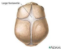
12 The skull at birth

13 The skull at birth
14 Bones of the Cranium continued Sphenoid Bone irregular bat-shaped bone forms part of the cranial floor Ethmoid Bone stabilizes the brain; forms the roof and sides of the nasal cavity Vomer nasal septum Palatine Bone posterior hard palate ( roof of mouth )
15 Bones of the Cranium Lateral View Coronal Suture Parietal Bone Temporal Bone Lambdoid Suture Squamous Suture Occipital Bone Frontal Bone Sphenoid Bone Ethmoid Bone
16 Bones of the Cranium - Frontal Coronal Suture Parietal Bone Frontal Bone Sphenoid Bone Ethmoid Bone Temporal Bone Vomer
17 Bones of the Cranium Inferior View Palatine Bone Sphenoid Bone Vomer Temporal Bone Parietal Bone Occipital Bone
18 Bones of the Cranium Horizontal View Frontal Bone Sphenoid Bone Ethmoid Bone Temporal Bone Parietal Bone Occipital Bone
19 Facial Bones Maxilla (2) anterior part of hard palate; upper jaw; articulates with all other facial bones except the mandible Zygomatic Bones (2) cheek bones Nasal Bones bridge of nose Lacrimal Bones tiny bones bearing tear ducts Mandible lower jaw
20 Bones of the Face Lateral View Coronal Suture Parietal Bone Temporal Bone Lambdoid Suture Squamous Suture Occipital Bone Frontal Bone Sphenoid Bone Ethmoid Bone Lacrimal Bone Nasal Bone Zygomatic Bone Maxilla Mandible
21 Bones of the Face - Frontal Coronal Suture Parietal Bone Frontal Bone Nasal Bone Sphenoid Bone Ethmoid Bone Lacrimal Bone Zygomatic Bone Maxilla Mandible Temporal Bone Vomer
22 Bones of the Face Inferior View Maxilla Palatine Bone Zygomatic Bone Maxilla Sphenoid Bone Vomer Temporal Bone Parietal Bone Occipital Bone
23 Teeth (32) Incisors Canines Pre-molars Molars Pre-molars Canines Incisors
24 Processes Any elevation or projection Can be part of a joint or a site of attachment for muscles, ligaments and tendons
25 Processes of the cranium Styloid process (temporal bone) Mastoid process (temporal bone) Zygomatic process (temporal bone) Occipital condyle (occipital bone) External occipital protuberance (occipital bone) Coronoid process (mandible) Condylar process (mandible) Mandibular process (mandible) Pterygoid process (sphenoid) Hamulus of the pterygoid process (sphenoid) PROCESS Protrusion for attachment of tendons and ligaments


26 Styloid Process Pointed piece of bone that extends down from the temporal bone just below the ear Attaches to ligaments that support the hyoid bone muscles that control the tongue and pharynx
27 Mastoid Process Built up area of the lower temporal bone where important neck muscles attach Muscle that rotate and elevate the head and clavicle attach here

28 Zygomatic Process of the Temporal Bone Connects temporal bone to facial bones (zygomatic bone)

29 Occipital Condyle The site on the occipital bone where skull meets vertebrae Atlas = the first vertebrae in the spinal column
30 External Occipital Protuberance Medial protrusion of the occipital bone Muscles that keep the head upright and allow the head to tilt backward attach here
 like a")

31 Coronoid Process (coronation day) like a crown Attachment point for muscle that closes the jaw
32 Condylar Process Forms a hinge with the temporal bone
33 Mandibular Process Smooth surface of the condylar process


34 The Sphenoid Bone Pterygoid Process and the hamulus of the pterygoid process
35 Process of the Skull Lateral View Coronal Suture Parietal Bone Temporal Bone Lambdoid Suture Squamous Suture Occipital Bone Zygomatic Process Frontal Bone Sphenoid Bone Ethmoid Bone Lacrimal Bone Nasal Bone Zygomatic Bone Maxilla Mastoid Process Styloid Process Mandible
36 Process of the Skull - Frontal Coronal Suture Parietal Bone Frontal Bone Nasal Bone Sphenoid Bone Ethmoid Bone Lacrimal Bone Zygomatic Bone Maxilla Mandible Temporal Bone Ethmoid Bone Vomer
37 Process of the Skull Inferior View Maxilla Palatine Bone Zygomatic Bone Zygomatic Process Maxilla Sphenoid Bone Vomer Styloid Process Mastoid Process Temporal Bone Occipital Condyle Parietal Bone Occipital Bone
38 Processes of the Skull Horizontal View Frontal Bone Sphenoid Bone Ethmoid Bone Temporal Bone Parietal Bone Occipital Bone

39 Name the Process
40 Name the Process
41 Foramina and Other Structures General terms to describe openings include Foramen, canal, meatus, fissure sinus
42 Foramina and Other Structures Supraorbital foramen (frontal) Infraorbital foramen (maxilla) Mental foramen (mandible) Foramen magnum (occipital) Jugular foramen (temporal) Carotid canal (temporal) External acoustic meatus (temporal) Mandibular foramen (mandible) Palatine foramen (palatine) Foramen lacerum (temporal) Foramen ovale (sphenoid) Foramen spinosum (sphenoid) Foramen rotundum (sphenoid) Stylomastoid foramen (temporal) FORAMEN, MEATUS, FISSURE, CANAL: Terms to describe openings for passage of nerves and blood vessels

43 Supraorbital foramen** above the orbit blood vessels and nerves that innervate the eyebrows and eyelids

44 Infraorbital foramen** below the orbit Facial nerves

45 Mental foramen** Distal/lateral opening for the mental nerve and vessels that innervate the lip

46 Mandibular foramen Proximal/medial opening for the mental nerve and vessels that innervate the lip and teeth

47 Palatine foramen Nerves that innervate the palate

48 External Auditory meatus** Opening that leads to the eardrum (tympanum)

49 Foramen magnum** Spinal cord
50 Jugular foramen** Jugular vein
51 Carotid Canal** Carotid artery

52 Foramen ovale** Trigeminal nerve mandibular branch


53 Foramen spinosum Nerves that innervate the meninges enter the brain

54 Foramen lacerum Fills with cartilage after birth

55 Foramen rotundum Trigeminal nerve: maxillary branch

56 Stylomastoid foramen Facial nerves exit skull

57 Foramina of the Skull Lateral View Coronal Suture Parietal Bone Temporal Bone Lambdoid Suture Squamous Suture Occipital Bone Zygomatic Process External Auditory Meatus Mastoid Process Styloid Process Mandible Frontal Bone Sphenoid Bone Ethmoid Bone Lacrimal Bone Nasal Bone Zygomatic Bone Maxilla Mandible Mental Foramen

58 Foramina of the Skull - Frontal Coronal Suture Parietal Bone Nasal Bone Sphenoid Bone Ethmoid Bone Lacrimal Bone Zygomatic Bone Maxilla Mandible Frontal Bone Supraorbital Foramen Superior Orbital Fissure Optic Canal Temporal Bone Infraaorbital Foramen Ethmoid Bone Vomer

59 Foramina of the Skull Inferior View Maxilla Palatine Bone Zygomatic Bone Zygomatic Process Vomer Styloid Process Mastoid Process Temporal Bone Parietal Bone Occipital Bone Maxilla Sphenoid Bone Foramen Ovale Carotid Canal Jugular Foramen Occipital Condyle Foramen Magnum

60 Foramina of the Skull Horizontal View Frontal Bone Sphenoid Bone Ethmoid Bone Optic Canal Temporal Bone Carotid Canal Foramen Ovale Jugular Foramen Parietal Bone Occipital Bone Foramen Magnum
61 Other Features Superior Nuchal Line Inferior Nuchal Line Mandibular foss
62 THE SKELETAL SYSTEM Continue with the vertebrae
 Subdivided based on vertebral")
63 Vertebral Column Consists of 26 bones 24 vertebrae Sacrum Coccyx Vertebrae separated by cartilage (intervertebral discs) Subdivided based on vertebral structure
64 Vertebrae Anatomy - general Vertebral body massive weight bearing portion Vertebral foramen the opening through which the spinal cord passes Articular processes extensions of the vertebrae that articulate with other bones or provide attachment for muscles Transverse processes Superior articular processes Inferior articular processes
65 Vertebrae Anatomy - General

66 Vertebrae Anatomy by region
 The first")

67 Cervical Region Seven cervical vertebrae (C1 to C7) The first cervical vertebrae (C1; atlas) articulates with the occipital condyle

68 Cervical region Anatomical Features oval concave body large vertebral foramen Stumpy spinous process with a notched tip Transverse foramina within the transverse processes Protect blood vessels supplying the brain

69 Cervical Vertebrae Anatomy

70 Cervical region Vertebral Foramen Vertebral Arch Spinous Process Lamina Superior Articular Processes Pedicle Superior Articular Facet Transverse Process Transverse Foramen Body
 Articulation of atlas and axis allows you to rotate your")
71 Cervical region The first two cervical vertebrae have unique characteristics C1 = ATLAS ( yes) Articulation of atlas with skull allows you to nod your head C2 = AXIS ( no ) Articulation of atlas and axis allows you to rotate your head
 Articulates with atlas Axis")
72 The Atlas and Axis Dens (odontoid process) Ligament Articulates with occipital condyles Atlas (C1) Articulates with atlas Axis (C2)


73 Thoracic Region Twelve cervical vertebrae (T1 to T12) Each thoracic vertebrae articulates with one or more pairs of ribs

74 Thoracic region Vertebrae Anatomy Heart shaped body More massive than cervical vertebrae Large, slender spinous processes that point inferiorly Costal facets for articulating with one or more pairs of ribs

75 Vertebral Anatomy thoracic region Spinous Process Transverse Process Lamina Vertebral Arch Pedicle Articular facets Pedicle Articular facets Vertebral Foramen Body
 The")

76 Lumbar Region Five lumbar vertebrae (L1 to L5) The fifth lumbar vertebrae articulates with the sacrum

77 Lumbar region Vertebrae Anatomy Vertebral body is thick and more oval than thoracic Massive, stumpy spinous process that projects posteriorly Bladelike transverse processes; no articulation for ribs Most massive, least mobile

78 Vertebral Anatomy lumbar region Spinous Process Vertebral Foramen Lamina Articular facets Transverse Process Pedicle Body


79 Sacrum Region sacrum Single bone formed by the fusion of 5 embryonic vertebrae
80 Sacrum Protects reproductive, digestive and excretory organs Attaches the axial skeleton to the appendicular skeleton Broad surface attachment of leg muscles Bones fuse shortly after puberty Prominent bulge (sacral promontory) is an important landmark in females during labor and delivery
81 Coccyx Coccyx Series of small fused vertebrae

82 Coccyx Attachment site for muscle that closes that anal opening Fusion of the bone is not complete until late in adulthood May eventually fuse with the sacrum
 Primary curves Thoracic and sacral curves")

83 Normal Spinal Curvature Four spinal curves (seen in lateral view) Primary curves Thoracic and sacral curves Appear late in fetal development Secondary curves Cervical curve as baby holds head up Lumbar curve as baby learns to stand

84 Normal Spinal Curvature


85 Spinal curvature development Cervical curvature develops as a baby learns to hold its head


86 Spinal curvature development Lumbar curvature develops as a baby learns to stand
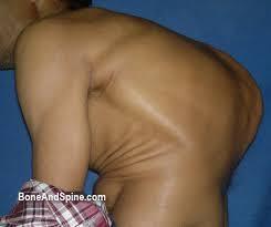
87 Abnormal spinal curvature Several abnormal conditions can arise during childhood and adolescence Kyphosis exaggerated thoracic curvature Lordosis exaggerated lumbar curvature Scoliosis abnormal lateral curvature
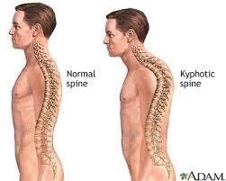
88 Kyphosis
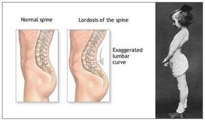
89 Lordosis
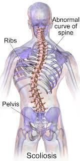
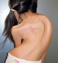
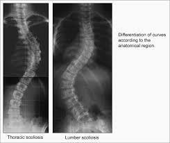
90 Scoliosis
91 THE SKELETAL SYSTEM Continue with the Thoracic Cage
92 Thoracic Cage Consists of the thoracic vertebrae, the ribs and the sternum Ribs + sternum = rib cage Provides bony support for the walls of the thoracic cavity Protects heart and lungs Serves as base for muscles involved in respiration
93 Ribs AKA costal bones Elongated flattened bones 12 pairs Ribs 1-7 = true ribs attach to the sternum by separate cartilage extensions Ribs 8-10 = false ribs; do not attach directly to the sternum; cartilages fuse before attachment Ribs = floating ribs; do not connect to sternum at all

94 Sternum AKA breastbone 3 parts 1. Manubrium = Broad triangular part articulates with clavicle 2. Elongated body articulates with ribs 3. Ziphoid process Damage to this process can puncture the liver CPR training places special emphasis on location of this part of the sternum to reduce damage during compressions

95 Floating Ribs Thoracic vertebrae True Ribs Manubrium Body Sternum Ziphoid Process Ribs False Ribs Costal Cartilage
Anatomy and Physiology. Bones, Sutures, Teeth, Processes and Foramina of the Human Skull
 Anatomy and Physiology Chapter 6 DRO Bones, Sutures, Teeth, Processes and Foramina of the Human Skull Name: Period: Bones of the Human Skull Bones of the Cranium: Frontal bone: forms the forehead and the
APPENDICULAR SKELETON 126 AXIAL SKELETON SKELETAL SYSTEM. Cranium. Skull. Face. Skull and associated bones. Auditory ossicles. Associated bones.
 SKELETAL SYSTEM 206 AXIAL SKELETON 80 APPENDICULAR SKELETON 26 Skull Skull and associated s 29 Cranium Face Auditory ossicles 8 4 6 Associated s Hyoid Thoracic cage 25 Sternum Ribs 24 Vertebrae 24 column
Structure Location Function
 Frontal Bone Cranium forms the forehead and roof of the orbits Occipital Bone Cranium forms posterior and inferior portions of the cranium Temporal Bone Cranium inferior to the parietal bone forms the
Biology 218 Human Anatomy. Adapted from Martini Human Anatomy 7th ed. Chapter 6 The Skeletal System: Axial Division
 Adapted from Martini Human Anatomy 7th ed. Chapter 6 The Skeletal System: Axial Division Introduction The axial skeleton: Composed of bones along the central axis of the body Divided into three regions:
Chapter 7. Skeletal System
 Chapter 7 Skeletal System 1 Skull A. The skull is made up of 22 bones: 8 cranial bones, 13 facial bones, and the mandible. B. The Cranium encloses and protects the brain, provides attachments for muscles,
ANATOMY & PHYSIOLOGY I Laboratory Version B Name Section. REVIEW SHEET Exercise 10 Axial Skeleton
 ANATOMY & PHYSIOLOGY I Laboratory Version B Name Section REVIEW SHEET Exercise 10 Axial Skeleton 1 POINT EACH. THE SKULL MULTIPLE CHOICE 1. The major components of the axial skeleton include the 7. The
Cranium Facial bones. Sternum Rib
 Figure 7.1 The human skeleton. Skull Thoracic cage (ribs and sternum) Cranium Facial bones Sternum Rib Bones of pectoral girdle Vertebral column Sacrum Vertebra Bones of pelvic girdle (a) Anterior view
Skeletal system. Prof. Abdulameer Al-Nuaimi. E. mail:
 Skeletal system Prof. Abdulameer Al-Nuaimi E-mail: a.al-nuaimi@sheffield.ac.uk E. mail: abdulameerh@yahoo.com Functions of Bone and The Skeletal System Support: The skeleton serves as the structural framework
Bones of the skull & face
 Bones of the skull & face Cranium= brain case or helmet Copyright The McGraw-Hill Companies, Inc. Permission required for reproduction or display. The cranium is composed of eight bones : frontal Occipital
Chapter 7 Part A The Skeleton
 Chapter 7 Part A The Skeleton Why This Matters Understanding the anatomy of the skeleton enables you to anticipate problems such as pelvic dimensions that may affect labor and delivery The Skeleton The
o Diaphysis o Area where red marrow is found o Area where yellow marrow is found o Epiphyseal plate AXIAL SKELETON Skull
 64 Anatomy & Physiology Coloring Workbook 7. Figure 5-2A is a midlevel, cross-sectional view of the diaphysis of the femur. Label the membrane that lines the cavity and the membrane that covers the outside
Human Anatomy and Physiology - Problem Drill 07: The Skeletal System Axial Skeleton
 Human Anatomy and Physiology - Problem Drill 07: The Skeletal System Axial Skeleton Question No. 1 of 10 Which of the following statements about the axial skeleton is correct? Question #01 A. The axial
Chapter 7: Skeletal System: Gross Anatomy
 Chapter 7: Skeletal System: Gross Anatomy I. General Considerations A. How many bones in an average adult skeleton? B. Anatomic features of bones are based on II. Axial Skeleton A. Skull 1. Functionally
Chapter 8A. The Skeletal System: The Axial Skeleton. The Skeletal System: The Axial Skeleton. Types of Bones. Types of Bones
 Chapter 8A The Skeletal System: The Axial Skeleton The Skeletal System: The Axial Skeleton 206 named bones Axial Skeleton 80 bones lie along longitudinal axis skull, hyoid, vertebrae, ribs, sternum, ear
The Axial Skeleton. C h a p t e r. PowerPoint Lecture Slides prepared by Jason LaPres Lone Star College - North Harris
 C h a p t e r 7 The Axial Skeleton PowerPoint Lecture Slides prepared by Jason LaPres Lone Star College - North Harris Copyright 2009 Pearson Education, Inc., publishing as Pearson Benjamin Cummings An
Anatomy and Physiology 1 Chapter 7 self quiz Pro, Dima Darwish,MD.
 Anatomy and Physiology 1 Chapter 7 self quiz Pro, Dima Darwish,MD. 1) How many bones make up the axial skeleton? A) 50 B) 60 C) 70 D) 80 E) 90 2) Which of the following is a function of the axial skeleton?
ACTIVITY 3: AXIAL SKELETON AND LONG BONE DISSECTION COW BONE DISSECTION
 ACTIVITY 3: AXIAL SKELETON AND LONG BONE DISSECTION Objectives: 1) How to get ready: Read Chapter 7, McKinley et al., Human Anatomy, 4e. All text references are for this textbook. Learning the meanings
The Skeletal System: Axial Skeleton
 The Skeletal System: Axial Skeleton The Big Idea The Axial Skeleton & Homeostasis The bones of the axial skeleton contribute to homeostasis by protecting many of the body s organs such as the brain, spinal
Chapter 7 The Skeletal System:The Axial Skeleton
 Chapter 7 The Skeletal System:The Axial Skeleton Axial Skeleton 80 bones lie along longitudinal axis skull, hyoid, vertebrae, ribs, sternum, ear ossicles Appendicular Skeleton 126 bones upper & lower limbs
YOU MUST BRING YOUR OWN GLOVES FOR THIS ACTIVITY.
 ACTIVITY 3: AXIAL SKELETON AND LONG BONE DISSECTION Objectives: 1) How to get ready: Read Chapter 7, McKinley et al., Human Anatomy, 5e. All text references are for this textbook. Learning the meanings
CHAPTER 7, PART II (BONES)
") Anatomy Name: CHAPTER 7, PART II (BONES) Entry #: INSTRUCTIONS: 1) READ Chapter 7, pg. 140-161. 2) Using the outline, make a note card for each underlined bone name or phrase. 3) On each note card, put
Anatomy Name: CHAPTER 7, PART II (BONES) Entry #: INSTRUCTIONS: 1) READ Chapter 7, pg. 140-161. 2) Using the outline, make a note card for each underlined bone name or phrase. 3) On each note card, put
Human Anatomy & Physiology I Dr. Sullivan Unit VIIIa The Axial Skeleton Chapter 8 (Sections )
") Human Anatomy & Physiology I Dr. Sullivan Unit VIIIa The Axial Skeleton Chapter 8 (Sections 8.1-8.3) I. Divisions of the skeletal system a) An adult human skeleton has 206 named bones b) Most are paired
Human Anatomy & Physiology I Dr. Sullivan Unit VIIIa The Axial Skeleton Chapter 8 (Sections 8.1-8.3) I. Divisions of the skeletal system a) An adult human skeleton has 206 named bones b) Most are paired
Skeletal System -Axial System. Chapter 7 Part A
 Skeletal System -Axial System Chapter 7 Part A Skeleton Learn: Names of the s. Identify specific landmarks that allow: Bones to fit into each other, Organs to fit into the cavities, Muscles to attach,
Biology 2401 The Skeletal System
 Biology 2401 The Skeletal System Purpose: The lab will describe the microscopic and gross anatomy of bone, identify bones of the body, and identify important bone markings. I. Overview of the Skeleton
Exercise 10. The Axial Skeleton
 Exercise 10 The Axial Skeleton The Axial Skeleton Consists of the skeletal structures found along the midline of the body. Includes the skull, hyoid, vertebrae, ribs, sternum, and sacrum. The cartilages
AXIAL SKELETON SKULL
 AXIAL SKELETON SKULL CRANIAL BONES (8 total flat bones w/ 2 paired) 1. Frontal forms forehead & upper portion of eyesocket (orbital) 2. Parietal paired bones; form superior & lateral walls of cranium 3.
Crafton Hills College Human Anatomy & Physiology Axial Skeleton
 A. Major Divisions Crafton Hills College Human Anatomy & Physiology Axial keleton 1. Axial: Part of skeleton lies along long axis of body 2. Appendicular: Bones & features of the appendages B. AXIAL KELETON
Axial Skeleton: Vertebrae and Thorax
 Axial Skeleton: Vertebrae and Thorax Function of the vertebral column (spine or backbone): 1) 2) 3) Composition of Vertebral column The vertebral column is formed by 33 individual vertebrae (some of which
Spring Written By: J. E. Sutton. Contents: I. Overview of the Skeleton: II. Appendicular Skeleton III. Axial Skeleton IV.
 Spring 2012 Written By: J. E. Sutton Contents: I. Overview of the Skeleton: II. Appendicular Skeleton III. Axial Skeleton IV. Articulations Overview of the Skeleton: I. Orientation to Human Skeleton: a.
The Skeletal System. Parts of the skeletal system. Bones (Skeleton) Joints Cartilages Ligaments
 Joints Cartilages Ligaments") The Skeletal System Parts of the skeletal system Bones (Skeleton) Joints Cartilages Ligaments Functions of the Bones Support Internal framework of the body Protection Skull and vertebrae protect brain
The Skeletal System Parts of the skeletal system Bones (Skeleton) Joints Cartilages Ligaments Functions of the Bones Support Internal framework of the body Protection Skull and vertebrae protect brain
TEST YOURSELF- Chapter 7
 TEST YOURSELF- Chapter 7 Cranial Bones 1. Give the name of the bone for each of the following markings. Some of the markings are found on more than one bone. List all that apply. Cranium a. Frontal squama:
Bone Flashcards for 10a
 Bone Flashcards for 0a CLAVICLE (collar bone). Sternal extremity (end) flat end. Acromial extremity (end) rounded end. SCAPULA (shoulder blade). Right or left scapula?. Superior border (superior margin).
Axial skeleton bones and markings
 Axial skeleton bones and markings Skull Cranial bones Frontal x 1 Supraorbital foramen Occipital x 1 Foramen magnum Occipital condyles Superior nuchal line Inferior nuchal line Anterior cranial fossa External
11/25/2012. Chapter 7 Part 2: Bones! Skeletal Organization. The Skull. Skull Bones to Know Cranium
 Chapter 7 Part 2: Bones! 5) Distinguish between the axial and appendicular skeletons and name the major parts of each 6) Locate and identify the bones and the major features of the bones that compose the
Copyright 2010 Pearson Education, Inc.
 E. VERTEBRAL COLUMN 1. The vertebral column extends from the skull to the pelvis and forms the vertical axis of the skeleton. 2. The vertebral column is composed of vertebrae that are separated by intervertebral
Skeletal system overview. Classification of Bones
 Skeletal system overview BIOL241 Lab #9 Classification of Bones Bone are identified by: shape internal tissues bone markings 1 1. Flat bones 2. Long bones 3. Short bones 4. Irregular bones 5. Sutural bones
Labs 9 and 10. Classification of Bones. Bone Shapes 1/05/13. Skeletal system overview. Bone are identified by:
 Labs 9 and 10 Skeletal system overview Classification of Bones Bone are identified by: shape internal tissues bone markings 1. Flat bones 2. Long bones 3. Short bones 4. Irregular bones 5. Sutural bones
BIO 137 AXIAL SKELETON BONE STUDY THE HUMAN SKELETON
 BIO 137 THE AXIAL SKELETON MARY CATHERINE FLATH, Ph.D. THE HUMAN SKELETON AXIAL SKULL HYOID THORACIC CAGE VERTEBRAL COLUMN APPENDICULAR PECTORAL GIRDLE UPPER LIMBS PELVIC GIRDLE LOWER LIMBS AXIAL SKELETON
Overview of the Skeleton: Bone Markings
 Name Overview of the Skeleton: Bone Markings Match the terms in column B with the appropriate description in column A. Column A 1. sharp, slender process* 2. small rounded projection* 3. narrow ridge of
Vertebral Column. Backbone consists of 26 vertebrae. Five vertebral regions. Cervical
 Vertebral Column Backbone consists of 26 vertebrae. Five vertebral regions Cervical vertebrae (7) in the neck. Thoracic vertebrae (12) in the thorax. Lumbar vertebrae (5) in the lower back. Sacrum (5,
Dr.Noor Hashem Mohammad Lecture (5)
") Dr.Noor Hashem Mohammad Lecture (5) 2016-2017 If the mandible is discarded, the anterior part of this aspect of the skull is seen to be formed by the hard palate. The palatal processes of the maxillae
Dr.Noor Hashem Mohammad Lecture (5) 2016-2017 If the mandible is discarded, the anterior part of this aspect of the skull is seen to be formed by the hard palate. The palatal processes of the maxillae
University of Palestine. Midterm Exam 2013/2014 Total Grade:
 Course No: DNTS2208 Course Title: Head and Neck Anatomy Date: 09/11/2013 No. of Questions: (50) Time: 1hour Using Calculator (No) University of Palestine Midterm Exam 2013/2014 Total Grade: Instructor
Bio 5/6 5 The Skeletal System Study Guide
 Name: THE SKELETAL SYSTEM: 5 The Skeletal System Study Guide Period: The skeleton is constructed of two of the most supportive tissues found in the human body - cartilage and bone. Besides supporting and
Skull-2. Norma Basalis Interna Norma Basalis Externa. Dr. Heba Kalbouneh Associate Professor of Anatomy and Histology
 Skull-2 Norma Basalis Interna Norma Basalis Externa Dr. Heba Kalbouneh Associate Professor of Anatomy and Histology Norma basalis interna Base of the skull- superior view The interior of the base of the
BONE CHALLENGE DANIL HAMMOUDI.MD
 BONE CHALLENGE DANIL HAMMOUDI.MD Bone Basic functions? A. support B. protection C. movement assistance in D. RBC formation-hemopoiesis E. mineral homeostasis +importance of calcium F. energy supply -yellow
Human Anatomy - Problem Drill 06: The Skeletal System Axial Skeleton & Articualtions
 Human Anatomy - Problem Drill 06: The Skeletal System Axial Skeleton & Articualtions Question No. 1 of 10 Instructions: (1) Read the problem and answer choices carefully, (2) Work the problems on paper
Copyright 2010 Pearson Education, Inc. Copyright 2010 Pearson Education, Inc. Figure Sectioned spinous process. Interspinous.
 PowerPoint Lecture Slides prepared by Janice Meeking, Mount Royal College C H A P T E R 7 The Skeleton: Part B Vertebral Column Transmits weight of trunk to lower limbs Surrounds and protects spinal cord
External Acoustic Meatus. Mastoid Process. Zygomatic Process. Temporal Bone
 Bone lab review 1. Frontal Bone 2. Supra-Orbital Foramen 3. Orbit (Orbital Cavity) 4. Superior Orbital Fissure 5. Inferior Orbital Fissure 6. Zygomatic Bone 7. Infra-Orbital Foramen 8. Maxilla 9. Mandible
Anatomy and Physiology II. Review Spine and Neck
 Anatomy and Physiology II Review Spine and Neck Spine regions How many cervical vertibrae are there? 7 The curvature is the cervical region posterior? Concave posterior How many thoracic? And curvature?
The Skull DANIL HAMMOUDI.MD
 The Skull DANIL HAMMOUDI.MD summary of bones/structures in Chapter 15 of the manual need tp be print as soon as possible http://www.mnsu.edu/emuseum/biology/humananatomy/skeletal/skul l/frontal/frontal.html
Lab Exercise #04 The Skeletal System Student Performance Objectives
 Lab Exercise #04 The Skeletal System Student Performance Objectives The material that you are required to learn in this exercise can be found in either the lecture text or the supplemental materials provided
Bone List Anatomy
 1 Frontal Bone Skull 2 Parietal Bone Skull 3 Occipital Bone Skull 4 Temporal Bone Skull 5 Coronal Suture Skull 6 Sagittal Suture Skull 7 Squamous suture Skull 8 Lambdoid Suture Skull 9 Surpaorbital Ridge
Important Parts of Bones
 Important Parts of Bones For 2015 Know: Humerus (posterior) Clavical Femur (Anterior) Foot Hand Mandible Os Coxa Scapula Skull (Anterior, Inferior, Lateral) Sternum Humerus (posterior) A. olecranon fossa
the Skeletal System provided by Academic Web Services Grand Canyon University
 Anatomy Resource Center Study Guides the Skeletal System HEAD & NECK REGIONAL VIEW SKULL BONES CRANIUM FACE SKULL LANDMARKS ANTERIOR SIDE SUPERIOR/INFERIOR VERTEBRAL COLUMN VERTEBRAL REGIONS CERVICAL C1
Biology 210 Chapter 8: Skeletal Tissues Supplement 1
 Biology 210 Chapter 8: Skeletal Tissues Supplement 1 By John McGill Material contributed by Beth Wyatt & Jack Bagwell DIVISIONS OF THE SKELETAL SYSTEM AXIAL SKELETON (80 BONES) Bones of the Head, Neck,
Anatomy images for MSS practical exam- 2019
 Anatomy images for MSS practical exam- 2019 Ilium Ischium Pubis Acetabulaum Iliac crest Iliac tubercle ASIS (muscle and ligament attached) AIIS (muscle attached) PSIS PIIS Ischial spine Ischial tuberosity
A. Incorrect! The axial skeleton includes bones of the skull, inner ear, chest and spinal column.
 Anatomy and Physiology - Problem Drill 07: The Skeletal System II No. 1 of 10 1. Which of the following statements about the axial skeleton is correct? A. The axial skeleton includes bones of the skull,
Dr. Sami Zaqout, IUG Medical School
 The skull The skull is composed of several separate bones united at immobile joints called sutures. Exceptions? Frontal bone Occipital bone Vault Cranium Sphenoid bone Zygomatic bones Base Ethmoid bone
Anatomy Skull and Spinal Cord
 1 Anatomy Skull and Spinal Cord 1. Skull The skull is anterior to the spinal column and is the bony structure that encases the brain. Its purpose is to protect the brain and allow attachments for the facial
SKELETON FUNCTIONS OF BONE:
 SKELETON FUNCTIONS OF BONE: SKELETON: 1. Performs a mechanical function in forming the skeletal support of the body and in forming a leverage system whereby work and movement are possible. 2. Serves as
Perpendicular Plate Zygomatic Bone. Mental Foramen Mandible
 Glabella Frontal Middle Nasal Concha Nasal Lacrimal Perpendicular Plate Zygomatic Inferior Nasal Concha Maxilla Mental Mandible Skull (anterior view) Squamosal Suture Coronal Suture Frontal Parietal Nasal
Musculoskeletal System (Part A-1) Module 7 -Chapter 10 Overview. Functions
 Module 7 -Chapter 10 Overview. Functions") Musculoskeletal System (Part A-1) Module 7 -Chapter 10 Overview Susie Turner, M.D. 1/8/13 Muscles Attachments Bones Bone types Surface features of bones Divisions of the skeletal system Joints or Articulations
Musculoskeletal System (Part A-1) Module 7 -Chapter 10 Overview Susie Turner, M.D. 1/8/13 Muscles Attachments Bones Bone types Surface features of bones Divisions of the skeletal system Joints or Articulations
Chapter 7 Part B The Skeleton
 Chapter 7 Part B The Skeleton 7.2 The Vertebral Column General Characteristics Extends from skull to pelvis Also called spine or spinal column Functions to transmit weight of trunk to lower limbs, surround
Skeletal System: Skull.
 Skeletal System: Skull www.fisiokinesiterapia.biz Bones of the Skull SPLANCHNOCRANIUM Nasal (2) Maxilla (2) Lacrimal (2) Zygomatic (2) Palatine (2) Inferior concha (2) Vomer Mandible NEUROCRANIUM Frontal
Skull basic structures. Neurocranium
 Assoc. Prof. Květuše Lovásová, M.V.D., PhD. Skull basic structures Skull consists of two groups of bones: neurocranium (bones forming the brain box) splanchnocranium (bones forming the facial skeleton)
Riverside Community College Anatomy & Physiology 2B SPRING 2012 EXAM #1-ABC (Nervous System)
") Riverside Community College Anatomy & Physiology 2B SPRING 2012 EXAM #1-ABC (Nervous System) Name: 1) This vertebra is an example of a(n). 1) A) thoracic B) axis C) atlas D) lumbar E) sacral 1 2) W hich
Riverside Community College Anatomy & Physiology 2B SPRING 2012 EXAM #1-ABC (Nervous System) Name: 1) This vertebra is an example of a(n). 1) A) thoracic B) axis C) atlas D) lumbar E) sacral 1 2) W hich
SKULL AS A WHOLE + ANTERIOR CRANIAL FOSSA
 SKULL AS A WHOLE + ANTERIOR CRANIAL FOSSA LEARNING OBJECTIVES At the end of this lecture, the student should be able to know: Parts of skeleton (axial and appendicular) Parts of skull Sutures of skull
SKULL / CRANIUM BONES OF THE NEUROCRANIUM (7) Occipital bone (1) Sphenoid bone (1) Temporal bone (2) Frontal bone (1) Parietal bone (2)
 Occipital bone (1) Sphenoid bone (1) Temporal bone (2) Frontal bone (1) Parietal bone (2)") Important! 1. Memorizing these pages only does not guarantee the succesfull passing of the midterm test or the semifinal exam. 2. The handout has not been supervised, and I can not guarantee, that these
Important! 1. Memorizing these pages only does not guarantee the succesfull passing of the midterm test or the semifinal exam. 2. The handout has not been supervised, and I can not guarantee, that these
An Introduction to the Axial Skeleton. Copyright 2009 Pearson Education, Inc., publishing as Pearson Benjamin Cummings
 An Introduction to the Axial Skeleton Copyright 2009 Pearson Education, Inc., publishing as Pearson Benjamin Cummings Terms: Structures of Bones Articulations: Contacts with other bones Landmarks (Bone
Skeletal System - Prelab 1
 Skeletal System - Prelab 1 1. Which bones contain the paranasal sinuses? What function do the sinuses serve? 2. What two areas are separated from each other by the hard palate? Name the two bones that
Skeletal System. Axial Division
 Skeletal System Axial Division The Axial Skeleton You will see that each bone has special features (overviewed in section I below) that provide Sites of Attachment (for muscles, ligaments, tendons, etc.)
Introduction to Local Anesthesia and Review of Anatomy
 5-Sep Introduction and Anatomy Review 12-Sep Neurophysiology and Pain 19-Sep Physiology and Pharmacology part 1 26-Sep Physiology and Pharmacology part 2 Introduction to Local Anesthesia and Review of
Lab 6, 7, 8: Skeletal System
 107 Lab 6, 7, 8: Skeletal System Adult Skull Bony orbit (FLEZMS) Frontal bone supraorbital foramen frontal sinus Lacrimal bone Ethmoid bone perpendicular plate of ethmoid middle nasal conchae cribriform
2. The vertebral arch is composed of pedicles (projecting from the body) and laminae (uniting arch posteriorly).
 and laminae (uniting arch posteriorly).") VERTEBRAL COLUMN 2018zillmusom I. VERTEBRAL COLUMN - functions to support weight of body and protect spinal cord while permitting movements of trunk and providing for muscle attachments. A. Typical vertebra
VERTEBRAL COLUMN 2018zillmusom I. VERTEBRAL COLUMN - functions to support weight of body and protect spinal cord while permitting movements of trunk and providing for muscle attachments. A. Typical vertebra
Chapter 7: Head & Neck
 Chapter 7: Head & Neck Osteology I. Overview A. Skull The cranium is composed of irregularly shaped bones that are fused together at unique joints called sutures The skull provides durable protection from
Nervous & Skeletal Systems. Virtual Science University
 Nervous & Skeletal Systems Virtual Science University 1 Nervous & Skeletal Systems Texas TEK B.10(A) The student will interpret the function of systems in organisms (humans) including the nervous and skeletal
Chapter 7: Skeletal System
 Chapter 7: Skeletal System The Skeletal System Introduction P. 182 Bone is an organ made up of tissues: It is made up of the following components. Cartilage Blood Nerves Bone Connective Bone Classification
University of Palestine. Midterm Exam 2013/2014 Total Grade:
 [ Course No: DNTS2208 Course Title: Head and Neck Anatomy Date: 17/11/1024 No. of Questions: (52) Time: 2hours Using Calculator (No) University of Palestine Midterm Exam 2013/2014 Total Grade: Instructor
Skeletal System. It s all about the bones!!!
 Skeletal System It s all about the bones!!! The Skeletal System in Action!! The Skeletal System in Action! https://www.youtube.com/watch?v=icwllrqkv cg&list=plzile25upgebvru0jneppcabh0fhktgt Q 1. FYI 5
Infratemporal fossa: Tikrit University college of Dentistry Dr.Ban I.S. head & neck Anatomy 2 nd y.
 Infratemporal fossa: This is a space lying beneath the base of the skull between the lateral wall of the pharynx and the ramus of the mandible. It is also referred to as the parapharyngeal or lateral pharyngeal
Bones Ethmoid bone Inferior nasal concha Lacrimal bone Maxilla Nasal bone Palatine bone Vomer Zygomatic bone Mandible
 splanchnocranium - Consists of part of skull that is derived from branchial arches - The facial bones are the bones of the anterior and lower human skull Bones Ethmoid bone Inferior nasal concha Lacrimal
Anatomy & Physiology Skeletal System Worksheet
 1. Name the five functions of the skeleton. c) d) e) Anatomy & Physiology Skeletal System Worksheet 2. The term for the shaft of a bone is:. 3. The bony struts found in spongy bone are called. 4. In ossification,
NOTES SKELETAL SYSTEM
 NOTES for the SKELETAL SYSTEM Anatomy & Physiology 2016 Johnson The Skeletal System I. System includes 4 basic parts: A. Bones (206 of em) B. Joints C. Cartilages D. Ligaments II. Bones have 5 basic functions:
The Thoracic Cage ANATOMY 2: THORACIC CAGE AND VERTEBRAL COLUMN
 ANATOMY 2: THORACIC CAGE AND VERTEBRAL COLUMN PSK 4U Mr. S. Kelly North Grenville DHS The Thoracic Cage 7 true ribs 3 false ribs 2 floating ribs Clavicle = collarbone Manubrium Sternum Xiphoid Process
VERTEBRAL COLUMN VERTEBRAL COLUMN
 VERTEBRAL COLUMN FUNCTIONS: 1) Support weight - transmits weight to pelvis and lower limbs 2) Houses and protects spinal cord - spinal nerves leave cord between vertebrae 3) Permits movements - *clinical
The SKELETAL System. The framework of bones and cartilage which protect organs, and provides a lever system that allows locomotion.
 The SKELETAL System The framework of bones and cartilage which protect organs, and provides a lever system that allows locomotion. Functions of the Skeletal System Support Protection Movement Facilitation
Lab Unit One Flashcards
 CLAVICLE (collar bone). Sternal extremity (end) flat end. Acromial extremity (end) rounded end.. Conoid tubercle near round end SCAPULA (shoulder blade). Right or left scapula?. Superior border (superior
BIOLOGY 113 LABORATORY Skeletal System
 BIOLOGY 113 LABORATORY Skeletal System Objectives Distinguish between the axial and appendicular skeleton. Distinguish between the cranium and facial skeleton. Locate and name the bones of the skull and
A&P 1 Skeletal Lab Week 1 Pre-lab Guide Skeletal Background Information
 A&P 1 Skeletal Lab Week 1 Pre-lab Guide Skeletal Background Information Note: These notes are very similar to those found in your lecture notes. This info is for BOTH LECTURE AND LAB! Read through them
2 skull, vertebral column, thoracic cage
 CHAPTER 7-SKELTON FILL-IN NOTES 2 skull, vertebral column, thoracic cage 3 Fig. 7.1 pg. 199 4 I. Skull: A. : Encloses and the brain - 8 bones B. : 14 bones Cranium A. Forehead (brain) Anterior part of
Bones of the Skull Lateral View
 Bones of the Skull Lateral View Frontal Bone Parietal Bone Occipital Bone Temporal Bone Sphenoid Bone Pterion Sutures of the Skull Lateral View Coronal Suture Lambdoid Suture Squamous Suture Sutures of
The Thoracic Cage. Role of the Thoracic Cage 2/13/2019. Anatomy 2: Thoracic Cage and Vertebral Column
 PSK 4U Mr. S. Kelly North Grenville DHS Anatomy 2: Thoracic Cage and Column The Thoracic Cage 7 true ribs 3 false ribs 2 floating ribs Clavicle = collarbone Manubrium Sternum Xiphoid Process 12 thoracic
PTERYGOPALATINE FOSSA
 PTERYGOPALATINE FOSSA Outline Anatomical Structure and Boundaries Foramina and Communications with other spaces and cavities Contents Pterygopalatine Ganglion Especial emphasis on certain arteries and
in compact bone, large vertical canals carrying blood vessels and nerves. in compact bone, large horizontal canals carrying blood vessels and nerves.
 Carl Christensen, PhD Skeletal System (Bones`) Bio. 2304 Human Anatomy 1. Identify a term for each of the following: shaft of a long bone ends of a long bone ossified remnant of the "growth plate" connective
Anatomy Made Easy MSS
 Anatomy Made Easy MSS part #1 هذا الملف يشمل تفريغ المحاضرة الثانية لعون بدءا من الصفحة 11 وحتى األخير Done By :MohamedA. Diabat Edited by Awn Academic team The Axial Skeleton The axial skeleton consist
Bones of the wrist and ankle Bones that form within tendons (e.g., patella)
") Skeletal System Review Surface Anatomy Dr. Gary Mumaugh Function of Bones Support form the framework that supports the body and cradles soft organs Protection provide a protective case for the brain, spinal
Skeletal System Review Surface Anatomy Dr. Gary Mumaugh Function of Bones Support form the framework that supports the body and cradles soft organs Protection provide a protective case for the brain, spinal
THEME 2. VERTEBRAE (GENERAL DATA). CERVICAL, THORACIC AND LUMBAR VERTEBRAE. SACRUM. COCCYX. THE VERTEBRAL COLUMN AS A WHOLE
. CERVICAL, THORACIC AND LUMBAR VERTEBRAE. SACRUM. COCCYX. THE VERTEBRAL COLUMN AS A WHOLE") THEME 2. VERTEBRAE (GENERAL DATA). CERVICAL, THORACIC AND LUMBAR VERTEBRAE. SACRUM. COCCYX. THE VERTEBRAL COLUMN AS A WHOLE Osteology of the Vertebral Column Bone Description vertebra Notes a vertebra
THEME 2. VERTEBRAE (GENERAL DATA). CERVICAL, THORACIC AND LUMBAR VERTEBRAE. SACRUM. COCCYX. THE VERTEBRAL COLUMN AS A WHOLE Osteology of the Vertebral Column Bone Description vertebra Notes a vertebra
Labs 6, 7, 8: Skeletal System
 153 Labs 6, 7, 8: Skeletal System Unit 6: Skeletal System: Bone tissue, Bones and Joints (p. 105-152) Ex. 6-1: Histology of Osseous Tissue, p. 113 Model: Osteon Tiss Lamella Osteocyte Lacunae Canaliculi
SHORT ANSWER. Write the word or phrase that best completes each statement or answers the question.
 Exam Name SHORT ANSWER. Write the word or phrase that best completes each statement or answers the question. Figure 7.1 Using Figure 7.1, match the following: 1) Articulates with hip bones of the pelvis.
bio4165 lab quiz 1 Posterior View Anterior View Lateral View Anterior View bio fall.quarter lab.quiz.1...page.1 of 6
 B A Posterior View D C E Lateral View bio.4165...fall.quarter.2005...lab.quiz.1...page.1 of 6 F I G 35 Posterior View H bio.4165...fall.quarter.2005...lab.quiz.1...page.2 of 6 J Posterior View L K Inferior