The Digestive System in the Head and Neck
|
|
|
- Charleen Hicks
- 6 years ago
- Views:
Transcription
1 The Digestive System in the Head and Neck

2 The Mouth The Lips The lips are two fleshy folds that surround the oral orifice They are covered on the outside by skin and are lined on the inside by mucous membrane the substance of the lips is made up by the orbicularis oris muscle and the muscles that radiate from the lips into the face Also included are the labial blood vessels and nerves, connective tissue, and many small salivary glands. The philtrum is the shallow vertical groove seen in the midline on the outer surface of the upper lip. Median folds of mucous membraneâ the labial frenulaeâ connect the inner surface of the lips to the gums.

3 The Mouth Cavity The mouth extends from the lips to the pharynx The entrance into the pharynx, the oropharyngeal isthmus, is formed on each side by the palatoglossal fold The mouth is divided into the vestibule and the mouth cavity proper.
4 Vestibule The vestibule lies between the lips and the cheeks externally and the gums and the teeth internally This slitlike space communicates with the exterior through the oral fissure between the lips when the jaws are closed, it communicates with the mouth proper behind the third molar tooth on each side. The vestibule is limited above and below by the reflection of the mucous membrane from the lips and cheeks to the gums. The lateral wall of the vestibule is formed by the cheek, which is made up by the buccinator muscle and is lined with mucous membrane. The tone of the buccinator muscle and that of the muscles of the lips keeps the walls of the vestibule in contact with one another The duct of the parotid salivary gland opens on a small papilla into the vestibule opposite the upper second molar tooth

5 Mouth Proper The mouth proper has a roof and a floor. The roof of the mouth is formed by the hard palate in front and the soft palate behind The floor is formed largely by the anterior two thirds of the tongue and by the reflection of the mucous membrane from the sides of the tongue to the gum of the mandible fold of mucous membrane called the frenulum of the tongue connects the undersurface of the tongue in the midline to the floor of the mouth Lateral to the frenulum, the mucous membrane forms a fringed fold, the plica fimbriata The submandibular duct of the submandibular gland opens onto the floor of the mouth on the summit of a small papilla on either side of the frenulum of the tongue The sublingual gland projects up into the mouth, producing a low fold of mucous membrane, the sublingual fold Numerous ducts of the gland open on the summit of the fold.
6 Mucous Membrane of the Mouth In the vestibule the mucous membrane is tethered to the buccinator muscle by elastic fibers in the submucosa prevent redundant folds of mucous membrane from being bitten between the teeth when the jaws are closed The mucous membrane of the gingiva, or gum, is strongly attached to the alveolar periosteum.
7 Sensory Innervation of the Mouth Roof: The greater palatine and nasopalatine nerves from the maxillary division of the trigeminal nerve Floor: The lingual nerve (common sensation), a branch of the mandibular division of the trigeminal nerve. The taste fibers travel in the chorda tympani nerve, a branch of the facial nerve. Cheek: The buccal nerve, a branch of the mandibular division of the trigeminal nerve (the buccinator muscle is innervated by the buccal branch of the facial nerve)
8 The Teeth The gingivae (gums) are specialized regions of the oral mucosa that surround the teeth and cover adjacent regions of the alveolar bone. Deciduous Teeth There are 20 deciduous teeth: four incisors, two canines, and four molars in each jaw They begin to erupt about 6 months after birth and have all erupted by the end of 2 years. The teeth of the lower jaw usually appear before those of the upper jaw.

9 Permanent Teeth There are 32 permanent teeth: four incisors, two canines, four premolars, and six molars in each jaw They begin to erupt at 6 years of age The last tooth to erupt is the third molar, which may happen between the ages of 17 and 30 The teeth of the lower jaw appear before those of the upper jaw.

10 The Tongue The tongue is a mass of striated muscle covered with mucous membrane The muscles attach the tongue to the styloid process and the soft palate above and to the mandible and the hyoid bone below The tongue is divided into right and left halves by a median fibrous septum.

11 Mucous Membrane of the Tongue The mucous membrane of the upper surface of the tongue can be divided into anterior and posterior parts by a V-shaped sulcus, the sulcus terminalis The apex of the sulcus projects backward and is marked by a small pit, the foramen cecum The apex of the sulcus projects backward and is marked by a small pit, the foramen cecum the foramen cecum is an embryologic remnant and marks the site of the upper end of the thyroglossal duct

12 Three types of papillae are present on the upper surface of the anterior two thirds of the tongue: the filiform papillae, the fungiform papillae, and the vallate papillae. The mucous membrane covering the posterior third of the tongue is devoid of papillae but has an irregular surface caused by the presence of underlying lymph nodules, the lingual tonsil. The mucous membrane on the inferior surface of the tongue is reflected from the tongue to the floor of the mouth In the midline anteriorly, the undersurface of the tongue is connected to the floor of the mouth by a fold of mucous membrane, the frenulum of the tongue On the lateral side of the frenulum, the deep lingual vein can be seen through the mucous membrane Lateral to the lingual vein, the mucous membrane forms a fringed fold called the plica fimbriata

13 Muscles of the Tongue The muscles of the tongue are divided into two types: intrinsic and extrinsic Intrinsic Muscles These muscles are confined to the tongue and are not attached to bone. They consist of longitudinal, transverse, and vertical fibers. Nerve supply: Hypoglossal nerve Action: Alter the shape of the tongue
14 Extrinsic Muscles These muscles are attached to bones and the soft palate They are the genioglossus (Superior genial spine of mandible), Protrudes apex of tongue through mouth the hyoglossus (Body and greater cornu of hyoid bone), Depresses tongue the styloglossus (Styloid process of temporal bone), Draws tongue upward and backward and the palatoglossus (Palatine aponeurosis), Pulls roots of tongue upward and backward, narrows oropharyngeal isthmus Insertion : Blends with eachother, the palatoglossus inserts at Side of tongue Nerve supply: Hypoglossal nerve

15 Movements of the Tongue Protrusion: The genioglossus muscles on both sides acting together Retraction: Styloglossus and hyoglossus muscles on both sides acting together Depression: Hyoglossus muscles on both sides acting together Retraction and elevation of the posterior third: Styloglossus and palatoglossus muscles on both sides acting together Shape changes: Intrinsic muscles
 Blood Supply The lingual artery, the tonsillar branch of the facial artery, and the ascending pharyngeal artery supply the tongue The veins drain")
16 Sensory Innervation Anterior two thirds: Lingual nerve branch of mandibular division of trigeminal nerve (general sensation) and chorda tympani branch of the facial nerve (taste) Posterior third: Glossopharyngeal nerve (general sensation and taste) Blood Supply The lingual artery, the tonsillar branch of the facial artery, and the ascending pharyngeal artery supply the tongue The veins drain into the internal jugular vein. Lymph Drainage Tip: Submental lymph nodes Sides of the anterior two thirds: Submandibular and deep cervical lymph nodes Posterior third: Deep cervical lymph nodes

17 The Palate The palate forms the roof of the mouth and the floor of the nasal cavity. It is divided into two parts: the hard palate in front and the soft palate behind. Hard Palate The hard palate is formed by the palatine processes of the maxillae and the horizontal plates of the palatine bones It is continuous behind with the soft palate. Soft Palate The soft palate is a mobile fold attached to the posterior border of the hard palate Its free posterior border presents in the midline a conical projection called the uvula. The soft palate is continuous at the sides with the lateral wall of the pharynx The soft palate is composed of mucous membrane, palatine aponeurosis, and muscles.

18 The mucous membrane covers the upper and lower surfaces of the soft palate. The palatine aponeurosis is a fibrous sheet attached to the posterior border of the hard palate It is the expanded tendon of the tensor veli palatini muscle.

19 Muscles of the Soft Palate The muscles of the soft palate are the tensor veli palatini, the levator veli palatini, the palatoglossus, the palatopharyngeus, and the musculus uvulae The muscle fibers of the tensor veli palatini converge as they descend from their origin to form a narrow tendon, which turns medially around the pterygoid hamulus The tendon, together with the tendon of the opposite side, expands to form the palatine aponeurosis When the muscles of the two sides contract, the soft palate is tightened so that the soft palate may be moved upward or downward as a tense sheet.

20 Levator veli palatini : O: Petrous part of temporal bone, auditory tube I: Palatine aponeurosis Innerv.: Pharyngeal plexus Action: Raises soft palate Tensor veli palatini Spine of sphenoid, auditory tube With muscle of other side, forms palatine aponeurosis Nerve to medial pterygoid from mandibular nerve Tenses soft palate

21 Palatopharyngeus Palatine aponeurosis Posterior border of thyroid cartilage Pharyngeal plexus Elevates wall of pharynx, pulls palatopharyngeal folds medially Musculus uvulae Posterior border of hard palate Mucous membrane of uvula Pharyngeal plexus Elevates uvula

22 Movements of the Soft Palate The pharyngeal isthmus (the communicating channel between the nasal and oral parts of the pharynx) is closed by raising the soft palate. Closure occurs during the production of explosive consonants in speech. The soft palate is raised by the contraction of the levator veli palatini on each side. At the same time, the upper fibers of the superior constrictor muscle contract and pull the posterior pharyngeal wall forward The palatopharyngeus muscles on both sides also contract so that the palatopharyngeal arches are pulled medially, like side curtains By this means the nasal part of the pharynx is closed off from the oral part.

23 Nerve Supply of the Palate The greater and lesser palatine nerves from the maxillary division of the trigeminal nerve enter the palate through the greater and lesser palatine foramina The nasopalatine nerve, also a branch of the maxillary nerve, enters the front of the hard palate through the incisive foramen. The glossopharyngeal nerve also supplies the soft palate Blood Supply of the Palate The greater palatine branch of the maxillary artery, the ascending palatine branch of the facial artery, and the ascending pharyngeal artery Lymph Drainage of the Palate Deep Cervical Lymph Nodes

24 The palatoglossal arch is a fold of mucous membrane containing the palatoglossus muscle, which extends from the soft palate to the side of the tongue The palatoglossal arch marks where the mouth becomes the pharynx. The palatopharyngeal arch is a fold of mucous membrane behind the palatoglossal arch runs downward and laterally to join the pharyngeal wall. The muscle contained within the fold is the palatopharyngeus muscle. The palatine tonsils, which are masses of lymphoid tissue, are located between the palatoglossal and palatopharyngeal arches

25 The Salivary Glands Parotid Gland The parotid gland is the largest salivary gland and is composed mostly of serous acini lies in a deep hollow below the external auditory meatus, behind the ramus of the mandible and in front of the sternocleidomastoid muscle The facial nerve divides the gland into superficial and deep lobes The parotid duct emerges from the anterior border of the gland and passes forward over the lateral surface of the masseter. It enters the vestibule of the mouth upon a small papilla opposite the upper second molar tooth Parasympathetic secretomotor supply arises from the glossopharyngeal nerve The nerves reach the gland via the tympanic branch, the lesser petrosal nerve, the otic ganglion, and the auriculotemporal nerve.


26 Submandibular Gland The submandibular gland consists of a mixture of serous and mucous acini It lies beneath the lower border of the body of the mandible divided into superficial and deep parts by the mylohyoid muscle The deep part of the gland lies beneath the mucous membrane of the mouth on the side of the tongue. The submandibular duct emerges from the anterior end of the deep part of the gland and runs forward beneath the mucous membrane of the mouth. It opens into the mouth on a small papilla, which is situated at the side of the frenulum of the tongue Parasympathetic secretomotor supply is from the facial nerve via the chorda tympani, and the submandibular ganglion The postganglionic fibers pass directly to the gland.
27 Anatomical relations Parotid Lies in the parotid bed that is formed by: the sternocleidomastoid muscle behind; the ramus of mandible in front; superiorly, the base of the trench is formed by the external acoustic meatus and the posterior aspect of the zygomatic arch. The parotid duct passes anteriorly across the external surface of the masseter muscle and then turns medially to penetrate the buccinator muscle of the cheek and open into the oral cavity adjacent to the crown of the second upper molar tooth The parotid gland encloses the external carotid artery, the retromandibular vein, and the origin of the extracranial part of the facial nerve [VII].
28 Submandibular gland the larger arm of the hook is directed forward in the horizontal plane below the mylohyoid muscle and is therefore outside the boundaries of the oral cavity-this larger superficial part of the gland is directly against a shallow impression on the medial side of the mandible (submandibular fossa) inferior to the mylohyoid line; the smaller arm of the hook (or deep part) of the gland loops around the posterior margin of the mylohyoid muscle to enter and lie within the floor of the oral cavity where it is lateral to the root of the tongue on the lateral surface of the hyoglossus muscle. The lingual nerve loops under the submandibular duct, crossing first the lateral side and then the medial side of the duct, as the nerve descends anteromedially through the floor of the oral cavity and then ascends into the tongue.

29 Sublingual Gland The sublingual gland lies beneath the mucous membrane (sublingual fold) of the floor of the mouth, close to the frenulum of the tongue It has both serous and mucous acini, with the latter predominating. The sublingual ducts (8 to 20 in number) open into the mouth on the summit of the sublingual fold Parasympathetic secretomotor supply is from the facial nerve via the chorda tympani, and the submandibular ganglion. Postganglionic fibers pass directly to the gland.

30 Sublingual and submandibular gland
31 The Pharynx The pharynx is situated behind the nasal cavities, the mouth, and the larynx and may be divided into nasal, oral, and laryngeal parts The pharynx is funnel shaped, its upper, wider end lying under the skull and its lower, narrow end becoming continuous with the esophagus opposite the sixth cervical vertebra
, the opening into the mouth, and the inlet of the larynx.")
32 The Pharynx The pharynx has a musculomembranous wall, which is deficient anteriorly. Here, it is replaced by the posterior openings into the nose (choanae), the opening into the mouth, and the inlet of the larynx. By means of the auditory tube, the mucous membrane is also continuous with that of the tympanic cavity.
33 Muscles of the Pharynx The muscles in the wall of the pharynx consist of the superior, middle, and inferior constrictor muscles whose fibers run in a somewhat circular direction, and the stylopharyngeus and salpingopharyngeus muscles, whose fibers run in a somewhat longitudinal direction.
34 Muscles of the Pharynx The three constrictor muscles extend around the pharyngeal wall to be inserted into a fibrous band or raphe that extends from the pharyngeal tubercle on the basilar part of the occipital bone of the skull down to the esophagus
35 Muscles of the Pharynx The three constrictor muscles overlap each other so that the middle constrictor lies on the outside of the lower part of the superior constrictor and the inferior constrictor lies outside the lower part of the middle constrictor
36 Muscles of the Pharynx (cont.) The lower part of the inferior constrictor, which arises from the cricoid cartilage, is called the cricopharyngeus muscle The fibers of the cricopharyngeus pass horizontally around the lowest and narrowest part of the pharynx and act as a sphincter
37 Muscles of the Pharynx (cont.) Killian's dehiscence is the area on the posterior pharyngeal wall between the upper propulsive part of the inferior constrictor and the lower sphincteric part, the cricopharyngeus.
38 Superior constrictor Muscles of the Pharynx O: Medial pterygoid plate, pterygoid hamulus, pterygomandibular ligament, mylohyoid line of mandible Ins: Pharyngeal tubercle of occipital bone, raphe in midline posteriorly Inerrv: Pharyngeal plexus Aids soft palate in closing off nasal pharynx, propels bolus downward

39 Middle constrictor Lower part of stylohyoid ligament, lesser and greater cornu of hyoid bone Pharyngeal raphe Pharyngeal plexus Propels bolus downward
40 Inferior constrictor Lamina of thyroid cartilage, cricoid cartilage Pharyngeal raphe Pharyngeal plexus Propels bolus downward
41 Cricopharyngeus Lowest fibers of inferior constrictor muscle Sphincter at lower end of pharynx Stylopharyngeus Styloid process of temporal bone Posterior border of thyroid cartilage Glossopharyngeal nerve Elevates larynx during swallowing
42 Salpingopharyngeus Auditory tube Blends with palatopharyngeus Pharyngeal plexus Elevates pharynx Palatopharyngeus Palatine aponeurosis Posterior border of thyroid cartilage Pharyngeal plexus Elevates wall of pharynx, pulls palatopharyngeal arch medially
43 The pharynx is divided into three parts: the nasal pharynx, the oral pharynx, and the laryngeal pharynx. Nasal Pharynx This lies above the soft palate and behind the nasal cavities In the submucosa of the roof is a collection of lymphoid tissue called the pharyngeal tonsil Interior of the Pharynx
44 The pharyngeal isthmus is the opening in the floor between the soft palate and the posterior pharyngeal wall On the lateral wall is the opening of the auditory tube, the elevated ridge of which is called the tubal elevation. Interior of the Pharynx
45 The pharyngeal recess is a depression in the pharyngeal wall behind the tubal elevation The salpingopharyngeal fold is a vertical fold of mucous membrane covering the salpingopharyngeus muscle. Interior of the Pharynx
46 Oral Pharynx This lies behind the oral cavity The floor is formed by the posterior one third of the tongue and the interval between the tongue and epiglottis In the midline is the median glossoepiglottic fold and on each side the lateral glossoepiglottic fold.
47 Oral Pharynx The depression on each side of the median glossoepiglottic fold is called the vallecula On the lateral wall on each side are the palatoglossal and the palatopharyngeal arches or folds and the palatine tonsils between them
48 interval between the two palatoglossal arches is called the oropharyngeal isthmus and marks the boundary between the mouth and pharynx. Oral Pharynx
49 Laryngeal Pharynx This lies behind the opening into the larynx The lateral wall is formed by the thyroid cartilage and the thyrohyoid membrane The piriform fossa is a depression in the mucous membrane on each side of the laryngeal inlet
50 Sensory Nerve Supply of the Pharyngeal Mucous Membrane Nasal pharynx: The maxillary nerve (V2) Oral pharynx: The glossopharyngeal nerve Laryngeal pharynx (around the entrance into the larynx): The internal laryngeal branch of the vagus nerve

51 Sensory Nerve Supply of the Pharyngeal Mucous Membrane Blood Supply of the Pharynx Ascending pharyngeal, tonsillar branches of facial arteries, and branches of maxillary and lingual arteries Lymph Drainage of the Pharynx Directly into the deep cervical lymph nodes or indirectly via the retropharyngeal or paratracheal nodes into the deep cervical nodes
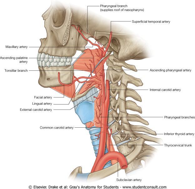
52
53 The Process of Swallowing (Deglutition) Masticated food is formed into a ball or bolus on the dorsum of the tongue and voluntarily pushed upward and backward against the undersurface of the hard palate This is brought about by the contraction of the styloglossus muscles on both sides, which pull the root of the tongue upward and backward The palatoglossus muscles then squeeze the bolus backward into the pharynx.
54 The Process of Swallowing (Deglutition) From this point onward the process of swallowing becomes an involuntary act. The nasal part of the pharynx is now shut off from the oral part of the pharynx by the elevation of the soft palate the pulling forward of the posterior wall of the pharynx by the upper fibers of the superior constrictor muscle, and the contraction of the palatopharyngeus muscles. This prevents the passage of food and drink into the nasal cavities
55 The larynx and the laryngeal part of the pharynx are pulled upward by the contraction of the stylopharyngeus, salpingopharyngeus, thyrohyoid, and palatopharyngeus muscles The main part of the larynx is thus elevated to the posterior surface of the epiglottis, and the entrance into the larynx is closed The laryngeal entrance is made smaller by the approximation of the aryepiglottic folds, and the arytenoid cartilages are pulled forward by the contraction of the aryepiglottic, oblique arytenoid, and thyroarytenoid muscles.
56 The bolus moves downward over the epiglottis, the closed entrance into the larynx, and reaches the lower part of the pharynx as the result of the successive contraction of the superior, middle, and inferior constrictor muscles Some of the food slides down the groove on either side of the entrance into the larynx, that is, down through the piriform fossae Finally, the lower part of the pharyngeal wall (the cricopharyngeus muscle) relaxes and the bolus enters the esophagus.
57 Palatine Tonsils The palatine tonsils are two masses of lymphoid tissue, each located in the depression on the lateral wall of the oral part of the pharynx between the palatoglossal and palatopharyngeal arches Each tonsil is covered by mucous membrane, and its free medial surface projects into the pharynx The surface is pitted by numerous small openings that lead into the tonsillar crypts. The tonsil is covered on its lateral surface by a fibrous capsule

58 Palatine Tonsils The capsule is separated from the superior constrictor muscle by loose areolar tissue and the external palatine vein descends from the soft palate in this tissue to join the pharyngeal venous plexus Lateral to the superior constrictor muscle lie the styloglossus muscle, the loop of the facial artery, and the internal carotid artery. The tonsil reaches its maximum size during early childhood, but after puberty it diminishes

59 The tonsillar branch of the facial artery. The veins pierce the superior constrictor muscle and join the external palatine, the pharyngeal, or the facial veins. Lymph Drainage of the Tonsil The upper deep cervical lymph nodes, just below and behind the angle of the mandible Blood Supply
 The pharyngeal tonsil in the roof of the nasopharynx forms the upper part,")
60 Waldeyer's Ring of Lymphoid Tissue The lymphoid tissue that surrounds the opening into the respiratory and digestive systems forms a ring The lateral part of the ring is formed by the palatine tonsils and tubal tonsils (lymphoid tissue around the opening of the auditory tube in the lateral wall of the nasopharynx) The pharyngeal tonsil in the roof of the nasopharynx forms the upper part, and the lingual tonsil on the posterior third of the tongue forms the lower part.
Subdivided into Vestibule & Oral cavity proper
 Extends from the lips to the oropharyngeal isthmus The oropharyngeal isthmus: Is the junction of mouth and pharynx. Is bounded: Above by the soft palate and the palatoglossal folds Below by the dorsum
Oral cavity : consist of two parts: the oral vestibule and the oral cavity proper. Oral vestibule : is slit like space between.
 Oral cavity Oral cavity : consist of two parts: the oral vestibule and the oral cavity proper Oral vestibule : is slit like space between the teeth, buccal gingiva, lips, and cheeks 1 Oral cavity Oral
-Ibrahim Al-Naser. -Dr Al- Muhtaseb. 1 P a g e
 -1 -Ibrahim Al-Naser - -Dr Al- Muhtaseb 1 P a g e The Digestive System The doctor started the lecture by talking about the class rules. The GI system is an organ system, it is divided into: The Alimentary
Dr.Ban I.S. head & neck anatomy 2 nd y. جامعة تكريت كلية طب االسنان املرحلة الثانية
 جامعة تكريت كلية طب االسنان التشريح مادة املرحلة الثانية أ.م.د. بان امساعيل صديق 6102-6102 1 The Palate The palate forms the roof of the mouth and the floor of the nasal cavity. It is divided into two
The Pharynx. Dr. Nabil Khouri MD. MSc, Ph.D
 The Pharynx Dr. Nabil Khouri MD. MSc, Ph.D Introduction The pharynx is the Musculo-fascial halfcylinder that links the oral and nasal cavities in the head to the larynx and esophagus in the neck Common
Anatomy 2. Parotid bed (V.imp): meaning that gland is sleeping on structures and they are:
: meaning that gland is sleeping on structures and they are:") Anatomy 2 Parotid Gland: "refer to previous sheet for extra details." Its pyramidal in shape, apex is toward pharynx. Its Medial surface is divided into Anterio-medial and posterio-medial and its posterio-medial
Anatomy 2 Parotid Gland: "refer to previous sheet for extra details." Its pyramidal in shape, apex is toward pharynx. Its Medial surface is divided into Anterio-medial and posterio-medial and its posterio-medial
Anatomy of Oral Cavity DR. MAAN AL-ABBASI
 Anatomy of Oral Cavity DR. MAAN AL-ABBASI By the end of this lecture you should be able to: 1. Differentiate different parts of the oral cavity 2. Describe the blood and nerve supply of mucosa and muscles
Prevertebral Region, Pharynx and Soft Palate
 Unit 20: Prevertebral Region, Pharynx and Soft Palate Dissection Instructions: Step1 Step 2 Step 1: Insert your fingers posterior to the sternocleidomastoid muscle, vagus nerve, internal jugular vein,
Today's lecture discuss : 1- the mouth. 5-the salivary glands
 Today's lecture discuss : 1- the mouth 3-the tongue 2-the teeth 4-the palates 5-the salivary glands ( u dnt have to refer to the slides, I've included everything in slides ( 1-27 ) except some figures.
Basic Anatomy and Physiology of the Lips and Oral Cavity. Dr. Faghih
 Basic Anatomy and Physiology of the Lips and Oral Cavity Dr. Faghih It is divided into seven specific subsites : 1. Lips 2. dentoalveolar ridges 3. oral tongue 4. retromolar trigone 5. floor of mouth 6.
Nose & Mouth OUTLINE. Nose. - Nasal Cavity & Its Walls. - Paranasal Sinuses. - Neurovascular Structures. Mouth. - Oral Cavity & Its Contents
 Dept. of Human Anatomy, Si Chuan University Zhou hongying eaglezhyxzy@163.com Nose & Mouth OUTLINE Nose - Nasal Cavity & Its Walls - Paranasal Sinuses - Neurovascular Structures Mouth - Oral Cavity & Its
THE INTERIOR OF THE PHARYNX. By Dr. Muhammad Imran Qureshi
 THE INTERIOR OF THE PHARYNX By Dr. Muhammad Imran Qureshi The Cavity The cavity of the pharynx is divided into: 1. The Nasal part (called Nasopharynx) 2. The Oral part (called the Oropharynx), 3. And the
- Reem Akiely. -Wardeh Al-Swalmeh. - Mohammad Al-Muhtaseb. 1 P a g e
 -2 - Reem Akiely -Wardeh Al-Swalmeh - Mohammad Al-Muhtaseb 1 P a g e The palate: * Hard palate * Soft palate the Uvula: is a muscular structure present In the midline of the soft palate (اللهاة) The Hard
Lec [8]: Mandibular nerve:
![Lec [8]: Mandibular nerve:](/thumbs/94/121295776.jpg "Lec [8]: Mandibular nerve:") Lec [8]: Mandibular nerve: The mandibular branch from the trigeminal ganglion lies in the middle cranial fossa lateral to the cavernous sinus. With the motor root of the trigeminal nerve [motor roots lies
Lec [8]: Mandibular nerve: The mandibular branch from the trigeminal ganglion lies in the middle cranial fossa lateral to the cavernous sinus. With the motor root of the trigeminal nerve [motor roots lies
ORAL CAVITY, ESOPHAGUS AND STOMACH
 ORAL CAVITY, ESOPHAGUS AND STOMACH 1 OBJECTIVES By the end of the lecture you should be able to: Describe the anatomy the oral cavity, (boundaries, parts, nerve supply). Describe the anatomy of the palate,
Infratemporal fossa: Tikrit University college of Dentistry Dr.Ban I.S. head & neck Anatomy 2 nd y.
 Infratemporal fossa: This is a space lying beneath the base of the skull between the lateral wall of the pharynx and the ramus of the mandible. It is also referred to as the parapharyngeal or lateral pharyngeal
بسم اهلل الرحمن الرحيم
 بسم اهلل الرحمن الرحيم Today we will talk about digestive system in the head & neck We have the mouth, teeth, tongue, palate & salivary glands all of these are included in this lecture *First we will start
Tikrit University collage of dentistry Dr.Ban I.S. head & neck anatomy 2 nd y. Lec [5] / Temporal fossa :
![Tikrit University collage of dentistry Dr.Ban I.S. head & neck anatomy 2 nd y. Lec [5] / Temporal fossa :](/thumbs/88/115294566.jpg "Tikrit University collage of dentistry Dr.Ban I.S. head & neck anatomy 2 nd y. Lec [5] / Temporal fossa :") Lec [5] / Temporal fossa : Borders of the Temporal Fossa: Superior: Superior temporal line. Inferior: gap between zygomatic arch and infratemporal crest of sphenoid bone. Anterior: Frontal process of the
Lec [5] / Temporal fossa : Borders of the Temporal Fossa: Superior: Superior temporal line. Inferior: gap between zygomatic arch and infratemporal crest of sphenoid bone. Anterior: Frontal process of the
Dr.Ban I.S. head & neck anatomy 2 nd y. جامعة تكريت كلية طب االسنان املرحلة الثانية أ.م.د. بان امساعيل صديق 6102/6102
 جامعة تكريت كلية طب االسنان التشريح مادة املرحلة الثانية أ.م.د. بان امساعيل صديق 6102/6102 Parotid region The part of the face in front of the ear and below the zygomatic arch is the parotid region. The
SCHOOL OF ANATOMICAL SCIENCES Mock Run Questions. 4 May 2012
 SCHOOL OF ANATOMICAL SCIENCES Mock Run Questions 4 May 2012 1. With regard to the muscles of the neck: a. the platysma muscle is supplied by the accessory nerve. b. the stylohyoid muscle is supplied by
Mohammad Mohtaseb. Nour Hussein. Faisal Nimri
 2 Mohammad Mohtaseb Nour Hussein Faisal Nimri Muscles of the tongue The tongue is a muscular organ and contains intrinsic and extrinsic muscles. The intrinsic muscle contains vertical, oblique, and transverse
Lips and labial mucosa
 Lips and labial mucosa External portion of the lips: the vermilion border and the skin Vermilion border : the exposed red portion of the lip, covered by mucous membrane, no mucous glands Boundary: the
PTERYGOPALATINE FOSSA
 PTERYGOPALATINE FOSSA Outline Anatomical Structure and Boundaries Foramina and Communications with other spaces and cavities Contents Pterygopalatine Ganglion Especial emphasis on certain arteries and
The Ear The ear consists of : 1-THE EXTERNAL EAR 2-THE MIDDLE EAR, OR TYMPANIC CAVITY 3-THE INTERNAL EAR, OR LABYRINTH 1-THE EXTERNAL EAR.
 The Ear The ear consists of : 1-THE EXTERNAL EAR 2-THE MIDDLE EAR, OR TYMPANIC CAVITY 3-THE INTERNAL EAR, OR LABYRINTH 1-THE EXTERNAL EAR Made of A-AURICLE B-EXTERNAL AUDITORY MEATUS A-AURICLE It consists
Temporal region. temporal & infratemporal fossae. Zhou Hong Ying Dept. of Anatomy
 Temporal region temporal & infratemporal fossae Zhou Hong Ying Dept. of Anatomy Temporal region is divided by zygomatic arch into temporal & infratemporal fossae. Temporal Fossa Infratemporal fossa Temporal
Oral Cavity and Pharynx. The Oral Cavity. The oral cavity is divided into two major portions: the vestibule and the cavum oris.
 11 Oral Cavity and Pharynx Persons who specialize in the care and treatment of the oral cavity have a great responsibility. The oral cavity participates actively in respiration, nutrition, and excretion
The PHARYNX. Dr. Nabil Khouri MD Ph.D
 The PHARYNX Dr. Nabil Khouri MD Ph.D PHARYNX Fibromuscular tube lined with mucous membrane extends from base of skull to lower border of cricoid cartilage (C-6). 12-14 cm long At the lower border of cricoid
Respiratory System. Cambridge University Press Concise Anatomy for Anaesthesia Andreas G. Erdmann Excerpt More information
 Respiratory System 1 The mouth DESCRIPTION The mouth extends from the lips (anterior) to the isthmus of the fauces (posterior). There are two sections: Vestibule slit-like cavity between the cheeks/lips
Upper Respiratory Tract
 Upper Respiratory Tract Lectures Objectives Describe the structure of nasal cavity including nasal septum. Describe the structure of lateral wall of nasal cavity including conchae and meatuses. Locate
Anatomy Sheet: Oral cavity Done by: rasha Rakan edited by: khansaa Mahmoud
 Anatomy Sheet: Oral cavity Done by: rasha Rakan edited by: khansaa Mahmoud The oral cavity has 2 parts: 1. Oral vestibule: outer part that consists of outside the teeth, between the teeth, the cheeks and
Veins of the Face and the Neck
 Veins of the Face and the Neck Facial Vein The facial vein is formed at the medial angle of the eye by the union of the supraorbital and supratrochlear veins. connected through the ophthalmic veins with
The Neck the lower margin of the mandible above the suprasternal notch and the upper border of the clavicle
 The Neck is the region of the body that lies between the lower margin of the mandible above and the suprasternal notch and the upper border of the clavicle below Nerves of the neck Cervical Plexus Is formed
Parotid Gland, Temporomandibular Joint and Infratemporal Fossa
 M1 - Anatomy Parotid Gland, Temporomandibular Joint and Infratemporal Fossa Jeff Dupree Sanger 9-057 jldupree@vcu.edu Parotid gland: wraps around the mandible positioned between the mandible and the sphenoid
Mohammad Hisham Al-Mohtaseb. Lina Mansour. Reyad Jabiri. 0 P a g e
 2 Mohammad Hisham Al-Mohtaseb Lina Mansour Reyad Jabiri 0 P a g e This is only correction for the last year sheet according to our record. If you already studied this sheet just read the yellow notes which
3-Deep fascia: is absent (except over the parotid gland & buccopharngeal fascia covering the buccinator muscle)
") The Face 1-Skin of the Face The skin of the face is: Elastic Vascular (bleed profusely however heal rapidly) Rich in sweat and sebaceous glands (can cause acne in adults) It is connected to the underlying
The Face 1-Skin of the Face The skin of the face is: Elastic Vascular (bleed profusely however heal rapidly) Rich in sweat and sebaceous glands (can cause acne in adults) It is connected to the underlying
Dr. Sami Zaqout Faculty of Medicine IUG
 Auricle External Ear External auditory meatus The Ear Middle Ear (Tympanic Cavity) Auditory ossicles Internal Ear (Labyrinth) Bony labyrinth Membranous labyrinth External Ear Auricle External auditory
Structure and Nerve Supply of The Larynx
 Kingdom of Bahrain Arabian Gulf University College of Medicine and Medical sciences Structure and Nerve Supply of The Larynx This presentation was originally prepared by: Dr. Kumar Notes were added by:
Larynx. Rudimentary. Behind the posterior surface : -stylopharyngeus - salpingopharyngeus -platopharyngeus
 Larynx The larynx is an organ that provides a protective sphincter at the inlet of the air passages and is responsible for voice production. It extends from C3-C6: *Posterior: the pharynx *Lateral: the
Face. Definition: The area between the two ears and from the chin to the eye brows. The muscles of the face
 Face Definition: The area between the two ears and from the chin to the eye brows. The muscles of the face The muscle of facial expression (include the muscle of the face and the scalp). All are derived
Dr.Noor Hashem Mohammad Lecture (5)
") Dr.Noor Hashem Mohammad Lecture (5) 2016-2017 If the mandible is discarded, the anterior part of this aspect of the skull is seen to be formed by the hard palate. The palatal processes of the maxillae
Dr.Noor Hashem Mohammad Lecture (5) 2016-2017 If the mandible is discarded, the anterior part of this aspect of the skull is seen to be formed by the hard palate. The palatal processes of the maxillae
ANTERIOR CERVICAL TRIANGLE (Fig. 2.1 )
") 2 Neck Anatomy ANTERIOR CERVICAL TRIANGLE (Fig. 2.1 ) The boundaries are: Lateral: sternocleidomastoid muscle Superior: inferior border of the mandible Medial: anterior midline of the neck This large triangle
2 Neck Anatomy ANTERIOR CERVICAL TRIANGLE (Fig. 2.1 ) The boundaries are: Lateral: sternocleidomastoid muscle Superior: inferior border of the mandible Medial: anterior midline of the neck This large triangle
Upper arch. 1Prosthodontics. Dr.Bassam Ali Al-Turaihi. Basic anatomy & & landmark of denture & mouth
 1Prosthodontics Lecture 2 Dr.Bassam Ali Al-Turaihi Basic anatomy & & landmark of denture & mouth Upper arch Palatine process of maxilla: it form the anterior three quarter of the hard palate. Horizontal
Temporal fossa Infratemporal fossa Pterygopalatine fossa Terminal branches of external carotid artery Pterygoid venous plexus
 Outline of content Temporal fossa Infratemporal fossa Pterygopalatine fossa Terminal branches of external carotid artery Pterygoid venous plexus Boundary Content Communication Mandibular division of trigeminal
Neck of Condylar. Process. Anterior Border of Ramus. Mandibular. Foramen. Posterior Border of Ramus Incisive Fossa.
 Learning Outcomes The Mandible Surface Anatomy Muscle Attachments The (FOM) Muscles of the FOM The Tongue Muscles of the Tongue The Submandibular Region Submandibular Gland Sublingual Gland Lingual The
Bisection of Head & Nasal Cavity 頭部對切以及鼻腔. 解剖學科馮琮涵副教授 分機
 Bisection of Head & Nasal Cavity 頭部對切以及鼻腔 解剖學科馮琮涵副教授 分機 3250 E-mail: thfong@tmu.edu.tw Outline: The structure of nose The concha and meatus in nasal cavity The openings of paranasal sinuses Canals, foramens
Lecture 07. Lymphatic's of Head & Neck. By: Dr Farooq Amanullah Khan PMC
 Lecture 07 Lymphatic's of Head & Neck By: Dr Farooq Amanullah Khan PMC Dated: 28.11.2017 Lymphatic Vessels Of the 800 lymph nodes in the human body, 300 are in the Head & neck region. The lymphatic vessels
3. The Jaw and Related Structures
 Overview and objectives of this dissection 3. The Jaw and Related Structures The goal of this dissection is to observe the muscles of jaw raising. You will also have the opportunity to observe several
Anatomic Relations Summary. Done by: Sohayyla Yasin Dababseh
 Anatomic Relations Summary Done by: Sohayyla Yasin Dababseh Anatomic Relations Lecture 1 Part-1 - The medial wall of the nose is the septum. - The vestibule lies directly inside the nostrils (Nares). -
Introduction. Key terms. Key terms. Key terms 10/12/2016 EQUINE UPPER DIGESTIVE SYSTEM. Learning Objectives for My Lectures. What are the functions?
 Learning Objectives for My Lectures Recognize the importance of the head. List the functions of the digestive system. EQUINE UPPER DIGESTIVE SYSTEM Dr. Fawzy Elnady Prof. of Anatomy and Embryology Cairo
Parotid Gland. Parotid Gland. Largest of 3 paired salivary glands (submandibular; sublingual) Ramus of Mandible. Medial pterygoid.
 Ramus of Mandible. Medial pterygoid.") Parotid region Parotid Gland Largest of 3 paired salivary glands (submandibular; sublingual) Ramus of Mandible Medial pterygoid Cross section of mandible Masseter D S SCM Parotid Gland Mastoid Process
Parotid region Parotid Gland Largest of 3 paired salivary glands (submandibular; sublingual) Ramus of Mandible Medial pterygoid Cross section of mandible Masseter D S SCM Parotid Gland Mastoid Process
Maxilla, ORBIT and infratemporal fossa. Neophytos C Demetriades MD, DDS, MSc Associate professor European University of Cyprus School of Medicine
 Maxilla, ORBIT and infratemporal fossa Neophytos C Demetriades MD, DDS, MSc Associate professor European University of Cyprus School of Medicine MAXILLA Superior, middle, and inferior meatus Frontal sinus
The Skull and Temporomandibular joint II Prof. Abdulameer Al-Nuaimi. E. mail:
 The Skull and Temporomandibular joint II Prof. Abdulameer Al-Nuaimi E-mail: a.al-nuaimi@sheffield.ac.uk E. mail: abdulameerh@yahoo.com Temporal fossa The temporal fossa is a depression on the temporal
Omran Saeed. Luma Taweel. Mohammad Almohtaseb. 1 P a g e
 2 Omran Saeed Luma Taweel Mohammad Almohtaseb 1 P a g e I didn t include all the photos in this sheet in order to keep it as small as possible so if you need more clarification please refer to slides In
APRIL
 APRIL - 2003 OCTOBER - 2003 February 2009 [KU 652] Sub. Code : 4131 FIRST B.D.S DEGREE EXAMINATION (Modified Regulations III) Paper I HUMAN ANATOMY, HISTOLOGY AND EMBRYOLOGY Time : Three hours
University of Palestine. Final Exam 1 st Semester 2014/2015 Total Grade: 60
 Question One: MCQ: 1- The coronal suture joins the a) frontal and parietal bones. b) left and right parietal bones. c) parietal and occipital bones. d) parietal, squamous temporal and greater wing of the
Dr.Ban I.S. head & neck anatomy 2 nd y جامعة تكريت كلية طب االسنان مادة التشريح املرحلة الثانية أ.م.د. بان امساعيل صديق 6102/6102
 جامعة تكريت كلية طب االسنان مادة التشريح املرحلة الثانية أ.م.د. بان امساعيل صديق 6102/6102 Pterygopalatine fossa: The pterygopalatine fossa is a cone-shaped depression, It is located between the maxilla,
Bony orbit Roof The orbital plate of the frontal bone Lateral wall: the zygomatic bone and the greater wing of the sphenoid
 Bony orbit Roof: Formed by: The orbital plate of the frontal bone, which separates the orbital cavity from the anterior cranial fossa and the frontal lobe of the cerebral hemisphere Lateral wall: Formed
Larynx - cartilaginous structure holding the vocal folds which protrude into airstream
 1! Larynx - cartilaginous structure holding the vocal folds which protrude into airstream 2! Flow increase - like thumb over garden hose Pressure drop - narrower space forces pressure drop due to speed
Gross Anatomy of the. TEMPORAL BONE, EXTERNAL EAR, and MIDDLE EAR
 Gross Anatomy of the TEMPORAL BONE, EXTERNAL EAR, and MIDDLE EAR M1 Gross and Developmental Anatomy 9:00 AM, December 11, 2008 Dr. Milton M. Sholley Professor of Anatomy and Neurobiology Assignment: Head
Structure Location Function
 Frontal Bone Cranium forms the forehead and roof of the orbits Occipital Bone Cranium forms posterior and inferior portions of the cranium Temporal Bone Cranium inferior to the parietal bone forms the
04 Development of the Face and Neck. Development of the Face Development of the neck
 04 Development of the Face and Neck Development of the Face Development of the neck Development of the face Overview of facial development The fourth week ~ the twelfth week of prenatal development Between
Cranial nerves.
 Cranial nerves eaglezhyxzy@163.com Key Points of Learning Name Components Passing through Peripheral distribution Central connection Function Cranial nerves Ⅰ olfactory Ⅱ optic Ⅲ occulomotor Ⅳ trochlear
Anatomy of the Trigeminal Nerve
 19 Anatomy of the Trigeminal Nerve.1 Introduction 0. The Central Part of the Trigeminal Nerve 1..1 Origin 1.. Trigeminal Nuclei.3 The Peripheral Part of the Trigeminal Nerve 4.3.1 Ophthalmic Nerve 4.3.
Bones Ethmoid bone Inferior nasal concha Lacrimal bone Maxilla Nasal bone Palatine bone Vomer Zygomatic bone Mandible
 splanchnocranium - Consists of part of skull that is derived from branchial arches - The facial bones are the bones of the anterior and lower human skull Bones Ethmoid bone Inferior nasal concha Lacrimal
Nose, Nasal cavity, Paranasal Sinuses & Pharynx
 Nose, Nasal cavity, Paranasal Sinuses & Pharynx Respiratory block-anatomy-lecture 2 Editing file Objectives At the end of the lecture, the students should be able to: Describe the boundaries of the nasal
Skull-2. Norma Basalis Interna Norma Basalis Externa. Dr. Heba Kalbouneh Associate Professor of Anatomy and Histology
 Skull-2 Norma Basalis Interna Norma Basalis Externa Dr. Heba Kalbouneh Associate Professor of Anatomy and Histology Norma basalis interna Base of the skull- superior view The interior of the base of the
Oral cavity landmarks
 By: Dr. Ahmed Rabah Oral cavity landmarks The knowledge of oral anatomy and physiology will help the operator and provides enough landmarks to act as positive guide during denture construction. This subject
Gross Anatomy of the. TEMPORAL BONE, EXTERNAL EAR, and MIDDLE EAR. Assignment: Head to Toe Temporomandibular Joint (TMJ)
") Gross Anatomy the TEMPORAL BONE, EXTERNAL EAR, and MIDDLE EAR M1 Gross and Developmental Anatomy 9:00 AM, December 11, 2008 Dr. Milton M. Sholley Pressor Anatomy and Neurobiology Assignment: Head to Toe
Gross Anatomy the TEMPORAL BONE, EXTERNAL EAR, and MIDDLE EAR M1 Gross and Developmental Anatomy 9:00 AM, December 11, 2008 Dr. Milton M. Sholley Pressor Anatomy and Neurobiology Assignment: Head to Toe
Anatomy and Physiology. Bones, Sutures, Teeth, Processes and Foramina of the Human Skull
 Anatomy and Physiology Chapter 6 DRO Bones, Sutures, Teeth, Processes and Foramina of the Human Skull Name: Period: Bones of the Human Skull Bones of the Cranium: Frontal bone: forms the forehead and the
Anatomy of the Pharynx and Oesophagus
 Anatomy of the Pharynx and Oesophagus EMBRYOLOGY Cephalocaudal and lateral folding result in the formation of an endodermally lined primitive gut. In its cephalic part this forms a blind ending tube, the
Dental Anatomy and Physiology for Clinical Dental Technicians. with Marnie Hayward
 Dental Anatomy and Physiology for Clinical Dental Technicians with Marnie Hayward Salivary glands Parotid Submandibular Sublingual Salivary glands position Parotid glands Lie below ear and behind angle
Anatomy: head and Neck (6 questions) 1. Prevertebral Flexor Musculature (lying in front of the vertebrae) include all, EXCEPT: Longus Colli.
 1. Prevertebral Flexor Musculature (lying in front of the vertebrae) include all, EXCEPT: Longus Colli.") Anatomy: head and Neck (6 questions) 1. Prevertebral Flexor Musculature (lying in front of the vertebrae) include all, EXCEPT: Longus Colli. Rectus Capitis Anterior. Rectus Capitis Lateralis. Rectus Capitis
Anatomy: head and Neck (6 questions) 1. Prevertebral Flexor Musculature (lying in front of the vertebrae) include all, EXCEPT: Longus Colli. Rectus Capitis Anterior. Rectus Capitis Lateralis. Rectus Capitis
human anatomy 2016 lecture fifteen Dr meethak ali ahmed neurosurgeon
 Cranial Nerves Organization of the Cranial Nerves The cranial nerves are named as follows: I. Olfactory II. Optic III. Oculomotor IV. Trochlear V. Trigeminal VI. Abducent VII. Facial VIII. Vestibulocochlear
REVIEW OF CLINICAL EMBRYOLOGY OF HEAD AND NECK
 REVIEW OF CLINICAL EMBRYOLOGY OF HEAD AND NECK OUTLINE - EMBRYOLOGY UNDERLYING CLINICAL CONDITIONS I. EARLY DEVELOPMENT OF FACE: CLEFT LIP, CLEFT PALATE, OBSTRUCTED NASOLACRIMAL DUCT II. BRANCHIAL ARCHES
The sebaceous glands (glands of Zeis) open directly into the eyelash follicles, ciliary glands (glands of Moll) are modified sweat glands that open
 open directly into the eyelash follicles, ciliary glands (glands of Moll) are modified sweat glands that open") The Orbital Region The orbits are a pair of bony cavities that contain the eyeballs; their associated muscles, nerves, vessels, and fat; and most of the lacrimal apparatus upper eyelid is larger and more
The Orbital Region The orbits are a pair of bony cavities that contain the eyeballs; their associated muscles, nerves, vessels, and fat; and most of the lacrimal apparatus upper eyelid is larger and more
*in general the blood supply of the nose comes from branches of the internal and external carotid arteries.
 In the previous lecture we talked about the anatomy of the nasal cavity, today we will talk about its blood supply, venous drainage, innervations, and finally about the paranasal sinuses. When we describe
HEAD & NECK ANATOMY - MCQ HEAD & NECK ANATOMY
 . ' HEAD & NECK ANATOMY I. Deep investing layer of cervical fascia splits to enclose: A. Sternocleidomastoid B. Trapezius C. Parotid gland D. Omohyoid 2. Regarding the prevertebral fascia, the following
Cranial Nerve VII - Facial Nerve. The facial nerve has 3 main components with distinct functions
 Cranial Nerve VII - Facial Nerve The facial nerve has 3 main components with distinct functions Somatic motor efferent Supplies the muscles of facial expression; posterior belly of digastric muscle; stylohyoid,
Tympanic Bulla Temporal Bone. Digastric Muscle. Masseter Muscle
 Superior view Hyoid Bone The hyoid bone does not articulate with any other bones. It is held in place by ligaments to the styloid process of the temporal bone and the thyroid cartilage of the larynx. It
Trigeminal Nerve Anatomy. Dr. Mohamed Rahil Ali
 Trigeminal Nerve Anatomy Dr. Mohamed Rahil Ali Trigeminal nerve Largest cranial nerve Mixed nerve Small motor root and large sensory root Motor root Nucleus of motor root present in the pons and medulla
MAXILLA, ORBIT & PTERYGOPALATINE FOSSA. Neophytos C Demetriades MD, DDS, MSc Associate professor European University of Cyprus School of Medicine
 MAXILLA, ORBIT & PTERYGOPALATINE FOSSA Neophytos C Demetriades MD, DDS, MSc Associate professor European University of Cyprus School of Medicine Maxilla MAXILLA Superior, middle, and inferior meatus Frontal
Trigeminal Nerve Worksheets, Distributions Page 1
 Trigeminal Nerve Worksheet #1 Distribution by Nerve Dr. Darren Hoffmann Dental Gross Anatomy, Spring 2013 We have drawn out each of the branches of CN V in lecture and you have an idea now for their basic
OPEN ACCESS ATLAS OF OTOLARYNGOLOGY, HEAD & NECK OPERATIVE SURGERY
 OPEN ACCESS ATLAS OF OTOLARYNGOLOGY, HEAD & NECK OPERATIVE SURGERY BUCCINATOR MYOMUCOSAL FLAP The Buccinator Myomucosal Flap is an axial flap, based on the facial and/or buccal arteries. It is a flexible
ORAL & PHARYNGEAL STRUCTURES
 ORAL & PHARYNGEAL STRUCTURES Pedro Amarante Andrade, PhD LCSC06 BIOSCIENCES FOR SPEECH AND LANGUAGE THERAPY 09/10/2017 1 Oral & pharyngeal structures Dentition THIS SESSION Tongue Taste & Sensation Tonsillar
Anatomy of the Airway
 Anatomy of the Airway Nagelhout, 5 th edition, Chapter 26 Morgan & Mikhail, 5 th edition, Chapter 23 Mary Karlet, CRNA, PhD Airway Anatomy The airway consists of the nose, pharynx, larynx, trachea, and
Muscles of mastication [part 1]
![Muscles of mastication [part 1]](/thumbs/76/73586850.jpg "Muscles of mastication [part 1]") Muscles of mastication [part 1] In this lecture well have the muscles of mastication, neuromuscular function, and its relationship to the occlusion morphology. The fourth determinant of occlusion is the
Muscles of mastication [part 1] In this lecture well have the muscles of mastication, neuromuscular function, and its relationship to the occlusion morphology. The fourth determinant of occlusion is the
Salivary Glands and Teeth. Dr. Nabil Khouri MD, Ph.D
 Salivary Glands and Teeth Dr. Nabil Khouri MD, Ph.D Anatomy and Histology of salivary glands Functions of Saliva Keeps the mouth moist Aids in swallowing Aids in speech Keeps the mouth and teeth clean
Dr. Sami Zaqout, IUG Medical School
 The skull The skull is composed of several separate bones united at immobile joints called sutures. Exceptions? Frontal bone Occipital bone Vault Cranium Sphenoid bone Zygomatic bones Base Ethmoid bone
Thyroid gland. importance. relations and connections. external laryngeal nerves. malformations.
 Thyroid gland 1. Recognize and understand the coverings of the thyroid gland and their clinical importance. 2. Recognize and understand the main parts of the thyroid gland and their locations, relations
University of Palestine. Midterm Exam 2013/2014 Total Grade:
 [ Course No: DNTS2208 Course Title: Head and Neck Anatomy Date: 17/11/1024 No. of Questions: (52) Time: 2hours Using Calculator (No) University of Palestine Midterm Exam 2013/2014 Total Grade: Instructor
Pharyngeal Apparatus. Pouches Endoderm Grooves Ectoderm Arch Neural Crest Somitomeres Aortic Arch - Vessel
 Pharyngeal Apparatus Pouches Endoderm Grooves Ectoderm Arch Neural Crest Somitomeres Aortic Arch - Vessel Segmental Organization Humans: Arch 1-4 prominent Arch 5 absent Arch 6 - transient First Arch Face
Oral Cavity, Soft Palate, Pharynx, and Larynx; Development of the Face and Palate
 Oral Cavity, Soft Palate, Pharynx, and Larynx; Development of the Face and Palate Think on this. The ability to eat and drink safely and efficiently is fundamental to our quality of life. The wide variety
Lungs a. d. b. c. e.
 Lungs d. e. Lungs Right superior lobe Right middle lobe Right inferior lobe d. Left superior lobe e. Left inferior lobe Sinuses d. Nasal Cavity & Sinuses g. g. i. Nasal Cavity & Sinuses g. h. d. f. e.
For the following questions, indicate the letter that corresponds to the SINGLE MOST APPROPRIATE ANSWER
 GROSS ANATOMY EXAMINATION May 15, 2000 For the following questions, indicate the letter that corresponds to the SINGLE MOST APPROPRIATE ANSWER 1. Pain associated with an infection limited to the middle
function - sensory & postganglionic sympathetic [communication from the internal carotid plexus in the cavernous sinus] innervation of the mucosa of
![function - sensory & postganglionic sympathetic [communication from the internal carotid plexus in the cavernous sinus] innervation of the mucosa of](/thumbs/74/71276096.jpg "function - sensory & postganglionic sympathetic [communication from the internal carotid plexus in the cavernous sinus] innervation of the mucosa of") Nerves I. Cranial nerves A. Olfactory (CN I) 1. Olfactory bulb 2. Olfactory tract B. Optic n. (CNII) function - carries visual sensory information from the neural retina to the diencephalon & midbrain
Nerves I. Cranial nerves A. Olfactory (CN I) 1. Olfactory bulb 2. Olfactory tract B. Optic n. (CNII) function - carries visual sensory information from the neural retina to the diencephalon & midbrain
Chapter 26: The temporomandibular joint, pharynx and larynx. The Temporomandibular Joint. Ligaments. (a) Capsular
 Capsular") Chapter 26: The temporomandibular joint, pharynx and larynx The Temporomandibular Joint This is a synovial joint of a condyloid (modified hinge) variety between the condyle of the mandible and the mandibular
Chapter 26: The temporomandibular joint, pharynx and larynx The Temporomandibular Joint This is a synovial joint of a condyloid (modified hinge) variety between the condyle of the mandible and the mandibular