Neck of Condylar. Process. Anterior Border of Ramus. Mandibular. Foramen. Posterior Border of Ramus Incisive Fossa.
|
|
|
- Sheila Griffith
- 5 years ago
- Views:
Transcription

 Foramen Coronoid Ramus of Mandible Lingula Mylohyoid Line Neck of Condylar Foramen Mandible Pterygoid Fovea")

 Mental Tubercle Inferior Border of Body Mental Foramen Mandible Ligaments Muscle Attachments The mental tubercle (a")


1 Learning Outcomes The Mandible Surface Anatomy Muscle Attachments The (FOM) Muscles of the FOM The Tongue Muscles of the Tongue The Submandibular Region Submandibular Gland Sublingual Gland Lingual The Head & Neck Parasympathetics Submandibular Region Mohammed A Al-Muharraqi MBChB, BDS, MSc, MRCS Glas, FFD RCS Irel, MFDS RCS Eng almuharraqi@doctors.org.uk Mandible Condylar Lingual (Genial) Foramen Coronoid Ramus of Mandible Lingula Mylohyoid Line Neck of Condylar Foramen Mandible Pterygoid Fovea Anterior Border of Ramus Alveolar Ridge Neck of Condylar Coronoid Oblique Line Head of Condylar Condylar Coronoid () Notch Angle of Mandible Alveolar Ridge Posterior Border of Ramus Incisive Fossa Ramus of Mandible Mylohyoid Groove Angle of Mandible Inferior Border of Body Submandibular Fossa Symphysis Menti (Median Ridge) Mental Protuberance Body of Mandible Sublingual Fossa Digastric Fossa Superior Mental Spine (Genial Tubercle) Inferior Mental Spine (Genial Tubercle) Mental Tubercle Inferior Border of Body Mental Foramen Mandible Ligaments Muscle Attachments The mental tubercle (a raised prominence at the mental symphysis) is a point of muscular attachment. The external surface of the ramus is covered by the attachment of the masseter muscle. On the inner surface of the body of the mandible, there is a horizontal mylohyoid line, which attaches the mylohyoid muscle. Above it, there is a shallow depression for the sublingual salivary gland and below it a deeper depression for the submandibular gland. At the anterior ends of the mylohyoid lines and superior to them, near the symphysis, there is the genial tubercles. Geniohyoid and Genioglossus attach here. The inner surface of the mandibular ramus is where the medial pterygoid muscle attaches. The sphenomandibular ligament attaches to the lingula (and superiorly to the sphenoid spine). The coronoid process is a triangular protuberance from the anterosuperior surface of the ramus. The temporalis muscle attaches here, predominantly on the deep surface. The condylar process is one of the important attachments for the lateral pterygoid muscle. 1


. The geniohyoid muscle (VII) elevates the hyoid bone and is a Thyrohyoid weak depressor of the mandible.")


2 Clavicle The hyoid bone is situated at the level of the third cervical vertebra and is suspended from the styloid processes of the temporal bones by the stylohyoid ligaments. The bone is horseshoe-shaped and consists of Inferior Border of Mandible a central body spanning the midline, with greater and lesser horns on each side. Muscles attached to the hyoid bone are: genio-glossus, genio-hyoid, hyoglossus, chondro-glossus, middle First Cervical Vertebra (Atlas) constrictor of the pharynx, mylohyoid, omo-hyoid, sterno-hyoid, stylo-hyoid, thyro-hyoid and the levator of the thyroid gland. Hyoid Bone Ligaments attached to the hyoid bone are: stylo-hyoid ligaments, Thyroid Cartilage thyro-hyoid membrane, hyoepiglottic ligament, lateral thyro- Cricoid Cartilage hyoid ligaments and the median thyro-hyoid ligament. Submandibular Ganglion Internal Jugular Accessory Hypoglossal The geniohyoid muscle originates from the inferior genial tubercle, found on the back of the symphysis menti of the mandible, the mental spine of the mandible. It passes backwards and slightly downwards Tongue to insert onto the anterior surface on the body of the hyoid bone s body. The innervation is shared with the thyrohyoid muscle, namely Artery the first cervical spinal nerve Sublingual Gland traveling with the hypoglossal Geniohyoid nerve. The blood supply is derived from the lingual artery (sublingual branch). The geniohyoid muscle (VII) elevates the hyoid bone and is a Thyrohyoid weak depressor of the mandible. Cricothyroid Common Carotid Thyroid Gland Genioglossus lies anteriorly near the median plane. It originates from the superior genial tubercle (mental spine) on the mandible. It fans out into the substance of the tongue. The superior fibers of the genioglossus muscle pass upwards and anteriorly towards the tip of the tongue. Genioglossus Transverse & Vertical receives its motor innervation from the hypoglossal nerve. The lingual Genioglossus artery and the submental branch of Styloglossus the facial artery supply this muscle. Geniohyoid The genioglossus muscle is a protractor and depressor of the Median Thyroid tongue. Ligament Styloglossus arises from the tip of the Thyrohyoid styloid process and the stylomandibular ligament. The Cricothyroid longitudinal & oblique parts of the muscle blend with the inferior longitudinal muscle of the tongue. It continues forwards towards the tip of the tongue. It is supplied by the hypoglossal nerve. The sublingual branch of the lingual artery supplies the muscle. Styloglossus retracts and elevates the tongue. Mylohyoid Tongue Levator of the Thyroid Gland Artery Anterior Belly of Digastric Internal Jugular (VII) Superior Belly of Omohyoid Sternohyoid Accessory Sternothyroid Inferior Belly of Omohyoid The digastric muscle consists of an anterior belly and a posterior belly connected by an intermediate tendon. The anterior belly is attached to the digastric fossa on the inferior border of the mandible. It runs downwards and backwards to the digastric tendon. It develops from the first branchial arch and so receives its motor supply from the mandibular division of the trigeminal nerve. It receives its blood supply from the submental branch of the facial artery. The posterior belly arises from the mastoid (digastric) notch behind the mastoid process. The posterior belly passes to the hyoid bone where it becomes the intermediate digastric tendon. The posterior belly of the digastric develops from the second branchial; hence its innervation is derived from the the facial nerve. Its arterial blood supply is from the posterior auricular and occipital arteries. The digastric muscle is involved in maintaining the stability of the hyoid bone and helps to raise the hyoid bone and the base of the tongue, but its prime function is to assist in depressing and retracting the mandible. Mylohyoid Levator of the Thyroid Gland Internal Jugular Accessory The mylohyoid muscle arises from the mylohyoid line of the mandible. The anterior fibers of the mylohyoid muscle interdigitate with the corresponding fibers on the Tongue opposite side to form a median raphe. This raphe is attached above to the symphysis menti of the mandible and below to the anterior surface of the hyoid bone. The Artery mylohyoid branch of the Sublingual Gland mandibular division of the Submandibular trigeminal nerve supplies the Gland muscle. The mylohyoid muscle (VII) receives its arterial supply from the sublingual branch of the lingual Thyrohyoid artery, the maxillary artery, the mylohyoid branch of the inferior Cricothyroid alveolar artery, and the submental Common Carotid branch of the facial artery. It raises Sternothyroid the floor of the mouth during the Thyroid Gland first stages of swallowing and helps to depress the mandible when the hyoid bone is fixed. Conversely, it aids in elevation of the hyoid bone. 2


, draining to the internal")


3 Supply to the Tongue The Tongue Lingual Artery The lingual artery is the main blood supply of the tongue, arising from the external carotid artery and coursing deep to hyoglossus. It branches extensively within the tongue. When resecting the tongue haemorrhage is more marked in the posterior region. Hence laser surgery or diathermy techniques are preferred. Venous drainage is by the lingual vein (superficial to hyoglossus), draining to the internal jugular vein. The lingual veins may be seen easily on the underside of the tongue, running just beneath the mucosa. These veins tend to become more prominent with age. Hypoglossal and Muscles of the Tongue Brainstem: Pons Cerebellum Brainstem: Medulla Hypoglossal Oblongata Root Cervical Ventral Root Superior Longitudinal Transverse & Vertical Styloid Inferior Longitudinal Genioglossus Spinal Accessory Styloglossus Symphysis Menti (Median Ridge) Hyoglossus *Hypoglossal (XII) Hyoid Bone Geniohyoid Hyoglossus The hyoglossus is a thin, quadrilateral muscle, which provides an important landmark in the floor of the mouth. It originates from the superior border of the greater horn of the hyoid bone. A part of the muscle is attached to the base of the lesser horn of the hyoid bone and has been called chondroglossus. At its origin, the hyoglossus muscle is separated from the attachment of the middle constrictor muscle of the pharynx by the lingual artery. It passes vertically upwards to insert into the side of the tongue. The hypoglossal nerve supplies the hyoglossus muscle. The sublingual branch of the lingual artery and the submental branch of the facial artery supply hyoglossus. Hyoglossus depresses the tongue. The submandibular duct and lingual nerve crosses hyoglossus as it forms the floor of the submandibular fossa. It is a key landmark in submandibular gland surgery. 3
 Hyoid Bone Lingual Artery Hypoglossal (XII) Lingual Mylohyoid (CNV)")
 Stylopharyngeus")
 Tongue Hypoglossal (XII) Palatoglossus Pharyngeal")
 expression & buccinator (VII) Levator palpebrae superioris")
 Abducent (VI) Strap group Ansa cervicalis Thyrohyoid C1 fibres on")
 appear.")
 may appear in patients who have little lip")


4 Hyoglossus Muscles of the Tongue Intrinsic Lingual Muscles Genioglossus Symphysis Menti (Median Ridge) Hyoid Bone Lingual Artery Hypoglossal (XII) Lingual Mylohyoid (CNV) Geniohyoid (C1 on XII) Hyoid Bone All Muscles of: Pharynx Are supplied by: Except: Which is/are supplied instead by: Pharyngeal plexus (IX, X, symp) Stylopharyngeus Cricopharyngeus Glossopharyngeal (IX) Recurrent laryngeal Palate Pharyngeal plexus Tensor palati Off n to med pterygoid (Vc) Tongue Hypoglossal (XII) Palatoglossus Pharyngeal plexus Mastication (Vc) Buccinator (VII) Larynx Recurrent laryngeal (X) Cricothyroid Ext br of superior laryngeal (X) expression & buccinator (VII) Levator palpebrae superioris Oculomotor (III) & sympathetic Eye Oculomotor (III) Superior oblique Lateral rectus Trochlear (IV) Abducent (VI) Strap group Ansa cervicalis Thyrohyoid C1 fibres on hypoglossal Sublingual Gland Sublingual Gland When saliva is retained or is extravasated into the adjacent tissues, mucocoeles (salivary cysts) appear. In the floor of the mouth they are called a ranula because they look like the belly of a frog. Excessive salivation (sialorrhoea) may appear in patients who have little lip control such as stroke patients or by parasympathetic stimulation. Conversely, sympathetic stimulation such as in the fight or flight response or by sympatheticomimetic drugs such as hyosine (used in motion sickness) causes a dry mouth. The paired sublingual glands are about twice the size of an almond. They are mainly mucous and have over a dozen ducts, half of which open into the submandibular gland and half of which open into the sublingual papilla directly. It lies in front of the anterior border of hyoglossus, between mylohyoid and genioglossus and makes a superficial depression in the mandible. 4


 duct is 4 to 5 cm long.")
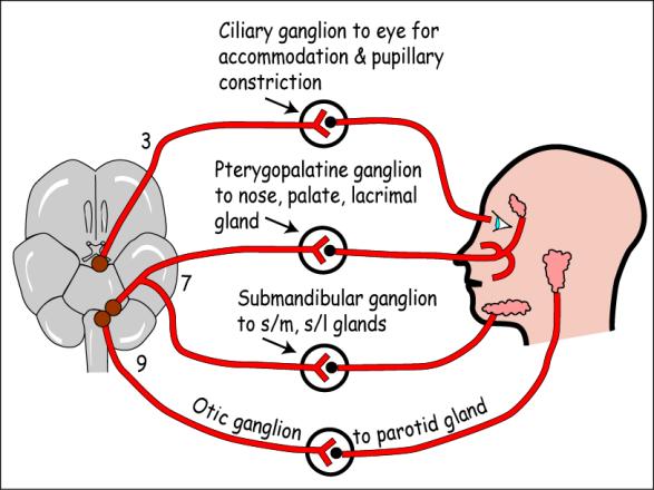
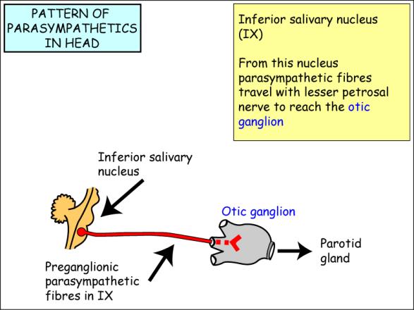
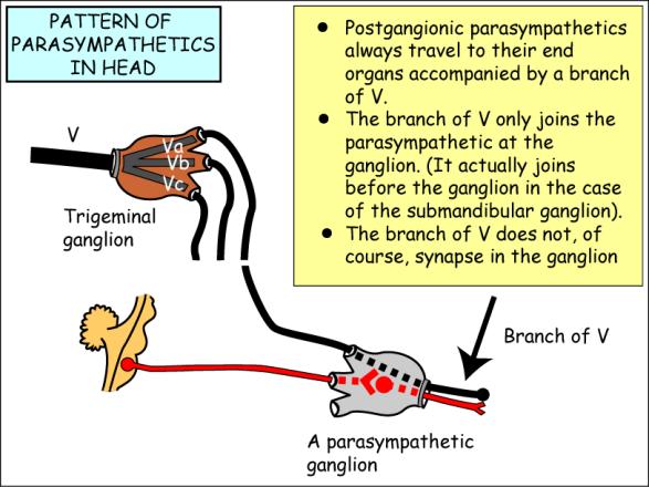
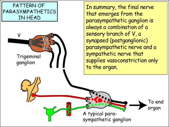
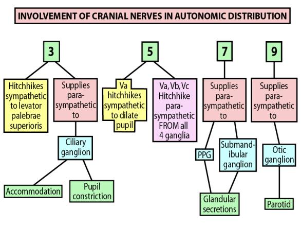
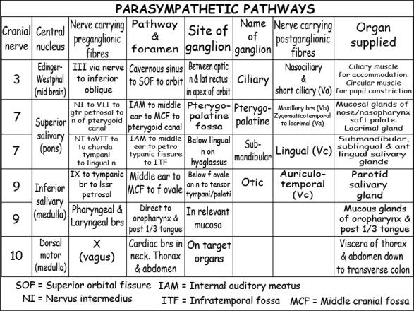
5 Submandibular Gland The submandibular gland is subject to all the diseases of the salivary glands including stones that form by precipitation on epithelial debris. They are not always calcific and so do not always appear on radiographs. However, sialography, when a dye is introduced into the gland, will reveal any blockage. The incision for removal of the gland must also be at least one finger's breath inferior to the lower border of the mandible to avoid damage to the mandibular branch of the facial nerve. Having incised platysma, the facial veins are the next obstacle. They are usually ligated and cut. Occasionally the facial artery requires ligation. It is important to tie off both ends securely as the artery can bleed from both points due to multiple anastomoses along its length. Finally care must be taken to identify the duct correctly and avoid damage to the lingual nerve. It is extremely important to identify the duct correctly before cutting it, because severing the lingual nerve will cause parasthesia of the anterior two thirds of the tongue which may not fully recover. Submandibular Gland The submandibular gland is irregularly shaped and about 2 x 3 x 3 cm in size. The gland consists of a larger superficial part and a smaller deep part. The two are continuous as they wrap around the posterior free edge of the mylohyoid muscle. The submandibular gland extends anteriorly as far as the anterior belly of the digastric and posteriorly as far as the stylomandibular ligament. Laterally, it lies within the submandibular fossa on the medial surface of the mandible. The submandibular (Wharton's) duct is 4 to 5 cm long. It leaves the deep part of the gland, runs anteriorly on the surface of hyoglossus (and deep to the mylohyoid), and terminates at the sublingual papilla in the floor of the mouth. The submandibular duct is accompanied by the lingual nerve the external surface of hyoglossus. The lingual nerve is crossed by the duct superiorly on its way into the hyoglossus, hence the rhyme: The lingual nerve takes a swerve, Around the hyoglossus, Says Wharton's duct to the nerve, "I think I'm going to cross it!" The Parasympathetic Ganglia in the Head 5
6 6
7 End 7
Oral cavity : consist of two parts: the oral vestibule and the oral cavity proper. Oral vestibule : is slit like space between.
 Oral cavity Oral cavity : consist of two parts: the oral vestibule and the oral cavity proper Oral vestibule : is slit like space between the teeth, buccal gingiva, lips, and cheeks 1 Oral cavity Oral
SCHOOL OF ANATOMICAL SCIENCES Mock Run Questions. 4 May 2012
 SCHOOL OF ANATOMICAL SCIENCES Mock Run Questions 4 May 2012 1. With regard to the muscles of the neck: a. the platysma muscle is supplied by the accessory nerve. b. the stylohyoid muscle is supplied by
The Neck the lower margin of the mandible above the suprasternal notch and the upper border of the clavicle
 The Neck is the region of the body that lies between the lower margin of the mandible above and the suprasternal notch and the upper border of the clavicle below Nerves of the neck Cervical Plexus Is formed
Learning Outcomes. The Carotid 20/02/2013. Scalp, Face, Parotid. Layers of the Scalp. The Parotid Gland. The Scalp. The Carotid The Facial Artery
 Learning Outcomes The Scalp Layers of the Scalp Bleeding from the Scalp The Carotid The Facial Artery Major Muscles of the Face and Jaw(s) Muscles of Mastication Muscles of Facial Expression The Parotid
ANTERIOR CERVICAL TRIANGLE (Fig. 2.1 )
") 2 Neck Anatomy ANTERIOR CERVICAL TRIANGLE (Fig. 2.1 ) The boundaries are: Lateral: sternocleidomastoid muscle Superior: inferior border of the mandible Medial: anterior midline of the neck This large triangle
2 Neck Anatomy ANTERIOR CERVICAL TRIANGLE (Fig. 2.1 ) The boundaries are: Lateral: sternocleidomastoid muscle Superior: inferior border of the mandible Medial: anterior midline of the neck This large triangle
Anterior triangle of neck
 Anterior triangle of neck Dept. of Anatomy Zhou Hong Ying Outline boundary and subdivisions of ant. triangle contents of the triangle Muscles: suprahyoid muscles, infrahyoid muscles Nerves: CNⅩ, CNⅪ, CNⅫ,
Larynx - cartilaginous structure holding the vocal folds which protrude into airstream
 1! Larynx - cartilaginous structure holding the vocal folds which protrude into airstream 2! Flow increase - like thumb over garden hose Pressure drop - narrower space forces pressure drop due to speed
Tikrit University collage of dentistry Dr.Ban I.S. head & neck anatomy 2 nd y. Lec [5] / Temporal fossa :
![Tikrit University collage of dentistry Dr.Ban I.S. head & neck anatomy 2 nd y. Lec [5] / Temporal fossa :](/thumbs/88/115294566.jpg "Tikrit University collage of dentistry Dr.Ban I.S. head & neck anatomy 2 nd y. Lec [5] / Temporal fossa :") Lec [5] / Temporal fossa : Borders of the Temporal Fossa: Superior: Superior temporal line. Inferior: gap between zygomatic arch and infratemporal crest of sphenoid bone. Anterior: Frontal process of the
Lec [5] / Temporal fossa : Borders of the Temporal Fossa: Superior: Superior temporal line. Inferior: gap between zygomatic arch and infratemporal crest of sphenoid bone. Anterior: Frontal process of the
The Pharynx. Dr. Nabil Khouri MD. MSc, Ph.D
 The Pharynx Dr. Nabil Khouri MD. MSc, Ph.D Introduction The pharynx is the Musculo-fascial halfcylinder that links the oral and nasal cavities in the head to the larynx and esophagus in the neck Common
Lec [8]: Mandibular nerve:
![Lec [8]: Mandibular nerve:](/thumbs/94/121295776.jpg "Lec [8]: Mandibular nerve:") Lec [8]: Mandibular nerve: The mandibular branch from the trigeminal ganglion lies in the middle cranial fossa lateral to the cavernous sinus. With the motor root of the trigeminal nerve [motor roots lies
Lec [8]: Mandibular nerve: The mandibular branch from the trigeminal ganglion lies in the middle cranial fossa lateral to the cavernous sinus. With the motor root of the trigeminal nerve [motor roots lies
Subdivided into Vestibule & Oral cavity proper
 Extends from the lips to the oropharyngeal isthmus The oropharyngeal isthmus: Is the junction of mouth and pharynx. Is bounded: Above by the soft palate and the palatoglossal folds Below by the dorsum
Dr.Ban I.S. head & neck anatomy 2 nd y. جامعة تكريت كلية طب االسنان املرحلة الثانية أ.م.د. بان امساعيل صديق 6102/6102
 جامعة تكريت كلية طب االسنان التشريح مادة املرحلة الثانية أ.م.د. بان امساعيل صديق 6102/6102 Parotid region The part of the face in front of the ear and below the zygomatic arch is the parotid region. The
Prevertebral Region, Pharynx and Soft Palate
 Unit 20: Prevertebral Region, Pharynx and Soft Palate Dissection Instructions: Step1 Step 2 Step 1: Insert your fingers posterior to the sternocleidomastoid muscle, vagus nerve, internal jugular vein,
Bones Ethmoid bone Inferior nasal concha Lacrimal bone Maxilla Nasal bone Palatine bone Vomer Zygomatic bone Mandible
 splanchnocranium - Consists of part of skull that is derived from branchial arches - The facial bones are the bones of the anterior and lower human skull Bones Ethmoid bone Inferior nasal concha Lacrimal
Anatomy: head and Neck (6 questions) 1. Prevertebral Flexor Musculature (lying in front of the vertebrae) include all, EXCEPT: Longus Colli.
 1. Prevertebral Flexor Musculature (lying in front of the vertebrae) include all, EXCEPT: Longus Colli.") Anatomy: head and Neck (6 questions) 1. Prevertebral Flexor Musculature (lying in front of the vertebrae) include all, EXCEPT: Longus Colli. Rectus Capitis Anterior. Rectus Capitis Lateralis. Rectus Capitis
Anatomy: head and Neck (6 questions) 1. Prevertebral Flexor Musculature (lying in front of the vertebrae) include all, EXCEPT: Longus Colli. Rectus Capitis Anterior. Rectus Capitis Lateralis. Rectus Capitis
Parotid Gland, Temporomandibular Joint and Infratemporal Fossa
 M1 - Anatomy Parotid Gland, Temporomandibular Joint and Infratemporal Fossa Jeff Dupree Sanger 9-057 jldupree@vcu.edu Parotid gland: wraps around the mandible positioned between the mandible and the sphenoid
Infratemporal fossa: Tikrit University college of Dentistry Dr.Ban I.S. head & neck Anatomy 2 nd y.
 Infratemporal fossa: This is a space lying beneath the base of the skull between the lateral wall of the pharynx and the ramus of the mandible. It is also referred to as the parapharyngeal or lateral pharyngeal
Dr.Ban I.S. head & neck anatomy 2 nd y. جامعة تكريت كلية طب االسنان املرحلة الثانية
 جامعة تكريت كلية طب االسنان التشريح مادة املرحلة الثانية أ.م.د. بان امساعيل صديق 6102-6102 1 The Palate The palate forms the roof of the mouth and the floor of the nasal cavity. It is divided into two
Alexander C Vlantis. Selective Neck Dissection 33
 05 Modified Radical Neck Dissection Type II Alexander C Vlantis Selective Neck Dissection 33 Modified Radical Neck Dissection Type II INCISION Various incisions can be used for a neck dissection. The incision
Surgical Anatomy of the Neck. M. J. Jurkiewicz, John Bostwick. Surgical Clinics of North America, Vol 54, No 6, December 1974.
 Surgical Anatomy of the Neck M. J. Jurkiewicz, John Bostwick Surgical Clinics of North America, Vol 54, No 6, December 1974. The radical neck dissection is a safe, effective therapeutic procedure for eradication
The Skull and Temporomandibular joint II Prof. Abdulameer Al-Nuaimi. E. mail:
 The Skull and Temporomandibular joint II Prof. Abdulameer Al-Nuaimi E-mail: a.al-nuaimi@sheffield.ac.uk E. mail: abdulameerh@yahoo.com Temporal fossa The temporal fossa is a depression on the temporal
Anatomy of Oral Cavity DR. MAAN AL-ABBASI
 Anatomy of Oral Cavity DR. MAAN AL-ABBASI By the end of this lecture you should be able to: 1. Differentiate different parts of the oral cavity 2. Describe the blood and nerve supply of mucosa and muscles
University of Palestine. Midterm Exam 2013/2014 Total Grade:
 Course No: DNTS2208 Course Title: Head and Neck Anatomy Date: 09/11/2013 No. of Questions: (50) Time: 1hour Using Calculator (No) University of Palestine Midterm Exam 2013/2014 Total Grade: Instructor
Neck-2. Dr. Heba Kalbouneh Associate Professor of Anatomy and Histology
 Neck-2 ` Dr. Heba Kalbouneh Associate Professor of Anatomy and Histology Triangles of the neck Side of the neck Midline Lower border of mandible Line between angle of mandible and mastoid Superior nuchal
Veins of the Face and the Neck
 Veins of the Face and the Neck Facial Vein The facial vein is formed at the medial angle of the eye by the union of the supraorbital and supratrochlear veins. connected through the ophthalmic veins with
Temporal region. temporal & infratemporal fossae. Zhou Hong Ying Dept. of Anatomy
 Temporal region temporal & infratemporal fossae Zhou Hong Ying Dept. of Anatomy Temporal region is divided by zygomatic arch into temporal & infratemporal fossae. Temporal Fossa Infratemporal fossa Temporal
University of Palestine. Midterm Exam 2013/2014 Total Grade:
 [ Course No: DNTS2208 Course Title: Head and Neck Anatomy Date: 17/11/1024 No. of Questions: (52) Time: 2hours Using Calculator (No) University of Palestine Midterm Exam 2013/2014 Total Grade: Instructor
Dr. Sami Zaqout Faculty of Medicine IUG
 Auricle External Ear External auditory meatus The Ear Middle Ear (Tympanic Cavity) Auditory ossicles Internal Ear (Labyrinth) Bony labyrinth Membranous labyrinth External Ear Auricle External auditory
3. The Jaw and Related Structures
 Overview and objectives of this dissection 3. The Jaw and Related Structures The goal of this dissection is to observe the muscles of jaw raising. You will also have the opportunity to observe several
human anatomy 2016 lecture fifteen Dr meethak ali ahmed neurosurgeon
 Cranial Nerves Organization of the Cranial Nerves The cranial nerves are named as follows: I. Olfactory II. Optic III. Oculomotor IV. Trochlear V. Trigeminal VI. Abducent VII. Facial VIII. Vestibulocochlear
Anatomy 2. Parotid bed (V.imp): meaning that gland is sleeping on structures and they are:
: meaning that gland is sleeping on structures and they are:") Anatomy 2 Parotid Gland: "refer to previous sheet for extra details." Its pyramidal in shape, apex is toward pharynx. Its Medial surface is divided into Anterio-medial and posterio-medial and its posterio-medial
Anatomy 2 Parotid Gland: "refer to previous sheet for extra details." Its pyramidal in shape, apex is toward pharynx. Its Medial surface is divided into Anterio-medial and posterio-medial and its posterio-medial
Tympanic Bulla Temporal Bone. Digastric Muscle. Masseter Muscle
 Superior view Hyoid Bone The hyoid bone does not articulate with any other bones. It is held in place by ligaments to the styloid process of the temporal bone and the thyroid cartilage of the larynx. It
The Neck. BY: Lina Abdullah & Rahaf Jreisat
 The Neck BY: Lina Abdullah & Rahaf Jreisat Boundaries of the Neck: generally from base of the skull to root of the neck Superior margin :From superior nuchal line of occipital bone up to mastoid process
function - sensory & postganglionic sympathetic [communication from the internal carotid plexus in the cavernous sinus] innervation of the mucosa of
![function - sensory & postganglionic sympathetic [communication from the internal carotid plexus in the cavernous sinus] innervation of the mucosa of](/thumbs/74/71276096.jpg "function - sensory & postganglionic sympathetic [communication from the internal carotid plexus in the cavernous sinus] innervation of the mucosa of") Nerves I. Cranial nerves A. Olfactory (CN I) 1. Olfactory bulb 2. Olfactory tract B. Optic n. (CNII) function - carries visual sensory information from the neural retina to the diencephalon & midbrain
Nerves I. Cranial nerves A. Olfactory (CN I) 1. Olfactory bulb 2. Olfactory tract B. Optic n. (CNII) function - carries visual sensory information from the neural retina to the diencephalon & midbrain
Basic Anatomy and Physiology of the Lips and Oral Cavity. Dr. Faghih
 Basic Anatomy and Physiology of the Lips and Oral Cavity Dr. Faghih It is divided into seven specific subsites : 1. Lips 2. dentoalveolar ridges 3. oral tongue 4. retromolar trigone 5. floor of mouth 6.
For the following questions, indicate the letter that corresponds to the SINGLE MOST APPROPRIATE ANSWER
 GROSS ANATOMY EXAMINATION May 15, 2000 For the following questions, indicate the letter that corresponds to the SINGLE MOST APPROPRIATE ANSWER 1. Pain associated with an infection limited to the middle
Larynx. Rudimentary. Behind the posterior surface : -stylopharyngeus - salpingopharyngeus -platopharyngeus
 Larynx The larynx is an organ that provides a protective sphincter at the inlet of the air passages and is responsible for voice production. It extends from C3-C6: *Posterior: the pharynx *Lateral: the
Mohammad Mohtaseb. Nour Hussein. Faisal Nimri
 2 Mohammad Mohtaseb Nour Hussein Faisal Nimri Muscles of the tongue The tongue is a muscular organ and contains intrinsic and extrinsic muscles. The intrinsic muscle contains vertical, oblique, and transverse
-Ibrahim Al-Naser. -Dr Al- Muhtaseb. 1 P a g e
 -1 -Ibrahim Al-Naser - -Dr Al- Muhtaseb 1 P a g e The Digestive System The doctor started the lecture by talking about the class rules. The GI system is an organ system, it is divided into: The Alimentary
Biology 218 Human Anatomy. Adapted from Martini Human Anatomy 7th ed. Chapter 10 The Muscular System Axial Musculature
 Adapted from Martini Human Anatomy 7th ed. Chapter 10 The Muscular System Axial Musculature Introduction The skeletal muscle of the body can be subdivided into: Axial musculature Muscles that position
Temporal fossa Infratemporal fossa Pterygopalatine fossa Terminal branches of external carotid artery Pterygoid venous plexus
 Outline of content Temporal fossa Infratemporal fossa Pterygopalatine fossa Terminal branches of external carotid artery Pterygoid venous plexus Boundary Content Communication Mandibular division of trigeminal
The Digestive System in the Head and Neck
 The Digestive System in the Head and Neck The Mouth The Lips The lips are two fleshy folds that surround the oral orifice They are covered on the outside by skin and are lined on the inside by mucous membrane
Upper arch. 1Prosthodontics. Dr.Bassam Ali Al-Turaihi. Basic anatomy & & landmark of denture & mouth
 1Prosthodontics Lecture 2 Dr.Bassam Ali Al-Turaihi Basic anatomy & & landmark of denture & mouth Upper arch Palatine process of maxilla: it form the anterior three quarter of the hard palate. Horizontal
Parotid Gland. Parotid Gland. Largest of 3 paired salivary glands (submandibular; sublingual) Ramus of Mandible. Medial pterygoid.
 Ramus of Mandible. Medial pterygoid.") Parotid region Parotid Gland Largest of 3 paired salivary glands (submandibular; sublingual) Ramus of Mandible Medial pterygoid Cross section of mandible Masseter D S SCM Parotid Gland Mastoid Process
Parotid region Parotid Gland Largest of 3 paired salivary glands (submandibular; sublingual) Ramus of Mandible Medial pterygoid Cross section of mandible Masseter D S SCM Parotid Gland Mastoid Process
Posterior Triangle of the Neck By Prof. Dr. Muhammad Imran Qureshi
 Posterior Triangle of the Neck By Prof. Dr. Muhammad Imran Qureshi For the purpose of anatomical description the neck is sub divided into two major triangles, the Anterior and the Posterior by muscle bellies
PTERYGOPALATINE FOSSA
 PTERYGOPALATINE FOSSA Outline Anatomical Structure and Boundaries Foramina and Communications with other spaces and cavities Contents Pterygopalatine Ganglion Especial emphasis on certain arteries and
Spinal nerves and cervical plexus Prof. Abdulameer Al Nuaimi. E mail: a.al E. mail:
 Spinal nerves and cervical plexus Prof. Abdulameer Al Nuaimi E mail: a.al nuaimi@sheffield.ac.uk E. mail: abdulameerh@yahoo.com Branches of ophthalmic artery Muscles of face A spinal nerve Spinal
THYROID & PARATHYROID. By Prof. Saeed Abuel Makarem & Dr. Sanaa Al-Sharawy
 THYROID & PARATHYROID By Prof. Saeed Abuel Makarem & Dr. Sanaa Al-Sharawy 1 OBJECTIVES By the end of the lecture, the student should be able to: Describe the shape, position, relations and structure of
Nose & Mouth OUTLINE. Nose. - Nasal Cavity & Its Walls. - Paranasal Sinuses. - Neurovascular Structures. Mouth. - Oral Cavity & Its Contents
 Dept. of Human Anatomy, Si Chuan University Zhou hongying eaglezhyxzy@163.com Nose & Mouth OUTLINE Nose - Nasal Cavity & Its Walls - Paranasal Sinuses - Neurovascular Structures Mouth - Oral Cavity & Its
3-Deep fascia: is absent (except over the parotid gland & buccopharngeal fascia covering the buccinator muscle)
") The Face 1-Skin of the Face The skin of the face is: Elastic Vascular (bleed profusely however heal rapidly) Rich in sweat and sebaceous glands (can cause acne in adults) It is connected to the underlying
The Face 1-Skin of the Face The skin of the face is: Elastic Vascular (bleed profusely however heal rapidly) Rich in sweat and sebaceous glands (can cause acne in adults) It is connected to the underlying
HEAD & NECK ANATOMY - MCQ HEAD & NECK ANATOMY
 . ' HEAD & NECK ANATOMY I. Deep investing layer of cervical fascia splits to enclose: A. Sternocleidomastoid B. Trapezius C. Parotid gland D. Omohyoid 2. Regarding the prevertebral fascia, the following
HEAD & NECK BY NUMBERS THIRD EDITION Copyright 2013, Anatomy Numbers (Phoenix, AZ) All rights reserved
 All rights reserved") HEAD & NECK BY NUMBERS THIRD EDITION Copyright 2013, Anatomy Numbers (Phoenix, AZ) All rights reserved No part of this publication may be reproduced or transmitted in any form or by any means, electronic
HEAD & NECK BY NUMBERS THIRD EDITION Copyright 2013, Anatomy Numbers (Phoenix, AZ) All rights reserved No part of this publication may be reproduced or transmitted in any form or by any means, electronic
Muscles of mastication [part 1]
![Muscles of mastication [part 1]](/thumbs/76/73586850.jpg "Muscles of mastication [part 1]") Muscles of mastication [part 1] In this lecture well have the muscles of mastication, neuromuscular function, and its relationship to the occlusion morphology. The fourth determinant of occlusion is the
Muscles of mastication [part 1] In this lecture well have the muscles of mastication, neuromuscular function, and its relationship to the occlusion morphology. The fourth determinant of occlusion is the
Biology 323 Human Anatomy for Biology Majors Week 10; Lecture 1; Tuesday Dr. Stuart S. Sumida. Cranial Nerves and Soft Tissues of the Skull
 Biology 323 Human Anatomy for Biology Majors Week 10; Lecture 1; Tuesday Dr. Stuart S. Sumida Cranial Nerves and Soft Tissues of the Skull FOREBRAIN MIDBRAIN HINDBRAIN Forebrain: Cerebrum Perception,
Maxilla, ORBIT and infratemporal fossa. Neophytos C Demetriades MD, DDS, MSc Associate professor European University of Cyprus School of Medicine
 Maxilla, ORBIT and infratemporal fossa Neophytos C Demetriades MD, DDS, MSc Associate professor European University of Cyprus School of Medicine MAXILLA Superior, middle, and inferior meatus Frontal sinus
Cranial nerves.
 Cranial nerves eaglezhyxzy@163.com Key Points of Learning Name Components Passing through Peripheral distribution Central connection Function Cranial nerves Ⅰ olfactory Ⅱ optic Ⅲ occulomotor Ⅳ trochlear
Lab Activity 11: Group I
 Lab Activity 11: Group I Muscles Martini Chapter 11 Portland Community College BI 231 Origin and Insertion Origin: The place where the fixed end attaches to a bone, cartilage, or connective tissue. Insertion:
This lab activity is aligned with Visible Body s Anatomy and Physiology app. Learn more at visiblebody.com/professors
 1 This lab activity is aligned with Visible Body s Anatomy and Physiology app. Learn more at visiblebody.com/professors 2 PRE-LAB EXERCISES A. Watch the video 13.1 Muscular System Overview and observe
Lecture 01. The Thyroid & Parathyroid Glands. By: Dr Farooq Khan PMC Date: 12 th March. 2018
 Lecture 01 The Thyroid & Parathyroid Glands By: Dr Farooq Khan PMC Date: 12 th March. 2018 INTRODUCTION LAYERS OF THE NECK The neck has four major compartments or layer which are enclosed by an outer musculofascial
Bony orbit Roof The orbital plate of the frontal bone Lateral wall: the zygomatic bone and the greater wing of the sphenoid
 Bony orbit Roof: Formed by: The orbital plate of the frontal bone, which separates the orbital cavity from the anterior cranial fossa and the frontal lobe of the cerebral hemisphere Lateral wall: Formed
Structure Location Function
 Frontal Bone Cranium forms the forehead and roof of the orbits Occipital Bone Cranium forms posterior and inferior portions of the cranium Temporal Bone Cranium inferior to the parietal bone forms the
Oral cavity landmarks
 By: Dr. Ahmed Rabah Oral cavity landmarks The knowledge of oral anatomy and physiology will help the operator and provides enough landmarks to act as positive guide during denture construction. This subject
ORAL CAVITY, ESOPHAGUS AND STOMACH
 ORAL CAVITY, ESOPHAGUS AND STOMACH 1 OBJECTIVES By the end of the lecture you should be able to: Describe the anatomy the oral cavity, (boundaries, parts, nerve supply). Describe the anatomy of the palate,
Face. Definition: The area between the two ears and from the chin to the eye brows. The muscles of the face
 Face Definition: The area between the two ears and from the chin to the eye brows. The muscles of the face The muscle of facial expression (include the muscle of the face and the scalp). All are derived
Anatomy and Physiology. Bones, Sutures, Teeth, Processes and Foramina of the Human Skull
 Anatomy and Physiology Chapter 6 DRO Bones, Sutures, Teeth, Processes and Foramina of the Human Skull Name: Period: Bones of the Human Skull Bones of the Cranium: Frontal bone: forms the forehead and the
By : Prof Saeed Abuel Makarem & Dr.Sanaa Alshaarawi
 By : Prof Saeed Abuel Makarem & Dr.Sanaa Alshaarawi OBJECTIVES By the end of the lecture, students shouldbe able to: List the nuclei of the deep origin of the trigeminal and facial nerves in the brain
A. The supraclavicular nerves supply sensory fibers to the skin of the clavicular area
 YR 1 GROSS ANATOMY WRITTEN EXAM 2 -- October 10, 1997. CHOOSE THE SINGLE BEST ANSWER FOR QUESTIONS 1-42. 1. Each of the following statements is CORRECT EXCEPT: A. The supraclavicular nerves supply sensory
The sebaceous glands (glands of Zeis) open directly into the eyelash follicles, ciliary glands (glands of Moll) are modified sweat glands that open
 open directly into the eyelash follicles, ciliary glands (glands of Moll) are modified sweat glands that open") The Orbital Region The orbits are a pair of bony cavities that contain the eyeballs; their associated muscles, nerves, vessels, and fat; and most of the lacrimal apparatus upper eyelid is larger and more
The Orbital Region The orbits are a pair of bony cavities that contain the eyeballs; their associated muscles, nerves, vessels, and fat; and most of the lacrimal apparatus upper eyelid is larger and more
Skull-2. Norma Basalis Interna Norma Basalis Externa. Dr. Heba Kalbouneh Associate Professor of Anatomy and Histology
 Skull-2 Norma Basalis Interna Norma Basalis Externa Dr. Heba Kalbouneh Associate Professor of Anatomy and Histology Norma basalis interna Base of the skull- superior view The interior of the base of the
OPEN ACCESS ATLAS OF OTOLARYNGOLOGY, HEAD & NECK OPERATIVE SURGERY
 OPEN ACCESS ATLAS OF OTOLARYNGOLOGY, HEAD & NECK OPERATIVE SURGERY BUCCINATOR MYOMUCOSAL FLAP The Buccinator Myomucosal Flap is an axial flap, based on the facial and/or buccal arteries. It is a flexible
Omran Saeed. Luma Taweel. Mohammad Almohtaseb. 1 P a g e
 2 Omran Saeed Luma Taweel Mohammad Almohtaseb 1 P a g e I didn t include all the photos in this sheet in order to keep it as small as possible so if you need more clarification please refer to slides In
Anatomy of the Trigeminal Nerve
 19 Anatomy of the Trigeminal Nerve.1 Introduction 0. The Central Part of the Trigeminal Nerve 1..1 Origin 1.. Trigeminal Nuclei.3 The Peripheral Part of the Trigeminal Nerve 4.3.1 Ophthalmic Nerve 4.3.
Anatomy and Physiology II. Review Spine and Neck
 Anatomy and Physiology II Review Spine and Neck Spine regions How many cervical vertibrae are there? 7 The curvature is the cervical region posterior? Concave posterior How many thoracic? And curvature?
Axial Muscles of the Head, Neck, and Back
 OpenStax-CNX module: m46484 1 Axial Muscles of the Head, Neck, and Back OpenStax College This work is produced by OpenStax-CNX and licensed under the Creative Commons Attribution License 4.0 By the end
OBJECTIVE: To obtain a fundamental knowledge of the root of the neck with respect to structure and function
 The root of the neck Jeff Dupree, Ph.D. e mail: jldupree@vcu.edu OBJECTIVE: To obtain a fundamental knowledge of the root of the neck with respect to structure and function READING ASSIGNMENT: Moore and
Head & Neck Contouring
 Head & Neck Contouring Presented by James Wheeler, MD Center for Cancer Care Goshen, IN 46526 September 12, 2014 Special Thanks to: Spencer Boulter, Director of Operations (AAMD) Adam Moore, RT(T), CMD
Lips and labial mucosa
 Lips and labial mucosa External portion of the lips: the vermilion border and the skin Vermilion border : the exposed red portion of the lip, covered by mucous membrane, no mucous glands Boundary: the
APRIL
 APRIL - 2003 OCTOBER - 2003 February 2009 [KU 652] Sub. Code : 4131 FIRST B.D.S DEGREE EXAMINATION (Modified Regulations III) Paper I HUMAN ANATOMY, HISTOLOGY AND EMBRYOLOGY Time : Three hours
Functional components
 Facial Nerve VII cranial nerve Emerges from Pons Two roots Functional components: 1. GSA (general somatic afferent) 2. SA (Somatic afferent) 3. GVE (general visceral efferent) 4. BE (Special visceral/branchial
INTRODUCTION: ANATOMY UNDERLYING CLINICAL TESTS OF CRANIAL NERVES
 INTRODUCTION: ANATOMY UNDERLYING CLINICAL TESTS OF CRANIAL NERVES CRANIAL NERVE I - OLFACTORY I - OLFACTORY NERVE - SMELL TEST: SMELL ODORS (note: not ammonia; pain in nasal cavity CN5 DAMAGE: LOSS OF
Lecture 07. Lymphatic's of Head & Neck. By: Dr Farooq Amanullah Khan PMC
 Lecture 07 Lymphatic's of Head & Neck By: Dr Farooq Amanullah Khan PMC Dated: 28.11.2017 Lymphatic Vessels Of the 800 lymph nodes in the human body, 300 are in the Head & neck region. The lymphatic vessels
- Reem Akiely. -Wardeh Al-Swalmeh. - Mohammad Al-Muhtaseb. 1 P a g e
 -2 - Reem Akiely -Wardeh Al-Swalmeh - Mohammad Al-Muhtaseb 1 P a g e The palate: * Hard palate * Soft palate the Uvula: is a muscular structure present In the midline of the soft palate (اللهاة) The Hard
Dr.Noor Hashem Mohammad Lecture (5)
") Dr.Noor Hashem Mohammad Lecture (5) 2016-2017 If the mandible is discarded, the anterior part of this aspect of the skull is seen to be formed by the hard palate. The palatal processes of the maxillae
Dr.Noor Hashem Mohammad Lecture (5) 2016-2017 If the mandible is discarded, the anterior part of this aspect of the skull is seen to be formed by the hard palate. The palatal processes of the maxillae
PCM1 Physical Exam Skills Session: Head and Neck FACILITATOR & STUDENT COPY
 PATIENT CENTERED MEDICINE - 1 GOALS & OUTCOMES: PCM1 Physical Exam Skills Session: Head and Neck FACILITATOR & STUDENT COPY 1. To introduce the applied anatomy relevant for the examination of the head
University of Palestine. Final Exam 1 st Semester 2014/2015 Total Grade: 60
 Question One: MCQ: 1- The coronal suture joins the a) frontal and parietal bones. b) left and right parietal bones. c) parietal and occipital bones. d) parietal, squamous temporal and greater wing of the
Thyroid gland. importance. relations and connections. external laryngeal nerves. malformations.
 Thyroid gland 1. Recognize and understand the coverings of the thyroid gland and their clinical importance. 2. Recognize and understand the main parts of the thyroid gland and their locations, relations
The Ear The ear consists of : 1-THE EXTERNAL EAR 2-THE MIDDLE EAR, OR TYMPANIC CAVITY 3-THE INTERNAL EAR, OR LABYRINTH 1-THE EXTERNAL EAR.
 The Ear The ear consists of : 1-THE EXTERNAL EAR 2-THE MIDDLE EAR, OR TYMPANIC CAVITY 3-THE INTERNAL EAR, OR LABYRINTH 1-THE EXTERNAL EAR Made of A-AURICLE B-EXTERNAL AUDITORY MEATUS A-AURICLE It consists
Tikrit University College of Dentistry Dr.Ban I.S. head & neck anatomy 2 nd y.
 Lec [3]/The scalp The scalp extends from the supraorbital margins anteriorly to the nuchal lines at the back of the skull and down to the temporal lines at the sides. The forehead, from eyebrows to hairline,
CERVICAL LYMPH NODES
 CERVICAL LYMPH NODES (ANATOMY & EXAMINATION) Hemant (DTCD 1 st YEAR) 1. Lymphatic Tissues: A Type of connective tissue that contains large numbers of lymphocytes. 2. Lymphatic Vessels: Are Tubes that assist
Anatomy images for MSS practical exam- 2019
 Anatomy images for MSS practical exam- 2019 Ilium Ischium Pubis Acetabulaum Iliac crest Iliac tubercle ASIS (muscle and ligament attached) AIIS (muscle attached) PSIS PIIS Ischial spine Ischial tuberosity
THIEME. Scalp and Superficial Temporal Region
 CHAPTER 2 Scalp and Superficial Temporal Region Scalp Learning Objectives At the end of the dissection of the scalp, you should be able to identify, understand and correlate the clinical aspects: Layers
Tracing the Cranial Nerves Osteologically
 CN I II III IV V 1 Supra-orbital ethmoidal nn. Ext. nasal V 2 Tracing the Cranial Nerves Osteologically Nucleus of Origin Olfactory tracts of frontal lobe of cerebrum Optic tracts from optic chiasma and
Alexander C Vlantis. Total Laryngectomy 57
 07 Total Laryngectomy Alexander C Vlantis Total Laryngectomy 57 Total Laryngectomy STEP 1 INCISION AND POSITION OF STOMA A superiorly based apron flap incision is marked with the horizontal limb placed
Human Anatomy and Physiology - Problem Drill 07: The Skeletal System Axial Skeleton
 Human Anatomy and Physiology - Problem Drill 07: The Skeletal System Axial Skeleton Question No. 1 of 10 Which of the following statements about the axial skeleton is correct? Question #01 A. The axial
SCHOOL OF ANATOMICAL SCIENCES. ANAT2020: HUMAN ANATOMY FOR MBBCH II, BHSc & BSc (Biomed Eng) END OF REGION TEST 2 MAY 2014
 END OF REGION TEST 2 MAY 2014") SCHOOL OF ANATOMICAL SCIENCES ANAT2020: HUMAN ANATOMY FOR MBBCH II, BHSc & BSc (Biomed Eng) END OF REGION TEST 2 MAY 2014 TOTAL TIME: 1 HOUR TOTAL MARKS: 50 Instructions: 1. Answer all questions. 2. Write
SCHOOL OF ANATOMICAL SCIENCES ANAT2020: HUMAN ANATOMY FOR MBBCH II, BHSc & BSc (Biomed Eng) END OF REGION TEST 2 MAY 2014 TOTAL TIME: 1 HOUR TOTAL MARKS: 50 Instructions: 1. Answer all questions. 2. Write