Considerations in Oncologic Resection (mandible & maxilla)
|
|
|
- Shanon Vernon Parrish
- 5 years ago
- Views:
Transcription
1 Considerations in Oncologic Resection (mandible & maxilla) Jeeve Kanagalingam MA, FRCS (ORL-HNS), FAMS Consultant ENT / Head & Neck Surgeon Tan Tock Seng Hospital Assistant Professor Lee Kong Chian School of Medicine
2 Oncological Resection of the Mandible Routes of spread Via dental sockets / pits (preferential) Direct invasion through periosteum Via mental and mandibular canals
3 J. S. Brown, H. Lewis-Jones. Evidence for imaging the mandible in the management of oral squamous cell carcinoma: a review. British Journal of Oral and Maxillofacial Surgery 2001; 39: Predicting mandibular invasion All modalities of imaging have their limitations A combination of scans e.g. OPG / Panorex and MRI may give a better yield Mental numbness is a very useful clinical sign
4 Marginal or Segmental Marginal mandibulectomy when tumour is abutting but not invading the mandible Segmental mandibulectomy when tumour invades the mandible
5 Does it matter? Retrospective study of 111 patients undergoing either segmental or marginal mandibulectomy Marginal if cortex involved or to achieve clear soft tissue margins Segmental if deeply invaded mandible Ranjan S Patel et al. The Prognostic Impact of Extent of Bone Invasion and Extent of Bone Resection in Oral Carcinoma. Laryngoscope 2008; 118:
6 Decision tree for mandibulectomy? Patient factors Height of mandible the aged and the edentulous Previous radiotherapy Technical factors Does tumour wrap around mandible making it difficult to achieve clear soft tissur margins? Will a marginal mandibulectomy leave sufficiently strong bone? Tumour factors Does the tumour invade the mandible

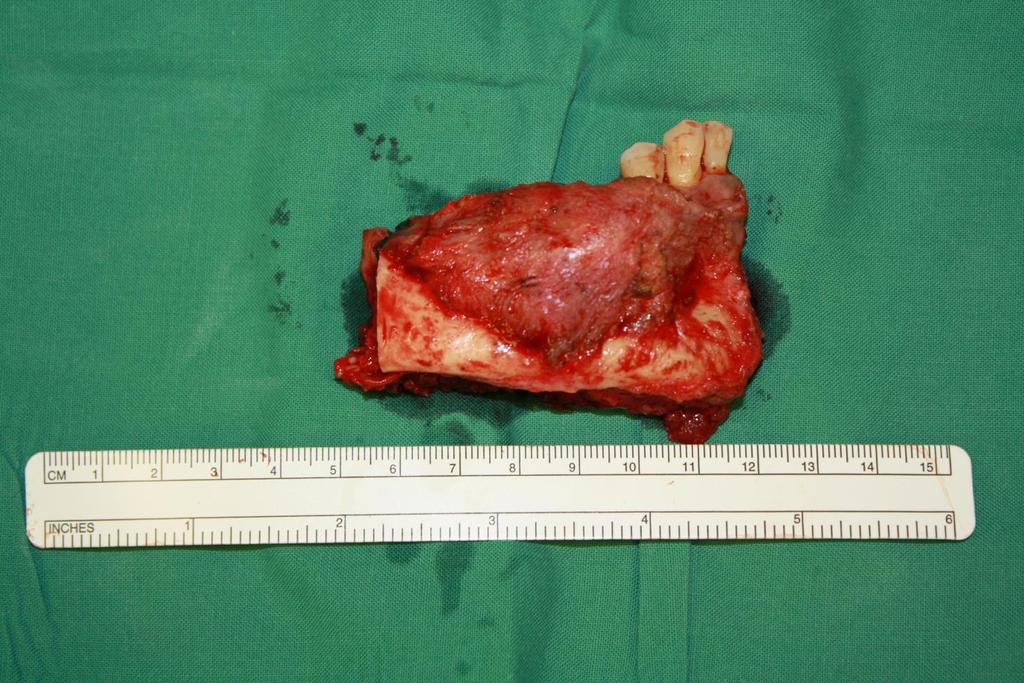

7 Mandibulectomy

8 Oncological Resection of the Maxilla Understand the anatomy of the maxilla and associated structures Understand the extent of the tumour and its behaviour Decide on the extent of maxillary resection Decide on the surgical approach Ohngren s Line
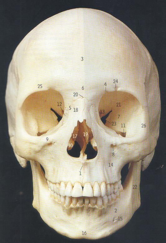

9 Anatomy


10 Anatomy


11 Imaging


12 Imaging
13 Approaches to the maxilla Endoscopic Per-oral Combined Lateral rhinotomy (Moure s) Mid-facial degloving Weber-Ferguson (WF) WF with lynch extension WF with Dieffenbach extension WF with supra and subciliary extension

14 Common facial incision

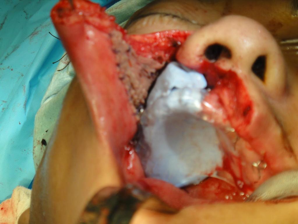
15 Resection of the maxilla Medial maxillectomy Subtotal maxillectomy Infrastructure maxillectomy Total maxillectomy Caldwell-Luc Maxillary swing

16 Lateral rhinotomy (Moure s incision) Relevant anatomy Angular vein Medial canthal ligament Lacrimal sac and nasolacrimal duct Trochlea for the tendon of the superior oblique Ethmoidal vessels 24, 12 and 6 rule Inferior orbital nerve Operative Otolaryngology. Bleach N et. al.

17 Lateral rhinotomy (Moure s incision) Headlight! Tarsorrhaphy Infiltration and nasal decongestant Plan incision carefully Cut down to bone avoiding angular vein Elevate periosteum with freers and blade Operative Otolaryngology. Bleach N et. al.
 Operative Otolaryngology. Bleach N et. al.")
18 If incision is extended along superior orbital rim, divide trochlea Retract and protect orbit with malleable retractors Work towards vessels, divide and ligate/cauterise Divide nasolacrimal duct tangentially Raise skin on face of maxilla to infraorbital nerve Lateral rhinotomy (Moure s incision) Operative Otolaryngology. Bleach N et. al.

19 Anterior ethmoidal artery (right)
20 Medial Maxillectomy
21 Medial Maxillectomy
22 Medial Maxillectomy
23 Medial Maxillectomy
24 Medial Maxillectomy


25 Medial Maxillectomy


26 Medial Maxillectomy post op


27 Medial Maxillectomy post op

28 Midfacial Degloving Cannot reach tumours with extension above the level of the medial canthus Can be combined with bicoronal flap Suitable for young children Operative Otolaryngology. Bleach N et. al.
 Operative Otolaryngology. Bleach N et. al.")
29 Midfacial Degloving 4 incision 1. Sublabial from one maxillary tuberosity to the other 2. Intercartilaginous incisions 3. Full transifixion incision 4. Vestibular incision (stepped to avoid stenosis) Operative Otolaryngology. Bleach N et. al.


30 Midfacial Degloving Operative Otolaryngology. Bleach N et. al.


31 Midfacial Degloving Midfacial Degloving Approach for Malignant Maxillary Tumors ASHRAF S. ZAGHLOUL, M.D*; M. AKRAM NOUH, M. and HISHAM ABD EL FATAH, M.D Cairo University.

32 Infrastructure Maxillectomy Tumours of the upper alveolus Often oral cavity SCCs so are aggressive


33


34

35

36 Subtotal Maxillectomy Removal of most of the maxilla Leaving some suprastructure for orbital support Best performed via a Weber-Ferguson incision with Dieffenbach extension


37 Subtotal Maxillectomy


38 Subtotal Maxillectomy


39 Subtotal Maxillectomy


40 Subtotal Maxillectomy

41 Subtotal Maxillectomy

42 Subtotal Maxillectomy


43 Subtotal Maxillectomy

44 Subtotal Maxillectomy


45 Subtotal Maxillectomy

46 Subtotal Maxillectomy

47 Subtotal Maxillectomy

48 Maxillary Swing For access to the nasopharynx in salvage nasopharyngectomy Allow for enbloc resection of paranasopharyngeal tissue

49 Sir Bobby Robson (18 February July 2009)
New modalities in the salvage of recurrent nasopharyngeal carcinoma
 New modalities in the salvage of recurrent nasopharyngeal carcinoma Dr Jeeve Kanagalingam FRCS Eng (ORL-HNS) Department of Otorhinolaryngology Tan Tock Seng Hospital SINGAPORE Nasopharyngeal carcinoma
OPEN ACCESS ATLAS OF OTOLARYNGOLOGY, HEAD & NECK OPERATIVE SURGERY
 OPEN ACCESS ATLAS OF OTOLARYNGOLOGY, HEAD & NECK OPERATIVE SURGERY INFERIOR MAXILLECTOMY Tumours of the hard palate and superior alveolus may be resected by inferior maxillectomy (Figure 1). A Le Fort
Jaw resection JAMES S BROWN. Contents. Resection of the mandible and maxilla. General principles. Applied anatomy
 39 Jaw resection JAMES S BROWN Contents Resection of the mandible and maxilla 391 General principles 391 Applied anatomy 391 Mandible 391 Maxilla 392 Mandibular resection for oral squamous cell carcinoma
Case Report Mid Facial Degloving Procedure: Managing A Case of Multiple Mid Face Fractures with Significant External Deformity
 55 Bangladesh J Otorhinolaryngol 2015; 21(1): 51-56 Case Report Mid Facial Degloving Procedure: Managing A Case of Multiple Mid Face Fractures with Significant External Deformity Akhil Chndra Biswas 1,
(loco-regional disease)
") (loco-regional disease) (oral cavity) (circumvillae papillae) (subsite) A (upper & lower lips) B (buccal membrane) C (mouth floor) D (upper & lower gingiva) E (hard palate) F (tongue -- anterior 2/3 rds
(loco-regional disease) (oral cavity) (circumvillae papillae) (subsite) A (upper & lower lips) B (buccal membrane) C (mouth floor) D (upper & lower gingiva) E (hard palate) F (tongue -- anterior 2/3 rds
Anatomy and Physiology. Bones, Sutures, Teeth, Processes and Foramina of the Human Skull
 Anatomy and Physiology Chapter 6 DRO Bones, Sutures, Teeth, Processes and Foramina of the Human Skull Name: Period: Bones of the Human Skull Bones of the Cranium: Frontal bone: forms the forehead and the
TOTAL MAXILLECTOMY, ORBITAL EXENTERATION
 TOTAL MAXILLECTOMY, ORBITAL EXENTERATION CC-BY-NC 3.0 Johan Fagan Total maxillectomy refers to surgical resection of the entire maxilla. Resection includes the floor and medial wall of the orbit and the
Structure Location Function
 Frontal Bone Cranium forms the forehead and roof of the orbits Occipital Bone Cranium forms posterior and inferior portions of the cranium Temporal Bone Cranium inferior to the parietal bone forms the
MALIGNANT TUMOURS OF THE JAWS
 MALIGNANT TUMOURS OF THE JAWS MALIGNANT TUMOURS OF THE JAWS Squamous cell carcinoma Osteogenic sarcoma Chondrosarcoma Fibrosarcoma Malignant lymphomas (incl. Burkitt s) Multiple myeloma Ameloblastoma Secondary
Skull Base Volume 12 Month. Patients. Anterior/Midline. Pituitary CSF Leak. Lateral. Craniocervical Junction
 UC SF 2 11/7/2009 Skull Base Surgery in 2009 Ivan El-Sayed MD, FACS Director- Otolaryngology Minimally Invasive Skull Base Surgery Program Department Otolaryngology-Head and Neck Surgery University of
1 Eyelids. Lacrimal Apparatus. Orbital Region. 3 The Orbit. The Eye
 1 1 Eyelids Orbital Region 2 Lacrimal Apparatus 3 The Orbit 4 The Eye 2 Eyelids The eyelids protect the eye from injury and excessive light by their closure. The upper eyelid is larger and more mobile
Dr. Esam Ahmad Z. Omar BDS, MSc-OMFS, FFDRCSI
 Anatomy of the Maxillary Sinus Dr. Esam Ahmad Z. Omar BDS, MSc-OMFS, FFDRCSI Assistant Professor & Consultant Oral&Maxillofacial Surgeon Anatomy of the Maxillary Sinus Diseases of Sinuses 1) Inflammatory:
Total Maxillectomy. A review. Dr T Balasubramanian
 Total Maxillectomy A review Dr T Balasubramanian Maxillectomy a review Introduction: The concept of maxillectomy was first described by Lazars in 1826. After this description it took nearly three years
The Facial Translocation Approach for Management of Head and Neck Cancer
 Review article: The Facial Translocation Approach for Management of Head and Neck Cancer 1Dr. Jaspreet Singh Badwal, 2 Dr. Upkardeep Singh, 3 Dr. Neha Bharti, 4 Dr. Shivani Garg, 5Dr. Simarpreet Singh
The modified endoscopic pre-lacrimal approach: how I do it
 Specialty Techniques Page 1 of 6 The modified endoscopic pre-lacrimal approach: how I do it Leslie T. Koh 1, Rataphol C. Dhepnorrarat 2 1 Department of Otolaryngology-Head & Neck Surgery, Changi General
Management of complications after laryngopharyngectomy
 Management of complications after laryngopharyngectomy Dr Jeeve Kanagalingam MA (Cambridge), BM BCh (Oxford), DLO, DOHNS, FRCS (ORL-HNS), FAMS Consultant ENT / Head and Neck Surgeon Tan Tock Seng Hospital
Surgery in Head and neck cancers.principles. Dr Diptendra K Sarkar MS,DNB,FRCS Consultant surgeon,ipgmer
 Surgery in Head and neck cancers.principles Dr Diptendra K Sarkar MS,DNB,FRCS Consultant surgeon,ipgmer Email:diptendrasarkar@yahoo.co.in HNC : common inclusives Challenges Anatomical preservation R0 Surgical
UPDATE ON RADIOTHERAPY
 1 Miriam Kleiter UPDATE ON RADIOTHERAPY Department for Companion Animals and Horses, Plattform Radiooncology and Nuclear Medicine, University of Veterinary Medicine Vienna Introduction Radiotherapy has
Long-Term Outcomes of Nasopharyngectomy Using Partial Maxillectomy Approach
 The Laryngoscope VC 2015 The American Laryngological, Rhinological and Otological Society, Inc. Long-Term Outcomes of Nasopharyngectomy Using Partial Maxillectomy Approach Li Shia Ng, MMed (ORL); Chwee
Juvenile Angiofibroma
 Juvenile Angiofibroma Disclaimer The pictures used in this presentation have been obtained from a number of sources. Their use is purely for academic and teaching purposes. The contents of this presentation
OPEN ACCESS ATLAS OF OTOLARYNGOLOGY, HEAD & NECK OPERATIVE SURGERY
 OPEN ACCESS ATLAS OF OTOLARYNGOLOGY, HEAD & NECK OPERATIVE SURGERY BUCCINATOR MYOMUCOSAL FLAP The Buccinator Myomucosal Flap is an axial flap, based on the facial and/or buccal arteries. It is a flexible
Temporal region. temporal & infratemporal fossae. Zhou Hong Ying Dept. of Anatomy
 Temporal region temporal & infratemporal fossae Zhou Hong Ying Dept. of Anatomy Temporal region is divided by zygomatic arch into temporal & infratemporal fossae. Temporal Fossa Infratemporal fossa Temporal
Oral cancer: Prognosis & Treatment. Dr. Hani Al Sheikh Radhi
 Oral cancer: Prognosis & Treatment Dr. Hani Al Sheikh Radhi Prognostic factors in Oral caner TNM staging T stage N stage M stage Site Histological Factors Vascular & Perineural Invasion Surgical Margins
The sebaceous glands (glands of Zeis) open directly into the eyelash follicles, ciliary glands (glands of Moll) are modified sweat glands that open
 open directly into the eyelash follicles, ciliary glands (glands of Moll) are modified sweat glands that open") The Orbital Region The orbits are a pair of bony cavities that contain the eyeballs; their associated muscles, nerves, vessels, and fat; and most of the lacrimal apparatus upper eyelid is larger and more
The Orbital Region The orbits are a pair of bony cavities that contain the eyeballs; their associated muscles, nerves, vessels, and fat; and most of the lacrimal apparatus upper eyelid is larger and more
Trigeminal Nerve Worksheets, Distributions Page 1
 Trigeminal Nerve Worksheet #1 Distribution by Nerve Dr. Darren Hoffmann Dental Gross Anatomy, Spring 2013 We have drawn out each of the branches of CN V in lecture and you have an idea now for their basic
The orbit-1. Dr. Heba Kalbouneh Assistant Professor of Anatomy and Histology
 The orbit-1 Dr. Heba Kalbouneh Assistant Professor of Anatomy and Histology Orbital plate of frontal bone Orbital plate of ethmoid bone Lesser wing of sphenoid Greater wing of sphenoid Lacrimal bone Orbital
PTERYGOPALATINE FOSSA
 PTERYGOPALATINE FOSSA Outline Anatomical Structure and Boundaries Foramina and Communications with other spaces and cavities Contents Pterygopalatine Ganglion Especial emphasis on certain arteries and
Chapter 7 Part A The Skeleton
 Chapter 7 Part A The Skeleton Why This Matters Understanding the anatomy of the skeleton enables you to anticipate problems such as pelvic dimensions that may affect labor and delivery The Skeleton The
Neck Dissection. Asst Professor Jeeve Kanagalingam MA (Cambridge), BM BCh (Oxford), MRCS (Eng), DLO, DOHNS, FRCS ORL-HNS (Eng), FAMS (ORL)
, BM BCh (Oxford), MRCS (Eng), DLO, DOHNS, FRCS ORL-HNS (Eng), FAMS (ORL)") Neck Dissection Asst Professor Jeeve Kanagalingam MA (Cambridge), BM BCh (Oxford), MRCS (Eng), DLO, DOHNS, FRCS ORL-HNS (Eng), FAMS (ORL) History radical neck Henry Butlin proposed enbloc removal of upper
Neck Dissection Asst Professor Jeeve Kanagalingam MA (Cambridge), BM BCh (Oxford), MRCS (Eng), DLO, DOHNS, FRCS ORL-HNS (Eng), FAMS (ORL) History radical neck Henry Butlin proposed enbloc removal of upper
Experience with malignant tumours of the maxillary sinus in the Department of Otolaryngology Universiti Kebangsaan Malaysia, Kuala Lumpur
 Med. J. Malaysia Vol. 44 No. 1 March 1989 Experience with malignant tumours of the maxillary sinus in the Department of Otolaryngology Universiti Kebangsaan Malaysia, Kuala Lumpur S. Lokman, MD (UKMalaysia)
Infratemporal fossa: Tikrit University college of Dentistry Dr.Ban I.S. head & neck Anatomy 2 nd y.
 Infratemporal fossa: This is a space lying beneath the base of the skull between the lateral wall of the pharynx and the ramus of the mandible. It is also referred to as the parapharyngeal or lateral pharyngeal
Cancer of the Oral Cavity
 The International Federation of Head and Neck Oncologic Societies Current Concepts in Head and Neck Surgery and Oncology Cancer of the Oral Cavity Ashok Shaha Principals of Management of Oral Cancer A)
Face. Definition: The area between the two ears and from the chin to the eye brows. The muscles of the face
 Face Definition: The area between the two ears and from the chin to the eye brows. The muscles of the face The muscle of facial expression (include the muscle of the face and the scalp). All are derived
Bones of the skull & face
 Bones of the skull & face Cranium= brain case or helmet Copyright The McGraw-Hill Companies, Inc. Permission required for reproduction or display. The cranium is composed of eight bones : frontal Occipital
Major Anatomic Components of the Orbit
 Major Anatomic Components of the Orbit 1. Osseous Framework 2. Globe 3. Optic nerve and sheath 4. Extraocular muscles Bony Orbit Seven Bones Frontal bone Zygomatic bone Maxillary bone Ethmoid bone Sphenoid
Head & Neck Case # 1
 DISCHARGE SUMMARY Head & Neck Case # 1 Date of Admission: 10/30/2010 Date of Discharge: 11/02/2010 Present Medical History: The patient is a 33-year-old lady with a history of right superior alveolar ridge
Introduction to Local Anesthesia and Review of Anatomy
 5-Sep Introduction and Anatomy Review 12-Sep Neurophysiology and Pain 19-Sep Physiology and Pharmacology part 1 26-Sep Physiology and Pharmacology part 2 Introduction to Local Anesthesia and Review of
CT of Maxillofacial Fracture Patterns. CT of Maxillofacial Fracture Patterns
 CT of Maxillofacial Fracture Patterns CT of Maxillofacial Fracture Patterns Stuart E. Mirvis, M.D., FACR Department of Radiology University of Maryland School of Medicine Viking 1 1976 MGS 2001 Technology
Bony orbit Roof The orbital plate of the frontal bone Lateral wall: the zygomatic bone and the greater wing of the sphenoid
 Bony orbit Roof: Formed by: The orbital plate of the frontal bone, which separates the orbital cavity from the anterior cranial fossa and the frontal lobe of the cerebral hemisphere Lateral wall: Formed
Maxilla, ORBIT and infratemporal fossa. Neophytos C Demetriades MD, DDS, MSc Associate professor European University of Cyprus School of Medicine
 Maxilla, ORBIT and infratemporal fossa Neophytos C Demetriades MD, DDS, MSc Associate professor European University of Cyprus School of Medicine MAXILLA Superior, middle, and inferior meatus Frontal sinus
Bones of the Skull Lateral View
 Bones of the Skull Lateral View Frontal Bone Parietal Bone Occipital Bone Temporal Bone Sphenoid Bone Pterion Sutures of the Skull Lateral View Coronal Suture Lambdoid Suture Squamous Suture Sutures of
Remember from the first year embryology Trilaminar disc has 3 layers: ectoderm, mesoderm, and endoderm
 Development of face Remember from the first year embryology Trilaminar disc has 3 layers: ectoderm, mesoderm, and endoderm The ectoderm forms the neural groove, then tube The neural tube lies in the mesoderm
A Study of Classification Systems for Maxillectomy Defects
 A Study of Classification Systems for Maxillectomy Defects Zubair Durrani FFDRCS, FRCS, FRCS (OMFS)* Syed Ghazanfar Hassan FFDRCS** Shomaila Ameer Alam BDS*** * Associate Professor & Consultant Oral and
Department of Otorhinolaryngology, Toho University, Omori-Nishi, Ota-ku, Tokyo , Japan
 Case Reports in Otolaryngology Volume 2015, Article ID 952923, 6 pages http://dx.doi.org/10.1155/2015/952923 Case Report Endoscopic Modified Medial Maxillectomy for Resection of an Inverted Papilloma Originating
Trigeminal Nerve (V)
") Trigeminal Nerve (V) Lecture Objectives Discuss briefly how the face is developed. Follow up the course of trigeminal nerve from its point of central connections, exit and down to its target areas. Describe
Trigeminal Nerve (V) Lecture Objectives Discuss briefly how the face is developed. Follow up the course of trigeminal nerve from its point of central connections, exit and down to its target areas. Describe
MAXILLARY INJECTION TECHNIQUE. Chinthamani Laser Dental Clinic
 MAXILLARY INJECTION TECHNIQUE Chinthamani Laser Dental Clinic Introduction A number of injection techniques are available to aid in providing clinically adequate anesthesia of the teeth and soft and hard
Anatomy images for MSS practical exam- 2019
 Anatomy images for MSS practical exam- 2019 Ilium Ischium Pubis Acetabulaum Iliac crest Iliac tubercle ASIS (muscle and ligament attached) AIIS (muscle attached) PSIS PIIS Ischial spine Ischial tuberosity
Inverted papilloma of the nasal cavity and paranasal sinuses: a study of 20 cases
 Original article: Inverted papilloma of the nasal cavity and paranasal sinuses: a study of 20 cases 1 Dr. Vijay Kumar Kalra, 2 Dr. Samar Pal Singh Yadav, 3 Dr. Swati 1Assistant Professor, 2 Senior Professor
Temporal fossa Infratemporal fossa Pterygopalatine fossa Terminal branches of external carotid artery Pterygoid venous plexus
 Outline of content Temporal fossa Infratemporal fossa Pterygopalatine fossa Terminal branches of external carotid artery Pterygoid venous plexus Boundary Content Communication Mandibular division of trigeminal
SUBJECT: CHANGES TO DENTAL SCHEDULE OF BENEFITS - APRIL 1, 2005 (Year 4 of the multi-year funding agreement) Kenora Robertson St.
 Kenora Robertson St.") Bulletin Bulletin Number 9075 Distribution Dental Surgeons Date April 8, 2005 Direct inquiries to Ministry of Health and Long-Term Care Processing Office (address below) SUBJECT: CHANGES TO DENTAL SCHEDULE
Bulletin Bulletin Number 9075 Distribution Dental Surgeons Date April 8, 2005 Direct inquiries to Ministry of Health and Long-Term Care Processing Office (address below) SUBJECT: CHANGES TO DENTAL SCHEDULE
Trigeminal Nerve Anatomy. Dr. Mohamed Rahil Ali
 Trigeminal Nerve Anatomy Dr. Mohamed Rahil Ali Trigeminal nerve Largest cranial nerve Mixed nerve Small motor root and large sensory root Motor root Nucleus of motor root present in the pons and medulla
Dr.Ban I.S. head & neck anatomy 2 nd y جامعة تكريت كلية طب االسنان مادة التشريح املرحلة الثانية أ.م.د. بان امساعيل صديق 6102/6102
 جامعة تكريت كلية طب االسنان مادة التشريح املرحلة الثانية أ.م.د. بان امساعيل صديق 6102/6102 Pterygopalatine fossa: The pterygopalatine fossa is a cone-shaped depression, It is located between the maxilla,
Transnasal Endoscopic Medial Maxillary Sinus Wall Transposition With Preservation of Structures
 The Laryngoscope VC 2015 The American Laryngological, Rhinological and Otological Society, Inc. Transnasal Endoscopic Medial Maxillary Sinus Wall Transposition With Preservation of Structures Alice Z.
Mick Spillane. Medical. Intensity-Modulated Radiotherapy for Sinonasal Tumors
 Mick Spillane Medical Formatted: Left Intensity-Modulated Radiotherapy for Sinonasal Tumors F Division of Radiotherapy, Department of Oncology (I. M., L. V., W. D. N.), and Division of Head and Neck Surgery,
Anatomic Relations Summary. Done by: Sohayyla Yasin Dababseh
 Anatomic Relations Summary Done by: Sohayyla Yasin Dababseh Anatomic Relations Lecture 1 Part-1 - The medial wall of the nose is the septum. - The vestibule lies directly inside the nostrils (Nares). -
MAXILLOFACIAL TRAUMA. The on-call maxillofacial surgeons can be contacted through the switchboard at the Southern General Hospital
 MAXILLOFACIAL TRAUMA The on-call maxillofacial surgeons can be contacted through the switchboard at the Southern General Hospital Mandibular Injuries Mechanism of injury Assault, falls, RTA-Direct trauma
Alexander C Vlantis. Selective Neck Dissection 33
 05 Modified Radical Neck Dissection Type II Alexander C Vlantis Selective Neck Dissection 33 Modified Radical Neck Dissection Type II INCISION Various incisions can be used for a neck dissection. The incision
Neuroradiology Case of the Day
 Neuroradiology Case of the Day 76 th CAR Annual Meeting, Montreal, Quebec April 27, 2013 Eugene Yu, MD Assistant Professor of Radiology and Otolaryngology-Head and Neck Surgery Head and Neck Imaging Princess
THREE-DIMENSIONAL COMPUTED TOMOGRAPIDC ANALYSIS FOR PLACEMENT OF MAXILLOFACIAL IMPLANTS AFTER MAXILLECTOMY
 Nagoya J. Med. Sci. 56. 69-79,1993 THREE-DIMENSIONAL COMPUTED TOMOGRAPIDC ANALYSIS FOR PLACEMENT OF MAXILLOFACIAL IMPLANTS AFTER MAXILLECTOMY MINORU VEDA, YOSHIHIRO SAWAKI, and TOSHIO KANEDA Department
PROBLEM RECOMMENDATION
 PREVENTION (MINIMIZING) IN ENDOSCOPIC Steven D. Schaefer, MD Professor and Chair Department of Otolaryngology PREVENTION AND Intraoperative Hemorrhage Loss of Orientation Inability to Identify/Preserve
Visibility of Maxillary and Mandibular Anatomical Landmarks in Digital Panoramic Radiographs: A Retrospective Study
 Visibility of Maxillary and Mandibular Anatomical Landmarks in Digital Panoramic Radiographs: A Retrospective Study Srisha Basappa, Smitha JD, Nishath Khanum*, Santosh Kanwar, Mahesh MS and Archana Patil
General Anatomy p. 1 Organization of the Human Body p. 1 Skeleton of the Human Body p. 4 Ossification of the Bones p. 6 Bone Structure p. 8 Joints p.
 General Anatomy p. 1 Organization of the Human Body p. 1 Skeleton of the Human Body p. 4 Ossification of the Bones p. 6 Bone Structure p. 8 Joints p. 10 Principal Joints (Immovable) p. 12 Synovial Joints
Skull basic structures. Neurocranium
 Assoc. Prof. Květuše Lovásová, M.V.D., PhD. Skull basic structures Skull consists of two groups of bones: neurocranium (bones forming the brain box) splanchnocranium (bones forming the facial skeleton)
NASAL FRACTURES. Andrew H. Murr, MD FACS Professor Chief of Service Department of Otolaryngology/ Head and Neck Surgery San Francisco General Hospital
 NASAL FRACTURES Andrew H. Murr, MD FACS Professor Chief of Service Department of Otolaryngology/ Head and Neck Surgery San Francisco General Hospital Roger Boles, M.D. Endowed Chair in Otolaryngology Education
Information and support
 13 11 20 Information and support Surgery for head and neck cancer Last reviewed June 2012 Contents Types of surgeries Surgery for oral cancer Surgery for salivary gland cancer Surgery for pharyngeal cancer
Traditional open surgery for advanced benign nasal tumours in an era of endoscopy: review of 38 cases.
 Traditional open surgery for advanced benign nasal tumours in an era of endoscopy: review of 38 cases. Akeem O Lasisi* and Aderemi A Adeosun Department of Otorhinolaryngology College of Medicine, University
Omran Saeed. Luma Taweel. Mohammad Almohtaseb. 1 P a g e
 2 Omran Saeed Luma Taweel Mohammad Almohtaseb 1 P a g e I didn t include all the photos in this sheet in order to keep it as small as possible so if you need more clarification please refer to slides In
NASAL SEPTUM ADENOID CYSTIC CARCINOMA: A CASE REPORT
 NASAL SEPTUM ADENOID CYSTIC CARCINOMA: A CASE REPORT Shu-Yu Tai, 1 Chen-Yu Chien, 2 Chih-Feng Tai, 2,4 Wen-Rei Kuo, 2,4 Wan-Ting Huang, 3 and Ling-Feng Wang 2,4 Departments of 1 Family Medicine, 2 Otolaryngology
Endoscopic medial maxillectomy
 Operative Techniques in Otolaryngology (2010) 21, 111-116 Endoscopic medial maxillectomy Kelly Cunningham, MD, Kevin C. Welch, MD From the Department of Otolaryngology - Head and Neck Surgery, Loyola University
Bisection of Head & Nasal Cavity 頭部對切以及鼻腔. 解剖學科馮琮涵副教授 分機
 Bisection of Head & Nasal Cavity 頭部對切以及鼻腔 解剖學科馮琮涵副教授 分機 3250 E-mail: thfong@tmu.edu.tw Outline: The structure of nose The concha and meatus in nasal cavity The openings of paranasal sinuses Canals, foramens
TRANSVERSE SECTION PLANE Scalp 2. Cranium. 13. Superior sagittal sinus
 TRANSVERSE SECTION PLANE 1 1. Scalp 2. Cranium 3. Superior sagittal sinus 4. Dura mater 5. Falx cerebri 6. Frontal lobes of the cerebrum 7. Middle meningeal artery 8. Cortex, grey matter 9. Cerebral vessels
Malignant growth Maxilla management an analysis
 ISSN: 2250-0359 Volume 3 Issue 2 2013 Malignant growth Maxilla management an analysis *Balasubramanian Thiagarajan *Geetha Ramamoorthy *Stanley Medical College Abstract: Malignant tumors involving maxilla
University of Palestine. Midterm Exam 2013/2014 Total Grade:
 [ Course No: DNTS2208 Course Title: Head and Neck Anatomy Date: 17/11/1024 No. of Questions: (52) Time: 2hours Using Calculator (No) University of Palestine Midterm Exam 2013/2014 Total Grade: Instructor
Squamous cell carcinoma of the maxillary sinus mimicking periodontitis
 Squamous cell carcinoma of the maxillary sinus mimicking periodontitis 저자저널명발행기관 NDSL URL Na, Ji Yeon ; Kang, Joo Hyun ; Choi, Seong-Ho ; Jeong, Ho-Gul ; Han, Sang-Sun 大韓齒科醫師協會誌 = The journal of the Korean
Epidemiology 3002). Epidemiology and Pathophysiology
. Epidemiology and Pathophysiology") Epidemiology Maxillofacial trauma or injuries are commonly encountered in the practice of emergency medicine and are presenting one of the most challenging problems to the attending surgeons or physicians
Epidemiology Maxillofacial trauma or injuries are commonly encountered in the practice of emergency medicine and are presenting one of the most challenging problems to the attending surgeons or physicians
Cranium Facial bones. Sternum Rib
 Figure 7.1 The human skeleton. Skull Thoracic cage (ribs and sternum) Cranium Facial bones Sternum Rib Bones of pectoral girdle Vertebral column Sacrum Vertebra Bones of pelvic girdle (a) Anterior view
Techniques of local anesthesia in the mandible
 Techniques of local anesthesia in the mandible The technique of choice for anesthesia of the mandible is the block injection and this is attributed to the absence of the advantages which are present in
MAXILLA, ORBIT & PTERYGOPALATINE FOSSA. Neophytos C Demetriades MD, DDS, MSc Associate professor European University of Cyprus School of Medicine
 MAXILLA, ORBIT & PTERYGOPALATINE FOSSA Neophytos C Demetriades MD, DDS, MSc Associate professor European University of Cyprus School of Medicine Maxilla MAXILLA Superior, middle, and inferior meatus Frontal
A longitudinal study of angular artery island flap, used for reconstruction of facial defects
 A longitudinal study of angular artery island flap, used for reconstruction of facial defects KEYWORDS: mid and upper facial defect, angular artery island flap, ipsilateral or contralateral, local flap,
Benign Neoplasms of the Nose
 Department of Otolaryngology Head and Neck Surgery Pursuing Wellness Through Teaching, Learning and Healing Benign Neoplasms of the Nose Ivan El Sayed, MD Disclosure Principal Investigator: Grant Support
OPEN ACCESS ATLAS OF OTOLARYNGOLOGY, HEAD & NECK OPERATIVE SURGERY
 OPEN ACCESS ATLAS OF OTOLARYNGOLOGY, HEAD & NECK OPERATIVE SURGERY ENDOSCOPIC DACRYOCYSTORHINOSTOMY (DCR) SURGICAL TECHNIQUE Hisham Wasl, Darlene Lubbe Endoscopic dacryocystorhinostomy (DCR) is a surgical
Core Curriculum Syllabus Emergencies in Otolaryngology-Head and Neck Surgery FACIAL FRACTURES
 Core Curriculum Syllabus Emergencies in Otolaryngology-Head and Neck Surgery A. General Considerations FACIAL FRACTURES Look for other fractures like skull and/or cervical spine fractures Test function
Bones Ethmoid bone Inferior nasal concha Lacrimal bone Maxilla Nasal bone Palatine bone Vomer Zygomatic bone Mandible
 splanchnocranium - Consists of part of skull that is derived from branchial arches - The facial bones are the bones of the anterior and lower human skull Bones Ethmoid bone Inferior nasal concha Lacrimal
The International Federation of Head and Neck Oncologic Societies. Current Concepts in Head and Neck Surgery and Oncology
 The International Federation of Head and Neck Oncologic Societies Current Concepts in Head and Neck Surgery and Oncology www.ifhnos.net The International Federation of Head and Neck Oncologic Societies
THE ANGULAR TRACT: AN ANATOMICAL
 British Journal of Oral Surgery (1981) 19, 116-120 0 The British Association of Oral Surgeons 0007-117X/81/00170116$02.00 THE ANGULAR TRACT: AN ANATOMICAL OF SURGICAL SIGNIFICANCE STRUCTURE HAITHEM A.
Endoscopic assisted harvest of the pedicled pectoralis major muscle flap
 British Journal of Plastic Surgery (2005) 58, 170 174 Endoscopic assisted harvest of the pedicled pectoralis major muscle flap Arif Turkmen*, A. Graeme B. Perks Plastic Surgery Department, Nottingham City
Non-endoscopic Mechanical Endonasal Dacryocystorhinostomy
 Surgical Technique Non-endoscopic Mechanical Endonasal Dacryocystorhinostomy Mohammad Etezad Razavi 1, MD; Morteza Noorollahian 2, MD; Alireza Eslampoor 1, MD 1 Khatam-al-Anbia Eye Research Center, Mashhad
The resident will be assigned to be on call with the Oral and Maxillofacial service. Call will be set according to PARO guidelines.
 Goals and Objectives for the Otolaryngology-Head & Neck Resident on the Oral and Maxillofacial Surgery (OMFS) Rotation St. Catharines General Hospital (1 four-week rotational block) During the second year
JOSE FRANCISCO GALLEGOS HERNANDEZ Hospital de Oncología, CMN SXXI. IMSS México City.
 JOSE FRANCISCO GALLEGOS HERNANDEZ Hospital de Oncología, CMN SXXI. IMSS México City. HNSCC with a global incidence of over 500,000 cases and 200,000 deaths annually is the leading cause of mortality and
Study of success rates in endoscopic dacryocystorhinostomy with and without stenting. dacryocystorhinostomy with and
 Original Research Article Study of success rates in endoscopic dacryocystorhinostomy with and without stenting Kirti Ambani 1, Niraj Suri 2, Hiren Parmar 3* 1 Assistant Professor, ENT Department, GMERS
Mohammad Hisham Al-Mohtaseb. Lina Mansour. Reyad Jabiri. 0 P a g e
 2 Mohammad Hisham Al-Mohtaseb Lina Mansour Reyad Jabiri 0 P a g e This is only correction for the last year sheet according to our record. If you already studied this sheet just read the yellow notes which
YOU MUST BRING YOUR OWN GLOVES FOR THIS ACTIVITY.
 ACTIVITY 3: AXIAL SKELETON AND LONG BONE DISSECTION Objectives: 1) How to get ready: Read Chapter 7, McKinley et al., Human Anatomy, 5e. All text references are for this textbook. Learning the meanings
BUILDING A. Achieving total reconstruction in a single operation. 70 OCTOBER 2016 // dentaltown.com
 BUILDING A MANDI Achieving total reconstruction in a single operation by Dr. Fayette C. Williams Fayette C. Williams, DDS, MD, FACS, is clinical faculty at John Peter Smith Hospital in Fort Worth, Texas,
Central Poorly Differentiated Adenocarcinoma of the Maxilla: Report of a Case
 Kobe J. Med. Sci., Vol. 49, No. 2, pp. 45-49, 2003 Central Poorly Differentiated Adenocarcinoma of the Maxilla: Report of a Case MASAHIRO UMEDA 1), SATOSHI YOKOO 1), YASUYUKI SHIBUYA 1), TAKAHIDE KOMORI
Malignant tumours of the maxillary complex: an 18-year review
 British Journal of Plastic Surgery (1998), 51,584-588 9 1998 The British Association of Plastic Surgeons I BRITISH JOURNAL OF PLASTIC SURGERY Malignant tumours of the maxillary complex: an 18-year review
Delayed diagnosis of oral squamous cell carcinoma following dental treatment
 ORAL doi 10.1308/003588413X13629960045599 T Singh 1, M Schenberg 2 1 Maxillofacial Surgery Unit, Dandenong Hospital, Melbourne, Australia 2 Honorary Senior Lecturer, Department of Surgery, Monash University,
1. BRIEF DESCRIPTION OF TRAINING
 RHINOLOGY 1. BRIEF DESCRIPTION OF TRAINING Exposure to clinical rhinology is provided in each of the four ORL years over the course of several rotations in a graduated approach. MEE General Otolaryngology