Angio-OCT. Degenerazione Maculare Legata all Eta. Giuseppe Querques
|
|
|
- Emma Wells
- 5 years ago
- Views:
Transcription

1 Angio-OCT Degenerazione Maculare Legata all Eta Giuseppe Querques Department of Ophthalmology, IRCCS Ospedale San Raffaele, University Vita Salute San Raffaele, Milan, Italy
2 Financial Disclosure ADVISORY BOARD MEMBER: CONSULTANT: Allergan Bayer Novartis Alimera sciences Allergan Bayer Bausch and Lomb Heidelberg Novartis Zeiss
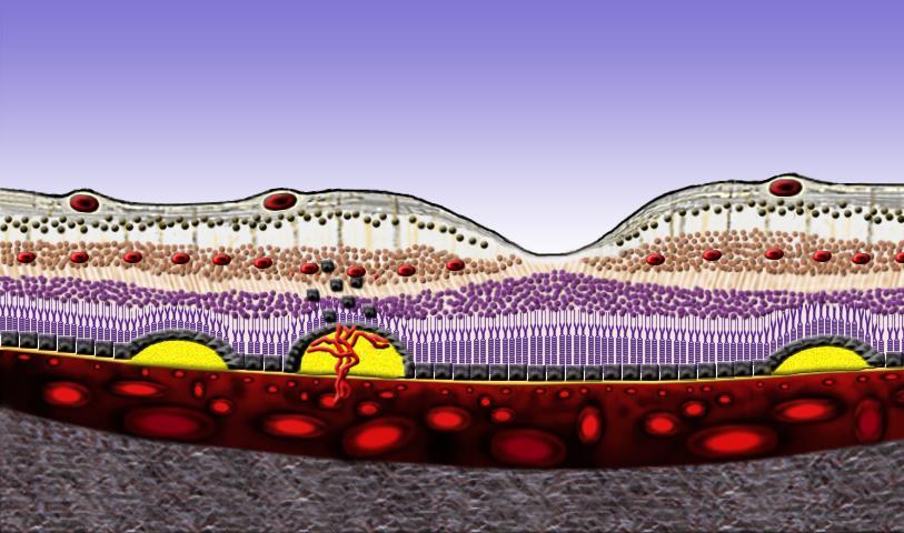
3 Classification of Neovascularization Classification made by GASS Type 1 > Sub-RPE > occult neovascularization

4 TYPE 1 NEO Courtesy of L. Yannuzzi

5 TYPE 1 NEO

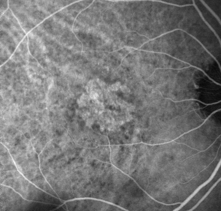
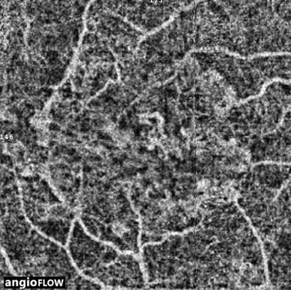
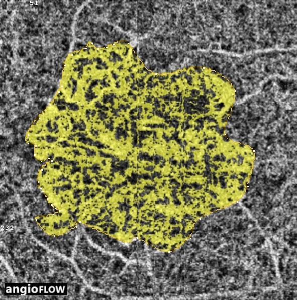
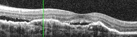
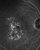
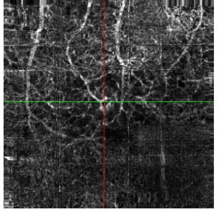
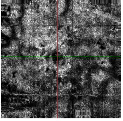
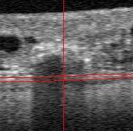
6 TYPE 1 NEO - OCTA

7 TYPE 1 NEO - OCTA

8 TYPE 1 NEO - OCTA

9 TYPE 1 NEO - OCTA
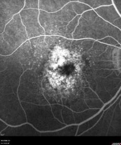
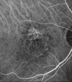
10 Classification of Neovascularization Classification made by GASS Type 2 > Sub-retinal > visible neovascularization
11 TYPE 2 NEO Courtesy of L. Yannuzzi

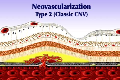

12 TYPE 2 NEO

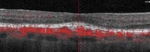
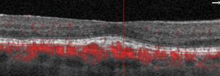
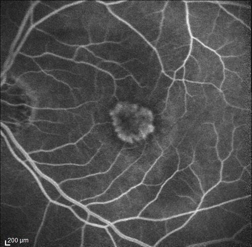
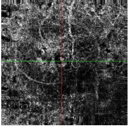
13 TYPE 2 NEO - OCTA

14 TYPE 2 NEO - OCTA
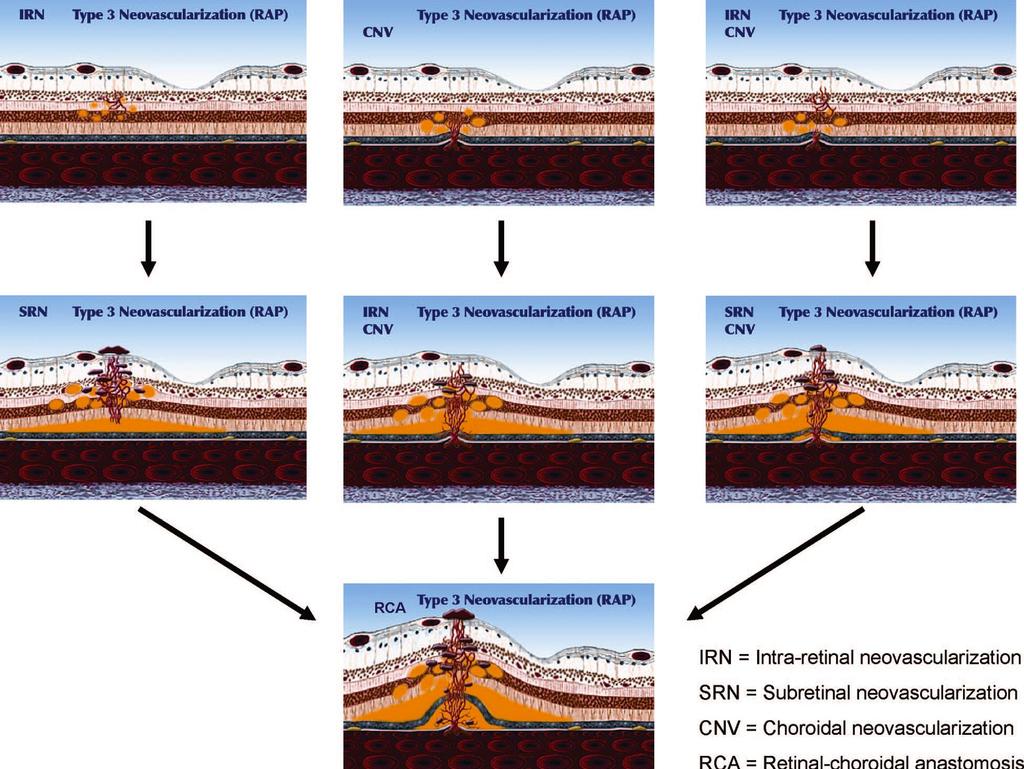
15 Classification of Neovascularization Classification made by GASS Type 3 Neovascularisation: introduced by Freund in 2008
16 Classification of Neovascularization Classification made by GASS Type 3 Neovascularisation: introduced by Freund in 2008 Several anatomical findings corresponding to type 3 neovascularization have been described in the past Hartnett 1992 «Abnormal deep retinal vascular complexe» Khun 1995 «Chorio-Retinal Anastomosis» (CRA) Yanuzzi 2001 «Retinal Angiomatous Proliferation» (RAP) Gass 2003 «Occult choroidal retinal anastomosis» (OCRA
17 Type 3 Neovascularization YANNUZZI PROPOSED THREE VARIANTS IN THE VASOGENIC PROCESS: 1. Initial focal retinal proliferation and progression 2. Focal retinal proliferation with preexisting or simultaneous choroidal proliferation 3. Initial focal choroidal proliferation and progression
18


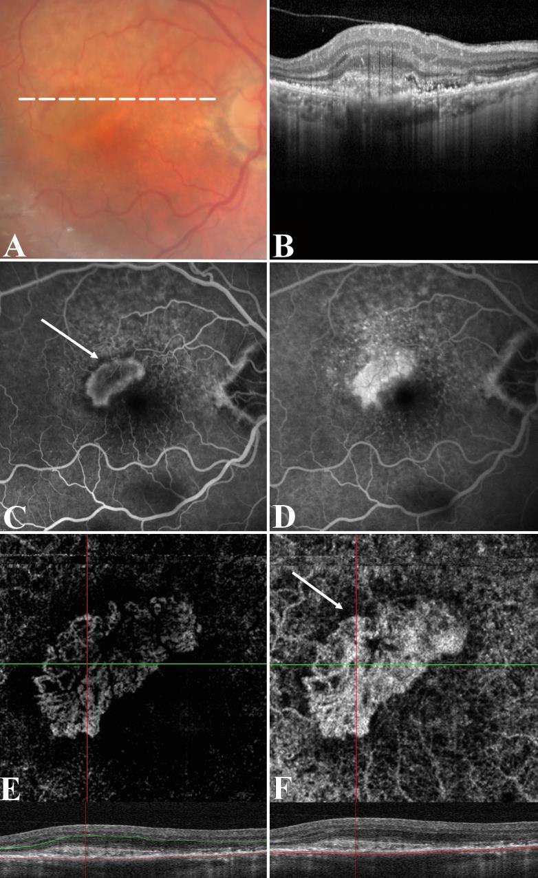
19 Multimodal Imaging of Type 3 Neo The intraretinal neovascular complex appears as a hyper-reflective lesion located in the outer retina adherent to the underlying RPE A focal discontinuity of the RPE band through which the hyperreflective intra retinal lesion communicated with the underlying material within drusen or drusenoid PED No evidence of a communication with the choroid

20 Multimodal Imaging of Type 3 Neo Primarily intraretinal proliferation and anastomoses between retinal vessels and evolving type 1 neovascular tissue within underlying drusen or drusenoid PEDs without evidence of anastomoses with the choroidal circulation Querques, Souied, Freund. Retina 2013
21 However, in vivo imaging does not allow us to conclusively rule out preexisting Type 1 neovascularization or even early RCA


22
23


24


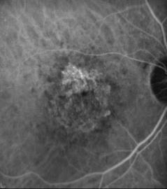
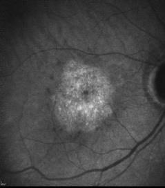
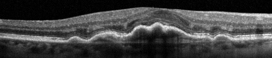
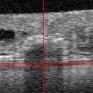
25 TYPE1 NEO quiescent
 is the Aleading cause of vision loss in patients above 50 years.")

, or large (. 125 mm). 7,8 Soft drusen are mound-like elevations, typically 63 mm to. 1.000 mm.")

 in the sub-rpe space (Type 1 neovascularization), or in the subretinal space (Type 2 neovascularization), 12,13 accompanied by")
 shows the deposition of material under the RPE.")
.")


.")
 of subclinical (without exudative retinal changes) focal")


26 Editorial Vascularized Drusen Slowly Progressive Type 1 Neovascularization Mimicking Drusenoid Retinal Pigment Epithelium Elevation ge-related macular degeneration (AMD) is the Aleading cause of vision loss in patients above 50 years. 1 Early AMD ischaracterized by the presence of macular drusen and retinal pigment epithelium (RPE) changes. Drusen are formed by material under the RPE (extracellular debris of varying composition located between the basal lamina of the RPE and the inner collagenous layer of the Bruch membrane in size) 2 6 ; they have been classically characterized as hard or soft, and small (, 63 mm), intermediate (. 63 mm but, 125 mm), or large (. 125 mm). 7,8 Soft drusen are mound-like elevations, typically 63 mm to mm. Different studies have indicated that large, soft, confluent drusen (but not small hard drusen) should be considered as the main factor associated with a greater risk for developing advanced AMD Almost 80% of patients with AMD with vision loss have the exudative advanced form of the disease, 11 which is characterized by the development of choroidal neovascularization (CNV) in the sub-rpe space (Type 1 neovascularization), or in the subretinal space (Type 2 neovascularization), 12,13 accompanied by exudative retinal changes; the remaining 20% of the patients with AMD who develop marked vision loss have the atrophic form of the disease. In soft drusen, optical coherence tomography (OCT) shows the deposition of material under the RPE. 6 Drusen are dynamic, with growth, regression of some drusen, and the formation of new drusen (that can occur simultaneously in the same macula). 14,15 Drusen growth is characterized by the continuous deposition of sub-rpe material (i.e., membranous debris). Drusen regression is characterized by reduced deposition and None of the authors have any financial/conflicting interests to disclose. Supplemental digital content is available for this article. Direct URL citations appear in the printed text and are provided in the HTML and PDF versions of this article on the journal s Web site ( elimination by macrophages of sub-rpe membranous debris, with material not removed that develops calcification. 16 We recently described a novel OCT finding appearing as multilaminar sub-rpe hyperreflectivity in eyes with regressing drusen, which we interpreted as layers of lipid mineralization (i.e., membranous debris developing calcification). 17 However, given the similitudes with the recently reported layers of tissue within vascularized pigment epithelium detachment (PED), which Spaide suggested to represent sub- RPE neovessels, 18 we did not exclude the presence (at least in some cases) of subclinical (without exudative retinal changes) focal neovascularization localized within regressing drusen as a possible explanation for the multilaminar sub-rpe hyperreflectivity. 17 The conflicting interpretation (layers of lipid mineralization vs. sub-rpe neovessels) isnot aproof of thelimitation of OCT technology but rather points out the ability of OCT technology to reveal novel sub-rpe anatomic details. Of note, regressing drusen typically portend the development of drusen-associated atrophy, and not the abnormal growth of newly formed vessels. However, Type 1 neovascularization (in the sub-rpe space), without exudative retinal changes, has been recently described under the definition of quiescent CNV. 19 Quiescent CNV was described on spectraldomain OCT (SD-OCT) as a stable irregularly slightly elevated RPE, without hyporeflective fluid accumulation in the intraretinal/subretinal space, showing a major axis in the horizontal plane. 19 Indeed, current high-resolution multimodal imaging, including splitspectrum amplitude-decorrelation angiography with OCT, strongly suggests that Type 1 neovascularization without exudative retinal changes may also present with different SD-OCT features than those reported for quiescent CNV (i.e., dome-shaped RPE elevations with or without multilaminar hyperreflectivity inside). Drusenoid RPE elevations presenting on SD-OCT multilaminar hyperreflectivity and collections of * *
 changes.")
, intermediate (. 63 mm but, 125 mm), or large (. 125 mm). 7,8 Soft drusen are mound-like elevations, typically 63 mm to.")
 should be considered as the main factor associated with a greater risk for developing advanced AMD.")
, or in the subretinal space (Type 2 neovascularization), 12,13 accompanied by exudative retinal changes; the remaining 20% of the patients with AMD who")
27 Editorial Vascularized Drusen Slowly Progressive Type 1 Neovascularization Mimicking Drusenoid Retinal Pigment Epithelium Elevation ge-related macular degeneration (AMD) is the Aleading cause of vision loss in patients above 50 years. 1 Early AMD ischaracterized by the presence of macular drusen and retinal pigment epithelium (RPE) changes. Drusen are formed by material under the RPE (extracellular debris of varying composition located between the basal lamina of the RPE and the inner collagenous layer of the Bruch membrane in size) 2 6 ; they have been classically characterized as hard or soft, and small (, 63 mm), intermediate (. 63 mm but, 125 mm), or large (. 125 mm). 7,8 Soft drusen are mound-like elevations, typically 63 mm to mm. Different studies have indicated that large, soft, confluent drusen (but not small hard drusen) should be considered as the main factor associated with a greater risk for developing advanced AMD Almost 80% of patients with AMD with vision loss have the exudative advanced form of the disease, 11 which is characterized by the development of choroidal neovascularization (CNV) in the sub-rpe space (Type 1 neovascularization), or in the subretinal space (Type 2 neovascularization), 12,13 accompanied by exudative retinal changes; the remaining 20% of the patients with AMD who develop marked vision loss have the atrophic form of the disease. In soft drusen, optical coherence tomography (OCT) shows the deposition of material under the RPE. 6 Drusen are dynamic, with growth, regression of some drusen, and the formation of new drusen (that can occur simultaneously in the same macula). 14,15 Drusen growth is characterized by the continuous deposition of sub-rpe material (i.e., membranous debris). Drusen regression is characterized by reduced deposition and None of the authors have any financial/conflicting interests to disclose. Supplemental digital content is available for this article. Direct URL citations appear in the printed text and are provided in the HTML and PDF versions of this article on the journal s Web site ( elimination by macrophages of sub-rpe membranous debris, with material not removed that develops calcification. 16 We recently described a novel OCT finding appearing as multilaminar sub-rpe hyperreflectivity in eyes with regressing drusen, which we interpreted as layers of lipid mineralization (i.e., membranous debris developing calcification). 17 However, given the similitudes with the recently reported layers of tissue within vascularized pigment epithelium detachment (PED), which Spaide suggested to represent sub- RPE neovessels, 18 we did not exclude the presence (at least in some cases) of subclinical (without exudative retinal changes) focal neovascularization localized within regressing drusen as a possible explanation for the multilaminar sub-rpe hyperreflectivity. 17 The conflicting interpretation (layers of lipid mineralization vs. sub-rpe neovessels) isnot aproof of thelimitation of OCT technology but rather points out the ability of OCT technology to reveal novel sub-rpe anatomic details. Of note, regressing drusen typically portend the development of drusen-associated atrophy, and not the abnormal growth of newly formed vessels. However, Type 1 neovascularization (in the sub-rpe space), without exudative retinal changes, has been recently described under the definition of quiescent CNV. 19 Quiescent CNV was described on spectraldomain OCT (SD-OCT) as a stable irregularly slightly elevated RPE, without hyporeflective fluid accumulation in the intraretinal/subretinal space, showing a major axis in the horizontal plane. 19 Indeed, current high-resolution multimodal imaging, including splitspectrum amplitude-decorrelation angiography with OCT, strongly suggests that Type 1 neovascularization without exudative retinal changes may also present with different SD-OCT features than those reported for quiescent CNV (i.e., dome-shaped RPE elevations with or without multilaminar hyperreflectivity inside). Drusenoid RPE elevations presenting on SD-OCT multilaminar hyperreflectivity and collections of 2433
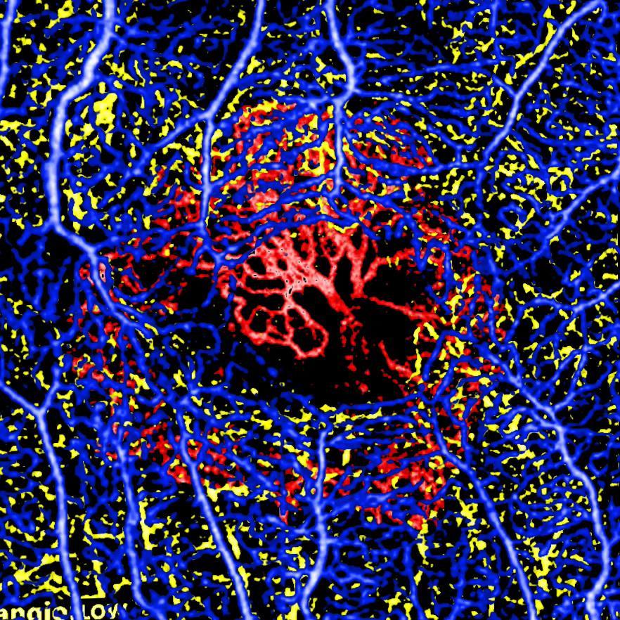
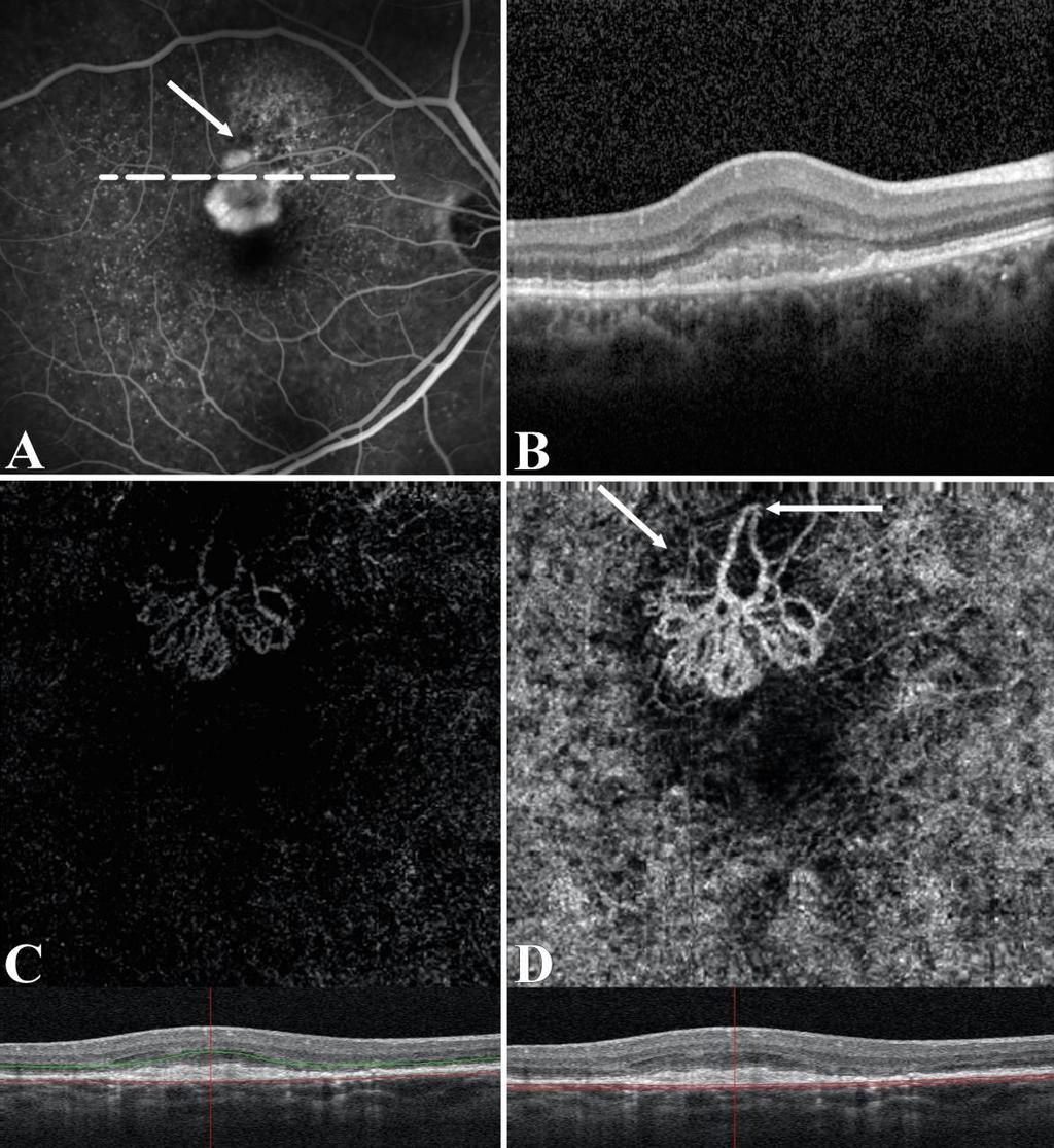
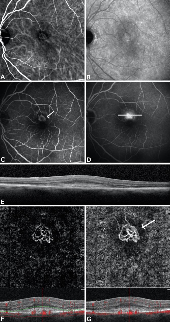
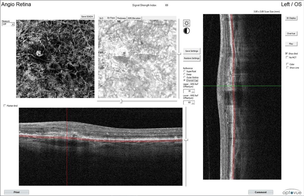
28 TYPE1 NEO quiescent OCTA We investigated the OCT-A features of treatment-naïve quiescent CNV in 22 AMD eyes and assessed its sensitivity and specificity for neovascular detection To estimate the sensitivity and specificity of OCT-A, an additional cohort of 22 eyes of 22 patients with drusenoid PED and no evidence of vascular network at ICGA, were merged to the study group as negative control group OCT-A was performed through AngioPlex CIRRUS HD- OCT model 5000 (Carl Zeiss Meditec, Inc., Dublin, USA), or using AngioVue RTVue XR Avanti (Optovue, Freemont, California, USA)

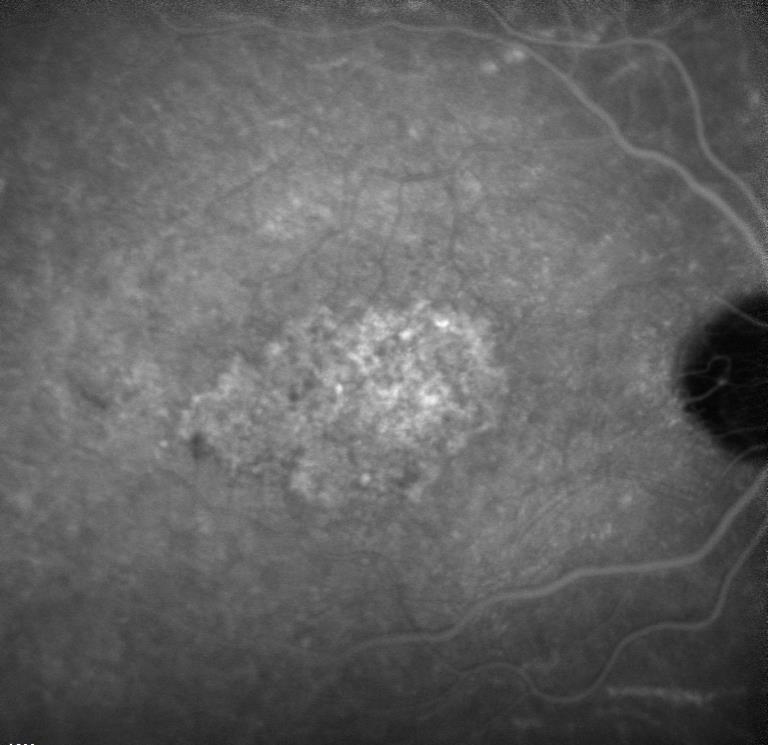
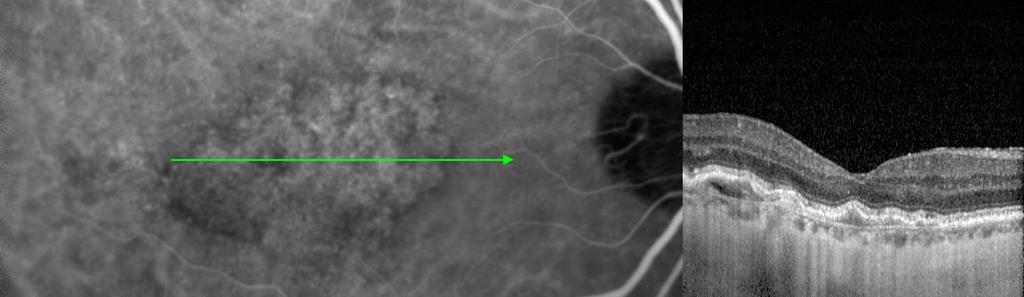

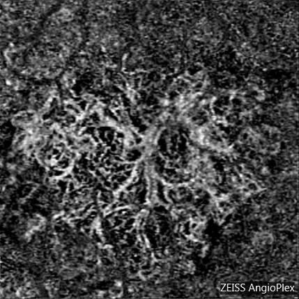

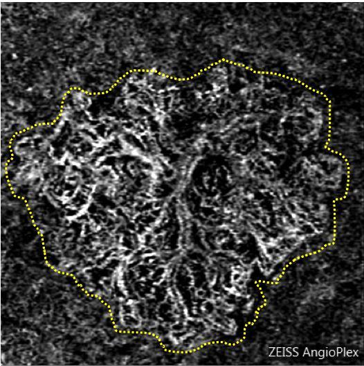

29 TYPE1 NEO quiescent OCTA
30 A TYPE1 NEO quiescent OCTA Two readers correctly identified on OCT-A quiescent CNVs in 18 out of 22 eyes, and correctly excluded all 22 eyes with AMD without CNV.


31 A TYPE1 NEO quiescent OCTA OCT-A sensitivity turned out to be 81.8%, and specificity was 100% and there was complete agreement among the readers
32
OCT Angiography in Primary Eye Care
 OCT Angiography in Primary Eye Care An Image Interpretation Primer Julie Rodman, OD, MS, FAAO and Nadia Waheed, MD, MPH Table of Contents Diabetic Retinopathy 3-6 Choroidal Neovascularization 7-9 Central
Type 1 Choroidal Neovascularization Lesion Size: Indocyanine Green Angiography Versus Optical Coherence Tomography Angiography
 Special Issue Type 1 Choroidal Neovascularization Lesion Size: Indocyanine Green Angiography Versus Optical Coherence Tomography Angiography Eliana Costanzo, 1,2 Alexandra Miere, 1 Giuseppe Querques, 1,3
Key words: Choroidal neovascularisation, Laser coagulation, Retinal imaging
 420 Mini Review The Role of Optical Coherence Tomography (OCT) in the Diagnosis and Management of Retinal Angiomatous Proliferation (RAP) in Patients with Age-related Macular Degeneration Antonio Polito,
Fluorescein Angiography
 Last revision: October 2011 by Luis Arias Fluorescein Angiography Authors: Luis Arias, MD Hospital Universitari de Bellvitge - University of Barcelona. Spain Jordi Monés, MD Institut de la Màcula i de
OCT Interpretation in Retinal Disease
 OCT Interpretation in Retinal Disease Jay M. Haynie, OD, FAAO Financial Disclosure I have received honoraria or am on the advisory board for the following companies: Carl Zeiss Meditec Advanced Ocular
Acquired vitelliform detachment in patients with subretinal drusenoid deposits (reticular pseudodrusen)
") Zurich Open Repository and Archive University of Zurich Main Library Strickhofstrasse 39 CH-8057 Zurich www.zora.uzh.ch Year: 2011 Acquired vitelliform detachment in patients with subretinal drusenoid
Zurich Open Repository and Archive University of Zurich Main Library Strickhofstrasse 39 CH-8057 Zurich www.zora.uzh.ch Year: 2011 Acquired vitelliform detachment in patients with subretinal drusenoid
Incorporating OCT Angiography Into Patient Care
 Incorporating OCT Angiography Into Patient Care Beth A. Steele, OD, FAAO OCT A: Introduction Isolates microvascular circulation from OCT image data Axial resolution = 5 microns (i.e. fine capillaries visible)
Optical Coherence Tomography in Diabetic Retinopathy. Mrs Samantha Mann Consultant Ophthalmologist Clinical Lead of SEL-DESP
 Optical Coherence Tomography in Diabetic Retinopathy Mrs Samantha Mann Consultant Ophthalmologist Clinical Lead of SEL-DESP Content OCT imaging Retinal layers OCT features in Diabetes Some NON DR features
In 1990 our group first described reticular pseudodrusen as a. Choroidal Changes Associated with Reticular Pseudodrusen. Retina
 Retina Choroidal Changes Associated with Reticular Pseudodrusen Giuseppe Querques, 1,2 Lea Querques, 1,2 Raimondo Forte, 1 Nathalie Massamba, 1 Florence Coscas, 1 and Eric H. Souied 1 PURPOSE. To analyze
Staphylomas in Pathologic Myopia With SS-OCT: A New Classification
 INSERT TO The 1st International Swept Source OCT & Angiography Conference took place in Madrid on Feb. 10-11, 2017. A cadre of renowned international retinal specialists presented new clinical findings
10/17/2017. FDA Approved. Zeiss AngioPlex TM Optovue AngioVue TM
 Images retinal microvasculature without dye injection Displays structure and function from a single imaging system Standard of Care-2011 DFE, Fundus Photos, VF 10-2, SD-OCT, FAF, or mferg 2016-AAO Baseline
Mark Dunbar: Disclosure
 Important Things to Understand About OCT Mark T. Dunbar, O.D., F.A.A.O. Bascom Palmer Eye Institute University of Miami, School of Medicine Mark Dunbar: Disclosure Optometry Advisory Board for: Allergan
The Quick Guide to OCT Mastery 50 Real Cases with Expert Analysis
 OPTICAL COHERENCE TOMOGRAPHY The Quick Guide to OCT Mastery 50 Real Cases with Expert Analysis VOL 1 Sanjay Sharma, MD, FRCS, MSc (Epid), MBA Ophthalmologist, Epidemiologist Queen s University, Canada
Clinical Study Optical Coherence Tomography Angiography in Retinal Vascular Diseases and Choroidal Neovascularization
 Hindawi Publishing Corporation Journal of Ophthalmology Volume 2015, Article ID 343515, 8 pages http://dx.doi.org/10.1155/2015/343515 Clinical Study Optical Coherence Tomography Angiography in Retinal
Stabilization of visual acuity with photodynamic therapy in eyes with chorioretinal anastomoses
 Graefe s Arch Clin Exp Ophthalmol (2004) 242:368 376 CLINICAL INVESTIGATION DOI 10.1007/s00417-003-0844-0 Rufino M. Silva José R. Faria de Abreu António Travassos José G. Cunha-Vaz Stabilization of visual
Advances in OCT Murray Fingeret, OD
 Disclosures Advances in OCT Murray Fingeret, OD Consultant Alcon, Allergan, Bausch & Lomb, Carl Zeiss Meditec, Diopsys, Heidelberg Engineering, Reichert, Topcon Currently Approved OCT Devices OCT Devices
연령연관황반변성에서망막혈관종성증식과동반된망막색소상피박리의임상양상과일차적인광역학치료의결과
 연령연관황반변성에서망막혈관종성증식과동반된망막색소상피박리의임상양상과일차적인광역학치료의결과 40 Table. Clinical characteristics and results of patients undergoing photodynamic therapy for retinal angiomatous proliferation Patients No. Age/ sex Eye
OCT Angiography. SriniVas Sadda, MD
 OCT Angiography SriniVas Sadda, MD Professor of Ophthalmology Director, Medical Retina Unit Ophthalmic Imaging Unit University of Southern California Los Angeles, California, USA Disclosure Consulting
HHS Public Access Author manuscript Ophthalmic Surg Lasers Imaging Retina. Author manuscript; available in PMC 2016 January 14.
 High-Speed Ultrahigh-Resolution OCT of Bruch s Membrane in Membranoproliferative Glomerulonephritis Type 2 Mehreen Adhi, MD, Sarah P. Read, MD, PhD, Jonathan J. Liu, PhD, James G. Fujimoto, PhD, and Jay
A view of the current and future role of optical coherence tomography in the management of age-related macular degeneration
 (2017) 31, 26 44 2017 Macmillan Publishers Limited, part of Springer Nature. All rights reserved 0950-222X/17 www.nature.com/eye REVIEW A view of the current and future role of optical coherence tomography
Posterior Segment Update
 Posterior Segment Update Featured Speaker: Dr. Kyle Cheatham, FAAO, DIP ABO DISCLOSURE STATEMENT We have no direct financial or proprietary interest in any companies, products or services mentioned in
ZEISS AngioPlex OCT Angiography. Clinical Case Reports
 Clinical Case Reports Proliferative Diabetic Retinopathy (PDR) Case Report 969 PROLIFERATIVE DIABETIC RETINOPATHY 1 1-year-old diabetic female presents for follow-up of proliferative diabetic retinopathy
R&M Solutions
 Mohamed Hosny El-Bradey, MD., Assistant Professor of Ophthalmology, Tanta University. Wael El Haig, MD., Professor of Ophthalmology. Zagazeeg University. 1 Myopic CNV is considered the most common vision
Age-related macular degeneration (AMD) is the leading cause
 is the leading cause") Special Issue Changes in Neovascular Lesion Hyperreflectivity After Anti-VEGF Treatment in Age-Related Macular Degeneration: An Integrated Multimodal Imaging Analysis Giuseppe Casalino, 1,2 Francesco Bandello,
Special Issue Changes in Neovascular Lesion Hyperreflectivity After Anti-VEGF Treatment in Age-Related Macular Degeneration: An Integrated Multimodal Imaging Analysis Giuseppe Casalino, 1,2 Francesco Bandello,
OPTICAL COHERENCE TOMOGRAPHY ANGIOGRAPHY OF THE RETINA AND OPTIC NERVE. Lindsay B. Howse, OD
 OPTICAL COHERENCE TOMOGRAPHY ANGIOGRAPHY OF THE RETINA AND OPTIC NERVE Lindsay B. Howse, OD drlindsayhowse@gmail.com None. FINANCIAL DISCLOSURES OUTLINE Introduction/How OCTA works OCTA Analysis Advantages
OCT Angiography. Financial Disclosures: Pre-Test: Which one is Correct?
 OCT Angiography Brandon Lujan, MD Medical Director, Casey Reading Center Assistant Professor of Ophthalmology Financial Disclosures: Genentech (Consultant, Grant support, Educational training) UC Berkeley
DOME SHAPED MACULOPATHY. Ιωάννης Ν. Βαγγελόπουλος Χειρ. Οφθαλμίατρος - Βόλος
 DOME SHAPED MACULOPATHY Ιωάννης Ν. Βαγγελόπουλος Χειρ. Οφθαλμίατρος - Βόλος DOME SHAPED MACULOPATHY-DEFINITIONS The entity Dome Shaped Macula ( DSM ) was first described by Gaucher and associates in 2008
Diagnosis in AMD. Managing your AMD Patients
 Managing your AMD Patients Robert W. Dunphy, O.D., F.A.A.O. Diagnosis in AMD Have suspicion Identify relative risk Conduct surveillance Biometry Utilize technology to facilitate detection of change / stability
APRIL 8th 2016 Therapy
 APRIL 8th 2016 Therapy 09.00-10.00 SESSION 1: Age-related Macular Degeneration Moderators: A. Brucker, E. Souied, M. Stirpe, F. Boscia 09.00-09.15 TBD A. Brucker 09.15-09.30 Anti-PDGF and CNV fibrosis
Retinal pigment epithelial detachments in the elderly:
 British Journal of Ophthalmology, 1985, 69, 397-403 Retinal pigment epithelial detachments in the elderly: classification and outcome A G CASSWELL, D KOHEN, AND A C BIRD From Moorfields Eye Hospital, City
Long-term Management of AMD. Motasem Al-latayfeh, MD Assistant Prof. Ophthalmology Hashemite University Jordan
 Long-term Management of AMD Motasem Al-latayfeh, MD Assistant Prof. Ophthalmology Hashemite University Jordan DEFINITION 1 Age-related macular degeneration (AMD) is a disorder of the macula characterized
Introducing ANGIOVUE ESSENTIAL. Built on the Avanti Widefield OCT Platform. OCT Angiography for Primary Eye Care
 Introducing ANGIOVUE ESSENTIAL Built on the Avanti Widefield OCT Platform OCT Angiography for Primary Eye Care Transform Your View of the Retina OCT Angiography (OCTA) is a quick non-invasive test that
evaluation of vitreoretinal adhesions in exudative AMD using optical coherence tomography
 evaluation of vitreoretinal adhesions in exudative AMD using optical coherence tomography Dr. Mahmoud Alaa Abouhusssein, FRCO Lecturer of ophthalmology, Alexandria university Dr. Amir Ramadan Gomaa, MD
ZEISS AngioPlex OCT Angiography Making the revolutionary, routine.
 ZEISS AngioPlex OCT Angiography Making the revolutionary, routine. The moment that revolutionary insight becomes routine. // OCT ANGIOGRAPHY MADE BY ZEISS CIRRUS with AngioPlex creates a new era in both
A Comparison Between Optical Coherence Tomography Angiography and Fluorescein Angiography for the Imaging of Type 1 Neovascularization.
 Thomas Jefferson University Jefferson Digital Commons Wills Eye Institute Papers Wills Eye Institute 7-1-2016 A Comparison Between Optical Coherence Tomography Angiography and Fluorescein Angiography for
A Treat and Extend Regimen Using Ranibizumab for Neovascular Age-Related Macular Degeneration
 A Treat and Extend Regimen Using Ranibizumab for Neovascular Age-Related Macular Degeneration Clinical and Economic Impact Omesh P. Gupta, MD, MBA, Gary Shienbaum, MD, Avni H. Patel, MD, Christopher Fecarotta,
November Volume 35 - Issue 11
 November 2015 - Volume 35 - Issue 11 pp: 2161-2431,e67-e72 Editorial Optical Coherence Tomography Angiography Spaide, Richard F.; Fujimoto, James G.; Waheed, Nadia K. Original Study IMAGE ARTIFACTS IN
We are IntechOpen, the world s leading publisher of Open Access books Built by scientists, for scientists. International authors and editors
 We are IntechOpen, the world s leading publisher of Open Access books Built by scientists, for scientists 3,700 108,500 1.7 M Open access books available International authors and editors Downloads Our
A population based study of macular choroidal neovascularization using optical coherence tomography in Eastern China
 EXPERIMENTAL AND THERAPEUTIC MEDICINE 8: 371-376, 2014 A population based study of macular choroidal neovascularization using optical coherence tomography in Eastern China JIE ZHAO, JUN HU, HAO LU and
Indocyanine Green Angiography and Optical Coherence Tomography Angiography of Choroidal Neovascularization in Age-Related Macular Degeneration
 Retina Indocyanine Green Angiography and Optical Coherence Tomography Angiography of Choroidal Neovascularization in Age-Related Macular Degeneration Chiara M. Eandi, 1 Antonio Ciardella, 2 Mariacristina
Original Policy Date
 MP 9.03.08 Photocoagulation of Macular Drusen Medical Policy Section Miscellaneous Policies Issue 12/2013 Original Policy Date 12/2013 Last Review Status/Date Reviewed with literature search/12/2013 Return
Flore De Bats, 1 Benjamin Wolff, 2,3 Martine Mauget-Faÿsse, 2 Claire Scemama, 2 and Laurent Kodjikian Introduction
 Case Reports in Medicine Volume 2013, Article ID 260237, 7 pages http://dx.doi.org/10.1155/2013/260237 Case Report B-Scan and En-Face Spectral-Domain Optical Coherence Tomography Imaging for the Diagnosis
OCT-Angiography Clinical Cases. OCT-Angiography Clinical Cases
 OCT-Angiography Clinical Cases OCT-Angiography Clinical Cases NIDEK RS-3000 Advance AngioScan Daniela Bacherini Andrea Sodi Stanislao Rizzo CONTENTS Page Authors 3 Introduction 4 Case 1 Case 2 Case 3 Case
Automated segmentation and analysis of layers and structures of human posterior eye
 University of Iowa Iowa Research Online Theses and Dissertations 2015 Automated segmentation and analysis of layers and structures of human posterior eye Li Zhang University of Iowa Copyright 2015 Li Zhang
Optical Coherence Tomography Angiography In Diagnosis Of Retinal Angiomatous Proliferation
 Optical Coherence Tomography Angiography In Diagnosis Of Retinal Angiomatous Proliferation Stepanov A, (1,4) Jiraskova N, 1,4) Lestak J, 2, 3, 4* 1. Department of Ophthalmology, University Hospital and
OCT Angiography The Next Frontier
 Choroid Retina avascular 5/13/2017 OCT Angiography The Next Frontier Pierce Kenworthy OD, FAAO June 9, 2017 OCT Angiography (OCTA) 2016 Non-invasive, motion contrast imaging Represents erythrocyte movement
Visualize. Analyze. Personalize. OCT + OCTA
 Visualize. Analyze. Personalize. OCT + OCTA A New Approach to Protecting Vision AngioVue OCT Angiography brings valuable new information to clinical practice. Non-invasive visualization of retinal vasculature.
NIH Public Access Author Manuscript JAMA Ophthalmol. Author manuscript; available in PMC 2013 September 10.
 NIH Public Access Author Manuscript Published in final edited form as: JAMA Ophthalmol. 2013 May ; 131(5): 693 694. doi:10.1001/jamaophthalmol.2013.692. Effect of Intravitreous Anti Vascular Endothelial
ATLAS OF OCT. Retinal Anatomy in Health & Pathology by Neal A. Adams, MD. Provided to you by:
 ATLAS OF OCT Retinal Anatomy in Health & Pathology by Neal A. Adams, MD Provided to you by: Atlas of OCT The OCT Atlas is written by Neal A. Adams, MD, and produced by Heidelberg Engineering, Inc. to help
OCT Image Analysis System for Grading and Diagnosis of Retinal Diseases and its Integration in i-hospital
 Progress Report for1 st Quarter, May-July 2017 OCT Image Analysis System for Grading and Diagnosis of Retinal Diseases and its Integration in i-hospital Milestone 1: Designing Annotation tool extraction
Citation. As Published Publisher. Version
 Effect of Intravitreous Anti Vascular Endothelial Growth Factor Therapy on Choroidal Thickness in Neovascular Age-Related Macular Degeneration Using Spectral-Domain The MIT Faculty has made this article
Optical coherence tomography (OCT) angiography
 angiography") CLINICAL TRIAL ENDPOINTS FOR OPTICAL COHERENCE TOMOGRAPHY ANGIOGRAPHY IN NEOVASCULAR AGE- RELATED MACULAR DEGENERATION EMILY D. COLE, BS,* DANIELA FERRARA, MD, PHD,* EDUARDO A. NOVAIS, MD,* RICARDO N.
CLINICAL TRIAL ENDPOINTS FOR OPTICAL COHERENCE TOMOGRAPHY ANGIOGRAPHY IN NEOVASCULAR AGE- RELATED MACULAR DEGENERATION EMILY D. COLE, BS,* DANIELA FERRARA, MD, PHD,* EDUARDO A. NOVAIS, MD,* RICARDO N.
Retinal Complications of Obstructive Sleep Apnea A Growing Concern!
 Retinal Complications of Obstructive Sleep Apnea A Growing Concern! Jay M. Haynie, OD, FAAO Financial Disclosure I have received honoraria or am on the advisory board for the following companies: Carl
Comparative Study of Experimental Choroidal Neovascularization by Optical Coherence Tomography and Histopathology
 Comparative Study of Experimental Choroidal Neovascularization by Optical Coherence Tomography and Histopathology Toshio Fukuchi, Kanji Takahashi, Masanobu Uyama and Miyo Matsumura Department of Ophthalmology,
APRIL 8th 2016 Therapy
 APRIL 8th 2016 Therapy 09.00-10.00 SESSION 1: Age-related Macular Degeneration Moderators: A. Brucker, E. Souied, M. Stirpe, R. Brancato 09.00-09.15 TBD A. Brucker 09.15-09.30 TBD E. Souied 09.30-09.45
Spectral-domain Optical Coherence Tomography Imaging of Age-related Macular Degeneration
 Imaging Spectral-domain Optical Coherence Tomography Imaging of Age-related Macular egeneration Carlos Alexandre de Amorim Garcia Filho, 1 Philip J Rosenfeld, 2 Zohar Yehoshua 3 and Giovanni Gregori 3
OCT Interpretation. Financial Disclosure. Jay M. Haynie, OD, FAAO. OCT Image Layers 7/21/2014
 OCT Interpretation Jay M. Haynie, OD, FAAO Financial Disclosure I have received honoraria or am on the advisory board for the following companies: Olympia Tacoma Renton Kennewick - Washington Carl Zeiss
Optical Coherence Tomography: Pearls for the Anterior Segment Surgeon Basic Science Michael Stewart, M.D.
 Optical Coherence Tomography: Pearls for the Anterior Segment Surgeon Basic Science Michael Stewart, M.D. Disclosure OCT Optical Coherence Tomography No relevant financial relationships I will refer to
The College of Optometrists - Learning outcomes for the Professional Certificate in Medical Retina
 Learning outcomes for the Professional Certificate in Medical Retina, incorporating diabetic retinopathy screening and age related macular degeneration The professional certificate is a prerequisite to
Fluorescein Leakage and Optical Coherence Tomography Features of Choroidal Neovascularization Secondary to Pathologic Myopia METHODS
 Retina Fluorescein Leakage and Optical Coherence Tomography Features of Choroidal Neovascularization Secondary to Pathologic Myopia Maurizio Battaglia Parodi, 1 Pierluigi Iacono, 2 Francesco Romano, 1
Identifying the Boundaries of Retinal Pigment Epithelial Detachments Using Two Spectral-Domain Optical Coherence Tomography Instruments
 CLINICAL SCIENCE Identifying the Boundaries of Retinal Pigment Epithelial Detachments Using Two Spectral-Domain Optical Coherence Tomography Instruments Fernando M. Penha, MD, PhD; Giovanni Gregori, PhD;
Clinical Characteristics of Polypoidal Choroidal Vasculopathy Associated with Chronic Central Serous Chorioretionopathy
 pissn: 1011-8942 eissn: 2092-9382 Korean J Ophthalmol 2012;26(1):15-20 http://dx.doi.org/10.3341/kjo.2012.26.1.15 Original Article Clinical Characteristics of Polypoidal Choroidal Vasculopathy Associated
FLUOCINOLONE ACETONIDE: STEROID LONG ACTING
 FLUOCINOLONE ACETONIDE: STEROID LONG ACTING Giuseppe Querques, MD PhD Department of Ophthalmology, IRCCS Ospedale San Raffaele, University Vita Salute San Raffaele, Milan, Italy Financial Disclosure ADVISORY
CENTRAL SEROUS CHORIORETINOPATHY (CSC) IS
 IS") Association Between the Efficacy of Half-Dose Photodynamic Therapy With Indocyanine Green Angiography and Optical Coherence Tomography Findings in the Treatment of Central Serous Chorioretinopathy MASSIMO
Association Between the Efficacy of Half-Dose Photodynamic Therapy With Indocyanine Green Angiography and Optical Coherence Tomography Findings in the Treatment of Central Serous Chorioretinopathy MASSIMO
Swept-Source OCT Angiography: SS OCT Angio TM
 Swept-Source OCT Angiography: SS OCT Angio TM Not available in all countries, please check with your distributor. 2015.09 Swept-Source OCT Angiography: SS OCT Angio TM Introduction Optical coherence tomography
Spontaneous Large Serous Retinal Pigment Epithelial Tear
 This is an Open Access article licensed under the terms of the Creative Commons Attribution-NonCommercial-NoDerivs 3.0 License (www.karger.com/oa-license), applicable to the online version of the article
Since /01/2014 Private practice Ophthalmology, Retinal Medical Specialty and AMD, private office
 Corinne GONZALEZ Ophthalmology (MD, Ph.D ) Retinal Medical Specialist SELARL CABINET DOCTEUR GONZALEZ 27, Boulevard des Minimes 31 200 Toulouse! 05 34 40 77 30 e-mail : cabinet.dr.gonzalez@wanadoo.fr Fax
Department of Ophthalmology, Kangnam Sacred Heart Hospital, College of Medicine, Hallym University, Seoul, Korea
 Department of Ophthalmology, Kangnam Sacred Heart Hospital, College of Medicine, Hallym University, Seoul, Korea Purpose: To investigate the factors that affect final vision following photodynamic therapy
Corinne GONZALEZ Ophthalmology (MD, Ph.D ) Retinal Medical Specialist
 Retinal Medical Specialist") Corinne GONZALEZ Ophthalmology (MD, Ph.D ) Retinal Medical Specialist PRESENT PROFESSIONAL POSITION OPHTALMOLOGIST DOCTOR, Private practice Retinal medical Specialist, DMLA in particular Installation year
Corinne GONZALEZ Ophthalmology (MD, Ph.D ) Retinal Medical Specialist PRESENT PROFESSIONAL POSITION OPHTALMOLOGIST DOCTOR, Private practice Retinal medical Specialist, DMLA in particular Installation year
Optical Coherence Tomograpic Features in Idiopathic Retinitis, Vasculitis, Aneurysms and Neuroretinitis (IRVAN)
") Columbia International Publishing Journal of Ophthalmic Research (2014) Research Article Optical Coherence Tomograpic Features in Idiopathic Retinitis, Vasculitis, Aneurysms and Neuroretinitis (IRVAN)
Columbia International Publishing Journal of Ophthalmic Research (2014) Research Article Optical Coherence Tomograpic Features in Idiopathic Retinitis, Vasculitis, Aneurysms and Neuroretinitis (IRVAN)
Senile disciform macular degeneration
 British Journal of Ophthalmology, 1977, 61, 141-147 Senile disciform macular degeneration in the second eye Z. GREGOR, A. C. BIRD, AND I. H. CHISHOLM From Moorfields Eye Hospital and Institute of Ophthalmology,
Thinking Beyond Wet Age-Related Macular Degeneration
 Ophthalmic Deliberations ISSN 0972-0200 Thinking Beyond Wet Age-Related Macular Degeneration Neha Goel, Atul Kumar, Mahesh P Shanmugam, Muna Bhende, Raja Narayanan, Hidetaka Matsumoto Atul Kumar MD, FAMS
Retinal Pigment Epithelial Detachment
 SURVEY OF OPHTHALMOLOGY VOLUME 52 NUMBER 3 MAY JUNE 2007 MAJOR REVIEW Retinal Pigment Epithelial Detachment Shiri Zayit-Soudry, MD, 1,3 Iris Moroz, MD, 2,3 and Anat Loewenstein, MD 1,3 1 Department of
Macular Morphology and Visual Acuity in the Comparison of Age-related Macular Degeneration Treatments Trials
 Macular Morphology and Visual Acuity in the Comparison of Age-related Macular Degeneration Treatments Trials Glenn J. Jaffe, MD, 1 Daniel F. Martin, MD, 2 Cynthia A. Toth, MD, 1 Ebenezer Daniel, MPH, PhD,
PLEX Elite 9000 from ZEISS Swept-Source OCT
 PLEX Elite 9000 from ZEISS Swept-Source OCT Uncovering the undiscovered. ZEISS PLEX Elite 9000 // INNOVATION MADE BY ZEISS 2 Ultra-wide angiography En face montage Image courtesy of Prof. G. Querques,
Prevalence, Natural Course, and Prognostic Role of Refractile Drusen in Age-Related Macular Degeneration
 Retina Prevalence, Natural Course, and Prognostic Role of Refractile Drusen in Age-Related Macular Degeneration Akio Oishi, 1 Sarah Thiele, 1 Jennifer Nadal, 2 Maho Oishi, 1 Monika Fleckenstein, 1 Matthias
S e q u e n t i a l M o r p h o l o g i c a l C h a n g e s i n the CNV Net after Intravitreal Anti-VEGF Evaluated with OCT Angiography
 O r i g i n a l P a p e r O p h t h a l m i c R e s 2 0 1 6 ; 5 5 : 1 4 5 1 5 1 D O I : 1 0. 1 1 5 9 / 0 0 0 4 4 2 6 7 1 R e c e i v e d : N o v e m b e r 1 9, 2 0 1 5 A c c e p t e d : N o v e m b e r
AperTO - Archivio Istituzionale Open Access dell'università di Torino
 AperTO - Archivio Istituzionale Open Access dell'università di Torino Artifacts in automatic retinal segmentation using different optical coherence tomography instruments. This is the author's manuscript
The role of OCT-A in retinal disease management
 Graefe's Archive for Clinical and Experimental Ophthalmology (2018) 256:2019 2026 https://doi.org/10.1007/s00417-018-4109-3 REVIEW ARTICLE The role of OCT-A in retinal disease management Francisco J. Rodríguez
Optical Coherence Tomography Features Preceding the Onset of Advanced Age-Related Macular Degeneration
 Clinical and Epidemiologic Research Optical Coherence Tomography Features Preceding the Onset of Advanced Age-Related Macular Degeneration Daniela Ferrara, 1 Rachel E. Silver, 2 Ricardo N. Louzada, 1,3
ARVO 2016 Annual Meeting Abstracts
 146 OCT Angiography 1 Sunday, May 01, 2016 3:15 PM 5:00 PM 6B Paper Session Program #/Board # Range: 947 953 Organizing Section: Retina Program Number: 947 Presentation Time: 3:15 PM 3:30 PM Comparisons
Efficacy of Anti-VEGF Agents in the Treatment of Age-Related Macular Degeneration
 Efficacy of Anti-VEGF Agents in the Treatment of Age-Related Macular Degeneration Marilita M. Moschos Abstract- Purpose: To evaluate by OCT and mf-erg the macular function in eyes with CNV due to ARMD
Characteristic Findings of Optical Coherence Tomography in Retinal Angiomatous Proliferation
 pissn: 1011-8942 eissn: 2092-9382 Korean J Ophthalmol 2013;27(5):351-360 http://dx.doi.org/10.3341/kjo.2013.27.5.351 Original rticle haracteristic Findings of Optical oherence Tomography in Retinal ngiomatous
Authors. Introduction. Introduction. Materials and Methods. Objective 10/27/2015
 Idiopathic Polypoidal Choroidal Vasculopathy (IPCV) in Thai Population Presenting with Choroidal Neovascularization (CNV) A multicenter study Authors Yonrawee Piyacomn 1, Chavakij Bhoomibunchoo 1, Yosanan
THE OPHTHALMOLOGIST S NEEDS FOR THE ANALYSIS OF THE RETINA
 biophotonics end-users needs THE OPHTHALMOLOGIST S NEEDS FOR THE ANALYSIS OF THE RETINA Dr Matonti Frédéric CHU Nord / INT AMU Marseille ANATOMY OF THE RETINA ANATOMY OF THE RETINA ANATOMY OF THE RETINA
P olypoidal choroidal vasculopathy (PCV), first described
, first described") 602 EXTENDED REPORT The origins of polypoidal choroidal vasculopathy M Yuzawa, R Mori, A Kawamura... Br J Ophthalmol 2005;89:602 607. doi: 10.1136/bjo.2004.049296 See end of article for authors affiliations...
602 EXTENDED REPORT The origins of polypoidal choroidal vasculopathy M Yuzawa, R Mori, A Kawamura... Br J Ophthalmol 2005;89:602 607. doi: 10.1136/bjo.2004.049296 See end of article for authors affiliations...
ANSWERING THE WHY? Clinicians discuss the latest imaging technologies for retina practice BY PETER K. KAISER, MD
 Insert to March 2018 Sponsored by MULTI-MODALITY IMAGING: LATEST EVOLUTIONS IN OCTA AND UWF As the array of safe and efficacious medical and surgical options for retinal diseases expands, so does the need
Retinal pigment epithelial atrophy over polypoidal choroidal vasculopathy lesions during ranibizumab monotherapy
 Hikichi et al. BMC Ophthalmology (2016) 16:55 DOI 10.1186/s12886-016-0237-x RESEARCH ARTICLE Retinal pigment epithelial atrophy over polypoidal choroidal vasculopathy lesions during ranibizumab monotherapy
Case Report Peripapillary Intrachoroidal Cavitation in Myopia Evaluated with Multimodal Imaging Comprising (En-Face) Technique
 Technique") Case Reports in Ophthalmological Medicine Volume 2015, Article ID 890876, 5 pages http://dx.doi.org/10.1155/2015/890876 Case Report Peripapillary Intrachoroidal Cavitation in Myopia Evaluated with Multimodal
Case Reports in Ophthalmological Medicine Volume 2015, Article ID 890876, 5 pages http://dx.doi.org/10.1155/2015/890876 Case Report Peripapillary Intrachoroidal Cavitation in Myopia Evaluated with Multimodal
OCT Angiography: An Upcoming Tool for Diagnosis and Treatment of Retinal Vascular Diseases
 E-ISSN 2454-2784 Recent Advances OCT Angiography: An Upcoming Tool for Diagnosis and Treatment of Retinal Vascular Diseases Purnima Sood 1, Nalini Saxena 2, Dinesh Talwar 3 1 Vitreo-Retina Consultant,
An atypical case of choroidal neovascularization associated with pseudoxanthoma elasticum treated with intravitreal bevacizumab: a case report
 Karampelas et al. BMC Research Notes 2013, 6:530 CASE REPORT Open Access An atypical case of choroidal neovascularization associated with pseudoxanthoma elasticum treated with intravitreal bevacizumab:
A deeper look at torpedo maculopathy
 CLINICAL AND EXPERIMENTAL REVIEW A deeper look at torpedo maculopathy Clin Exp Optom 2017; 100: 563 568 Casey Hamm* OD Diana Shechtman OD FAAO Sherrol Reynolds OD FAAO *College of Optometry, University
Fluorescence Lifetimes of Drusen in Age-Related Macular Degeneration. Chantal Dysli, Rahel Fink, Sebastian Wolf, and Martin S.
 Retina Fluorescence Lifetimes of Drusen in Age-Related Macular Degeneration Chantal Dysli, Rahel Fink, Sebastian Wolf, and Martin S. Zinkernagel Department of Ophthalmology, Inselspital, Bern University
Deeper visualizations for intervening with confidence.
 CIRRUS OCT with AngioPlex from ZEISS Making the revolutionary routine New vascular quantification Deeper visualizations for intervening with confidence. CIRRUS OCT with AngioPlex from ZEISS can be a much
Reproducibility of Choroidal Thickness Measurements Across Three Spectral Domain Optical Coherence Tomography Systems
 Reproducibility of Choroidal Thickness Measurements Across Three Spectral Domain Optical Coherence Tomography Systems The MIT Faculty has made this article openly available. Please share how this access
Central venous occlusion
 Central venous occlusion Central venous occlusion (right eye) There are dark haemorrhages at the macula and all over the retina. Choroidal haemangioma A choroidal haemangioma has salmon pink colour. There
Department of Ophthalmology, University Hospital of Santiago de Compostela, Ramon Baltar S/N, Santiago de Compostela, Spain 2
 Case Reports in Medicine Volume 2012, Article ID 897097, 10 pages doi:10.1155/2012/897097 Case Report Verteporfin Photodynamic Therapy Combined with Intravitreal Ranibizumab for Polypoidal Choroidal Vasculopathy
Will OCT-Angiography replace FA?
 ASL Roma A PRESIDIO TERRITORIALE NUOVO REGINA MARGHERITA AMBULATORIO PATOLOGIE RETINICHE Resp. Dott.ssa SUSANNA CATALANO CENTRO ITALIANO MACULA Will OCT-Angiography replace FA? Marco Rispoli, Luca di Antonio,
The Role of Phenotype in Selectively Enriching Patients for Clinical Studies
 The Role of Phenotype in Selectively Enriching Patients for Clinical Studies Developing Treatments for Dry Age-Related Macular Degeneration (AMD) Workshop November 15, 2014 National Academy of Sciences