The Role of Phenotype in Selectively Enriching Patients for Clinical Studies
|
|
|
- Gwenda Florence Hall
- 5 years ago
- Views:
Transcription
 Workshop November 15, 2014 National Academy of")

1 The Role of Phenotype in Selectively Enriching Patients for Clinical Studies Developing Treatments for Dry Age-Related Macular Degeneration (AMD) Workshop November 15, 2014 National Academy of Sciences Building, Lecture Room 2101 Constitution Ave., N.W., Washington, DC Philip J. Rosenfeld, MD, PhD Professor of Ophthalmology Bascom Palmer Eye Institute University of Miami Miller School of Medicine
2 Financial Disclosures Acucela: Consultant/Research Grant Advanced Cell Technology: Research grant Alcon: Consultant Alexion: Research grant/consultant Bayer Pharmaceuticals: Consultant Boehringer Ingelheim: Consultant Carl Zeiss Meditec: Research grant Chengdu Kanghong Biotech: Consultant Genentech: Research grant/study advisory board GlaxoSmithKline: Research grant/consultant Healios K.K.: Consultant Oraya: Consultant Hoffman-La Roche: Study advisory board Vision Medicines: Consultant Xcovery Vision: Study advisory board
3 Dry AMD Treatments: Therapeutic Goals Prevent vision loss Slow the loss of vision Restore lost vision
4 Visual acuity Dry AMD Treatments: Visual Acuity as an Endpoint? - Vision loss takes years - Unrealistic short-term clinical trial endpoint - Vision loss may not correlate with disease progression - Surrogate outcome/endpoint needed for clinical trials


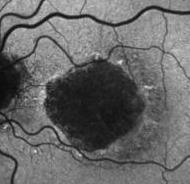
5 Baseline 20/25 6 Months 20/25 Autofluorescence Growth of GA with no loss of vision +2.4 mm 2 OCT Fundus Image 3.0 mm mm 2


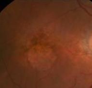
6 Baseline 20/30 6 Months 20/125 Autofluorescence Growth of GA with loss of vision +2.4 mm 2 OCT Fundus Image 3.8 mm mm 2


7 Baseline 20/ mm 2 Week 26 20/125 Vision loss disease progression +2.4 mm 2 Baseline 20/25 Week 26 20/25
8 Baseline 20/40 * +2.4 mm 2 Week 26 20/125 * Central vision loss depends on proximity of GA to foveal center* * Baseline 20/ mm 2 * Week 26 20/25
9 Phenotype Enrichment Depends on The Surrogate Endpoint Surrogate anatomic endpoints: - Growth of geographic atrophy (color, autofluorescence, or OCT en face imaging) - Progression to neovascular AMD - Change in drusen area and/or volume - Progression from drusen to GA (in AREDS, 95% of GA had drusen) - Progression of AREDS severity scale - Changes in retinal/rpe/choroidal anatomy using a variety of imaging strategies
10 Phenotype Enrichment Depends on The Surrogate Endpoint Surrogate anatomic endpoints: - Growth of geographic atrophy (color, autofluorescence, or OCT en face imaging) - Progression to neovascular AMD - Change in drusen area and/or volume - Progression from drusen to GA (in AREDS, 95% of GA had drusen) - Progression of AREDS severity scale - Changes in retinal/rpe/choroidal anatomy using a variety of imaging strategies
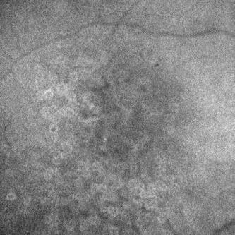
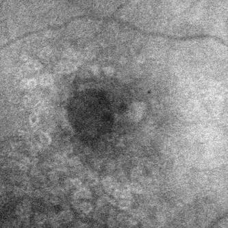
11 Growth of Geographic Atrophy Phenotype enrichment: Autofluorescence patterns (e.g. banded) Bilateral vs. unilateral GA Disruption/atrophy of photoreceptors at margins of GA imaged by OCT Decreased retinal sensitivity at margins of GA measured by microperimetry Low luminance deficits Presence/absence of reticular pseudodrusen (subretinal drusenoid deposits) or decreased choroidal thickness Delayed dark adaptation
12 Growth of Geographic Atrophy Phenotype enrichment: Autofluorescence patterns (e.g. banded) Bilateral vs. unilateral GA Disruption/atrophy of photoreceptors by OCT imaging at margins of G Decreased retinal sensitivity by microperimetry at margins of GA Low luminance deficits Presence/absence of reticular pseudodrusen (subretinal drusenoid deposits) or decreased choroidal thickness Delayed dark adaptation
13 Enlargement Rates of GA using Fundus Autofluorescence (FAF) FAF Patterns None ER = 0.02 mm 2 /yr FAF Patterns Diffuse ER = 1.71 mm 2 /yr Focal ER = 0.36 mm 2 /yr Banded ER = 2.52 mm 2 /yr Patchy ER = 1.84 mm 2 /yr Diffuse, Trickling ER = 3.78 mm 2 /yr Holz et al, Am J Ophthalmol 2007; 143:
 Baseline 2 years later")

 and 7 DA (if multifocal then 1")
14 Example: Phase II and Phase III Lampalizumab Trials (Genentech/Roche) Baseline 2 years later Enrollment criteria: Bilateral GA Presence of hyperautofluorescence of either banded or diffuse patterns adjacent to the area of GA Area of GA: 1 disc area [DA]) and 7 DA (if multifocal then 1 focal lesion 0.5 DA)

15 Size vs. Unifocality vs. Multifocality Larger lesions appear to grow faster Multifocal lesions appear to grow faster Strategies to account for growth rate differences: Square root transformation of area measurements Correction for circularity index
16 AREDS Color Photo Measurements: Change in area of GA over 4 years Change in Area (mm 2 ) Baseline Lesion Size LARGE >4 DA MEDIUM DA SMALL DA Time ( Years ) Growth rate depends on baseline lesion size AREDS Report Number 26 Arch Ophthalmol. 2009; 127 (9) :


17 2 Years 1.7 Years Geographic Atrophy: The Growth Rate Dilemma - Growth rate increases with lesion size and multifocality - As lesions grow larger, they grow faster - Multifocality/multilobularity changes as lesions grow - Variability in test-retest measurements increases as the area of GA increases - What s the solution for designing clinical trials?
 2 r 1 r 1 r 2")

18 Square Root Transformation Strategy: Difference in Areas = Difference in Radii Area = 2 πr = r π r 2 ( r r1) 2 r 1 r 1 r 2 Growth rate independent of baseline size Test-retest measurements independent of size Yehoshua et al. Ophthalmology, April 2011; 118:
19 AREDS Database: Growth of GA over 4 Years (Courtesy of Emily Chew and Rick Ferris) Change in Area (mm 2 ) Time (Y) Feuer et al., JAMA Ophthalmology, Jan Change in Square Root of Area (mm) Time (Y) Growth rate no longer depends on size
 Difference in")

20 AREDS Color Fundus Database: Growth of GA over 4 Years (Courtesy of Emily Chew and Rick Ferris) Difference in Area Measurements Difference in the Square Root Area Measurements Confirms size range of GA for clinical trials
21 Non-Circularity Index (NCI) Helps Predict Progression of GA Ophthalmology 2013;120: Definition of NCI: Actual Area Perimeter Area Actual perimeter = 2πr p r p = radius of a circle with a perimeter equal to the perimeter from the actual GA Perimeter Area = π r p 2 If the GA lesion is a circle, then the NCI = 1
22 Non-Circularity Index (NCI) Helps Predict Progression of GA Ophthalmology 2013;120: Less circular (multifocal/multilobular) More circular
23 Growth of Geographic Atrophy Phenotype enrichment: Autofluorescence patterns (e.g. banded) Bilateral vs. unilateral GA Disruption/atrophy of photoreceptors at margins of GA imaged by OCT Decreased retinal sensitivity by microperimetry at margins of GA Low luminance deficits Presence/absence of reticular pseudodrusen (subretinal drusenoid deposits) or decreased choroidal thickness Delayed dark adaptation
:338-345.")
24 Abnormal Anatomy and Visual Function Extends Beyond the Margin of GA Histopathology: Photoreceptor atrophy at variable distances from edge of GA Geographic Atrophy: A Histopathological Assessment Bird et al., JAMA Ophthalmol. 2014;132(3): Loss of photoreceptors 1400 μm from the edge of GA Intact RPE Electrophysiology and microperimetry: Photoreceptor dysfunction identified away from the edge of the GA Bhutto I, Lutty G. Understanding AMD: relationships between the photoreceptor/retinal pigment epithelium/bruch's membrane/choriocapillaris complex. Molecular aspects of medicine 2012;33:
25 SD-OCT Imaging of the Outer Retina Can Show Disrupted Photoreceptors and Predict Progression of GA Ongoing prospective SD-OCT study Eyes with GA secondary to AMD Size of GA between 0.5 DA (1.8 mm 2 ) and 7 DAs (18 mm 2 ) Followed for at least 1 year Nunes RP et al. Predicting the progression of geographic atrophy in age-related macular degeneration with SD-OCT en face imaging of the outer retina. Ophthalmic Surg Lasers Imaging Retina 2013;44(4):


26 Outer Retinal IS/OS/EZ Slab En Face Image Bottom red line = 20 μm above RPE Top red line = 40 μm above RPE

27 20μm thick slab containing the IS/OS/EZ boundary


28 20μm thick slab containing the IS/OS/EZ boundary
29 En Face Projection: IS/OS/EZ Region

30 IS/OS/EZ Slab En Face Image Top red line = 40 μm above RPE B-scan through fovea Black line = RPE Outer Retinal IS/OS/EZ Slab En Face Image Bottom red line = 20 μm above RPE Location of B-scan


31 OD Case #1 Color Images OS Heidelberg Autofluorescence Images Sub-RPE Slab En Face Images (GA) IS/OS/EZ Slab En Face Images (Focal Pattern) ARR
 Baseline IS/OS/EZ")

32 Growth of GA Over 1 Year: Focal Pattern Baseline 6 Months 12 Months Sub-RPE Slab En Face Images (GA) Baseline IS/OS/EZ Slab En Face Images (Focal Pattern) Baseline GA Baseline GA + Growth at 6 months Baseline GA + Growth at 12 months ARR

 Magnified IS/OS/EZ Slab En Face")
33 Correlation between B-Scan and Outer Retinal IS/OS/EZ Slab En Face Image A Location of B-scan B B-scan Corresponding to Slab Image C IS/OS/EZ Slab En Face Image (Focal Pattern) Magnified IS/OS/EZ Slab En Face Image ARR


34 Correlation between B-Scan and Outer Retinal IS/OS/EZ Slab En Face Image A Location of B-scan B B-scan Corresponding to Slab Image C IS/OS/EZ Slab En Face Image (Focal Pattern) Magnified IS/OS/EZ Slab En Face Image ARR


35 Correlation between B-Scan and Outer Retinal IS/OS/EZ Slab En Face Image Location of B-scan A B B-scan Corresponding to Slab Image C IS/OS/EZ Slab En Face Image (Focal Pattern) Magnified IS/OS/EZ Slab En Face Image Growth of IS/OS defect being used in ongoing CNTF trial in MacTel2

36 Loss of IS/OS/EZ Integrity Corresponds to Decreased Microperimetric Retinal Sensitivity IS/OS/EZ Grading Wu Z, Ayton LN, Luu CD, Guymer RH. Relationship between retinal microstructures on optical coherence tomography and microperimetry in age-related macular degeneration. Ophthalmology 2014;121(7):

37 Microperimetric Retinal Sensitivity Decreased Away from the Edge of GA Loss of retinal sensitivity corresponds to perilesional area with increased autofluorescence Holz FG, Strauss EC, Schmitz-Valckenberg S, van Lookeren Campagne M. Geographic atrophy: clinical features and potential therapeutic approaches. Ophthalmology 2014;121(5):
38 Growth of Geographic Atrophy Phenotype enrichment: Autofluorescence patterns (e.g. banded) Bilateral vs. unilateral GA Disruption/atrophy of photoreceptors by OCT imaging at margins of GA Decreased retinal sensitivity by microperimetry at margins of GA Low luminance deficits Presence/absence of reticular pseudodrusen (subretinal drusenoid deposits) or decreased choroidal thickness Delayed dark adaptation


39 Low Luminance Deficit Testing Normal Luminance ETDRS Acuity Low Luminance ETDRS Acuity Low Luminance Deficit = Normal Luminance VA score - Low Luminance VA Score Sunness JS, Rubin GS, Broman A, Applegate CA, Bressler NM, Hawkins BS. Ophthalmology Sep;115(9): log unit neutral density filter (filter lowers luminance by 100-fold), Kodak Wratten filter; Kodak, Rochester, NY
40 COMPLETE Study: Geographic atrophy Low Luminance Deficit Predicts Growth Rate Yehoshua Z, et al. Systemic complement inhibition with eculizumab for geographic atrophy in age-related macular degeneration: the COMPLETE study. Ophthalmology 2014;121(3): Non-central GA with VA 20/60 (Pearson correlation r=0.38, p=0.007)
41 Growth of Geographic Atrophy Phenotype enrichment: Autofluorescence patterns (e.g. banded) Bilateral vs. unilateral GA Disruption/atrophy of photoreceptors by OCT imaging at margins of GA Decreased retinal sensitivity by microperimetry at margins of GA Low luminance deficits Presence/absence of reticular pseudodrusen (subretinal drusenoid deposits) or decreased choroidal thickness Delayed dark adaptation
42 Phenotype Enrichment Depends on The Surrogate Endpoint Surrogate anatomic endpoints: - Growth of geographic atrophy (color, autofluorescence, or OCT en face imaging) - Progression to neovascular AMD - Change in drusen area and/or volume - Progression from drusen to GA (in AREDS, 95% of GA had drusen) - Progression of AREDS severity scale - Changes in retinal/rpe/choroidal anatomy using a variety of imaging strategies
 5 intermediate (>64 μm) or large (>125 μm) or confluent soft drusen with hyperpigmentation Prevent progression")

43 Anecortave Acetate Risk Reduction Trial (Alcon Research. Ltd/Novartis) 5 intermediate (>64 μm) or large (>125 μm) or confluent soft drusen with hyperpigmentation Prevent progression from high-risk intermediate AMD (soft drusen, pigment hyperplasia within 3000μm) to wet AMD Wet AMD in fellow eye Incidence of sight-threatening CNV in 4 years estimated at 33% Ferris FL, et al. A simplified severity scale for age-related macular degeneration: AREDS Report No. 18. Arch Ophthalmol 2005;123(11):
44 Anecortave Acetate Risk Reduction Trial (Alcon Research. Ltd/Novartis) 2596 patients enrolled worldwide At Month 48: Estimated 80% power to detect a 30% reduction in CNV Estimated 92% power to detect a 35% reduction in CNV After interim analysis at 2 years, study stopped, never published Successful enrollment proves feasibility of this surrogate study design Ferris FL, et al. A simplified severity scale for age-related macular degeneration: AREDS Report No. 18. Arch Ophthalmol 2005;123(11):
45 Phenotype Enrichment Depends on The Surrogate Endpoint Surrogate anatomic endpoints: - Growth of geographic atrophy (color, autofluorescence, or OCT en face imaging) - Progression to neovascular AMD - Change in drusen area and/or volume - Progression from drusen to GA (in AREDS, 95% of GA had drusen) - Progression of AREDS severity scale - Changes in retinal/rpe/choroidal anatomy using a variety of imaging strategies
46 Treatment of Drusen with Laser: Slow Progression to VA loss, CNV, and GA Laser treatment of drusen to prevent progression to advanced age-related macular degeneration (Review) Parodi MB, Virgili G, EvansJR The Cochrane Library 2009, Issue 3 Thisisareprint of acochranereview, prepared and maintained by TheCochraneCollaboration and published in TheCochraneLibrary 2009, Issue3 Laser treatment of drusen to prevent progression to advanced age-related macular degeneration (Review) Copyright 2009 The Cochrane Collaboration. Published by John Wiley & Sons, Ltd. 9 studies, 2216 subjects randomized No evidence that disappearance of drusen reduced risk of developing CNV, GA or visual acuity loss

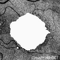
47 Cirrus SD-OCT Measurement of Drusen using the 200 X 200 Raster Scan Pattern: 6mm X 6mm 200 A-scans 200 A-scans 30 micron 30 micron 40,000 A-scans Equal distances between A-scans and B-scans
 200 X 200 raster scan measures 6mm X 6 mm on the")
48 Cirrus SD-OCT Fundus Scanning Pattern 200 X 200 A-scans (6mm X 6mm) 200 X 200 raster scan measures 6mm X 6 mm on the macula
49 Segmentation Algorithms

50 Segmentation Algorithms RPE Segmentation Also available on the Topcon SDOCT instrument


")
51 Measuring RPE Elevations: Subtract RPE Floor from RPE Elevations RPE Elevations Virtual RPE Floor RPE Elevations Automated RPE Floor RPE Difference Map (Drusen) Area and Volume Measurements


52 Drusen: Volume and Area Measurements RPE Segmentation Area: 5.21 mm 2 Volume: mm 3 Zeiss Cirrus SDOCT, Ver. 6.0 software
 Mean Volume = 0.202mm 3 (SD=0.002) Gregori et al. Ophthalmology.")
53 Reproducibility of Drusen Measurements 103 eyes from 74 patient 5 separate SD-OCT scans at the same visit Highly reproducible Mean Area = 3.49mm 2 (SD=0.04) Mean Volume = 0.202mm 3 (SD=0.002) Gregori et al. Ophthalmology Jul;118(7):1373-9
54 Natural History of Drusen in the Absence of Any Geographic Atrophy Using SDOCT Imaging 143 eyes Followed up to 24 months Different progression patterns observed - Increase: 48%/yr - Stable: 40%/yr - Decrease: 12%/yr Yehoshua et al., 2011, Ophthalmology 118(12):
55 Natural History of Drusen in the Absence of Any Geographic Atrophy Using SDOCT Imaging 143 eyes Followed up to 24 months Different progression patterns observed - Increase: 48%/yr - Stable: 40%/yr - Decrease: 12%/yr Yehoshua et al., 2011, Ophthalmology 118(12):


56 Increase in Drusen Area and Volume: 48%/yr Color OCT B-Scan RPE Map Hybrid Drusen Map Baseline Area: 3.28 mm 2 Vol: mm 3 Month 12 Area: 3.94 mm 2 Vol: mm 3 Yehoshua et al., 2011, Ophthalmology 118(12):
57 Decrease in Drusen Area and Volume: 3 Possible Outcomes Formation of geographic atrophy Formation of CNV No significant anatomic abnormality identified Yehoshua et al., 2011, Ophthalmology 118(12):


58 Decrease in Drusen Area and Volume with Formation of GA: 4.5%/yr Color Autofluorescence OCT B-Scan RPE Map Hybrid Drusen Map Baseline Area: 3.75 mm 2 Vol: mm 3 VA: 20/30 Month 12 Area: 0.55 mm 2 Vol: mm 3 VA: 20/63 GA Area:1.28 mm 2 Yehoshua et al., 2011, Ophthalmology 118(12): _OD


59 Decrease in Drusen Area and Volume with Formation of CNV: 3.5%/yr Color Baseline OCT B-Scan RPE Map Hybrid Drusen Map Area: 2.56mm 2 Vol: 0.181mm 3 Month 12 Drusen & CNV Area: 2.41mm 2 Vol: 0.100mm 3 Yehoshua et al., 2011, Ophthalmology 118(12):


60 Decrease in Drusen Volume > 50% Without Formation of GA or CNV: 4%/yr Color OCT B-Scan RPE Map Hybrid Drusen Map Baseline Area: 3.03mm 2 Vol: 0.222mm 3 Month 6 Area: 0.032mm 2 Vol: 0 mm 3 Month 12 Area: 0.03 mm 2 Vol: 0 mm 3 Yehoshua et al., 2011, Ophthalmology 118(12):
61 Placement of 3 mm and 5 mm Diameter Circles Centered on the Fovea Automatic algorithm registers the OCT fundus image with color fundus image Gregori et al. Ophthalmology Jul;118(7):1373-9
62 Placement of 3 mm and 5 mm Diameter Circles Centered on the Fovea Automatic algorithm registers the OCT fundus image with color fundus image Gregori et al. Ophthalmology Jul;118(7):1373-9

63 Placement of 3 mm and 5 mm Diameter Circles Centered on the Fovea Automatic algorithm registers the OCT fundus image with color fundus image Gregori et al. Ophthalmology Jul;118(7):1373-9


64 Quantification of Drusen with the 3 mm and 5 mm Diameter Circles Centered on the Fovea Gregori et al. Ophthalmology Jul;118(7):1373-9
65 Decrease in Drusen Volume > 50% Without Formation of GA or CNV as a Clinical Trial Endpoint Yehoshua et al., 2011, Ophthalmology 118(12):
66 Phase II Eculizumab Study Bascom Palmer Eye Institute COMPLETE Study Design Inclusion: High Risk Drusen OR Geographic Atrophy Drusen Cohort N = 30 2:1 Randomization Eculizumab/Placebo 26 weeks GA Cohort N = 30 2:1 Randomization Followup through one year ClinicalTrials.gov Identifier: NCT
67 Phase II Eculizumab Study: COMPLETE Study Design Bascom Palmer Eye Institute Drusen Cohort N = 30 Visual Acuity: 20/63 or better Drusen Volume mm 3 No evidence of GA


68 Color Fundus Topcon AF Heidelberg AF Fluorescein angiography Heidelberg OCT OCT Fundus Image RPE map Area : 4.29 mm 2 Area : 2.28 mm 2 Area : 4.02 mm 2 Vol : mm 3 Vol : mm 3 Vol : mm 3 Zeiss Cirrus Ver. 6.0 software P1264_OD_100809
69 Phase II Eculizumab Study Bascom Palmer Eye Institute Can Eculizumab Decrease Drusen Volume > 50% Without Formation of GA or CNV as a Clinical Trial Endpoint? Garcia et al., 2014, OSLI-RETINA, January/February Vol.45, No. 1
70 Cube root volume at week 26 COMPLETE Study: Drusen Outcomes Drusen Volume Change at 26 Weeks B Study + fellow eyes (n=37) A 50% decrease in drusen volume over 26 weeks Cube root volume at baseline Active vs placebo for drusen: p=0.15 Outcome effectively ruled out a 22% or greater Two eyes showing decreased volume were placebo treated success rate Two eyes for developing reducing CNV were drusen placebo treated volume - based on the 95% confidence interval between treatment groups
71 COMPLETE Study: Change in Drusen Volume Over 52 weeks Week 26 Outcome Week 52 Outcome Study Study + fellow eyes eyes (n=27) (n=37) Study + fellow eyes (n=37) 50% decrease in drusen volume over 26 weeks 50% decrease in drusen volume over 52 weeks Change in drusen volumes over 26 and 52 Two eyes showing decreased volume were placebo treated weeks consistent with natural history data Two eyes developing CNV were placebo treated


")


72 Week 52 LLD 09 Week 26 LLD 10 Baseline LLD 19 COMPLETE Study: Drusen Examples VA 20/16 (91 letters) Increase in Drusen Volume Vol= 0.12 mm 3 VA 20/12.5 (95 letters) Vol= 0.15 mm 3 VA 20/16 (93 letters) Vol= 0.18 mm BHH


")


73 Week 52 LLD 13 Week 26 LLD 13 Week 12 LLD 14 Baseline LLD 10 COMPLETE Study: Drusen Examples VA 20/16 (92 letters) Decrease in Drusen Volume VA 20/16 (90 letters) Vol= 0.2 mm 3 VA 20/16 (94 letters) Vol= mm 3 VA 20/16 (91 letters) Vol= mm 3 Vol= mm BJJ


74 Week 52 LLD 14 Week 26 LLD 22 Week 24 Baseline LLD 16 COMPLETE Study: Drusen Examples 20/40 (72 letters) Conversion to CNV 20/40 (69 letters) Ranibizumab # 1 Subretinal fluid and serous PED 20/40 (73 letters) 20/32 (75 letters) s/p Ranibizumab X 4 2 placebo eyes developed CNV (p=0.13) AMM
75 89 Drusen Cohort: Primary Study Question Drusen Cohort - Biostatistician: Trial is a success - But, drug failed to meet primary endpoint
76 Novel Composite Clinical Trial Endpoint In drusen-only eyes: - Growth is more common than regression - Growth leads to GA - Growth leads to CNV - Perhaps, a composite endpoint is best
77 Novel Composite Clinical Trial Endpoint In drusen-only eyes, failure defined as: - Growth of drusen volume/area - Formation of CNV - Conversion of drusen to GA
78 Novel Composite Clinical Trial Endpoint Goal of therapy = Prevent failure - Prevent growth of drusen volume/area - Prevent formation of CNV - Prevent conversion of drusen to GA
79 Novel Composite Clinical Trial Endpoint Normal (placebo) failure rate is 60% Garcia et al., 2014, OSLI-RETINA, January/February Vol.45, No. 1
80 Novel Composite Clinical Trial Endpoint Based on natural history data, which was validated in the COMPLETE Study: - For a study with 90% power - to detect a 50% reduction in failure rate - only 62 pts. needed per treatment arm Why study drusen progression rather than enlargement of GA?
81 Drusen Progression as an Endpoint Earlier stage disease than GA Treat earlier and preserve more vision Better defined population than GA? May be at a stage influenced more broadly by therapies (e.g. complement inhibition) Could prevent progression to CNV If treatment slows growth of GA, would it necessarily be effective in slowing progression of drusen to GA? Could open possibility of treating even earlier based on genetics plus phenotype
82 Important Take-Home Messages Phenotype enrichment depends on endpoint Growth of GA is the most commonly used surrogate endpoint for dry AMD trials Enrichment strategies include hyper-af patterns, size, complexity, and genetics Limitations of GA include analysis of growth rate and its late stage (too late?) Surrogate endpoints using earlier stages (e.g. drusen) attractive for Phase 2 studies
83 Possible Future Scenarios Treatment successfully slows or stops the progression of GA - What s the labeled indication? - When will treatment be initiated? - Will early treatment prevent progression to GA or CNV? Treatment fails to slow or stop GA - Could treatment have prevented the progression to GA or formation of CNV? - If so, then goal would be to treat as early as possible
84
Geographic atrophy (GA) is a significant cause of progressive. Predictive Value of Outer Retina En Face OCT Imaging for Geographic Atrophy Progression
 is a significant cause of progressive. Predictive Value of Outer Retina En Face OCT Imaging for Geographic Atrophy Progression") Retina Predictive Value of Outer Retina En Face OCT Imaging for Geographic Atrophy Progression Audrey Giocanti-Auregan, 1,2 Ramin Tadayoni, 2,3 Franck Fajnkuchen, 1,2,4 Pauline Dourmad, 4 Stéphanie Magazzeni,
Retina Predictive Value of Outer Retina En Face OCT Imaging for Geographic Atrophy Progression Audrey Giocanti-Auregan, 1,2 Ramin Tadayoni, 2,3 Franck Fajnkuchen, 1,2,4 Pauline Dourmad, 4 Stéphanie Magazzeni,
Clinical Trial Endpoints for Macular Diseases
 Clinical Trial Endpoints for Macular Diseases Developed in collaboration Learning Objective Upon completion, participants should be able to: Summarize types of biomarkers of progression and treatment response
Dry AMD: the regulatory view
 EMA Ophthalmology Workshop 2011 Dry AMD: the regulatory view Marco Coassin, MD PhD University of Rome Campus Bio-Medico - Italy In this presentation Personal views Previous scientific advices No currently
Mark Dunbar: Disclosure
 Important Things to Understand About OCT Mark T. Dunbar, O.D., F.A.A.O. Bascom Palmer Eye Institute University of Miami, School of Medicine Mark Dunbar: Disclosure Optometry Advisory Board for: Allergan
Spectral-domain Optical Coherence Tomography Imaging of Age-related Macular Degeneration
 Imaging Spectral-domain Optical Coherence Tomography Imaging of Age-related Macular egeneration Carlos Alexandre de Amorim Garcia Filho, 1 Philip J Rosenfeld, 2 Zohar Yehoshua 3 and Giovanni Gregori 3
Fundus Autofluorescence. Jonathan A. Micieli, MD Valérie Biousse, MD
 Fundus Autofluorescence Jonathan A. Micieli, MD Valérie Biousse, MD The retinal pigment epithelium (RPE) has many important functions including phagocytosis of the photoreceptor outer segments Cone Rod
Optical Coherence Tomography: Pearls for the Anterior Segment Surgeon Basic Science Michael Stewart, M.D.
 Optical Coherence Tomography: Pearls for the Anterior Segment Surgeon Basic Science Michael Stewart, M.D. Disclosure OCT Optical Coherence Tomography No relevant financial relationships I will refer to
Posterior Segment Age-related Macular Degeneration
 Posterior Segment Age-related Macular egeneration Spectral-domain Optical oherence Tomography Imaging of Age-related Macular egeneration arlos Alexandre de Amorim Garcia Filho, M, 1 Philip J Rosenfeld,
Long-term Management of AMD. Motasem Al-latayfeh, MD Assistant Prof. Ophthalmology Hashemite University Jordan
 Long-term Management of AMD Motasem Al-latayfeh, MD Assistant Prof. Ophthalmology Hashemite University Jordan DEFINITION 1 Age-related macular degeneration (AMD) is a disorder of the macula characterized
OCT Interpretation in Retinal Disease
 OCT Interpretation in Retinal Disease Jay M. Haynie, OD, FAAO Financial Disclosure I have received honoraria or am on the advisory board for the following companies: Carl Zeiss Meditec Advanced Ocular
Original Policy Date
 MP 9.03.08 Photocoagulation of Macular Drusen Medical Policy Section Miscellaneous Policies Issue 12/2013 Original Policy Date 12/2013 Last Review Status/Date Reviewed with literature search/12/2013 Return
Diagnosis in AMD. Managing your AMD Patients
 Managing your AMD Patients Robert W. Dunphy, O.D., F.A.A.O. Diagnosis in AMD Have suspicion Identify relative risk Conduct surveillance Biometry Utilize technology to facilitate detection of change / stability
Acquired vitelliform detachment in patients with subretinal drusenoid deposits (reticular pseudodrusen)
") Zurich Open Repository and Archive University of Zurich Main Library Strickhofstrasse 39 CH-8057 Zurich www.zora.uzh.ch Year: 2011 Acquired vitelliform detachment in patients with subretinal drusenoid
Zurich Open Repository and Archive University of Zurich Main Library Strickhofstrasse 39 CH-8057 Zurich www.zora.uzh.ch Year: 2011 Acquired vitelliform detachment in patients with subretinal drusenoid
Angio-OCT. Degenerazione Maculare Legata all Eta. Giuseppe Querques
 Angio-OCT Degenerazione Maculare Legata all Eta Giuseppe Querques Department of Ophthalmology, IRCCS Ospedale San Raffaele, University Vita Salute San Raffaele, Milan, Italy Financial Disclosure ADVISORY
OCT Angiography in Primary Eye Care
 OCT Angiography in Primary Eye Care An Image Interpretation Primer Julie Rodman, OD, MS, FAAO and Nadia Waheed, MD, MPH Table of Contents Diabetic Retinopathy 3-6 Choroidal Neovascularization 7-9 Central
Comparison of Geographic Atrophy Measurements from the OCT Fundus Image and the Sub-RPE Slab Image
 CLINICAL SCIENCE Comparison of Geographic Atrophy Measurements from the OCT Fundus Image and the Sub-RPE Slab Image Zohar Yehoshua, MD, MHA; Carlos Alexandre A. Garcia Filho, MD; Fernando M. Penha, MD,
ZEISS AngioPlex OCT Angiography Overview ZEISS OCT Angiography
 ZEISS AngioPlex OCT Angiography Overview ZEISS OCT Angiography California, ZEISS AngioPlex Ultra-clear visualization of microvascular blood flow using non-invasive OCT angiography 2 AngioPlex OCT Angiography
Optical Coherence Tomography in Diabetic Retinopathy. Mrs Samantha Mann Consultant Ophthalmologist Clinical Lead of SEL-DESP
 Optical Coherence Tomography in Diabetic Retinopathy Mrs Samantha Mann Consultant Ophthalmologist Clinical Lead of SEL-DESP Content OCT imaging Retinal layers OCT features in Diabetes Some NON DR features
R&M Solutions
 Mohamed Hosny El-Bradey, MD., Assistant Professor of Ophthalmology, Tanta University. Wael El Haig, MD., Professor of Ophthalmology. Zagazeeg University. 1 Myopic CNV is considered the most common vision
Multimodal Imaging of Geographic Atrophy
 A PROMOTIONAL SUPPLEMENT TO June 2017 C u t t i n g - e d g e A d v a n c e m e n t s Multimodal Imaging of Geographic Atrophy PROCEEDINGS FROM A SYMPOSIUM HELD DURING THE EURETINA MEETING OF THE EUROPEAN
Fundus Autofluorescence
 Brittany Bateman, BS Fundus autofluorescence imaging is used to record fluorescence that may occur naturally in ocular structures or as a byproduct of a disease process. This technique allows the topographic
Fundus autofluorescence in exudative age-related macular degeneration
 Fundus autofluorescence in exudative age-related macular degeneration Q. Peng*, Y. Dong* and P.Q. Zhao Department of Ophthalmology, Xinhua Hospital Affiliated to Shanghai JiaoTong University School of
Course # Getting to Know Your OCT
 Course # 140 Getting to Know Your OCT Course Title: Lecturer: Getting to Know Your OCT Brad Sutton, OD, FAAO IU School of Optometry Financial Disclosures No financial disclosures Optical Coherence Tomography-OCT
ZEISS AngioPlex OCT Angiography. Clinical Case Reports
 Clinical Case Reports Proliferative Diabetic Retinopathy (PDR) Case Report 969 PROLIFERATIVE DIABETIC RETINOPATHY 1 1-year-old diabetic female presents for follow-up of proliferative diabetic retinopathy
Identifying the Boundaries of Retinal Pigment Epithelial Detachments Using Two Spectral-Domain Optical Coherence Tomography Instruments
 CLINICAL SCIENCE Identifying the Boundaries of Retinal Pigment Epithelial Detachments Using Two Spectral-Domain Optical Coherence Tomography Instruments Fernando M. Penha, MD, PhD; Giovanni Gregori, PhD;
Why Is Imaging Critical in My Uveitis Practice?
 Why Is Imaging Critical in My Uveitis Practice? Dilraj S. Grewal, MD Developed in collaboration Imaging Is the Backbone of Uveitis Workup and Monitoring Treatment Response FP FAF B- scan Multimodal Imaging
Advances in OCT Murray Fingeret, OD
 Disclosures Advances in OCT Murray Fingeret, OD Consultant Alcon, Allergan, Bausch & Lomb, Carl Zeiss Meditec, Diopsys, Heidelberg Engineering, Reichert, Topcon Currently Approved OCT Devices OCT Devices
OCT Interpretation. Financial Disclosure. Jay M. Haynie, OD, FAAO. OCT Image Layers 7/21/2014
 OCT Interpretation Jay M. Haynie, OD, FAAO Financial Disclosure I have received honoraria or am on the advisory board for the following companies: Olympia Tacoma Renton Kennewick - Washington Carl Zeiss
Widefield Retinal Imaging with Auto Fluorescence Technology in the Optometric Practice
 Widefield Retinal Imaging with Auto Fluorescence Technology in the Optometric Practice This course will define ultra-widefield retinal imaging and autofluorescence for the attendee. Will show how it is
Comparison of Neovascular Lesion Area Measurements From Different Swept-Source OCT Angiographic Scan Patterns in Age-Related Macular Degeneration
 Retina Comparison of Neovascular Lesion Area Measurements From Different Swept-Source OCT Angiographic Scan Patterns in Age-Related Macular Degeneration Fang Zheng, 1,2 Qinqin Zhang, 3 Elie H. Motulsky,
Retina Conference. Janelle Fassbender, MD, PhD University of Louisville Department of Ophthalmology and Visual Sciences 09/04/2014
 Retina Conference Janelle Fassbender, MD, PhD University of Louisville Department of Ophthalmology and Visual Sciences 09/04/2014 Subjective CC/HPI: 64 year old Caucasian female referred by outside ophthalmologist
HHS Public Access Author manuscript Ophthalmic Surg Lasers Imaging Retina. Author manuscript; available in PMC 2016 January 14.
 High-Speed Ultrahigh-Resolution OCT of Bruch s Membrane in Membranoproliferative Glomerulonephritis Type 2 Mehreen Adhi, MD, Sarah P. Read, MD, PhD, Jonathan J. Liu, PhD, James G. Fujimoto, PhD, and Jay
Cirrus TM HD-OCT. Details defi ne your decisions
 Cirrus TM HD-OCT Details defi ne your decisions 2 With high-defi nition OCT Carl Zeiss Meditec takes you beyond standard spectral domain Built on 10 years experience at the vanguard of innovation, Carl
A Comparative Study of Age Related Macular Degeneration In Relation To SD-OCTand Fundus Photography.
 IOSR Journal of Dental and Medical Sciences (IOSR-JDMS) e-issn: 2279-0853, p-issn: 2279-0861.Volume 14, Issue 11 Ver. III (Nov. 2015), PP 33-37 www.iosrjournals.org A Comparative Study of Age Related Macular
Swept-Source OCT Angiography: SS OCT Angio TM
 Swept-Source OCT Angiography: SS OCT Angio TM Not available in all countries, please check with your distributor. 2015.09 Swept-Source OCT Angiography: SS OCT Angio TM Introduction Optical coherence tomography
Efficacy of Anti-VEGF Agents in the Treatment of Age-Related Macular Degeneration
 Efficacy of Anti-VEGF Agents in the Treatment of Age-Related Macular Degeneration Marilita M. Moschos Abstract- Purpose: To evaluate by OCT and mf-erg the macular function in eyes with CNV due to ARMD
Optical Coherence Tomography (OCT) in Uveitis Piergiorgio Neri, BMedSc, MD, PhD Head Ocular Immunology Unit
 in Uveitis Piergiorgio Neri, BMedSc, MD, PhD Head Ocular Immunology Unit") The Eye Clinic Polytechnic University of Marche Head: Prof Alfonso Giovannini November, 1991 Optical Coherence Tomography (OCT) in Uveitis Piergiorgio Neri, BMedSc, MD, PhD Head Ocular Immunology Unit
The Eye Clinic Polytechnic University of Marche Head: Prof Alfonso Giovannini November, 1991 Optical Coherence Tomography (OCT) in Uveitis Piergiorgio Neri, BMedSc, MD, PhD Head Ocular Immunology Unit
DOME SHAPED MACULOPATHY. Ιωάννης Ν. Βαγγελόπουλος Χειρ. Οφθαλμίατρος - Βόλος
 DOME SHAPED MACULOPATHY Ιωάννης Ν. Βαγγελόπουλος Χειρ. Οφθαλμίατρος - Βόλος DOME SHAPED MACULOPATHY-DEFINITIONS The entity Dome Shaped Macula ( DSM ) was first described by Gaucher and associates in 2008
NIH Public Access Author Manuscript JAMA Ophthalmol. Author manuscript; available in PMC 2013 September 10.
 NIH Public Access Author Manuscript Published in final edited form as: JAMA Ophthalmol. 2013 May ; 131(5): 693 694. doi:10.1001/jamaophthalmol.2013.692. Effect of Intravitreous Anti Vascular Endothelial
Year 4 Results For a Phase 1 Trial of Voretigene Neparvovec in Biallelic RPE65- Mediated Inherited Retinal Disease
 8:00 AM Year 4 Results For a Phase 1 Trial of Voretigene Neparvovec in Biallelic RPE65- Mediated Inherited Retinal Disease Albert M. Maguire, MD OBJECTIVE Assess maintenance of functional vision/visual
Macular Morphology and Visual Acuity in the Comparison of Age-related Macular Degeneration Treatments Trials
 Macular Morphology and Visual Acuity in the Comparison of Age-related Macular Degeneration Treatments Trials Glenn J. Jaffe, MD, 1 Daniel F. Martin, MD, 2 Cynthia A. Toth, MD, 1 Ebenezer Daniel, MPH, PhD,
Choroidal Mapping; a Novel Approach for Evaluating Choroidal Thickness and Volume
 Imaging Technique Choroidal Mapping; a Novel Approach for Evaluating Choroidal Thickness and Volume Jila Noori 1, MD; Mohammad Riazi Esfahani 1,2, MD Fedra Hajizadeh 2, MD; Mohammad-Mehdi Zaferani 1, MD
Incorporating OCT Angiography Into Patient Care
 Incorporating OCT Angiography Into Patient Care Beth A. Steele, OD, FAAO OCT A: Introduction Isolates microvascular circulation from OCT image data Axial resolution = 5 microns (i.e. fine capillaries visible)
A view of the current and future role of optical coherence tomography in the management of age-related macular degeneration
 (2017) 31, 26 44 2017 Macmillan Publishers Limited, part of Springer Nature. All rights reserved 0950-222X/17 www.nature.com/eye REVIEW A view of the current and future role of optical coherence tomography
OCT Angiography. SriniVas Sadda, MD
 OCT Angiography SriniVas Sadda, MD Professor of Ophthalmology Director, Medical Retina Unit Ophthalmic Imaging Unit University of Southern California Los Angeles, California, USA Disclosure Consulting
High Resolution Imaging in Patients with Retinal Dystrophies
 High Resolution Imaging in Patients with Retinal Dystrophies Ophthalmic Photographers Society Annual Midyear Meeting April 2, 213 Jacque Duncan, M.D. UCSF Department of Ophthalmology How can retinal imaging
Geographic atrophy (GA) is the atrophic late-stage manifestation
 is the atrophic late-stage manifestation") Clinical and Epidemiologic Research Semiautomated Image Processing Method for Identification and Quantification of Geographic Atrophy in Age-Related Macular Degeneration Steffen Schmitz-Valckenberg, 1
Clinical and Epidemiologic Research Semiautomated Image Processing Method for Identification and Quantification of Geographic Atrophy in Age-Related Macular Degeneration Steffen Schmitz-Valckenberg, 1
Citation. As Published Publisher. Version
 Effect of Intravitreous Anti Vascular Endothelial Growth Factor Therapy on Choroidal Thickness in Neovascular Age-Related Macular Degeneration Using Spectral-Domain The MIT Faculty has made this article
Restricted Summed-Area Projection for Geographic Atrophy Visualization in SD-OCT Images
 Article Restricted Summed-Area Projection for Geographic Atrophy Visualization in SD-OCT Images DOI: 10.1167/tvst.4.5.2 Qiang Chen 1, Sijie Niu 1, Honglie Shen 1, Theodore Leng 3, Luis de Sisternes 2,
Geographic atrophy (GA) represents the atrophic late-stage
 represents the atrophic late-stage") Clinical Trials Progression of Age-Related Geographic Atrophy: Role of the Fellow Eye Monika Fleckenstein, 1,2 Steffen Schmitz-Valckenberg, 1,2 Christine Adrion, 3 Sivatharisini Visvalingam, 1 Arno P.
Clinical Trials Progression of Age-Related Geographic Atrophy: Role of the Fellow Eye Monika Fleckenstein, 1,2 Steffen Schmitz-Valckenberg, 1,2 Christine Adrion, 3 Sivatharisini Visvalingam, 1 Arno P.
Cirrus TM HD-OCT. Details define your decisions
 Cirrus TM HD-OCT Details define your decisions 2 With high-definition OCT Carl Zeiss Meditec takes you beyond standard spectral domain Built on 10 years experience at the vanguard of innovation, Carl Zeiss
8/6/17. Disclosures Aerie Pharmaceuticals Alcon BioTissue Diopsys Optovue Shire
 Nathan Lighthizer, O.D., F.A.A.O. Associate Professor Assistant Dean for Clinical Care Director of Continuing Education Chief of Specialty Care Clinics Oklahoma College of Optometry Tahlequah, OK lighthiz@nsuok.edu
RETINA 2018 OBJECTIVES OCT VERY USEFUL INFORMATION SAFE AND FRIENDLY 1/11/2018 KELLY MITCHELL
 RETINA 2018 KELLY MITCHELL OBJECTIVES HIGHLIGHT NEW DIAGNOSTIC & TREATMENT OPTIONS REVIEW DIAGNOSTIC KEYS OF SELECT RETINAL DISEASES DISCUSS USE OF IMAGING AND REFERRAL RECOURSES FOR PATIENT BENEFIT OCT
Clinically Significant Macular Edema (CSME)
") Clinically Significant Macular Edema (CSME) 1 Clinically Significant Macular Edema (CSME) Sadrina T. Shaw OMT I Student July 26, 2014 Advisor: Dr. Uwaydat Clinically Significant Macular Edema (CSME) 2
Clinically Significant Macular Edema (CSME) 1 Clinically Significant Macular Edema (CSME) Sadrina T. Shaw OMT I Student July 26, 2014 Advisor: Dr. Uwaydat Clinically Significant Macular Edema (CSME) 2
Spontaneous Large Serous Retinal Pigment Epithelial Tear
 This is an Open Access article licensed under the terms of the Creative Commons Attribution-NonCommercial-NoDerivs 3.0 License (www.karger.com/oa-license), applicable to the online version of the article
OCT Angiography. Financial Disclosures: Pre-Test: Which one is Correct?
 OCT Angiography Brandon Lujan, MD Medical Director, Casey Reading Center Assistant Professor of Ophthalmology Financial Disclosures: Genentech (Consultant, Grant support, Educational training) UC Berkeley
FROM OUTDATED TO UPDATED Eminence-Based Medicine
 FROM OUTDATED TO UPDATED Eminence-Based Medicine Evidence-Based Medicine A REVIEW OF KEY CLINICAL TRIALS Anthony DeWilde, OD FAAO 1 EMINENCE BASED MEDICINE 2 EVIDENCE BASED MEDICINE 3 4 CLINICAL TRIALS
Despite our growing knowledge of the
 OVERVIEW OF AVAILABLE TREATMENTS FOR NEOVASCULAR AGE-RELATED MACULAR DEGENERATION * Carl D. Regillo, MD ABSTRACT Although potential treatments for neovascular age-related macular degeneration represent
Advanced retinal pigment epithelium analysis by SD-OCT to monitor dry AMD progression
 special report Advanced retinal pigment epithelium analysis by SD-OCT to monitor dry AMD progression This article provides a short overview of developments in optical coherence tomography (OCT) technology
Method for comparing visual field defects to local RNFL and RGC damage seen on frequency domain OCT in patients with glaucoma.
 Method for comparing visual field defects to local RNFL and RGC damage seen on frequency domain OCT in patients with glaucoma. Donald C. Hood 1,2,* and Ali S. Raza 1 1 Department of Psychology, Columbia
Intraocular Radiation Therapy for Age-Related Macular Degeneration
 Medical Policy Manual Medicine, Policy No. 134 Intraocular Radiation Therapy for Age-Related Macular Degeneration Next Review: April 2019 Last Review: June 2018 Effective: August 1, 2018 IMPORTANT REMINDER
IQ 532 Micropulse Green Laser treatment for Refractory Chronic Central Serous Retinopathy
 Cronicon OPEN ACCESS EC OPHTHALMOLOGY Case Report IQ 532 Micropulse Green Laser treatment for Refractory Chronic Central Serous Retinopathy Fawwaz Al Mamoori* Medical Retina Department, Eye Specialty Hospital,
Ganglion cell analysis by optical coherence tomography (OCT) Jonathan A. Micieli, MD Valérie Biousse, MD
 Jonathan A. Micieli, MD Valérie Biousse, MD") Ganglion cell analysis by optical coherence tomography (OCT) Jonathan A. Micieli, MD Valérie Biousse, MD Figure 1. Normal OCT of the macula (cross section through the line indicated on the fundus photo)
Ganglion cell analysis by optical coherence tomography (OCT) Jonathan A. Micieli, MD Valérie Biousse, MD Figure 1. Normal OCT of the macula (cross section through the line indicated on the fundus photo)
Fluorescein Angiography
 Last revision: October 2011 by Luis Arias Fluorescein Angiography Authors: Luis Arias, MD Hospital Universitari de Bellvitge - University of Barcelona. Spain Jordi Monés, MD Institut de la Màcula i de
The Quick Guide to OCT Mastery 50 Real Cases with Expert Analysis
 OPTICAL COHERENCE TOMOGRAPHY The Quick Guide to OCT Mastery 50 Real Cases with Expert Analysis VOL 1 Sanjay Sharma, MD, FRCS, MSc (Epid), MBA Ophthalmologist, Epidemiologist Queen s University, Canada
Diabetic Retinopathy Clinical Research Network
 Diabetic Retinopathy Clinical Research Network Comparison of Time Domain OCT and Spectral Domain OCT Retinal Thickness Measurement in Diabetic Macular Edema Version 1.0 June 16, 2009 comparison of td vs
Case Report Peripapillary Intrachoroidal Cavitation in Myopia Evaluated with Multimodal Imaging Comprising (En-Face) Technique
 Technique") Case Reports in Ophthalmological Medicine Volume 2015, Article ID 890876, 5 pages http://dx.doi.org/10.1155/2015/890876 Case Report Peripapillary Intrachoroidal Cavitation in Myopia Evaluated with Multimodal
Case Reports in Ophthalmological Medicine Volume 2015, Article ID 890876, 5 pages http://dx.doi.org/10.1155/2015/890876 Case Report Peripapillary Intrachoroidal Cavitation in Myopia Evaluated with Multimodal
Flore De Bats, 1 Benjamin Wolff, 2,3 Martine Mauget-Faÿsse, 2 Claire Scemama, 2 and Laurent Kodjikian Introduction
 Case Reports in Medicine Volume 2013, Article ID 260237, 7 pages http://dx.doi.org/10.1155/2013/260237 Case Report B-Scan and En-Face Spectral-Domain Optical Coherence Tomography Imaging for the Diagnosis
Retinal pigment epithelial detachments in the elderly:
 British Journal of Ophthalmology, 1985, 69, 397-403 Retinal pigment epithelial detachments in the elderly: classification and outcome A G CASSWELL, D KOHEN, AND A C BIRD From Moorfields Eye Hospital, City
Management of Neovascular AMD
 Kapusta AMD Part 1 Management of Neovascular AMD Dr. Michael A. Kapusta, MD, FRCSC Ophthalmologist in Chief Jewish General Hospital Vitreoretinal Surgeon 1 FINANCIAL DISCLOSURES Consulting honoraria Bayer,
Overview. Macular OCT Artifact Study
 Imaging Artifacts Sarah Moyer, CRA, OCT-C Director, Ophthalmic Imaging Kittner Eye Center University of North Carolina Chapel Hill, NC Disclose financial interest now Overview Sarah s Thoughts on Artifacts
Use of Aflibercept in cases of recurrent or refractory neovascular AMD previously treated with other antiangiogenic drug: anatomo-functional outcomes
 Use of Aflibercept in cases of recurrent or refractory neovascular AMD previously treated with other antiangiogenic drug: anatomo-functional outcomes Daniel E. Charles, MD, Martin Charles, MD, Gisela Jelusich,
SUMMARY. Heather Casparis, MD,* and Neil M. Bressler, MD MARINA AND ANCHOR
 The following are summaries of selected presentations and posters from the American Society of Retina Specialists and European VitreoRetinal Society Annual Meeting held September 9 13, 2006, in Cannes,
11/29/2016 MACULAR MALADIES: TYPICAL & ATYPICAL CASES
 MACULAR MALADIES: TYPICAL & ATYPICAL CASES Dawn Pewitt, OD, FAAO Triad Eye Institute, Grove, OK Dpewitt@triadeye.com Disclosure Statement: No financial disclosures COPE 51218-PS Please silence all mobile
ATLAS OF OCT. Retinal Anatomy in Health & Pathology by Neal A. Adams, MD. Provided to you by:
 ATLAS OF OCT Retinal Anatomy in Health & Pathology by Neal A. Adams, MD Provided to you by: Atlas of OCT The OCT Atlas is written by Neal A. Adams, MD, and produced by Heidelberg Engineering, Inc. to help
A population based study of macular choroidal neovascularization using optical coherence tomography in Eastern China
 EXPERIMENTAL AND THERAPEUTIC MEDICINE 8: 371-376, 2014 A population based study of macular choroidal neovascularization using optical coherence tomography in Eastern China JIE ZHAO, JUN HU, HAO LU and
Prevalence, Natural Course, and Prognostic Role of Refractile Drusen in Age-Related Macular Degeneration
 Retina Prevalence, Natural Course, and Prognostic Role of Refractile Drusen in Age-Related Macular Degeneration Akio Oishi, 1 Sarah Thiele, 1 Jennifer Nadal, 2 Maho Oishi, 1 Monika Fleckenstein, 1 Matthias
ACTIVATED OR NOT? RETINAL CASE PRESENTATION Shorye Payne, MD Medical Retinal Specialist Robley Rex VA Eye Clinic
 ACTIVATED OR NOT? RETINAL CASE PRESENTATION Shorye Payne, MD Medical Retinal Specialist Robley Rex VA Eye Clinic C We anticipate that the future management of posterior uveal melanoma (PUM) will focus
Title: OCT Analysis Workshop: Interpretation of OCT printouts
 Title: OCT Analysis Workshop: Interpretation of OCT printouts Authors: David Yang, OD, FAAO Staff Optometrist, VA Palo Alto Health Care System Associate Clinical Professor, UC Berkeley School of Optometry
Although photocoagulation and photodynamic PROCEEDINGS PEGAPTANIB SODIUM FOR THE TREATMENT OF AGE-RELATED MACULAR DEGENERATION *
 PEGAPTANIB SODIUM FOR THE TREATMENT OF AGE-RELATED MACULAR DEGENERATION Evangelos S. Gragoudas, MD ABSTRACT In December 24, the US Food and Drug Administration (FDA) approved pegaptanib sodium. Pegaptanib
In 1990 our group first described reticular pseudodrusen as a. Choroidal Changes Associated with Reticular Pseudodrusen. Retina
 Retina Choroidal Changes Associated with Reticular Pseudodrusen Giuseppe Querques, 1,2 Lea Querques, 1,2 Raimondo Forte, 1 Nathalie Massamba, 1 Florence Coscas, 1 and Eric H. Souied 1 PURPOSE. To analyze
Optical Coherence Tomography Features Preceding the Onset of Advanced Age-Related Macular Degeneration
 Clinical and Epidemiologic Research Optical Coherence Tomography Features Preceding the Onset of Advanced Age-Related Macular Degeneration Daniela Ferrara, 1 Rachel E. Silver, 2 Ricardo N. Louzada, 1,3
OCT Angiography The Next Frontier
 Choroid Retina avascular 5/13/2017 OCT Angiography The Next Frontier Pierce Kenworthy OD, FAAO June 9, 2017 OCT Angiography (OCTA) 2016 Non-invasive, motion contrast imaging Represents erythrocyte movement
An atypical case of choroidal neovascularization associated with pseudoxanthoma elasticum treated with intravitreal bevacizumab: a case report
 Karampelas et al. BMC Research Notes 2013, 6:530 CASE REPORT Open Access An atypical case of choroidal neovascularization associated with pseudoxanthoma elasticum treated with intravitreal bevacizumab:
Fundus Autofluorescence and its PRACTICAL applications: Retina Beyond the Color. Start to think about this. Disclosure 5/21/2015
 Fundus Autofluorescence and its PRACTICAL applications: Retina Beyond the Color Jeffry D. Gerson, O.D., F.A.A.O Olathe, KS jgerson@hotmail.com Start to think about this. Disclosure I have worked with/consulted
OCT Image Analysis System for Grading and Diagnosis of Retinal Diseases and its Integration in i-hospital
 Progress Report for1 st Quarter, May-July 2017 OCT Image Analysis System for Grading and Diagnosis of Retinal Diseases and its Integration in i-hospital Milestone 1: Designing Annotation tool extraction
THE OPHTHALMOLOGIST S NEEDS FOR THE ANALYSIS OF THE RETINA
 biophotonics end-users needs THE OPHTHALMOLOGIST S NEEDS FOR THE ANALYSIS OF THE RETINA Dr Matonti Frédéric CHU Nord / INT AMU Marseille ANATOMY OF THE RETINA ANATOMY OF THE RETINA ANATOMY OF THE RETINA
Geographic atrophy (GA), a late-stage manifestation of various
, a late-stage manifestation of various") Clinical Trials Fundus Autofluorescence and Spectral-Domain Optical Coherence Tomography Characteristics in a Rapidly Progressing Form of Geographic Atrophy Monika Fleckenstein, 1 Steffen Schmitz-Valckenberg,
Clinical Trials Fundus Autofluorescence and Spectral-Domain Optical Coherence Tomography Characteristics in a Rapidly Progressing Form of Geographic Atrophy Monika Fleckenstein, 1 Steffen Schmitz-Valckenberg,
Yasser R. Serag, MD Tamer Wasfi, MD El- Saied El-Dessoukey, MD Magdi S. Moussa, MD Anselm Kampik, MD
 Microperimetric Evaluation of Brilliant Blue G- assisted Internal Limiting Membrane Peeling By Yasser R. Serag, MD Tamer Wasfi, MD El- Saied El-Dessoukey, MD Magdi S. Moussa, MD Anselm Kampik, MD The internal
Ocular imaging in acquired retinopathy with multiple myeloma
 Ocular imaging in acquired retinopathy with multiple myeloma ABDELRAHMAN GABER SALMAN MD- FRCS (GLASG)- MRCS (ED) ASSOCIATE PROFESSOR AIN SHAMS UNIVERSITY EVRS 2015 Immunogammopathies Immunogammopathies
Retinal rejuvenation a restorative treatment breakthrough
 Retinal rejuvenation a restorative treatment breakthrough AGE-RELATED MACULAR DEGENERATION (AMD) CLINICALLY SIGNIFICANT MACULAR EDEMA (CSME) Helping the world see clearly 2 2RT FROM ELLEX Combating some
Posterior Segment Update
 Posterior Segment Update Featured Speaker: Dr. Kyle Cheatham, FAAO, DIP ABO DISCLOSURE STATEMENT We have no direct financial or proprietary interest in any companies, products or services mentioned in
Mariam Raouf Fadel M.B., B.Ch. M.Sc., Cairo University. A thesis. Submitted by. For partial fulfillment of. MD Degree in Ophthalmology
 Correlation of fundus autofluorescence and spectral domain OCT findings of the macula with visual outcome after successful repair of rhegmatogenous retinal detachment A thesis Submitted by Mariam Raouf
ISPUB.COM. Photopsia post flu: A case of MEWDS. S Baisakhiya, S Dulani, S Lele INTRODUCTION CASE HISTORY
 ISPUB.COM The Internet Journal of Ophthalmology and Visual Science Volume 8 Number 1 Photopsia post flu: A case of MEWDS S Baisakhiya, S Dulani, S Lele Citation S Baisakhiya, S Dulani, S Lele. Photopsia
Reproducibility of Choroidal Thickness Measurements Across Three Spectral Domain Optical Coherence Tomography Systems
 Reproducibility of Choroidal Thickness Measurements Across Three Spectral Domain Optical Coherence Tomography Systems The MIT Faculty has made this article openly available. Please share how this access
Case Report Optical Coherence Tomography Angiography of Macular Telangiectasia Type 2 with Associated Subretinal Neovascular Membrane
 Hindawi Case Reports in Ophthalmological Medicine Volume 2017, Article ID 8186134, 4 pages https://doi.org/10.1155/2017/8186134 Case Report Optical Coherence Tomography Angiography of Macular Telangiectasia
Retinal pigment epithelial atrophy over polypoidal choroidal vasculopathy lesions during ranibizumab monotherapy
 Hikichi et al. BMC Ophthalmology (2016) 16:55 DOI 10.1186/s12886-016-0237-x RESEARCH ARTICLE Retinal pigment epithelial atrophy over polypoidal choroidal vasculopathy lesions during ranibizumab monotherapy
RETINA REVEALED. Dynamic Developments in AMD Diagnosis and Treatment (2014) The Dawn of: Pharmaco-Genetics (aka Nutrigenomics) Jerome Sherman, OD
 The Dawn of: Pharmaco-Genetics (aka Nutrigenomics) Jerome Sherman, OD") RETINA REVEALED Dynamic Developments in AMD Diagnosis and Treatment (2014) The Dawn of: Pharmaco-Genetics (aka Nutrigenomics) Jerome Sherman, OD Disclosures: Jerome Sherman Dr. Sherman has lectured, received
RETINA REVEALED Dynamic Developments in AMD Diagnosis and Treatment (2014) The Dawn of: Pharmaco-Genetics (aka Nutrigenomics) Jerome Sherman, OD Disclosures: Jerome Sherman Dr. Sherman has lectured, received
Photocoagulation of disciform macular lesions
 British Journal of Ophthalmology, 1979, 63, 669-673 Photocoagulation of disciform macular lesions with krypton laser A. C. BIRD AND R. H. B. GREY From the Institute of Ophthalmology, Moorfields Eye Hospital,
Opthea Initiates OPT-302 Diabetic Macular Edema Clinical Trial
 ASX and Media Release 3 January 2018 Opthea Initiates OPT-302 Diabetic Macular Edema Clinical Trial Melbourne, Australia; January 3 2018 Opthea Limited (ASX:OPT), a late stage biopharmaceutical company