OPTICAL COHERENCE TOMOGRAPHY ANGIOGRAPHY OF THE RETINA AND OPTIC NERVE. Lindsay B. Howse, OD
|
|
|
- May Garrison
- 5 years ago
- Views:
Transcription
1 OPTICAL COHERENCE TOMOGRAPHY ANGIOGRAPHY OF THE RETINA AND OPTIC NERVE Lindsay B. Howse, OD
2 None. FINANCIAL DISCLOSURES
3 OUTLINE Introduction/How OCTA works OCTA Analysis Advantages and Disadvantages of OCTA Applications of OCTA Diabetic retinopathy Choroidal Diseases Vascular occlusions and Macular Telantagiectasias Retinitis Pigmentosa Optic Neuropathies and Glaucoma
 http://www.arn.")
4 RETINAL AND CHOROIDAL BLOOD SUPPLY Central Retinal Artery (direct) Choroid (diffusion)


5 CENTRAL RETINAL ARTERY Campbell et al 2017

6 CHOROIDAL VASCULATURE Ibrahim, M.N., Agarwal, S., Vupparaboina, K.K., Chhablani, J., Richhariya, A., & Jana, S. (2017). Segmenting and Labeling blood vessels in choroidal Haller's layer: A multiple target tracking approach IEEE EMBS International Conference on Biomedical & Health Informatics (BHI),
.")
7 OPTICAL COHERENCE TOMOGRAPHY ANGIOGRAPHY (OCTA) Non-invasive imaging technology that allows in vivo visualization of the retinal and choroidal vasculatures, including the peripapillary network. Retina Q. Zhang et al., J. Biomed. Opt., 20, (2015). Optic Nerve Density Maps


8 2016 Birth of OCTA THE GROWTH OF OCT SDOCT 2007 TDOCT 2002 Birth of the OCT 1995 Slide Courtesy of Diana Shechtman, OD
 B-scan y Clusters of OCT B-scans.")
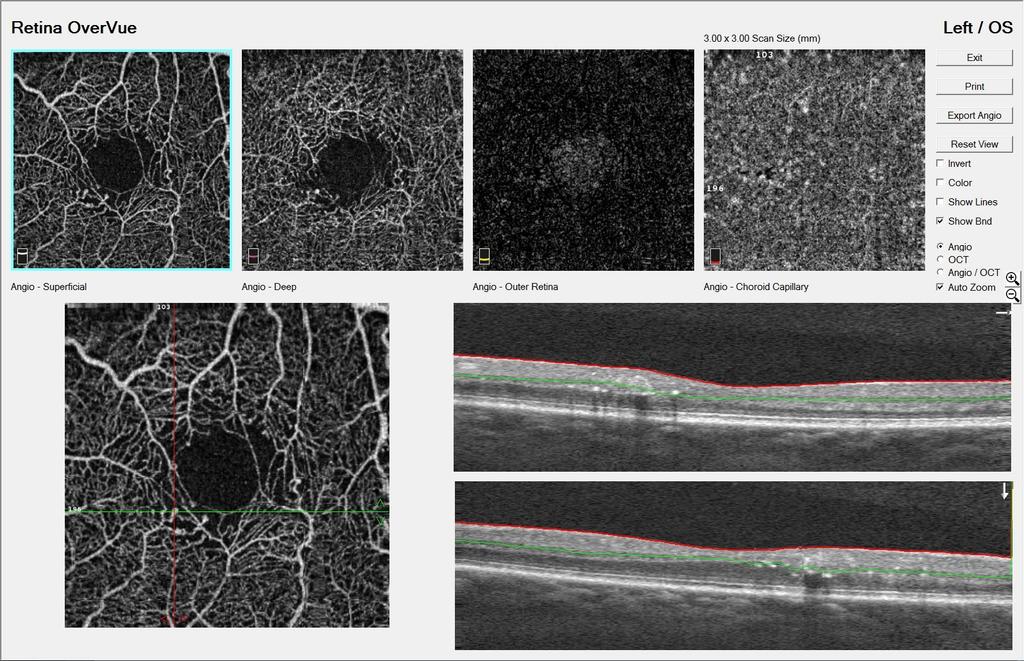
9 OCTA Dual modality- en face flow information + cross sec structure info simultaneously & depth coded Detects motion of scattering particles such as red-blood cells within sequential OCT B-scan Voxels performed repeatedly at the same location of the retina very fast Acquisition with FastTrac Data Processing powered by OMAG C (decorrelation) B-scan y Clusters of OCT B-scans. Each cluster b-scan acquired in the same position on the retina anatomy doesn't t change OCT=anatomy Blood flow OCT B-scan. Each cluster generates one Blood flow scan is the only change = movement OCTA = vascular AngioPlex Map. Process pixels Reconstructed map of the perfused microvasculature within the retina and choroid.

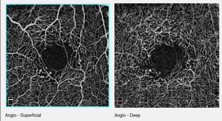
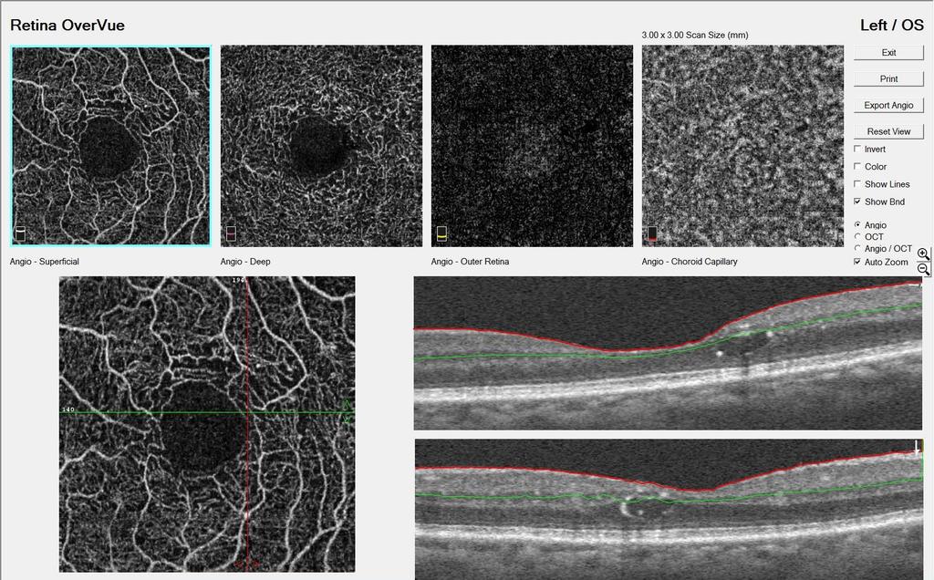
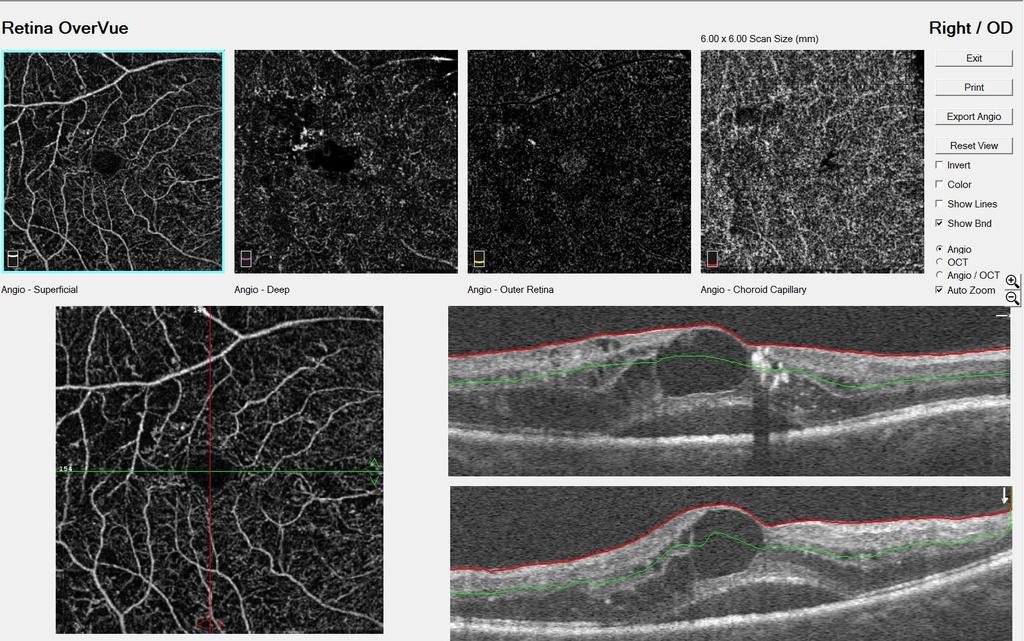
10 Full retina slab RETINAL SEGMENTATIONS Superficial Retina (ILM to IPL) Deep Retina IPL to OPL Avascular Retina Below OPL Color Depth Retinal Map can help you determine which section you need to look more carefully combines superficial, deep and avascular maps ILM to RPE OCTA Maps are flattened 2D representations vasculature. Color Represents Depth in Tissue Slide Courtesy of Diana Shechtman, OD


11 CHOROIDAL VASCULATURE Normal should be dense and homogenous Choriocapillaris Choroid Drusen Choroidal atrophy Slide Courtesy of Diana Shechtman, OD
12 TECHNOLOGY AngioVue (Optovue) AngioPlex (Zeiss) Spectralis OCT Angiography (Heidelberg)
13 OCTA ANALYSIS

14 NORMAL OCTA Superficial Retina (ILM to IPL) Deep Retina IPL to OPL Avascular Retina Below OPL Choriocapillaris Choroid Slide Courtesy of Diana Shechtman, OD


15 NORMAL OCTA Capillary Density
16 COMPARISON TO FLUORESCEIN ANGIOGRAPHY AND ICG Advantages of OCTA Non-invasive imaging, no use of exogenous dye No risk of adverse affect (from dye) Fast! Takes 3-4 seconds compared to several minutes Repeatability Higher resolution Able to visualize fine detail of vasculature at all retinal layers because not obscured by leaking dye Provides more precise depth localization and delineation of lesions
17 COMPARISON TO FLUORESCEIN ANGIOGRAPHY AND ICG Disadvantages of OCTA Cannot visualize active leakage Not able to visualize vessels that have no flow or slower flow than detection threshold of OCTA Limited field of view still developing widefield imaging Susceptible to artifacts (motion, blink and shadowing)
18 APPLICATIONS OF OCTA
19 DIABETIC RETINOPATHY
20 DIABETIC RETINOPATHY Mostly affects the superficial capillary plexus (SCP) Easier to identify vascular anomalies associated with DR: Microaneurysms Retinal capillary dropout Enlargement and distortion of foveal avascular zone Vascular loops Neovascularization
21 DIABETIC RETINOPATHY Study by Carlos et al eyes with DM with no DR and 28 control eyes of healthy subjects OCTA able to image foveal microvascular changes not detected by clinical examination. FAZ remodeling more in diabetic than in control eyes (36% vs 11%) More capillary non profusion in diabetic eyes (21% vs 4%) Conclusion: diabetic eyes showed statistically significant FAZ enlargement compared to healthy eyes, irrespective of presence of DR.
22 DIABETIC RETINOPATHY: MICROANEURYSMS
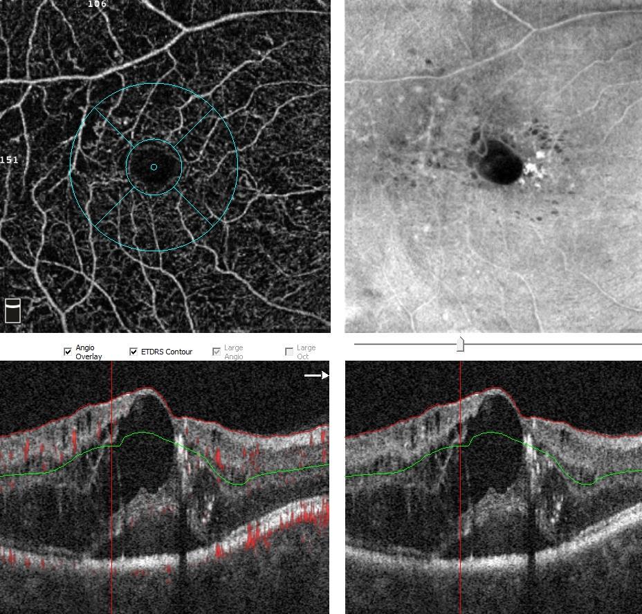
23 NVD


24 BUT MY PATIENT SEES 20/ YO African American Male Type 2 DM x 10 years Best corrected vision 20/20 OD, OS


25 BUT MY PATIENT SEES 20/20.
26 OCTA DETECTION OF MICROVASCULAR CHANGES IN DM

27 OCTA DETECTION OF MICROVASCULAR CHANGES IN DM

28 OCTA DETECTION OF MICROVASCULAR CHANGES IN DM
29 PROLIFERATIVE DIABETIC RETINOPATHY

30 AGE-RELATED MACULAR DEGENERATION
 but sensitivity was low")
31 Sambhav et al 2017 AGE RELATED MACULAR DEGENERATION OCTA is great tool for monitoring Dry to Wet conversion Recognize subtle CNVM and get treatment promptly OCTA provides ability to visualize choroidal neovascularization in avascular layer or choriocapillaris Study with 48 eyes with confirmed CNV, specificity of CNV detection on OCTA compared to FA was high (91%) but sensitivity was low (50%). (Carlos et al. 2015)

 Modified classification from J.")

32 TYPES OF CNVM Type 1: Occult (beneath the RPE layer) Type 2: Classic (above the RPE layer and has adjacent SRF leakage) Type 3: RAP (Retinal Angiomatous Proliferation) Modified classification from J. Jung and K.B. Freund. All OCT-Angiography images have been obtained using the AngioVue OCT system from Optovue (Fremont, California)

33 TYPES OF CNVM Modified classification from J. Jung and K.B. Freund. All OCT-Angiography images have been obtained using the AngioVue OCT system from Optovue (Fremont, California)

34 POLYPOIDAL CHOROIDAL VASCULOPATHY
35 POLYPOIDAL CHOROIDAL VASCULOPATHY
 with")


36 POLYPOIDAL CHOROIDAL VASCULOPATHY 1982 by Yannuzzi: Characterized by branching vascular network (BVN) with adjacent polypoidal lesions (dilations/polyps) at the terminal ends Multiple, recurrent serosanguineous detachments of the RPE (PED) and/or neurosensory retina Associated with secondary bleeding or leakage from the polypoidal lesions. ICG Serosanguineous PED
. Comparison of indocyanine green angiography and optical coherence tomographic angiography in polypoidal choroidal vasculopathy.")
37 POLYPOIDAL CHOROIDAL VASCULOPATHY ICGA VS OCTA? ICG OCTA: Outer Retina OCTA: Choroidal Capillaries Polyps detected: 100% by ICGA 85% by OCTA BVN detected: 70% ICGA 70% OCTA Takayama, K., Ito, Y., Kaneko, H., Kataoka, K., Sugita, T., Maruko, R., Terasaki, H. (2017). Comparison of indocyanine green angiography and optical coherence tomographic angiography in polypoidal choroidal vasculopathy. Eye, 31(1),

38 CHRONIC CENTRAL SEROUS CHORIORETINOPATHY

 for detection of CNV in eyes with chronic")
39 CHRONIC CSCR OCTA is helpful in identifying active CNVM Bonini Filho et al reports that OCTA has high sensitivity and specificity (compared to FA) for detection of CNV in eyes with chronic CSCR

40 MACULAR TELANGIECTASIAS

41 MACULAR TELANGIECTASIA A congenital or developmental vascular disorder Exudative dilations of perifoveal retinal capillaries Type 1:Aneurysmal Males, unilateral, 4 th or 5 th decade VA 20/40 or better Dilation of capillaries, aneurysms, leakage and non-profusion to temporal macula Limited to SCP and DCP

42 MACULAR TELANGIECTASIA Microaneurysms, capillary outbursts, vascular abnormalities and sclerotic vessels easy to visualize in SCP and DCP on OCTA Early identification leads to prompt treatment and appropriate blood work

43 RETINAL VASCULAR OCCLUSIONS
44 RETINAL VASCULAR OCCLUSIONS CRVO and BRVO is thrombosis of the retinal vein leading to impaired capillary profusion and retinal ischemia Kashani et al report findings in 26 eyes with RVO. They showed OCTA findings were consistent with clinical, anatomic and FA findings. Areas of ischemia are well delinated on OCTA and correspond with areas seen on FA. SCP and DCP can be separated allowing for better appreciation of lesions affecting primarily middle retina. OCTA often used as adjunct tool to characterize vascular occlusions (Wu, Villegas, and Kovach 2018)
45 CASE: COMBINED CRAO AND CRVO 69 YO Female presented to ER with sudden, painless vision loss after cataract surgery with retrobulbar anesthesia OS BCVA: 20/40 OD, HM OS APD OS Anterior segment OS : corneal edema, tr cell 1+ flare Posterior segment OS: Mild disc edema, macular edema, whitening of the macula, subtle tortuosity of vessels, flame-shaped hemes and cotton wool spots in all quadrants (Wu, Villegas, and Kovach 2018)
46 CASE: COMBINED CRAO AND CRVO SD-OCT OS: Hyperreflectivity and edema of the inner retina with disruption of ellipsoid zone OCTA OS: Absence of flow in foveal and perifoveal area in SCP and DCP. Normal choriocapillaris and choroid. (Wu, Villegas, and Kovach 2018)
47 CENTRAL RETINAL VEIN OCCLUSION


48 CENTRAL RETINAL VEIN OCCLUSION


49 CENTRAL RETINAL VEIN OCCLUSION
50 RETINITIS PIGMENTOSA
51 RETINITIS PIGMENTOSA RP demonstrates alterations in all macular vasculature, mostly reduction in SCP and DCP. Reduction in blood flow occurs early in disease and can lead to ischemia, retinal damage and cell death Vessel density abnormalities at the level of the DCP appear to be directly related to macular function and visual potential
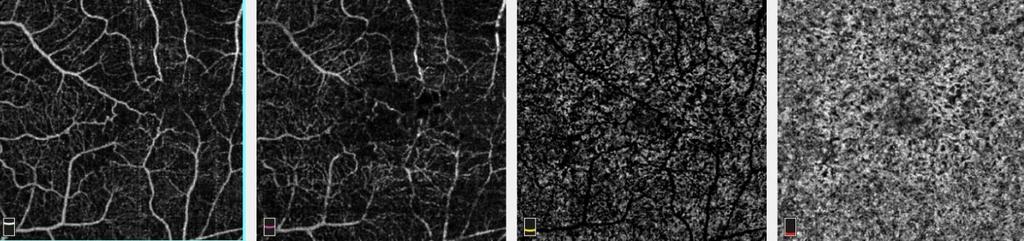
52 OCTA Macular Capillary Density at Different Stages of RP Normal Early RP Moderate RP Advanced RP SCP DCP Choriocapillaris


53 Mild RP SCP DCP Outer Retina Choriocapillaris OD OS OD OD OS


54 Moderate RP SCP DCP Outer Retina Choriocapillaris OD OS OD OS


55 Severe RP SCP DCP Outer Retina Choriocapillaris OD OS OD OS

56 GLAUCOMA AND OPTIC NEUROPATHIES
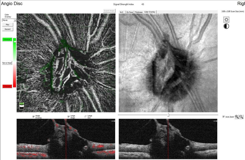
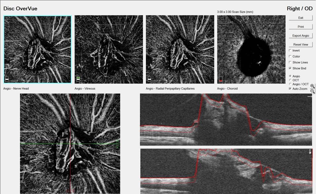
57 BLOOD SUPPLY TO ONH ONH supplied by two main sources: Central retinal artery =superficial layers (NFL) Posterior ciliary artery = deeper layers (prelaminar, lamina cribosa, and retrolaminar regions)


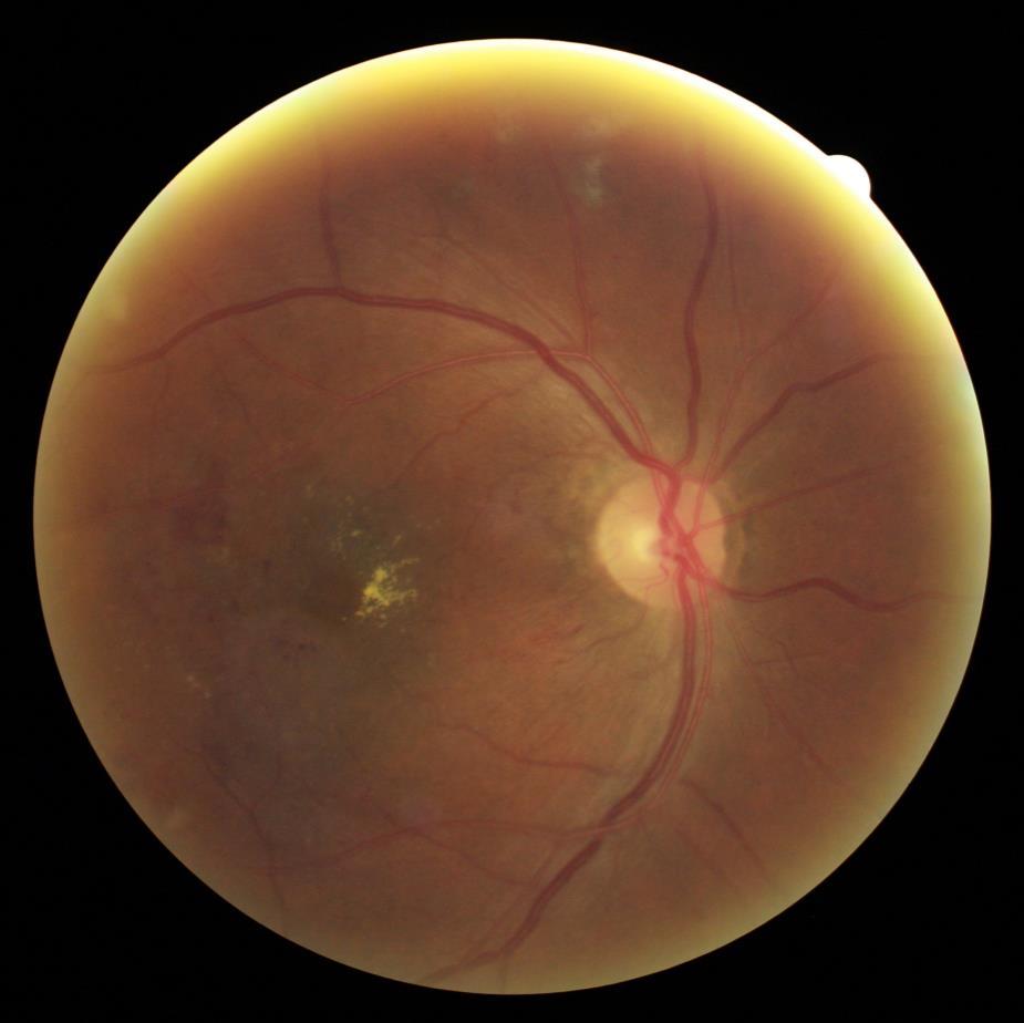
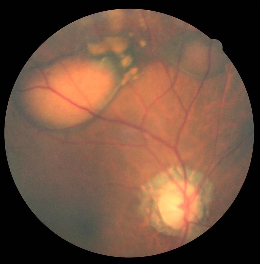
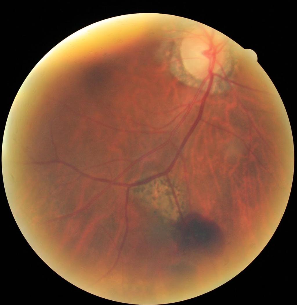
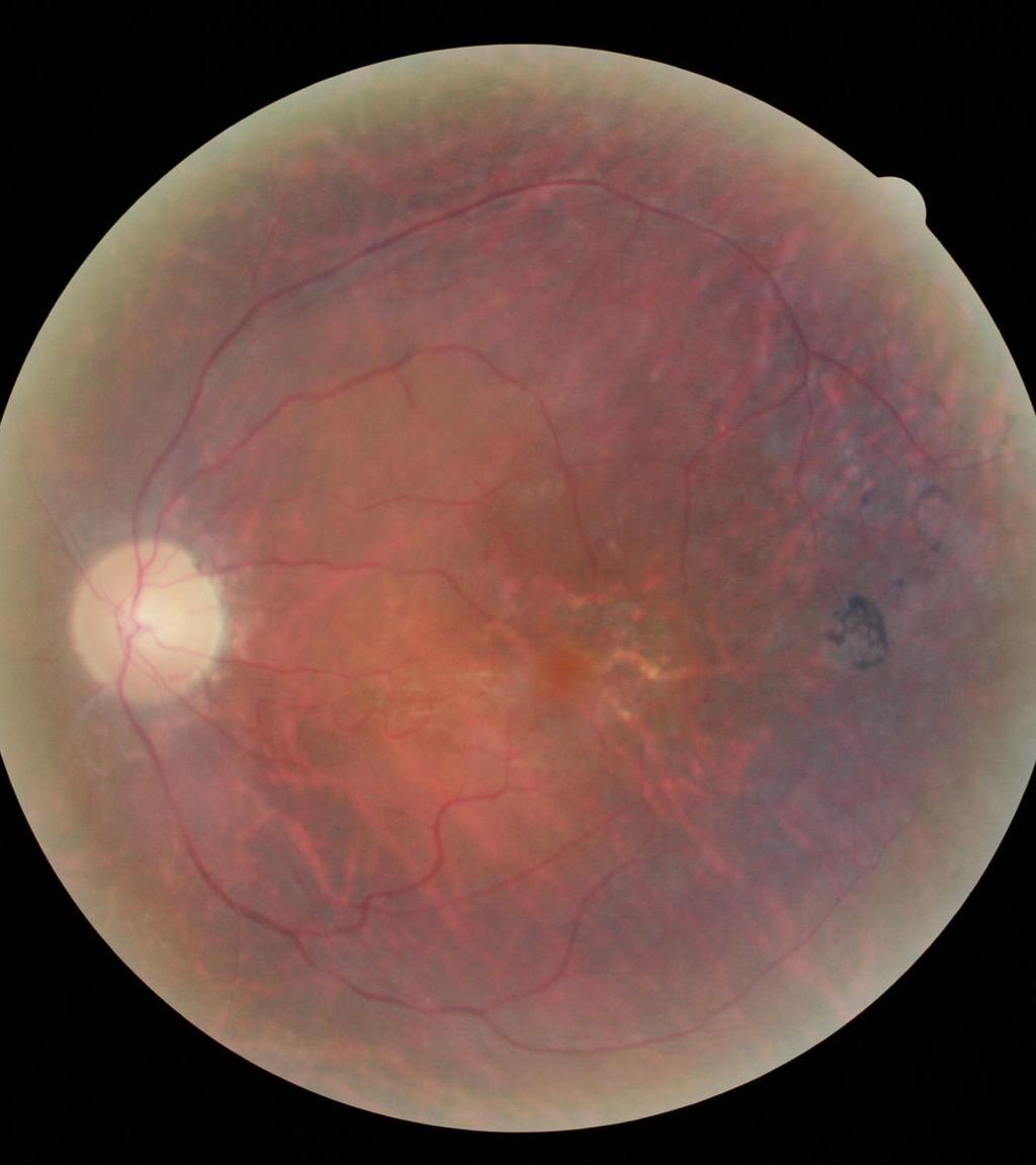
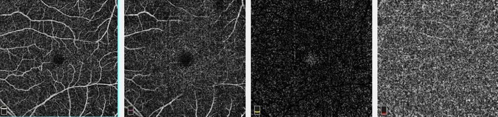
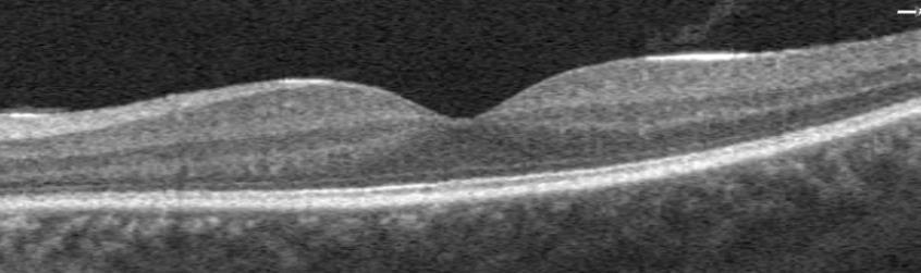
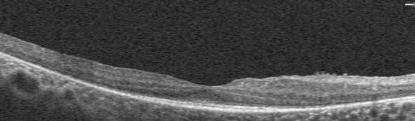
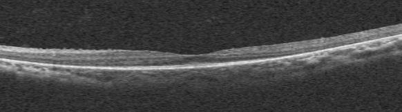
58 CASE: NON-ARTERITIC ANTERIOR ISCHEMIC OPTIC NEUROPATHY 72 yo Caucasian male presented with chronic NAION H/O: NAION occurred in right eye 13 years ago followed by a similar event in the left eye the following year. Medical Hx: HIV, diabetes type 2, and hypertension. BCVA: 20/20-3 OD, 20/25 OS. Mild APD OS. Mild R-G color deficiency OS, while the right eye was normal. Anterior segment findings were unremarkable except for a small posterior subcapsular cataract in the visual axis of the left eye. Fundus examination revealed superior temporal pallor of the right optic nerve head and generalize pallor of the left optic nerve head. Both optic nerves had distinct margins and 0.1 C/R ratio.


59 CASE: NON-ARTERITIC ANTERIOR ISCHEMIC OPTIC NEUROPATHY


60 CASE: NON-ARTERITIC ANTERIOR ISCHEMIC OPTIC NEUROPATHY
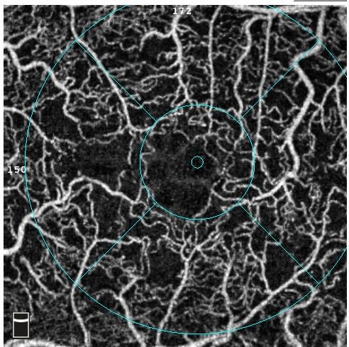
61 CASE: NON-ARTERITIC ANTERIOR ISCHEMIC OPTIC NEUROPATHY OCT angiography provides high resolution imaging of the optic nerve and peripapillary vasculature that spatially corresponds to optic nerve atrophy, retinal nerve fiber layer thickness and visual field defects in cases of chronic optic neuropathy such as NAION. Optic neuropathies are a heterogeneous group of optic nerve disorders that require different management within different timelines, but yet many have similar ophthalmoscopic presentation, usually as optic disc edema or atrophy. The characterization of these various disorders using OCTA could provide a tool that would facilitate their differential diagnosis, allowing for prompt and accurate management of the condition.
62 TAKE HOME POINTS
63 TAKE HOME POINTS OCTA is an innovated technology that can provide insight into the pathophysiology of retinal and optic nerve diseases OCTA can aid in diagnosis and prompt management of a variety of retinal and optic nerve diseases
64 REFERENCES Campbell, J. P., Zhang, M., Hwang, T. S., Bailey, S. T., Wilson, D. J., Jia, Y., & Huang, D. (2017). Detailed Vascular Anatomy of the Human Retina by Projection-Resolved Optical Coherence Tomography Angiography. Scientific Reports, 7, Samara, W. A., Shahlaee, A., Sridhar, J., Khan, M. A., Ho, A. C., & Hsu, J. (2016). Quantitative Optical Coherence Tomography Angiography Features and Visual Function in Eyes With Branch Retinal Vein Occlusion. American Journal of Ophthalmology, 166, Sambhav, K., Grover, S., & Chalam, K. V. (2017). The application of optical coherence tomography angiography in retinal diseases. Survey of Ophthalmology, 62(6), Venugopal, J. P., Rao, H. L., Weinreb, R. N., Pradhan, Z. S., Dasari, S., Riyazuddin, M., Webers, C. A. (2018). Repeatability of vessel density measurements of optical coherence tomography angiography in normal and glaucoma eyes. British Journal of Ophthalmology, 102(3),
Incorporating OCT Angiography Into Patient Care
 Incorporating OCT Angiography Into Patient Care Beth A. Steele, OD, FAAO OCT A: Introduction Isolates microvascular circulation from OCT image data Axial resolution = 5 microns (i.e. fine capillaries visible)
OCT Angiography in Primary Eye Care
 OCT Angiography in Primary Eye Care An Image Interpretation Primer Julie Rodman, OD, MS, FAAO and Nadia Waheed, MD, MPH Table of Contents Diabetic Retinopathy 3-6 Choroidal Neovascularization 7-9 Central
Visualize. Analyze. Personalize. OCT + OCTA
 Visualize. Analyze. Personalize. OCT + OCTA A New Approach to Protecting Vision AngioVue OCT Angiography brings valuable new information to clinical practice. Non-invasive visualization of retinal vasculature.
OCT Angiography The Next Frontier
 Choroid Retina avascular 5/13/2017 OCT Angiography The Next Frontier Pierce Kenworthy OD, FAAO June 9, 2017 OCT Angiography (OCTA) 2016 Non-invasive, motion contrast imaging Represents erythrocyte movement
ZEISS AngioPlex OCT Angiography Making the revolutionary, routine.
 ZEISS AngioPlex OCT Angiography Making the revolutionary, routine. The moment that revolutionary insight becomes routine. // OCT ANGIOGRAPHY MADE BY ZEISS CIRRUS with AngioPlex creates a new era in both
FA vs. OCTA? The status of OCTA, today. Fukuoka, JSOS 2016 Gerd Klose. Korobelnik J Fr Ophthalmol (2015)
") FA vs. OCTA? The status of OCTA, today Korobelnik J Fr Ophthalmol (2015) Fukuoka, JSOS 2016 Gerd Klose 1 2 FA / ICGA a well-founded Gold standard! Benefits Useful for many pathologies High contrast, detailed
FA vs. OCTA? The status of OCTA, today Korobelnik J Fr Ophthalmol (2015) Fukuoka, JSOS 2016 Gerd Klose 1 2 FA / ICGA a well-founded Gold standard! Benefits Useful for many pathologies High contrast, detailed
OCT Interpretation in Retinal Disease
 OCT Interpretation in Retinal Disease Jay M. Haynie, OD, FAAO Financial Disclosure I have received honoraria or am on the advisory board for the following companies: Carl Zeiss Meditec Advanced Ocular
10/17/2017. FDA Approved. Zeiss AngioPlex TM Optovue AngioVue TM
 Images retinal microvasculature without dye injection Displays structure and function from a single imaging system Standard of Care-2011 DFE, Fundus Photos, VF 10-2, SD-OCT, FAF, or mferg 2016-AAO Baseline
OCT Angiography. SriniVas Sadda, MD
 OCT Angiography SriniVas Sadda, MD Professor of Ophthalmology Director, Medical Retina Unit Ophthalmic Imaging Unit University of Southern California Los Angeles, California, USA Disclosure Consulting
Is OCT-A Needed As An Investigative Tool During The Management Of Diabetic Macular Edema
 Is OCT-A Needed As An Investigative Tool During The Management Of Diabetic Macular Edema Ayman M Khattab MD, FRCS Professor of Ophthalmology Cairo University Diabetic Macular Edema (DME) Diabetic macular
ZEISS AngioPlex OCT Angiography Overview ZEISS OCT Angiography
 ZEISS AngioPlex OCT Angiography Overview ZEISS OCT Angiography California, ZEISS AngioPlex Ultra-clear visualization of microvascular blood flow using non-invasive OCT angiography 2 AngioPlex OCT Angiography
OCT Angiography. Financial Disclosures: Pre-Test: Which one is Correct?
 OCT Angiography Brandon Lujan, MD Medical Director, Casey Reading Center Assistant Professor of Ophthalmology Financial Disclosures: Genentech (Consultant, Grant support, Educational training) UC Berkeley
ZEISS AngioPlex OCT Angiography. Clinical Case Reports
 Clinical Case Reports Proliferative Diabetic Retinopathy (PDR) Case Report 969 PROLIFERATIVE DIABETIC RETINOPATHY 1 1-year-old diabetic female presents for follow-up of proliferative diabetic retinopathy
Swept-Source OCT Angiography: SS OCT Angio TM
 Swept-Source OCT Angiography: SS OCT Angio TM Not available in all countries, please check with your distributor. 2015.09 Swept-Source OCT Angiography: SS OCT Angio TM Introduction Optical coherence tomography
Deeper visualizations for intervening with confidence.
 CIRRUS OCT with AngioPlex from ZEISS Making the revolutionary routine New vascular quantification Deeper visualizations for intervening with confidence. CIRRUS OCT with AngioPlex from ZEISS can be a much
Introducing ANGIOVUE ESSENTIAL. Built on the Avanti Widefield OCT Platform. OCT Angiography for Primary Eye Care
 Introducing ANGIOVUE ESSENTIAL Built on the Avanti Widefield OCT Platform OCT Angiography for Primary Eye Care Transform Your View of the Retina OCT Angiography (OCTA) is a quick non-invasive test that
Dr/ Marwa Abdellah EOS /16/2018. Dr/ Marwa Abdellah EOS When do you ask Fluorescein angiography for optic disc diseases???
 When do you ask Fluorescein angiography for optic disc diseases??? 1 NORMAL OPTIC DISC The normal optic disc on fluorescein angiography is fluorescent due to filling of vessels arising from the posterior
Optical Coherence Tomography in Diabetic Retinopathy. Mrs Samantha Mann Consultant Ophthalmologist Clinical Lead of SEL-DESP
 Optical Coherence Tomography in Diabetic Retinopathy Mrs Samantha Mann Consultant Ophthalmologist Clinical Lead of SEL-DESP Content OCT imaging Retinal layers OCT features in Diabetes Some NON DR features
OCT-Angiography Clinical Cases. OCT-Angiography Clinical Cases
 OCT-Angiography Clinical Cases OCT-Angiography Clinical Cases NIDEK RS-3000 Advance AngioScan Daniela Bacherini Andrea Sodi Stanislao Rizzo CONTENTS Page Authors 3 Introduction 4 Case 1 Case 2 Case 3 Case
Mark Dunbar: Disclosure
 Important Things to Understand About OCT Mark T. Dunbar, O.D., F.A.A.O. Bascom Palmer Eye Institute University of Miami, School of Medicine Mark Dunbar: Disclosure Optometry Advisory Board for: Allergan
Will OCT-Angiography replace FA?
 ASL Roma A PRESIDIO TERRITORIALE NUOVO REGINA MARGHERITA AMBULATORIO PATOLOGIE RETINICHE Resp. Dott.ssa SUSANNA CATALANO CENTRO ITALIANO MACULA Will OCT-Angiography replace FA? Marco Rispoli, Luca di Antonio,
Visualize. Analyze. Personalize. OCT + OCTA. with
 Visualize. Analyze. Personalize. OCT + OCTA with Avanti Widefield OCT with AngioVue OCTA Imaging Comprehensive Structural and Functional Imaging in a Single Imaging Platform Comprehensive OCT Imaging The
The Quick Guide to OCT Mastery 50 Real Cases with Expert Analysis
 OPTICAL COHERENCE TOMOGRAPHY The Quick Guide to OCT Mastery 50 Real Cases with Expert Analysis VOL 1 Sanjay Sharma, MD, FRCS, MSc (Epid), MBA Ophthalmologist, Epidemiologist Queen s University, Canada
Disclosures. Definitions. Goals. Imaging and glaucoma 3/22/2016
 Pinakin Davey OD, PhD, FAAO Professor and Director of Research Disclosures Principal investigator for ivue OCT trial Principal investigator Topcon FDA trials for Maestro and OCT 2000 Consultant for Topcon
OCT Angiography: The Next Step in Retinal Imaging Jonathan Zelenak D.O.
 OCT Angiography: The Next Step in Retinal Imaging Jonathan Zelenak D.O. Hillsdale Hospital Michigan State University Overview Evolution of OCT How does OCT angiography work? Clinical examples Potential
Advances in OCT Murray Fingeret, OD
 Disclosures Advances in OCT Murray Fingeret, OD Consultant Alcon, Allergan, Bausch & Lomb, Carl Zeiss Meditec, Diopsys, Heidelberg Engineering, Reichert, Topcon Currently Approved OCT Devices OCT Devices
Angio-OCT. Degenerazione Maculare Legata all Eta. Giuseppe Querques
 Angio-OCT Degenerazione Maculare Legata all Eta Giuseppe Querques Department of Ophthalmology, IRCCS Ospedale San Raffaele, University Vita Salute San Raffaele, Milan, Italy Financial Disclosure ADVISORY
Non-arteritic anterior ischemic optic neuropathy (NAION) with segmental optic disc edema. Jonathan A. Micieli, MD Valérie Biousse, MD
 with segmental optic disc edema. Jonathan A. Micieli, MD Valérie Biousse, MD") Non-arteritic anterior ischemic optic neuropathy (NAION) with segmental optic disc edema Jonathan A. Micieli, MD Valérie Biousse, MD A 75 year old white woman lost vision in the inferior part of her visual
Non-arteritic anterior ischemic optic neuropathy (NAION) with segmental optic disc edema Jonathan A. Micieli, MD Valérie Biousse, MD A 75 year old white woman lost vision in the inferior part of her visual
PART 1: GENERAL RETINAL ANATOMY
 PART 1: GENERAL RETINAL ANATOMY General Anatomy At Ora Serrata At Optic Nerve Head Fundoscopic View Of Normal Retina What Is So Special About Diabetic Retinopathy? The WHO definition of blindness is
Optical Coherence Tomography: Pearls for the Anterior Segment Surgeon Basic Science Michael Stewart, M.D.
 Optical Coherence Tomography: Pearls for the Anterior Segment Surgeon Basic Science Michael Stewart, M.D. Disclosure OCT Optical Coherence Tomography No relevant financial relationships I will refer to
The diagnostic value of optical coherence tomography angiography in diabetic retinopathy: a systematic review
 https://doi.org/10.1007/s10792-018-1034-8 (0456789().,-volV) (0456789().,-volV) REVIEW The diagnostic value of optical coherence tomography angiography in diabetic retinopathy: a systematic review David
World Sight Day Case Studies. Mark Frost Screening Manager South East London DESP
 World Sight Day 2015 Case Studies Mark Frost Screening Manager South East London DESP Introduction All of the following cases have been identified in our screening programme over the last 3 years. The
SOUTH-EAST EUROPEAN JOURNAL of OPHTHALMOLOGY 2015; 1 (1) 34 40
 34 40") Review article SOUTH-EAST EUROPEAN JOURNAL of OPHTHALMOLOGY 2015; 1 (1) 34 40 Retinal nerve fiber layer versus peripapillary capillary density assessment A powerful tool for detecting optic nerve head
Review article SOUTH-EAST EUROPEAN JOURNAL of OPHTHALMOLOGY 2015; 1 (1) 34 40 Retinal nerve fiber layer versus peripapillary capillary density assessment A powerful tool for detecting optic nerve head
OCT Angiography: An Upcoming Tool for Diagnosis and Treatment of Retinal Vascular Diseases
 E-ISSN 2454-2784 Recent Advances OCT Angiography: An Upcoming Tool for Diagnosis and Treatment of Retinal Vascular Diseases Purnima Sood 1, Nalini Saxena 2, Dinesh Talwar 3 1 Vitreo-Retina Consultant,
Clinical Study Optical Coherence Tomography Angiography in Retinal Vascular Diseases and Choroidal Neovascularization
 Hindawi Publishing Corporation Journal of Ophthalmology Volume 2015, Article ID 343515, 8 pages http://dx.doi.org/10.1155/2015/343515 Clinical Study Optical Coherence Tomography Angiography in Retinal
Clinical Case Presentation. Branch Retinal Vein Occlusion. Sarita M. Registered Nurse Whangarei Base Hospital
 Clinical Case Presentation on Branch Retinal Vein Occlusion Sarita M. Registered Nurse Whangarei Base Hospital Introduction Case Study Pathogenesis Clinical Features Investigations Treatment Follow-up
Go With the Flow: An OCT Angiography Primer Lorne Yudcovitch, OD, MS, FAAO
 Go With the Flow: An OCT Angiography Primer Lorne Yudcovitch, OD, MS, FAAO yudcovil@pacificu.edu OCT Angiography (OCTA) History 2000 - First Doppler flowimetry OCT on human retina 2005 Speckle analysis
Leo Semes, OD, FAAO UAB Optometry
 Leo Semes, OD, FAAO UAB Optometry Safe; inert Has long track record - over 45 years Mixes with plasma and highlights blood vessel compromise Using specific exciting (490 nm)and absorption (510 nm) filters
11/29/2016 MACULAR MALADIES: TYPICAL & ATYPICAL CASES
 MACULAR MALADIES: TYPICAL & ATYPICAL CASES Dawn Pewitt, OD, FAAO Triad Eye Institute, Grove, OK Dpewitt@triadeye.com Disclosure Statement: No financial disclosures COPE 51218-PS Please silence all mobile
November Volume 35 - Issue 11
 November 2015 - Volume 35 - Issue 11 pp: 2161-2431,e67-e72 Editorial Optical Coherence Tomography Angiography Spaide, Richard F.; Fujimoto, James G.; Waheed, Nadia K. Original Study IMAGE ARTIFACTS IN
Cirrus TM HD-OCT. Details defi ne your decisions
 Cirrus TM HD-OCT Details defi ne your decisions 2 With high-defi nition OCT Carl Zeiss Meditec takes you beyond standard spectral domain Built on 10 years experience at the vanguard of innovation, Carl
Why Is Imaging Critical in My Uveitis Practice?
 Why Is Imaging Critical in My Uveitis Practice? Dilraj S. Grewal, MD Developed in collaboration Imaging Is the Backbone of Uveitis Workup and Monitoring Treatment Response FP FAF B- scan Multimodal Imaging
Case Report Optical Coherence Tomography Angiography of Macular Telangiectasia Type 2 with Associated Subretinal Neovascular Membrane
 Hindawi Case Reports in Ophthalmological Medicine Volume 2017, Article ID 8186134, 4 pages https://doi.org/10.1155/2017/8186134 Case Report Optical Coherence Tomography Angiography of Macular Telangiectasia
Course # Getting to Know Your OCT
 Course # 140 Getting to Know Your OCT Course Title: Lecturer: Getting to Know Your OCT Brad Sutton, OD, FAAO IU School of Optometry Financial Disclosures No financial disclosures Optical Coherence Tomography-OCT
Optical Coherence Tomograpic Features in Idiopathic Retinitis, Vasculitis, Aneurysms and Neuroretinitis (IRVAN)
") Columbia International Publishing Journal of Ophthalmic Research (2014) Research Article Optical Coherence Tomograpic Features in Idiopathic Retinitis, Vasculitis, Aneurysms and Neuroretinitis (IRVAN)
Columbia International Publishing Journal of Ophthalmic Research (2014) Research Article Optical Coherence Tomograpic Features in Idiopathic Retinitis, Vasculitis, Aneurysms and Neuroretinitis (IRVAN)
ANSWERING THE WHY? Clinicians discuss the latest imaging technologies for retina practice BY PETER K. KAISER, MD
 Insert to March 2018 Sponsored by MULTI-MODALITY IMAGING: LATEST EVOLUTIONS IN OCTA AND UWF As the array of safe and efficacious medical and surgical options for retinal diseases expands, so does the need
Retinal Complications of Obstructive Sleep Apnea A Growing Concern!
 Retinal Complications of Obstructive Sleep Apnea A Growing Concern! Jay M. Haynie, OD, FAAO Financial Disclosure I have received honoraria or am on the advisory board for the following companies: Carl
Ganglion cell analysis by optical coherence tomography (OCT) Jonathan A. Micieli, MD Valérie Biousse, MD
 Jonathan A. Micieli, MD Valérie Biousse, MD") Ganglion cell analysis by optical coherence tomography (OCT) Jonathan A. Micieli, MD Valérie Biousse, MD Figure 1. Normal OCT of the macula (cross section through the line indicated on the fundus photo)
Ganglion cell analysis by optical coherence tomography (OCT) Jonathan A. Micieli, MD Valérie Biousse, MD Figure 1. Normal OCT of the macula (cross section through the line indicated on the fundus photo)
Principle of OCT. Reading Between the Lines: OCT Interpretation. Initial Concept. Advantage: High Resolution Cross Section Images
 Principle of OCT Reading Between the Lines: OCT Interpretation Mohammad Rafieetary, OD, FAAO mrafieetary@charlesretina.com Introduction Optical Biopsy Morphologic Evaluation of Live Tissue Measurements
What Is O.C.T. and Why Should I Give A Rip? OCT & Me How Optical Coherence Tomography Changed the Life of a Small Town Optometrist 5/19/2014
 OCT & Me How Optical Coherence Tomography Changed the Life of a Small Town Optometrist Email: myoder@wcoil.com Mark A. Yoder, O.D. 107 N. Main Street PO Box 123 Bluffton, OH 45817 @yoderod 115.02 Histoplasma
Evaluation of efficacy of eplerenone in the management of chronic central serous choroidoretinopathy
 Original article: Evaluation of efficacy of eplerenone in the management of chronic central serous choroidoretinopathy Dr. Sushant Madaan* Department of Ophthalmology, NIMS Medical College and Hopsital,Jaipur,
OCCLUSIVE VASCULAR DISORDERS OF THE RETINA
 OCCLUSIVE VASCULAR DISORDERS OF THE RETINA Learning outcomes By the end of this lecture the students would be able to Classify occlusive vascular disorders (OVD) of the retina. Correlate the clinical features
CAPILLARY NETWORK ANOMALIES IN BRANCH RETINAL VEIN OCCLUSION ON OPTICAL COHERENCE TOMOGRAPHY ANGIOGRAPHY
 CAPILLARY NETWORK ANOMALIES IN BRANCH RETINAL VEIN OCCLUSION ON OPTICAL COHERENCE TOMOGRAPHY ANGIOGRAPHY MARCO RISPOLI, MD, MARIA CRISTINA SAVASTANO, MD, PHD, BRUNO LUMBROSO, MD Purpose: To analyze the
Neuropathy (NAION) and Avastin. Clinical Assembly of the AOCOO-HNS Foundation May 9, 2013
 and Avastin. Clinical Assembly of the AOCOO-HNS Foundation May 9, 2013") Non Arteritic Ischemic Optic Neuropathy (NAION) and Avastin Shalom Kelman, MD Clinical Assembly of the AOCOO-HNS Foundation May 9, 2013 Anterior Ischemic Optic Neuropathy Acute, painless, visual loss,
Non Arteritic Ischemic Optic Neuropathy (NAION) and Avastin Shalom Kelman, MD Clinical Assembly of the AOCOO-HNS Foundation May 9, 2013 Anterior Ischemic Optic Neuropathy Acute, painless, visual loss,
Diabetic Retinopathy
 Diabetic Retinopathy Diabetes can be classified into type 1 diabetes mellitus and type 2 diabetes mellitus, formerly known as insulin-dependent diabetes mellitus, and non-insulin diabetes mellitus, respectively.
ANGIO OCT IMAGING OF MACULAR VASCULATURE IN DIABETIC MACULAR EDEMA BEFORE AND AFTER MACULAR SURGERY
 17th EVRS Meeting September 14-17, 2017 Teatro della Pergola FLORENCE - ITALY ANGIO OCT IMAGING OF MACULAR VASCULATURE IN DIABETIC MACULAR EDEMA BEFORE AND AFTER MACULAR SURGERY G. Macrì, G. Pacelli, V.
OCT Fundal Angiography Initial Experience The new era in Medical Retina Imaging Based on Cirrus 5000 AngioPlex 2016 Model Sheena George & Nicholas
 OCT Fundal Angiography Initial Experience The new era in Medical Retina Imaging Based on Cirrus 5000 AngioPlex 2016 Model Sheena George & Nicholas Lee Consultants Ophthalmologist at The Hillingdon Hospital
EyePACS Grading System (Part 2): Detecting Presence and Severity of Background (Non-Proliferative) Diabetic Retinopathy Lesion
: Detecting Presence and Severity of Background (Non-Proliferative) Diabetic Retinopathy Lesion") EyePACS Grading System (Part 2): Detecting Presence and Severity of Background (Non-Proliferative) Diabetic Retinopathy Lesion George Bresnick MD MPA Jorge Cuadros OD PhD Anatomy of the eye: 3 Normal Retina
EyePACS Grading System (Part 2): Detecting Presence and Severity of Background (Non-Proliferative) Diabetic Retinopathy Lesion George Bresnick MD MPA Jorge Cuadros OD PhD Anatomy of the eye: 3 Normal Retina
Building The Retina Company
 Building The Retina Company Optos devices produce ultra-widefield (UWF ), high resolution images (optomap ) of approximately 82% (200 ) of the retina. A single optomap can document the retina from the
Title: OCT Analysis Workshop: Interpretation of OCT printouts
 Title: OCT Analysis Workshop: Interpretation of OCT printouts Authors: David Yang, OD, FAAO Staff Optometrist, VA Palo Alto Health Care System Associate Clinical Professor, UC Berkeley School of Optometry
Clinically Significant Macular Edema (CSME)
") Clinically Significant Macular Edema (CSME) 1 Clinically Significant Macular Edema (CSME) Sadrina T. Shaw OMT I Student July 26, 2014 Advisor: Dr. Uwaydat Clinically Significant Macular Edema (CSME) 2
Clinically Significant Macular Edema (CSME) 1 Clinically Significant Macular Edema (CSME) Sadrina T. Shaw OMT I Student July 26, 2014 Advisor: Dr. Uwaydat Clinically Significant Macular Edema (CSME) 2
Retinal Capillary Network and Foveal Avascular Zone in Eyes with Vein Occlusion and Fellow Eyes Analyzed With Optical Coherence Tomography Angiography
 Retinal Capillary Network and Foveal Avascular Zone in Eyes with Vein Occlusion and Fellow Eyes Analyzed With Optical Coherence Tomography Angiography The MIT Faculty has made this article openly available.
OCT Interpretation. Financial Disclosure. Jay M. Haynie, OD, FAAO. OCT Image Layers 7/21/2014
 OCT Interpretation Jay M. Haynie, OD, FAAO Financial Disclosure I have received honoraria or am on the advisory board for the following companies: Olympia Tacoma Renton Kennewick - Washington Carl Zeiss
 measure of your overall performance. An isolated glucose test is helpful to let you know what your sugar level is at one moment, but it doesn t tell you whether or not your diabetes is under adequate control
The Evaluation of Diabetic Macular Ischemia Using Optical Coherence Tomography Angiography
 Retina The Evaluation of Diabetic Macular Ischemia Using Optical Coherence Tomography Angiography Patrick D. Bradley, 1 Dawn A. Sim, 1 Pearse A. Keane, 1 João Cardoso, 1,2 Rupesh Agrawal, 1 Adnan Tufail,
The role of OCT-A in retinal disease management
 Graefe's Archive for Clinical and Experimental Ophthalmology (2018) 256:2019 2026 https://doi.org/10.1007/s00417-018-4109-3 REVIEW ARTICLE The role of OCT-A in retinal disease management Francisco J. Rodríguez
Cirrus TM HD-OCT. Details define your decisions
 Cirrus TM HD-OCT Details define your decisions 2 With high-definition OCT Carl Zeiss Meditec takes you beyond standard spectral domain Built on 10 years experience at the vanguard of innovation, Carl Zeiss
Spontaneous Large Serous Retinal Pigment Epithelial Tear
 This is an Open Access article licensed under the terms of the Creative Commons Attribution-NonCommercial-NoDerivs 3.0 License (www.karger.com/oa-license), applicable to the online version of the article
Venous Occlusive Diseases
 Venous Occlusive Diseases Bruce R. Saran, MD Adjunct Assistant Clinical Professor of Medicine Scheie Eye Institute University of Pennsylvania School of Medicine Philadelphia, PA -a division of: RVO Demographics
Mild NPDR. Moderate NPDR. Severe NPDR
 Diabetic retinopathy Diabetic retinopathy is the most common cause of blindness in adults aged 35-65 years-old. Hyperglycaemia is thought to cause increased retinal blood flow and abnormal metabolism in
Vascular Disease Ocular Manifestations of Systemic Hypertension
 Vascular Disease Ocular Manifestations of Systemic Hypertension Maynard L. Pohl, OD, FAAO Pacific Cataract & Laser Institute 10500 NE 8 th Street, Suite 1650 Bellevue, WA 98004 USA 425-462-7664 Cerebrovascular
The retinal function imager and clinical applications
 Su and Garg Eye and Vision (2018) 5:20 https://doi.org/10.1186/s40662-018-0114-1 REVIEW Open Access The retinal function imager and clinical applications Daniel Su and Sunir Garg * Abstract Background:
OCT Angiography: The Newest Frontier for the Revolutionary Technology
 Supplement April 2015 OCT Angiography: The Newest Frontier for the Revolutionary Technology OCT Angiography is a new non-invasive, motion contrast micro-vascular imaging modality. Based on two patented
연령연관황반변성에서망막혈관종성증식과동반된망막색소상피박리의임상양상과일차적인광역학치료의결과
 연령연관황반변성에서망막혈관종성증식과동반된망막색소상피박리의임상양상과일차적인광역학치료의결과 40 Table. Clinical characteristics and results of patients undergoing photodynamic therapy for retinal angiomatous proliferation Patients No. Age/ sex Eye
! Honoraria. " Kemin " Nicox " Review of Optometry " Optometric Management " VSP. ! Scientific Advisory Boards
 Financial Disclosure: JP Choroid Chronicles Joseph J. Pizzimenti, OD, FAAO pizzimen@nova.edu! Honoraria " Kemin " Nicox " Review of Optometry " Optometric Management " VSP! Scientific Advisory Boards "
Age-related Macular Degeneration Update
 Age-related Macular Degeneration Update AMD: The Burden of Disease Carlo J. Pelino, OD, FAAO cpelino@salus.edu Joseph J. Pizzimenti, OD, FAAO pizzimen@nova.edu Course Goals Statement of the problem Epidemiology
Case Report Peripapillary Intrachoroidal Cavitation in Myopia Evaluated with Multimodal Imaging Comprising (En-Face) Technique
 Technique") Case Reports in Ophthalmological Medicine Volume 2015, Article ID 890876, 5 pages http://dx.doi.org/10.1155/2015/890876 Case Report Peripapillary Intrachoroidal Cavitation in Myopia Evaluated with Multimodal
Case Reports in Ophthalmological Medicine Volume 2015, Article ID 890876, 5 pages http://dx.doi.org/10.1155/2015/890876 Case Report Peripapillary Intrachoroidal Cavitation in Myopia Evaluated with Multimodal
IN NICU OCT UTILIZES A CONCEPT KNOWN AS INTERFEROMETRY APPLICATIONS FOR OCT THE PRIMARY USE IN THE EYE - RETINA
 2016 25 YEARS OF OPTICAL COHERENCE TOMOGRAPHY OPTICAL COHERENCE TOMOGRAPHY IN NICU Marcin Stopa, MD, PhD, FEBO Department of Ophthalmology, Chair of Ophthalmology and Optometry. Poznan University of Medical
Case Report: Indocyanine Green Dye Leakage from Retinal Artery in Branch Retinal Vein Occlusion
 Case Report: Indocyanine Green Dye Leakage from Retinal Artery in Branch Retinal Vein Occlusion Hiroki Fujita, Kyoko Ohno-Matsui, Soh Futagami and Takashi Tokoro Department of Visual Science, Tokyo Medical
The College of Optometrists - Learning outcomes for the Professional Certificate in Medical Retina
 Learning outcomes for the Professional Certificate in Medical Retina, incorporating diabetic retinopathy screening and age related macular degeneration The professional certificate is a prerequisite to
Neuro-Ocular Grand Rounds Anthony B. Litwak,OD, FAAO VA Medical Center Baltimore, Maryland
 Neuro-Ocular Grand Rounds Anthony B. Litwak,OD, FAAO VA Medical Center Baltimore, Maryland Dr. Litwak is on the speaker and advisory boards for Alcon and Zeiss Meditek COMMON OPTIC NEUROPATHIES THAT CAN
The Human Eye. Cornea Iris. Pupil. Lens. Retina
 The Retina Thin layer of light-sensitive tissue at the back of the eye (the film of the camera). Light rays are focused on the retina then transmitted to the brain. The macula is the very small area in
CENTRAL SEROUS CHORIORETINOPATHY (CSC) IS
 IS") Association Between the Efficacy of Half-Dose Photodynamic Therapy With Indocyanine Green Angiography and Optical Coherence Tomography Findings in the Treatment of Central Serous Chorioretinopathy MASSIMO
Association Between the Efficacy of Half-Dose Photodynamic Therapy With Indocyanine Green Angiography and Optical Coherence Tomography Findings in the Treatment of Central Serous Chorioretinopathy MASSIMO
Simply the best OCT & OCTA image quality.
 Avanti Widefield OCT with AngioVue OCT Angiography Simply the best OCT & OCTA image quality. Dear Friends of Optovue, Since introducing Spectral Domain OCT to the ophthalmology market in 2006, Optovue
Research Article Diabetic Macular Ischemia Diagnosis: Comparison between Optical Coherence Tomography Angiography and Fluorescein Angiography
 Ophthalmology Volume 2016, Article ID 3989310, 6 pages http://dx.doi.org/10.1155/2016/3989310 Research Article Diabetic Macular Ischemia Diagnosis: Comparison between Optical Coherence Tomography Angiography
Role of Fluorescein angiography in evaluation of posterior segment disorders
 Original Article Role of Fluorescein angiography in evaluation of posterior segment disorders Arvind R, Surendar S 2, Ch. Jagan Mohan Rao 3 Associate Professor, 2 Postgraduate student, 3 Senior resident,
Neuro-Ocular Grand Rounds
 Neuro-Ocular Grand Rounds Anthony B. Litwak,OD, FAAO VA Medical Center Baltimore, Maryland Dr. Litwak is on the speaker and advisory boards for Alcon and Zeiss Meditek COMMON OPTIC NEUROPATHIES THAT CAN
Experience Spectacular Retinal Imaging with the new NIDEK F-10 Digital Ophthalmoscope
 Experience Spectacular Retinal Imaging with the new NIDEK F-10 Digital Ophthalmoscope The F-10 was developed to give Ophthalmologists a high definition (HD) diagnostic imaging system. Designed to provide
Diagnosis in AMD. Managing your AMD Patients
 Managing your AMD Patients Robert W. Dunphy, O.D., F.A.A.O. Diagnosis in AMD Have suspicion Identify relative risk Conduct surveillance Biometry Utilize technology to facilitate detection of change / stability
What You Should Know About Acute Macular Neuroretinopathy
 What You Should Know About Acute Macular Neuroretinopathy David J. Browning MD, PhD Chong Lee BS Acute macular neuroretinopathy is a condition characterized by the sudden, painless onset of paracentral
Moving forward with a different perspective
 Moving forward with a different perspective The Leader In Vision Diagnostics Offers A New Perspective Marco has served the eyecare community by offering exceptional lane products and automated high tech
OCT Image Analysis System for Grading and Diagnosis of Retinal Diseases and its Integration in i-hospital
 Progress Report for1 st Quarter, May-July 2017 OCT Image Analysis System for Grading and Diagnosis of Retinal Diseases and its Integration in i-hospital Milestone 1: Designing Annotation tool extraction
Chris Brown, M.D. Eye Specialty Group, PLC Continuing Education Series
 Chris Brown, M.D. Eye Specialty Group, PLC 2018 Continuing Education Series Disclaimer I have no financial interests in this lecture or any information discussed therein Objectives Fluorescein Angiogram
Choroidal Mapping; a Novel Approach for Evaluating Choroidal Thickness and Volume
 Imaging Technique Choroidal Mapping; a Novel Approach for Evaluating Choroidal Thickness and Volume Jila Noori 1, MD; Mohammad Riazi Esfahani 1,2, MD Fedra Hajizadeh 2, MD; Mohammad-Mehdi Zaferani 1, MD
Fluorescein Angiography
 Last revision: October 2011 by Luis Arias Fluorescein Angiography Authors: Luis Arias, MD Hospital Universitari de Bellvitge - University of Barcelona. Spain Jordi Monés, MD Institut de la Màcula i de
COEXISTENCE OF OPTIC NERVE HEAD DRUSEN
 COEXISTENCE OF OPTIC NERVE HEAD DRUSEN AND COMBINED HAMARTOMA OF THE RETINA AND RETINAL PIGMENT EPITHELIUM IN A TAIWANESE MALE Yo-Chen Chang 1 and Rong-Kung Tsai 2,3 1 Department of Ophthalmology, Kaohsiung
Do You See What I See!!! Shane R. Kannarr, OD
 Do You See What I See!!! Shane R. Kannarr, OD skannarr@kannarreyecare.com Define Specialty Testing Additional Test to: Prove/Disprove Diagnosis To monitor progression of a condition To document a condition
THE ROLE OF anti-vegf IN DIABETIC RETINOPATHY AND AGE RELATED MACULAR DEGENERATION
 THE ROLE OF anti-vegf IN DIABETIC RETINOPATHY AND AGE RELATED MACULAR DEGENERATION MOESTIDJAB DEPARTMENT OF OPHTHALMOLOGY SCHOOL OF MEDICINE AIRLANGGA UNIVERSITY DR SOETOMO HOSPITAL SURABAYA INTRODUCTION
TOPCON EURETINA Clinical Advances and Applications With Swept Source OCT and Angiography. JANUARY/FEBRUARY 2019 VOL. 17, NO.
 SUPPLEMENT TO SPONSORED BY JANUARY/FEBRUARY 2019 VOL. 17, NO. 1 TOPCON EURETINA 2018 Clinical Advances and Applications With Swept Source OCT and Angiography. This supplement summarizes highlights from
Adaptive Optics and OCTA: Update on Retinal Imaging. Judy E. Kim, MD Professor of Ophthalmology Medical College of Wisconsin
 Adaptive Optics and OCTA: Update on Retinal Imaging Judy E. Kim, MD Professor of Ophthalmology Medical College of Wisconsin Financial Disclosure Advisory Board Alimera Science, Allergan, Bayer, Novartis