Case Report Catastrophic Antiphospholipid Syndrome Presenting as Bilateral Central Retinal Artery Occlusions
|
|
|
- Rosalind Weaver
- 5 years ago
- Views:
Transcription
1 Hindawi Publishing Corporation Case Reports in Ophthalmological Medicine Volume 2015, Article ID , 5 pages Case Report Catastrophic Antiphospholipid Syndrome Presenting as Bilateral Central Retinal Artery Occlusions Steven S. Saraf, Yogin P. Patel, Ankit Desai, and Uday R. Desai Department of Ophthalmology, Henry Ford Hospital, Detroit, MI, USA Correspondence should be addressed to Steven S. Saraf; ssaraf1@hfhs.org Received 14 December 2014; Accepted 12 January 2015 Academic Editor: Maurizio Battaglia Parodi Copyright 2015 Steven S. Saraf et al. This is an open access article distributed under the Creative Commons Attribution License, which permits unrestricted use, distribution, and reproduction in any medium, provided the original work is properly cited. A previously healthy 22-year-old African American woman presented with bilateral vision loss associated with headache. Her ocular examination was significant for bilateral retinal arterial boxcarring, retinal whitening, retinal hemorrhages, and cherry red spots. She was diagnosed with bilateral central retinal artery occlusions and was hospitalized due to concomitant diagnosis of stroke and hypercoagulable state. She was also found to be in heart failure and kidney failure. Rheumatology was consulted and she was diagnosed with catastrophic antiphospholipid syndrome in association with systemic lupus erythematosus. Approximately 7 months after presentation, the patient s vision improved and remained stable at 20/200 and 20/ Introduction Catastrophic antiphospholipid syndrome (CAPS) is a rare autoimmune entity consisting of life threatening intravascular thrombosis leading to multiorgan failure. Immune complex formation results in widespread platelet aggregation, coagulation, and immune response. The cascade of events is often triggered by a preceding insult such as infection, surgery, or medication change. The disease may present as a primary condition or in association with an underlying process, most commonly systemic lupus erythematosus (SLE). Specificdiagnosticcriteriaincludetheinvolvementofatleast three organ systems, evidence of intravascular thrombosis, andpresenceofantiphospholipidantibodies[1]. Ocular involvement is rare, with only 5.8% of cases reporting retinal findings [2]. To our knowledge, there has been one case report in the literature describing CAPS associated with choroidal thrombosis with ultimately good visual prognosis [3]. We present a rare case of CAPS in a previously healthy 22-year-old African American woman presenting with bilateral CRAO with profound vision loss. 2. Case Report A previously healthy 22-year-old African American woman presented with progressive vision loss and headache. She denied any trauma, pain, pain with eye movements, flashes, or floaters but complained of lethargy. A head CT showed a subacute infarct of the posteroparietal and occipital lobes. The patient was hospitalized and a hypercoagulability work-up was ordered. Initial laboratory testing showed anemia, thrombocytopenia, elevated creatinine, and positive antiphospholipid antibodies. Echocardiography revealed valvular vegetations. Ophthalmic evaluation demonstrated best corrected visual acuity of hand motions in the right eye and 20/200 in the left eye. An afferent pupillary defect was appreciated in the right eye. Extraocular movements were full. Confrontational visual fields showed global depression in the right eye and inferior hemifield depression in the left. Tonometry was 15 and 11 by applanation in the right and left eyes, respectively. Slit-lamp examination of the right eye was remarkable for anteriorvitreousredcellswhilethelefteyewasquiet.dilated fundus exam revealed pale optic nerves, disc hemorrhages, disc neovascularization, arterial attenuation, and arterial boxcarring. Multiple retinal hemorrhages, retinal whitening, and a cherry red spot were noted in both eyes (Figure 1). Fluorescein angiography showed extensive retinal nonperfusion bilaterally (Figure 2). Macular optical coherence tomography showed bilateral cystoid macular edema with distortion of the foveal contour (Figure 3).


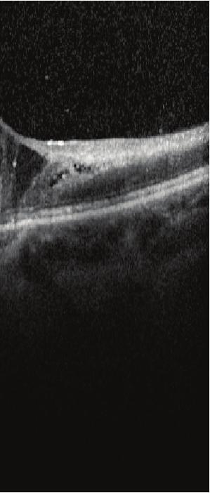
2 2 Case Reports in Ophthalmological Medicine Figure 1: Color fundus photos of each eye show bilateral retinal arterial boxcarring, retinal whitening, intraretinal and preretinal hemorrhages, and cherry red spots. Figure 2: Fluorescein angiography of both eyes in the late phases shows minimal perfusion to the peripapillary areas with late leakage of the filling vessels. OCT 20 (5.9 mm) ART (9) Q: 27 [HS] OCT 20 (5.9 mm) ART (9) Q: 29 [HS] OD OS Figure 3: Optical coherence tomography of maculae shows cystoid macular edema and loss of foveal contour.
3 Case Reports in Ophthalmological Medicine 3 Table 1: Revised diagnostic criteria for systemic lupus erythematosus as defined by SLICC. SLICC diagnostic criteria for systemic lupus erythematosus [4] Requirements include at least 4 criteria At least one criterion must be met in the clinical and laboratory categories Clinical criteria Laboratory criteria Acute cutaneous lupus ANA Chronic cutaneous lupus Anti-DNA Oral or nasal ulcers Anti-Sm Nonscarring alopecia Antiphospholipid antibodies Arthritis Low complement (C3, C4, and CH50) Serositis Direct Coombs test Renal Neurologic Hemolytic anemia Leukopenia Thrombocytopenia High urine protein-to-creatinine ratio or red blood cell casts. Seizures, psychosis, mononeuritis multiplex, myelitis, peripheral or cranial neuropathy, and acute confusional state, in the absence of other explainable causes. Review of systems revealed a recent 30-pound weight gain, difficulty focusing, hair loss, skin changes, and constipation. Rheumatological work-up was significant for positive ANA, anti-smith antibodies, cardiolipin antibodies, lupus anticoagulant test, and RPR. Given the clinical presentation, laboratory work-up, and imaging, the patient was diagnosed with SLE, antiphospholipid antibody syndrome, and catastrophic antiphospholipid syndrome (Tables 1, 2, and 3). The patient was given IV methylprednisolone followed by an oral prednisone taper. She was later initiated on mycophenolate mofetil. She underwent panretinal photocoagulation. Her kidney failure recovered while in the hospital and she later required a mitral valve replacement. 3. Discussion Though there can be overlap, catastrophic antiphospholipid syndrome is a distinct entity from primary antiphospholipid syndrome (APS) and systemic lupus erythematosus. CAPS was first described by Asherson in 1992 as a multiorgan vasoocclusive crisis characterized by small vessel thromboses. By definition, it must involve at least three organ systems accompanied by high levels of cardiolipin antibodies or lupus anticoagulant (Table 3). Often seen, though not necessary for diagnosis, are thrombocytopenia and microangiopathic hemolytic anemia [5]. CAPS differs from anti-phospholipid antibodysyndromeduetoitsacutenatureandthesizeof vessels involved. In addition, CAPS can lead to an acute episode of widespread small vessel thrombosis, significant morbidity, and treatment in the ICU setting. The CAPS registry shows that the most commonly affected organ systems include the kidneys (73%), lungs (59%), brain (56%), heart (50%), and skin (45%). Retinal involvement is relatively uncommon and has been reported in 5.8% of cases. There is a 39% mortality rate with causes of death commonly involving large vessel occlusions in the brain and acute respiratory distress syndrome. Patients are often conscious at the time of presentation but may rapidly deteriorate and become comatose [2]. The etiology of CAPS is primarily speculative as it is a difficult disease to study given its rarity and its likely underdiagnosis in the ICU setting. One theory is the two-hit hypothesis where the patient has a known underlying predisposition including HLA genotype or other prothrombotic conditions such as malignancy, vasculitis, SLE, or other rheumatologic conditions [6]. The second hit can then be identified in approximately 50% of cases. This may include infection (22%), trauma or surgery (13%), anticoagulation withdrawal (7.2%), neoplasia (6.8%), and lupus flares (3%) [2]. In the case of our patient, no inciting trigger was identified. An alternate theory is the thrombotic storm hypothesis described by Kitchens, who postulated that patients with preexisting hypercoagulability may spontaneously form a thrombus leading to an overall increase in systemic thrombogenic factors, depletion of natural anticoagulant proteins, andanincreaseinplasminogenactivatorinhibitors.this thrombotic milieu becomes self-perpetuating and leads to widespread small vessel thrombosis [7]. A third theory states that infections may induce thrombotic activity via molecular mimicry. Infection leads to a transient increase in circulating antiphospholipid antibodies from activated B cells. T cells are directly activated by toxins released by bacteria such as Staphylococcus and Enterococcus species leading to further stimulation of antibody production and eventual widespread thrombosis. The primary focus of treatment is to suppress thrombotic activity and immunologic contributions to the prothrombotic state. Treatment usually begins with intravenous anticoagulation with transition to warfarin once a therapeutic INR is reached. Traditionally, patients with APS with venous thrombosis are maintained at an INR of 2-3, but arterial or recurrent thrombosis requires an INR >3 [8]. No definite criteria exist for CAPS, but our patient has been maintained at INR 2-3 without recurrence. Long term immunosuppression can be achieved by intravenous immunoglobulin (IVIG), corticosteroids, or immunologic agents such as mycophenolate mofetil and cyclophosphamide [2]. Our case highlights the importance of interdisciplinary care with internal medicine, rheumatology, and cardiology. To our knowledge, there has been one other case report describing profound retinal changes related to CAPS. Her ocular symptoms resolved entirely after systemic treatment with IVIG and corticosteroids [3]. In contrast, our patient was thought to require intervention with panretinal photocoagulation due to the profound degree of retinal ischemia, in addition to corticosteroids and immune modulators. However, there are no established guidelines on the management of retinal vasoocclusive disease related to CAPS. Further attention and study are warranted to understand the consequences of CAPS in the visual system and elucidate the need for intervention by the ophthalmologist.
4 4 Case Reports in Ophthalmological Medicine Table 2: Sapporo criteria for diagnosis of antiphospholipid antibody syndrome. Diagnosis of antiphospholipid antibody syndrome [9] Requirements include at least one clinical and one laboratory criterion In the absence of an underlying cause, the diagnosis of primary antiphospholipid syndrome is made In the presence of SLE, the diagnosis of secondary antiphospholipid syndrome is made Clinical criteria Vascular thrombosis One or more episodes of venous, arterial, or small vessel thrombosis without evidence of thrombosis in surrounding tissues Pregnancy morbidity Unexplained fetal death at>10 weeks of gestation in an otherwise normal fetus One or more premature births before 34 weeks of gestation due to eclampsia, preeclampsia, or placental insufficiency in a morphologically normal neonate Threeormoreconsecutiveunexplained <10-week pregnancy losses Laboratory criteria IgG or IgM anticardiolipin antibodies Greater than 40 GPL or MPL units or >99th percentile for the testing laboratory Detected on two or more occasions at least six weeks apart IgG or IgM beta 2-glycoprotein I antibodies Titer greater than 99th percentile for the testing laboratory Lupus anticoagulant activity Positive study is based on guidelines established by Scientific Standardization Committee (SCC) [10] Detected on two or more occasions at least 6 weeks apart Table 3: Diagnostic criteria for diagnosis of catastrophic antiphospholipid syndrome as defined by International Congress on Antiphospholipid Antibodies Task Force. Diagnostic criteria for catastrophic antiphospholipid syndrome (CAPS) [2] AllfourcriteriamustbemetfordefinitediagnosisofCAPS Probable diagnosis of CAPS includes three criteria Diagnostic criteria (1) Evidence of involvement of 3 organs, systems, and/or tissues (2) Development of manifestations within a 1-week span (3) Presence of antiphospholipid antibodies Lupus anticoagulant (positive study is based on guidelines established by SCC) Anticardiolipin antibodies (in titers higher than 40 GPL) Beta 2-glycoprotein I antibodies (in titers higher than 40 GPL) (4) Findings unexplained by other diagnoses Scientific Standardization Committee [10]. Conflict of Interests The authors report no conflict of interests. No outside assistance was obtained in the execution of this research. References [1] A. Nayer and L. M. Ortega, Catastrophic antiphospholipid syndrome: a clinical review, JournalofNephropathology,vol.3, no.1,pp.9 17,2014. [2]R.Cervera,I.Rodríguez-Pintó, S. Colafrancesco et al., 14th international congress on antiphospholipid antibodies task force report on catastrophic antiphospholipid syndrome, Autoimmunity Reviews,vol.13,no.7,pp ,2014. [3] M. Rehak, P. Meier, E. Bühner, S. Petros, and P. Wiedemann, Occlusion of choroidal vessels in a patient with catastrophic antiphospholipid syndrome, Acta Ophthalmologica,vol.89,no. 6, pp , [4] M. Petri, A.-M. Orbai, G. S. Alarcón et al., Derivation and validation of the Systemic Lupus International Collaborating Clinics classification criteria for systemic lupus erythematosus, Arthritis & Rheumatism,vol.64,no.8,pp ,2012. [5] R. A. Asherson, The catastrophic antiphospholipid syndrome, Journal of Rheumatology,vol.19,no.4,pp ,1992. [6] R. A. Asherson and R. Cervera, Catastrophic antiphospholipid syndrome, Current Opinion in Hematology, vol. 7, no. 5, pp , [7]C.S.Kitchens, Thromboticstorm:whenthrombosisbegets thrombosis, The American Journal of Medicine, vol.104,no.4, pp , [8] D. Puente, G. Pombo, and R. Forastiero, Current management of antiphospholipid syndrome-related thrombosis, Expert
5 Case Reports in Ophthalmological Medicine 5 Review of Cardiovascular Therapy, vol.7,no.12,pp , [9] J. A. Gómez-Puerta and R. Cervera, Diagnosis and classification of the antiphospholipid syndrome, Journal of Autoimmunity,vol.48-49,pp.20 25,2014. [10] J. T. Brandt, L. K. Barna, and D. A. Triplett, Laboratory identification of lupus anticoagulants: results of the Second International Workshop for Identification of Lupus Anticoagulants. On behalf of the Subcommittee on Lupus Anticoagulants/ Antiphospholipid Antibodies of the ISTH, Thrombosis and Haemostasis,vol.74,no.6,pp ,1995.
Manifestation of Antiphospholipid Syndrome among Saudi patients :examining the applicability of sapporo Criteria
 Manifestation of Antiphospholipid Syndrome among Saudi patients :examining the applicability of sapporo Criteria Farjah H AlGahtani Associate professor,md,mph Leukemia,Lymphoma in adolescent,thromboembolic
Mortality in the Catastrophic Antiphospholipid Syndrome
 ARTHRITIS & RHEUMATISM Vol. 54, No. 8, August 2006, pp 2568 2576 DOI 10.1002/art.22018 2006, American College of Rheumatology Mortality in the Catastrophic Antiphospholipid Syndrome Causes of Death and
Sindrome da anticorpi antifosfolipidi: clinica e terapia. Vittorio Pengo Clinical Cardiology, Padova, Italy
 Sindrome da anticorpi antifosfolipidi: clinica e terapia Vittorio Pengo Clinical Cardiology, Padova, Italy Revised Classification Criteria for the Antiphospholipid Syndrome J Thromb Haemost 2006;4:295-306
LUPUS CAN DO EVERYTHING, BUT NOT EVERYTHING IS LUPUS LUPUS 101 SLE SUBSETS AUTOIMMUNE DISEASE 11/4/2013 HOWARD HAUPTMAN, MD IDIOPATHIC DISCOID LUPUS
 LUPUS 101 LUPUS CAN DO EVERYTHING, BUT NOT EVERYTHING IS LUPUS HOWARD HAUPTMAN, MD IDIOPATHIC DISCOID LUPUS SLE SUBSETS SUBACUTE CUTANEOUS LUPUS DRUG INDUCED LUPUS NEONATAL LUPUS LATE ONSET LUPUS ANTI-PHOSPHOLIPID
Systemic Lupus Erythematosus
 Systemic Lupus Erythematosus Marc C. Hochberg, MD, MPH Professor of Medicine and Head, Division of Rheumatology University of Maryland School of Medicine CASE: HISTORY A 26-year-old woman is seen for migratory
Definition Chronic autoimmune disease The body s immune system starts attacking itself Can affect most organs and tissues in the body Brain, lungs, he
 LIVING WITH SYSTEMIC LUPUS ERYTHEMATOSUS Stacy Kennedy, M.D.,M.B.A. Rowan Diagnostic Clinic Salisbury, N.C. May 11, 2013 Agenda What is lupus Who is affected Causes of lupus Symptoms and organ involvement
SLE and the Antiphospholipid Syndrome
 SLE and the Antiphospholipid Syndrome Susan Y. Ritter MD, PhD Associate Physician Division of Rheumatology, Immunology and Allergy Department of Medicine Brigham and Women s Hospital Instructor in Medicine
Case Report Catastrophic Antiphospholipid Syndrome
 Case Reports in Rheumatology Volume 2016, Article ID 4161439, 4 pages http://dx.doi.org/10.1155/2016/4161439 Case Report Catastrophic Antiphospholipid Syndrome Rawhya R. El-Shereef, 1 Zein El-Abedin, 2
Optical Coherence Tomograpic Features in Idiopathic Retinitis, Vasculitis, Aneurysms and Neuroretinitis (IRVAN)
") Columbia International Publishing Journal of Ophthalmic Research (2014) Research Article Optical Coherence Tomograpic Features in Idiopathic Retinitis, Vasculitis, Aneurysms and Neuroretinitis (IRVAN)
Columbia International Publishing Journal of Ophthalmic Research (2014) Research Article Optical Coherence Tomograpic Features in Idiopathic Retinitis, Vasculitis, Aneurysms and Neuroretinitis (IRVAN)
OCCLUSIVE VASCULAR DISORDERS OF THE RETINA
 OCCLUSIVE VASCULAR DISORDERS OF THE RETINA Learning outcomes By the end of this lecture the students would be able to Classify occlusive vascular disorders (OVD) of the retina. Correlate the clinical features
Case Report Lower Limb Ischemia: Aortoiliac Thrombosis Related to Antiphospholipid Syndrome (APS) Case Report and Review of the Literature
 Case Report and Review of the Literature") Case Reports in Surgery Volume 2013, Article ID 536971, 4 pages http://dx.doi.org/10.1155/2013/536971 Case Report Lower Limb Ischemia: Aortoiliac Thrombosis Related to Antiphospholipid Syndrome (APS) Case
Case Reports in Surgery Volume 2013, Article ID 536971, 4 pages http://dx.doi.org/10.1155/2013/536971 Case Report Lower Limb Ischemia: Aortoiliac Thrombosis Related to Antiphospholipid Syndrome (APS) Case
Catastrophic antiphospholipid syndrome: A rare but serious complication of antiphospholipid antibody syndrome Sandhu VK*; Singh H
 Open Journal of Clinical & Medical Case Reports Volume 3 (2017) Issue 4 ISSN 2379-1039 Catastrophic antiphospholipid syndrome: A rare but serious complication of antiphospholipid antibody syndrome Sandhu
The Antiphospholipid Syndrome
 The Antiphospholipid Syndrome 1 / 6 2 / 6 3 / 6 The Antiphospholipid Syndrome Antiphospholipid antibody syndrome (commonly called antiphospholipid syndrome or APS) is an autoimmune disease present mostly
Summary Article: Lupus (Systemic Lupus Erythematosus) from Harvard Medical School Health Topics A-Z
 from Harvard Medical School Health Topics A-Z") Topic Page: Systemic Lupus Erythematosus Summary Article: Lupus (Systemic Lupus Erythematosus) from Harvard Medical School Health Topics A-Z What Is It? Lupus is thought to develop when the immune system
Topic Page: Systemic Lupus Erythematosus Summary Article: Lupus (Systemic Lupus Erythematosus) from Harvard Medical School Health Topics A-Z What Is It? Lupus is thought to develop when the immune system
Lupus as a risk factor for cardiovascular disease
 Lupus as a risk factor for cardiovascular disease SØREN JACOBSEN Department Rheumatology, Rigshospitalet Søren Jacobsen Main sponsors: Gigtforeningen Novo Nordisk Fonden Rigshospitalet Disclaimer: Novo
9/25/2013 SYSTEMIC LUPUS ERYTHEMATOSUS (SLE)
") SYSTEMIC LUPUS ERYTHEMATOSUS (SLE) 1 Other Types of Lupus Discoid Lupus Erythematosus Lupus Pernio --- Sarcoidosis Lupus Vulgaris --- Tuberculosis of the face Manifestations of SLE Fever Rashes Arthritis
SYSTEMIC LUPUS ERYTHEMATOSUS (SLE) 1 Other Types of Lupus Discoid Lupus Erythematosus Lupus Pernio --- Sarcoidosis Lupus Vulgaris --- Tuberculosis of the face Manifestations of SLE Fever Rashes Arthritis
The Human Eye. Cornea Iris. Pupil. Lens. Retina
 The Retina Thin layer of light-sensitive tissue at the back of the eye (the film of the camera). Light rays are focused on the retina then transmitted to the brain. The macula is the very small area in
Non-arteritic anterior ischemic optic neuropathy (NAION) with segmental optic disc edema. Jonathan A. Micieli, MD Valérie Biousse, MD
 with segmental optic disc edema. Jonathan A. Micieli, MD Valérie Biousse, MD") Non-arteritic anterior ischemic optic neuropathy (NAION) with segmental optic disc edema Jonathan A. Micieli, MD Valérie Biousse, MD A 75 year old white woman lost vision in the inferior part of her visual
Non-arteritic anterior ischemic optic neuropathy (NAION) with segmental optic disc edema Jonathan A. Micieli, MD Valérie Biousse, MD A 75 year old white woman lost vision in the inferior part of her visual
AN INTERESTING CASE OF RESPIRATORY DISTRESS. Dr.V.Akila Devi DNB PG Southern Railway Headquarters Hospital Chennai
 AN INTERESTING CASE OF RESPIRATORY DISTRESS Dr.V.Akila Devi DNB PG Southern Railway Headquarters Hospital Chennai 11 month old female infant 1 st born to parents of NC marriage referred from Kolkatta H/O:
Conflict of Interest. Systemic Lupus Erythematosus and the Antiphospholipid Syndrome Bonnie L. Bermas, MD Brigham and Women s Hospital.
 Systemic Lupus Erythematosus and the Antiphospholipid Syndrome Bonnie L. Bermas, MD Brigham and Women s Hospital Conflict of Interest Disclosures: None Overview Diagnostic Classification Criteria of SLE
Antiphospholipid antibodies
 CARDIOLOGY PATIENT PAGE CARDIOLOGY PATIENT PAGE Antiphospholipid Antibodies Caron P. Misita, PharmD; Stephan Moll, MD Antiphospholipid antibodies (APLAs) are proteins that may be present in the blood and
Thrombophilia. Dr. A Sarrafnejad PhD Dep. Immunology School of public health TUMS
 Autoimmune Thrombophilia Dr. A Sarrafnejad PhD Dep. Immunology School of public health TUMS Saraf@sina.tums.ac.ir Acquired Thrombophilia HIT PNH Cyckle cell Anemia Myeloproliferative lf Diseases Thrombocytosis
Antiphospholipid Syndrome Handbook
 Antiphospholipid Syndrome Handbook Maria Laura Bertolaccini, Oier Ateka-Barrutia, and Munther A. Khamashta Antiphospholipid Syndrome Handbook Maria Laura Bertolaccini, MD, PhD Lupus Research Unit The Rayne
UNDERSTANDING SYSTEMIC LUPUS ERYTHEMATOSUS
 UNDERSTANDING SYSTEMIC LUPUS ERYTHEMATOSUS Stacy Kennedy, M.D.,M.B.A. October 20, 2012 Agenda What is lupus Who is affected Causes of lupus Symptoms and organ involvement Diagnosis Treatment Pregnancy
Why Is Imaging Critical in My Uveitis Practice?
 Why Is Imaging Critical in My Uveitis Practice? Dilraj S. Grewal, MD Developed in collaboration Imaging Is the Backbone of Uveitis Workup and Monitoring Treatment Response FP FAF B- scan Multimodal Imaging
Current View of the Treatment of Antiphospholipid Syndrome
 GROUPE HOSPITALIER PITIE SALPETRIERE Current View of the Treatment of Antiphospholipid Syndrome Pr Zahir AMOURA Department of Internal Medicine French National Reference center for SLE and APS Hôpital
Case Follow Up. Sepi Jooniani PGY-1
 Case Follow Up Sepi Jooniani PGY-1 Triage 54 year old M Pt presents to prelim states noticed today he had reddness to eyes, states worse in R eye. Pt denies any pain or itching. No further complaints.
Most Common Hemostasis Consults: Thrombocytopenia
 Most Common Hemostasis Consults: Thrombocytopenia Cindy Neunert, MS MSCS Assistant Professor, Pediatrics CUMC Columbia University TSHNA Meeting, April 15, 2016 Financial Disclosures No relevant financial
Purtscher s retinopathy: A case of severe bilateral visual loss due to chest compression
 O P E N A C C E S S Case study Purtscher s retinopathy: A case of severe bilateral visual loss due to chest compression Shakeel P Hashim, Maha M El Shafei, Zakia M Al Ansari Hamad Medical Corporation,
Review Article. Catastrophic Antiphospholipid Syndrome. Introduction. Etiopathogenesis. Man-Chi Leung and Woon-Leung Ng
 Review Article Catastrophic Antiphospholipid Syndrome Man-Chi Leung and Woon-Leung Ng Abstract: Keywords: The catastrophic antiphospholipid syndrome is a potentially life threatening rheumatological condition
Vascular Disease Ocular Manifestations of Systemic Hypertension
 Vascular Disease Ocular Manifestations of Systemic Hypertension Maynard L. Pohl, OD, FAAO Pacific Cataract & Laser Institute 10500 NE 8 th Street, Suite 1650 Bellevue, WA 98004 USA 425-462-7664 Cerebrovascular
Policy. Section: Medicine Effective Date: January 15, 2015 Subsection: Pathology/Laboratory Original Policy Date: December 5, 2014 Subject:
 Last Review Status/Date: December 2014 Page: 1 of 10 Summary Systemic lupus erythematosus (SLE) is an autoimmune connective tissue disease that can be difficult to diagnose because patients often present
Case Report Antiphospholipid Syndrome: Multiple Manifestations in a Single Patient A High Suspicion Is Still Needed
 Hindawi Case Reports in Medicine Volume 2017, Article ID 5797041, 4 pages https://doi.org/10.1155/2017/5797041 Case Report Antiphospholipid Syndrome: Multiple Manifestations in a Single Patient A High
Rare Presentation of Ocular Toxoplasmosis
 Case Report Rare Presentation of Ocular Toxoplasmosis Rakhshandeh Alipanahi MD From Department of Ophthalmology, Nikookari Eye Hospital, Tabriz University of Medical Sciences, Tabriz, Iran. Correspondence:
Neuropsychiatric Systemic Lupus Erythematosus (NPSLE) Case presentations and topic discussion The Rheumatology Unit UMMC experience
 Case presentations and topic discussion The Rheumatology Unit UMMC experience") Neuropsychiatric Systemic Lupus Erythematosus (NPSLE) Case presentations and topic discussion The Rheumatology Unit UMMC experience References Sanna G, Bertolaccini ML. Neuropsychiatric manifestations
Neuropsychiatric Systemic Lupus Erythematosus (NPSLE) Case presentations and topic discussion The Rheumatology Unit UMMC experience References Sanna G, Bertolaccini ML. Neuropsychiatric manifestations
AMPLIADO SÍNDROME ANTIFOSFOLÍPIDO SÍNDROME DE SJÖGREN María José Cuadrado Lozano Clínica Universidad de Navarra-Madrid
 AMPLIADO SÍNDROME ANTIFOSFOLÍPIDO SÍNDROME DE SJÖGREN María José Cuadrado Lozano Clínica Universidad de Navarra-Madrid PLASMABLAST PROLIFERATION IS ASSOCIATED WITH TLR7 POLYMORPHISMS AND UPREGULATION OF
Are there still any valid indications for thrombophilia screening in DVT?
 Carotid artery stenosis and risk of stroke Are there still any valid indications for thrombophilia screening in DVT? Armando Mansilha MD, PhD, FEBVS Faculty of Medicine of University of Porto Munich, 2016
Anaesthesiologic management of a parturient with a known antiphospholipid syndrome
 Anaesthesiologic management of a parturient with a known antiphospholipid syndrome Pascal Vuilleumier Universitätsklinik für Anästhesie und Schmerztherapie Case presentation: Mrs A. B. 1975 Early monday
Neuro-Ocular Grand Rounds
 Neuro-Ocular Grand Rounds Anthony B. Litwak,OD, FAAO VA Medical Center Baltimore, Maryland Dr. Litwak is on the speaker and advisory boards for Alcon and Zeiss Meditek COMMON OPTIC NEUROPATHIES THAT CAN
Insights into the DX of Pediatric SLE
 Insights into the DX of Pediatric SLE Dr. John H. Yost Pediatric Rheumatology Children s Hospital at Dartmouth Assistant Professor of Medicine Geisel School of Medicine at Dartmouth john.h.yost@hitchcock.org
Diabetic Retinopathy
 Diabetic Retinopathy Diabetes can be classified into type 1 diabetes mellitus and type 2 diabetes mellitus, formerly known as insulin-dependent diabetes mellitus, and non-insulin diabetes mellitus, respectively.
Neuro-Ocular Grand Rounds Anthony B. Litwak,OD, FAAO VA Medical Center Baltimore, Maryland
 Neuro-Ocular Grand Rounds Anthony B. Litwak,OD, FAAO VA Medical Center Baltimore, Maryland Dr. Litwak is on the speaker and advisory boards for Alcon and Zeiss Meditek COMMON OPTIC NEUROPATHIES THAT CAN
Policy. Background
 Last Review Status/Date: December 2016 Page: 1 of 11 Summary Systemic lupus erythematosus (SLE) is an autoimmune connective tissue disease that can be difficult to diagnose because patients often present
Case Report Embolic Stroke as the Initial Manifestation of Systemic Lupus Erythematosus
 Case Reports in Rheumatology Volume 2015, Article ID 373201, 5 pages http://dx.doi.org/10.1155/2015/373201 Case Report Embolic Stroke as the Initial Manifestation of Systemic Lupus Erythematosus Reshma
Thrombophilia. Diagnosis and Management. Kevin P. Hubbard, DO, FACOI
 Thrombophilia Diagnosis and Management Kevin P. Hubbard, DO, FACOI Clinical Professor of Medicine Kansas City University of Medicine and Biosciences-College of Osteopathic Medicine Kansas City, Missouri
PART 1: GENERAL RETINAL ANATOMY
 PART 1: GENERAL RETINAL ANATOMY General Anatomy At Ora Serrata At Optic Nerve Head Fundoscopic View Of Normal Retina What Is So Special About Diabetic Retinopathy? The WHO definition of blindness is
Chronic Refractory Uveitis in a Patient with Childhood-Onset Cyclic Neutropenia
 155 This is an Open Access article licensed under the terms of the Creative Commons Attribution- NonCommercial-NoDerivs 3.0 License (www.karger.com/oa-license), applicable to the online version of the
ZEISS AngioPlex OCT Angiography. Clinical Case Reports
 Clinical Case Reports Proliferative Diabetic Retinopathy (PDR) Case Report 969 PROLIFERATIVE DIABETIC RETINOPATHY 1 1-year-old diabetic female presents for follow-up of proliferative diabetic retinopathy
A60-year-old woman was admitted to
 CMAJ Early release, published at www.cmaj.ca on December 5, 2011. Subject to revision. Practice What is your call? Rash and loss of vision in a 60-year-old woman with systemic lupus erythematosus Monica
High Impact Rheumatology
 High Impact Rheumatology Systemic Lupus Erythematosus Bernard Rubin, DO MPH Case 1: History A 45-year-old woman presents with severe dyspnea and cough. She was in excellent health until 4 weeks ago when
Intravitreal Triamcinolone Acetonide for Macular Edema in HLA-B27 Negative Ankylosing Spondylitis
 105 This is an Open Access article licensed under the terms of the Creative Commons Attribution- NonCommercial-NoDerivs 3.0 License (www.karger.com/oa-license), applicable to the online version of the
Clinically Significant Macular Edema (CSME)
") Clinically Significant Macular Edema (CSME) 1 Clinically Significant Macular Edema (CSME) Sadrina T. Shaw OMT I Student July 26, 2014 Advisor: Dr. Uwaydat Clinically Significant Macular Edema (CSME) 2
Clinically Significant Macular Edema (CSME) 1 Clinically Significant Macular Edema (CSME) Sadrina T. Shaw OMT I Student July 26, 2014 Advisor: Dr. Uwaydat Clinically Significant Macular Edema (CSME) 2
DISCLOSURE. Presented by: Merav Sendowski, MD Oregon Health and Science University
 Thrombophilia! DISCLOSURE Presented by: Merav Sendowski, MD Oregon Health and Science University Created by: Thomas Deloughery, MD Oregon Health and Science University Current Relevant Financial Relationship(s)
DOWNLOAD OR READ : POSITIVE OPTIONS FOR ANTIPHOSPHOLIPID SYNDROME APS SELF HELP AND TREATMENT PDF EBOOK EPUB MOBI
 DOWNLOAD OR READ : POSITIVE OPTIONS FOR ANTIPHOSPHOLIPID SYNDROME APS SELF HELP AND TREATMENT PDF EBOOK EPUB MOBI Page 1 Page 2 positive options for antiphospholipid syndrome aps self help and treatment
What You Should Know About Acute Macular Neuroretinopathy
 What You Should Know About Acute Macular Neuroretinopathy David J. Browning MD, PhD Chong Lee BS Acute macular neuroretinopathy is a condition characterized by the sudden, painless onset of paracentral
Diagnosis and treatment of diabetic retinopathy. Blake Cooper MD Ophthalmologist Vitreoretinal Surgeon Retina Associates Kansas City
 Diagnosis and treatment of diabetic retinopathy Blake Cooper MD Ophthalmologist Vitreoretinal Surgeon Retina Associates Kansas City Disclosures Consulted for Novo Nordisk 2017,2018. Will be discussing
Situaciones estresantes en el lupus
 Situaciones estresantes en el lupus Munther A Khamashta MD FRCP PhD Director: Lupus Research Unit Barcelona, Noviembre 2008 What is Lupus? Lupus is a neurological disease and sometimes affects other organs
Living with Lupus: An Insider s Perspective
 Living with Lupus: An Insider s Perspective Pamela Thorpe, MD, FACP Lupus Foundation of America, Inc. Philadelphia Tri-State Chapter Volunteer May 2014 My Own Story Is it Lupus Yet? The What What is this?
Recurrent intraocular hemorrhage secondary to cataract wound neovascularization (Swan Syndrome)
") Recurrent intraocular hemorrhage secondary to cataract wound neovascularization (Swan Syndrome) John J. Chen MD, PhD; Young H. Kwon MD, PhD August 6, 2012 Chief complaint: Recurrent vitreous hemorrhage,
Recurrent intraocular hemorrhage secondary to cataract wound neovascularization (Swan Syndrome) John J. Chen MD, PhD; Young H. Kwon MD, PhD August 6, 2012 Chief complaint: Recurrent vitreous hemorrhage,
.,Dr Ali Alkazzaz Babylon collage of medicine 2016
 .,Dr Ali Alkazzaz Babylon collage of medicine 2016 Lupus history Lupus is the Latin word for wolf 1 st used medically in the 10 th century Described clinically in the 19 th century Butterfly rash in 1845
Clinical Study A Study on Clinical and Pathologic Features in Lupus Nephritis with Mainly IgA Deposits and a Literature Review
 Clinical and Developmental Immunology Volume 2013, Article ID 289316, 5 pages http://dx.doi.org/10.1155/2013/289316 Clinical Study A Study on Clinical and Pathologic Features in Lupus Nephritis with Mainly
A Patient s Guide to Diabetic Retinopathy
 Diabetic Retinopathy A Patient s Guide to Diabetic Retinopathy 840 Walnut Street, Philadelphia PA 19107 www.willseye.org Diabetic Retinopathy 1. Definition Diabetic retinopathy is a complication of diabetes
Clinical Case Presentation. Branch Retinal Vein Occlusion. Sarita M. Registered Nurse Whangarei Base Hospital
 Clinical Case Presentation on Branch Retinal Vein Occlusion Sarita M. Registered Nurse Whangarei Base Hospital Introduction Case Study Pathogenesis Clinical Features Investigations Treatment Follow-up
Section: Medicine Effective Date: January 15, 2016 Subsection: Pathology/Laboratory Original Policy Date: December 5, 2014 Subject:
 Last Review Status/Date: December 2015 Page: 1 of 11 Summary Systemic lupus erythematosus (SLE) is an autoimmune connective tissue disease that can be difficult to diagnose because patients often present
Fundus Autofluorescence. Jonathan A. Micieli, MD Valérie Biousse, MD
 Fundus Autofluorescence Jonathan A. Micieli, MD Valérie Biousse, MD The retinal pigment epithelium (RPE) has many important functions including phagocytosis of the photoreceptor outer segments Cone Rod
Neovascular Glaucoma Associated with Cilioretinal Artery Occlusion Combined with Perfused Central Retinal Vein Occlusion
 Neovascular Glaucoma Associated with Cilioretinal Artery Occlusion Combined with Perfused Central Retinal Vein Occlusion Man-Seong Seo,* Jae-Moon Woo* and Jeong-Jin Seo *Department of Ophthalmology, Chonnam
Misdiagnosed Vogt-Koyanagi-Harada (VKH) disease and atypical central serous chorioretinopathy (CSC)
 disease and atypical central serous chorioretinopathy (CSC)") HPTER 12 Misdiagnosed Vogt-Koyanagi-Harada (VKH) disease and atypical central serous chorioretinopathy (S) linical Features VKH disease is a bilateral granulomatous panuveitis often associated with exudative
HPTER 12 Misdiagnosed Vogt-Koyanagi-Harada (VKH) disease and atypical central serous chorioretinopathy (S) linical Features VKH disease is a bilateral granulomatous panuveitis often associated with exudative
Supplementary Online Content
 Supplementary Online Content Park KH, Kim YK, Woo SJ, et al. Iatrogenic occlusion of the ophthalmic artery after cosmetic facial filler injections: a national survey by the Korean Retina Society. JAMA
ONE of the following:
 Medical Coverage Policy Belimumab (Benlysta) EFFECTIVE DATE: 01 01 2012 POLICY LAST UPDATED: 11 21 2017 OVERVIEW Belimumab (Benlysta ) is indicated for the treatment of adult patients with active, autoantibody-positive,
Case Report Increase in Central Retinal Edema after Subthreshold Diode Micropulse Laser Treatment of Chronic Central Serous Chorioretinopathy
 Case Reports in Ophthalmological Medicine Volume 2015, Article ID 813414, 4 pages http://dx.doi.org/10.1155/2015/813414 Case Report Increase in Central Retinal Edema after Subthreshold Diode Micropulse
Scleritis LEN V KOH OD
 Scleritis LEN V KOH OD 2014 PUCO 1 Introduction A painful, destructive, and potentially blinding disorder Highly symptomatic High association with systemic disease Immunosuppresssive agents 2014 PUCO 2
Macular Hole Associated with Vogt-Koyanagi-Harada Disease at the Acute Uveitic Stage
 Published online: September 15, 2015 2015 The Author(s) Published by S. Karger AG, Basel 1663 2699/15/0063 0328$39.50/0 This article is licensed under the Creative Commons Attribution-NonCommercial 4.0
Brampton Hurontario Street Brampton, ON L6Y 0P6
 Diabetic Retinopathy What is Diabetic Retinopathy Diabetic retinopathy is one of the leading causes of blindness world-wide. Diabetes damages blood vessels in many organs of the body including the eyes.
Grand Rounds: Interesting and Exemplary Cases From Guanajuato and Djibouti
 Learning Community: January 25, 2015 Grand Rounds: Interesting and Exemplary Cases From Guanajuato and Djibouti JORGE CUADROS, OD, PHD EyePACS In Guanajuato Program started in 2007 Cameras go from clinic
Diabetic Management beyond traditional risk factors and LDL-C control: Can we improve macro and microvascular risks?
 Retinopathy Diabetes has a negative effect on eyes in many ways, increasing the risk of cataracts for example, but the most common and serious ocular complication of diabetes is retinopathy. Diabetic retinopathy
Development of SLE among Possible SLE Patients Seen in Consultation: Long-Term Follow-Up. Disclosures. Background. Evidence-Based Medicine.
 Development of SLE among Patients Seen in Consultation: Long-Term Follow-Up Abstract # 1699 May Al Daabil, MD Bonnie L. Bermas, MD Alexander Fine Hsun Tsao Patricia Ho Joseph F. Merola, MD Peter H. Schur,
EyePACS Grading System (Part 2): Detecting Presence and Severity of Background (Non-Proliferative) Diabetic Retinopathy Lesion
: Detecting Presence and Severity of Background (Non-Proliferative) Diabetic Retinopathy Lesion") EyePACS Grading System (Part 2): Detecting Presence and Severity of Background (Non-Proliferative) Diabetic Retinopathy Lesion George Bresnick MD MPA Jorge Cuadros OD PhD Anatomy of the eye: 3 Normal Retina
EyePACS Grading System (Part 2): Detecting Presence and Severity of Background (Non-Proliferative) Diabetic Retinopathy Lesion George Bresnick MD MPA Jorge Cuadros OD PhD Anatomy of the eye: 3 Normal Retina
Central Retinal Artery Occlusion in a Healthy Pregnant Woman: A Suspected Case of Susac Syndrome
 Bahrain Medical Bulletin, Vol. 36, No. 4, December 2014 Central Retinal Artery Occlusion in a Healthy Pregnant Woman: A Suspected Case of Susac Syndrome Muhammad Shabbir Khan, FCPS (Pak.), FRCS (Glasg.)*
Diabetic Retinopathy A Presentation for the Public
 Diabetic Retinopathy A Presentation for the Public Ray M. Balyeat, MD The Eye Institute Tulsa, Oklahoma The Healthy Eye Light rays enter the eye through the cornea, pupil and lens. These light rays are
Thrombophilia: To test or not to test
 Kenneth Bauer, MD Harvard Medical School, Boston, MA Professor of Medicine VA Boston Healthcare System Chief, Hematology Section Beth Israel Deaconess Medical Center, Boston, MA Director, Thrombosis Clinical
Seong Joon Ahn *, Jooyoung Joung, Sang Hyup Lee and Byung Ro Lee *
 Ahn et al. BMC Ophthalmology (2018) 18:310 https://doi.org/10.1186/s12886-018-0985-x CASE REPORT Open Access Intravitreal dexamethasone implant therapy for the treatment of cystoid macular Oedema due to
Sequential non-arteritic anterior ischemic optic neuropathy (NAION) Jonathan A. Micieli, MD Valérie Biousse, MD
 Jonathan A. Micieli, MD Valérie Biousse, MD") Sequential non-arteritic anterior ischemic optic neuropathy (NAION) Jonathan A. Micieli, MD Valérie Biousse, MD A 68 year old white woman had a new onset of floaters in her right eye and was found to have
Sequential non-arteritic anterior ischemic optic neuropathy (NAION) Jonathan A. Micieli, MD Valérie Biousse, MD A 68 year old white woman had a new onset of floaters in her right eye and was found to have
Disclosures. Sneddon Syndrome: Untangling the Web. Case Presentation. MRI head w/ and w/o contrast. Further Workup. Further Workup 10/26/2017
 Sneddon Syndrome: Untangling the Web Sadhana Murali, MD, PGY-4 University of Wisconsin-Madison WNS Conference October 27-28, 2017 Disclosures I have no relevant financial relationships with any commercial
Interferon-Associated Retinopathy: Communicating with Internal Medicine Ari Wes, Esther S. Hong, MD, and Thomas A. Oetting, MS, MD
 Interferon-Associated Retinopathy: Communicating with Internal Medicine Ari Wes, Esther S. Hong, MD, and Thomas A. Oetting, MS, MD July 26, 2010 Chief Complaint: New floaters in both eyes. History of Present
Is OCT-A Needed As An Investigative Tool During The Management Of Diabetic Macular Edema
 Is OCT-A Needed As An Investigative Tool During The Management Of Diabetic Macular Edema Ayman M Khattab MD, FRCS Professor of Ophthalmology Cairo University Diabetic Macular Edema (DME) Diabetic macular
Moncef Khairallah, MD
 Moncef Khairallah, MD Department of Ophthalmology, Fattouma Bourguiba University Hospital Faculty of Medicine, University of Monastir Monastir, Tunisia INTRODUCTION IU: anatomic form of uveitis involving
Neuropathy (NAION) and Avastin. Clinical Assembly of the AOCOO-HNS Foundation May 9, 2013
 and Avastin. Clinical Assembly of the AOCOO-HNS Foundation May 9, 2013") Non Arteritic Ischemic Optic Neuropathy (NAION) and Avastin Shalom Kelman, MD Clinical Assembly of the AOCOO-HNS Foundation May 9, 2013 Anterior Ischemic Optic Neuropathy Acute, painless, visual loss,
Non Arteritic Ischemic Optic Neuropathy (NAION) and Avastin Shalom Kelman, MD Clinical Assembly of the AOCOO-HNS Foundation May 9, 2013 Anterior Ischemic Optic Neuropathy Acute, painless, visual loss,
DOACs in SPECIAL POPULATIONS
 DOACs in SPECIAL POPULATIONS Ann K Wittkowsky PharmD, CACP, FASHP, FCCP Clinical Professor University of Washington School of Pharmacy Director, Anticoagulation Services UWMedicine Department of Pharmacy
Hemolytic uremic syndrome: Investigations and management
 Hemolytic uremic syndrome: Investigations and management SAWAI Toshihiro M.D., Ph.D. Department of Pediatrics, Shiga University of Medical Science Otsu, JAPAN AGENDA TMA; Thrombotic micro angiopathy STEC-HUS;
Choroidal Neovascularization in Sympathetic Ophthalmia
 Choroidal Neovascularization in Sympathetic Ophthalmia Lucia Sobrin, Miguel Cordero Coma, C. Stephen Foster Case Report A 49-year-old man presented after a ruptured globe repair of his left eye status
Neurology Case Presentation. Rawan Albadareen, MD 12/20/13
 Neurology Case Presentation Rawan Albadareen, MD 12/20/13 Case presentation A 49 y.o. female presented to the ED after an episode of zigzagging w a jagged bright light crossing through her Rt visual field
Serum Biomarker Panel Testing for Systemic Lupus Erythematosus
 Serum Biomarker Panel Testing for Systemic Lupus Erythematosus Policy Number: 2.04.123 Last Review: 8/2017 Origination: 8/2015 Next Review: 8/2018 Policy Blue Cross and Blue Shield of Kansas City (Blue
Lupus anticoagulant and anticardiolipin antibodies in SLE with secondary Antiphospholipid Antibody Syndrome
 Turk J Hematol 2007; 24:69-74 Turkish Society of Hematology RESEARCH ARTICLE Lupus anticoagulant and anticardiolipin antibodies in SLE with secondary Antiphospholipid Antibody Syndrome Shveta Garg, Annamma
Optic Disk Pit with Sudden Central Visual Field Scotoma
 Optic Disk Pit with Sudden Central Visual Field Scotoma The Harvard community has made this article openly available. Please share how this access benefits you. Your story matters. Citation Published Version
SWISS SOCIETY OF NEONATOLOGY. Neonatal cerebral infarction
 SWISS SOCIETY OF NEONATOLOGY Neonatal cerebral infarction May 2002 2 Mann C, Neonatal and Pediatric Intensive Care Unit, Landeskrankenhaus und Akademisches Lehrkrankenhaus Feldkirch, Austria Swiss Society
Case Report Optic Disk Pit with Sudden Central Visual Field Scotoma
 Case Reports in Ophthalmological Medicine Volume 2016, Article ID 1423481, 4 pages http://dx.doi.org/10.1155/2016/1423481 Case Report Optic Disk Pit with Sudden Central Visual Field Scotoma Nikol Panou
Antiphospholipid Antibody Syndrome: Management Issues for the Hematologist
 Antiphospholipid Antibody Syndrome: Management Issues for the Hematologist Wisconsin Institute of Discovery Karen Rossi/Bristol-Myers Squibb Morey A. Blinder, MD Washington University, St. Louis, MO March
OCULAR MANIFESTATIONS OF SYSTEMIC DISEASES THUCANH MULTERER, MD
 OCULAR MANIFESTATIONS OF SYSTEMIC DISEASES THUCANH MULTERER, MD UNDERGRADUATE: Philadelphia College of Pharmacy and Science 1996 MEDICAL SCHOOL: MCP Hahnemann School of Medicine, Philadelphia PA 2000 RESIDENCY:
Pearls, Pitfalls and Advances in Neuro-Ophthalmology
 Pearls, Pitfalls and Advances in Neuro-Ophthalmology Nancy J. Newman, MD Emory University Atlanta, GA Consultant for Gensight Biologics, Santhera Data Safety Monitoring Board for Quark AION Study Medical-legal
Disclosures. Rheumatological Approaches to Differential Diagnosis, Physical Examination, and Interpretation of Studies. None
 Rheumatological Approaches to Differential Diagnosis, Physical Examination, and Interpretation of Studies Sarah Goglin MD Assistant Professor of Medicine Division of Rheumatology Disclosures None 1 [footer