Characterization of the Role of Transferrin receptor 1 (Tfr1) in the Intestinal Epithelium, Pancreas, and Skin. Alan Chen
|
|
|
- Bartholomew Long
- 5 years ago
- Views:
Transcription
1 Characterization of the Role of Transferrin receptor 1 (Tfr1) in the Intestinal Epithelium, Pancreas, and Skin by Alan Chen Department of Pharmacology and Cancer Biology Duke University Date: Approved: Nancy Andrews, Supervisor David Kirsch Terry Lechler Rodger Liddle Dissertation submitted in partial fulfillment of the requirements for the degree of Doctor of Philosophy in the Department of Pharmacology and Cancer Biology in the Graduate School of Duke University 2015
2 ABSTRACT Characterization of the Role of Transferrin receptor 1 (Tfr1) in the Intestinal Epithelium, Pancreas, and Skin by Alan Chen Department of Pharmacology and Cancer Biology Duke University Date: Approved: Nancy Andrews, Supervisor David Kirsch Terry Lechler Rodger Liddle An abstract of a dissertation submitted in partial fulfillment of the requirements for the degree of Doctor of Philosophy in the Department of Pharmacology and Cancer Biology in the Graduate School of Duke University 2015
3 Copyright by Alan Chen 2015
4 Abstract Characterization of the role of transferrin receptor 1 (Tfr1) in the intestinal epithelium, pancreas and skin Transferrin receptor 1 (Tfr1) serves as a receptor for transferrin, an iron-binding protein in the blood, in its canonical role of iron assimilation. Tfr1 is expressed ubiquitously in many tissues and is believed to be required for iron uptake by most cells (1). The Tfr1 global knockout mouse highlights the requirement for Tfr1 in erythrocyte precursors (2). The erythron is the tissue with the highest iron requirement, to enable hemoglobin production (3). Tfr1-null embryos die by embryonic day 12.5 with anemia, which has been assumed to cause lethality of the knockout mice. Due to the embryonic lethality of the mice, the role of Tfr1 has not been well characterized in other tissues in vivo. This thesis examines the role of Tfr1 in other tissues through the generation and characterization of conditional knockout mouse models of Tfr1 deletion in the intestinal epithelium, pancreas, and skin. Tfr1 is expressed on the basolateral surface of proliferating cells in the intestinal epithelium. Deletion of Tfr1 specifically in the intestinal epithelium resulted in the loss of intestinal epithelial homeostasis, loss of proliferation, lipid accumulation, gene expression indicating epithelial to mesenchymal transition of intestinal epithelial cells, and early neonatal lethality. These phenotypes were mostly alleviated by forced expression of a mutant Tfr1 allele which is unable to bind to iron-loaded transferrin, iv
5 suggesting that Tfr1 has a novel role independent of its canonical iron-assimilatory ability. Deletion of Tfr1 in the pancreas resulted in juvenile death due to perturbed homeostasis of both endocrine and exocrine tissues, resulting in symptoms associated with pancreatitis and diabetes. No diabetic phenotype was detected in the conditional knockout mouse model of Tfr1 deletion specifically in β-cells, suggesting that the primary effect of the loss of Tfr1 was limited to the exocrine tissue. Deletion of Tfr1 in the epidermis of the skin caused neonatal lethality with abnormal hair follicle morphology and a significant reduction in dermal adipocytes. These results indicate that the loss of Tfr1 has pleiotropic effects, depending on the cell type affected. Furthermore, Tfr1 appears to have non-canonical functions in the intestinal epithelium, a novel discovery. v
6 Dedication This body of work is dedicated to my parents, Mr. and Mrs. David and Jiyan Chen. Their sacrifices gave me the opportunity to be the person that I am today. vi
7 Contents Abstract... iv List of Figures... viii Acknowledgements... iix 1. General Introduction The transferrin receptor is essential for intestinal homeostasis Introduction Materials and methods Results Discussion The transferrin receptor in the pancreas Introduction Materials and Methods Results Discussion The transferrin receptor in the skin Introduction Materials and Methods Results Discussion References Biography vii
8 List of Figures Figure 1. Phenotype of Tfr1 IEC-KO mice Figure 2. Tfr1 is required for homeostasis in the developing intestinal epithelium Figure 3. Tfr1 is required for adult intestinal epithelial homeostasis Figure 4. Iron-loading does not restore viability in adults Figure 5. Tfr1 R654A rescues Tfr1 IEC-KO mice Figure 6. Induction of EMT-associated genes in Tfr1 IEC-KO IECs Figure 7. Altered lipid metabolism in Tfr1 IEC-KO IECs Figure 8. Disrupted exocrine tissue and reduced islets in Tfr1 Panc-KO pancreas Figure 9. Fewer β-cells in Tfr1 Panc-KO pancreas Figure 10. Reduced serum insulin levels in Tfr1 Panc-KO mice Figure 11. Development of hyperglycemia in Tfr1 Panc-KO mice Figure 12. Presence of mild fibrosis in Tfr1 Panc-KO pancreas Figure 13. Tfr1 Panc-KO mice have steatorrhea Figure 14. Islets of Langerhans appear intact in Tfr1 Beta-KO pancreas Figure 15. Tfr1 Beta-KO mice have normal serum glucose levels Figure 16. Tfr1 Beta-KO mice have increased autophagy Figure 17. Tfr1 Skin-KO pups at fail to thrive Figure 18. General epidermis architecture remains intact in Tfr1 Skin-KO skin epithelium, but lack of adipose layer and abnormal proliferation is observed in root sheaths Figure 19. Levels of keratin 6 are not elevated in Tfr1 Skin-KO skin epithelium Figure 20. Increase in apoptosis in Tfr1 Skin-KO skin epithelium at P viii
9 Acknowledgements Thank you to my wonderful mentor Dr. Nancy Andrews and my entire committee for their support. Thanks to my committee members for their advice and help with numerous reagents. Thanks for Adriana Donovan and Renee Ned for their work on the Tfr1 fl/fl ;Villin-Cre mouse. Finally, thank you to Michael Landowski for his work on the skin histology. ix
10 1. General Introduction Iron, as an ion or in the form of heme or Fe-S clusters, is required for the function of various essential proteins such as hemoglobin, ribonucleotide reductase, and components of the electron transport chain. Free iron molecules are highly catalytic and can promote the creation of reactive oxidative species, resulting in damage to proteins, lipids, and nucleic acids. At physiological conditions, iron present in the Fe(III) form, is highly insoluble in aqueous solutions. To alleviate these issues, transferrin, a very abundant plasma protein, is able to bind and stabilize iron in the serum. Iron-loaded transferrin binds to Tfr1 at the cell surface and is internalized through receptor-mediated, clathrin-dependent endocytosis. As the ph within the early endosome decreases to ~5.5, iron is released from transferrin and trafficked across the endosomal membrane. Transmembrane transport is carried out by the iron transporter DMT1 in erythroid precursors and possibly other cell types. Apo-transferrin, complexed with Tfr1, is recycled back to the cell surface. Due to the higher extracellular ph of ~7.4, apotransferrin dissociates from Tfr1 and reenters the serum for the next around of iron acquisition. This process is termed the transferrin cycle. Tfr1 is a homodimer capable of binding up to two molecules of transferrin, while transferrin is capable of bind up to two molecules of iron. Tfr1 is a kda glycoprotein which is post-transcriptionally regulated in an iron-dependent manner in non-erythroid tissues. In the case of low intracellular iron, iron regulatory proteins bind to iron response elements at the 3 UTR of the Tfr1 mrna, inhibiting degradation of the 1
11 transcript and increasing levels of Tfr1 for iron acquisition. In the case of high intracellular iron, these iron regulatory proteins are unable to bind to the iron response elements, allowing degradation of the transcript and a resulting decrease in Tfr1 protein. 2
12 2. The transferrin receptor is essential for intestinal homeostasis 2.1 Introduction Tfr1 is a cell surface protein which binds and takes up iron-loaded transferrin from the serum through receptor-mediated endocytosis (the transferrin cycle), and is critical for iron acquisition by erythroid precursors. Tfr1 is expressed at low levels in most tissues, but more highly expressed in the placenta, erythroid precursors, activated lymphocytes, osteoclasts, microglia, intestinal crypt cells, cultured cells and many cancers (4). We previously generated and characterized global Tfr1-/- mouse embryos. Tfr1-/- mice developed severe anemia due to impaired proliferation of erythroid precursors, and died by embryonic day We further showed that hematopoietic cells require Tfr1 to develop but most other tissues appeared not to require it (5). Additionally, mice with severe deficiency of the ligand, transferrin, accumulated iron in non-hematopoietic tissues (6). Together these results demonstrated that Tfr1 is not universally required for iron uptake. Diverse, non-canonical functions of Tfr1 and homologous proteins have also been elucidated. In the liver Tfr1 interacts with Hfe, an atypical major histocompatibility class I-like protein that is mutated in most patients with hemochromatosis, to sequester Hfe and thus modulate expression of hepcidin, an important iron-regulatory hormone (7). In celiac disease, Tfr1 is improperly localized the apical surface of mature enterocytes where it can bind to and transport dietary IgA (8). Tfr1 can also serve as a cell surface 3
13 receptor for pathogenic New World hemorrhagic fever arenaviruses (9) and parvoviruses (10), which make use of its endocytic cycle. Tfr1 has a non-functional carboxypeptidase domain of uncertain function and two close mammalian homologs Tfr2, which can function as a transferrin receptor but appears to primarily act as a sensor of iron stores (11) and Folh1, a folate hydrolase (12) -- as well as several other, more distant homologs (13). Tfr1 has been implicated in T cell receptor signaling (14,15), though its role remains uncertain. Endocytosis of Tfr1 requires a cytoplasmic tetrapeptide internalization sequence ( 20 YTRF 23 ) (16). Tfr1 endocytosis has been described as constitutive because it is not ligand-dependent, but it is regulated by Src phosphorylation of dynamin and cortactin (17). Interestingly, Src also phosphorylates the tyrosine residue in the endocytosis motif (18). This promiscuous motif has also been shown to be the interaction site for the autophagy protein Gabarap (19), which in turn interacts with the anti-apoptotic protein Bcl2 (20), and also for the exosome protein Alix [(Pdcd6ip) (21). Tfr1 is localized on the basolateral surface of intestinal epithelial cells (IECs) in the crypts (22), the proliferative niche of the intestinal epithelium (23). Binding of transferrin to the basolateral surface of IECs was decreased through dietary iron overload in mice (24). Conversely, binding was increased during iron deficiency, suggesting that Tfr1 might have a role in maintaining iron homeostasis within IECs. In an attempt to better understand its function in the intestine, we developed conditional knockout mice 4
14 using Cre recombinase expressed throughout the intestinal epithelium. These mutant mice had a severe, early lethal phenotype that appears to be from loss of a novel function of Tfr1, not involving iron assimilation. 2.2 Materials and methods Mouse breeding Tfr1 IEC-KO mice were generated by crossing Tfr1 fl/fl mice (129 background) with villin-cre mice (129 background). Resulting progeny were intercrossed to generate Tfr1 fl/+ ; villin-cre mice in the F2 generation, which were used as parents to produce the F3 generation used in experiments. Tfr1 iiec-ko mice were generated by crossing Tfr1 fl/fl mice (129 background) with villin-creert2 mice (Bl/6 background). Resulting progeny were intercrossed to generate Tfr1 flfl ; villin-creert2 mice in the F2 generation, which were used as parents to produce the F3 generation used in experiments. Tfr1 IEC-KO ;Rosa26 Tfr1R654A/+ mice were generated by crossing Tfr1 flfl ; villin-cre mice (129 background) with Rosa26 Tfr1R654A/+ mice (129 background). Resulting progeny were intercrossed to generate Tfr1 flfl ; villin-cre mice in the F2 generation, which were used as parents to produce the F3 generation used in experiments. Wildtype controls for both mouse strains were Tfr1 fl/fl or Tfr1 fl/+,which have no phenotype. 5
15 Histology Duodenal or colonic tissue was dissected from pups and fixed overnight in 10% formalin followed by 70% ethanol. Tissue pieces were embedded in paraffin and sectioned longitudinally. Hematoxylin and eosin staining was performed after sections were deparaffinized and rehydrated. Ki67 staining was performed was performed after antibody retrieval by boiling the slide in 10mM sodium citrate buffer ph 6.0. Immunofluorescence and Oil-Red-O staining Duodenal or colonic tissue was dissected from pups, embedded in OCT media, and snap frozen in liquid nitrogen. Duodenal or colonic tissue was then sectioned longitudinally, fixed with ice cold acetone for 15 minutes, and permeabilized with 0.1% Tween-20. Sections were stained with mouse anti-human Tfr1 (Invitrogen), goat antimouse Alexafluor 488 secondary (Life Technologies), and DAPI. Sections were also stained with Oil-Red-O to assess neutral lipid content. Tamoxifen injection Tamoxifen injections were performed through daily intraperitoneal injections, with a single dose (3mg/40g body weigh) for 5 days. Iron-dextran injection 6
16 A single dose of iron-dextran (1mg) was performed through daily intraperitoneal injection, followed by tamoxifen injection after 5 days. Microarray Crypt-enriched IECs were harvested from duodenum and proximal colon as previously described. RNA extraction was performed using QIAGEN RNeasy kit and used with Genechip Mouse Genome Array (Affymetrix). Statistical analysis P-values were determined for RT-PCR values by student s t-test. Error bars represent standard error of the mean. 7
17 2.3 Results Neonatal lethality in mice with conditional deletion of Tfr1 in IECs In order to investigate the role of Tfr1 in IECs and to circumvent the embryonic lethality of the global Tfr1-/- mice, we generated a IEC-specific conditional knockout mouse model (Tfr1 IEC-KO ) by crossing Tfr1 fl/fl mice with villin-cre transgenic mice (25). The villin-cre transgene is known to be expressed in all IECs, including stem cells, by embryonic day 12.5 (E12.5). The resulting Tfr1 IEC-KO pups were born in Mendelian ratios. Tfr1 IEC-KO pups were indistinguishable from their wild type (Tfr1 fl/fl and Tfr1 fl/+ ) littermates at birth. However, they failed to thrive and were severely runted by postnatal day 1 (P1) (Figure 1A). Lethality was observed in Tfr1 IEC-KO pups beginning from P1 to P3, at which time 100% of the mice had died (Figure 1B). The stomachs of P1 Tfr1 IEC- KO pups were distended and full of milk. Milk was also detected in the intestinal lumen (not shown and Figure 1C). The lengths of both the intestine and colon were shorter in Tfr1 IEC-KO pups by P1. Histology of other tissues appeared normal (not shown). An enriched population of intervillous IECs and colonic crypts was isolated and used to validate loss of Tfr1 transcript and protein (Figure 1D, E). The small amount of Tfr1 detected can likely be attributed to non-iec contaminating cells collected during the isolation process. 8
18 Conditional deletion of Tfr1 in IECs results in the loss of intestinal epithelial homeostasis and proliferation To confirm previous reports of Tfr1 localization in the intestinal epithelium, and to determine whether Tfr1 was present in the intestines of Tfr1 IEC-KO mice, we examined duodenal sections for immunofluorescence using an anti-tfr1 antibody. As expected, Tfr1 was found be enriched on the basolateral surface of P0 IECs in control mice, with the highest expression localized to cells residing in the intervillous region, but it was undetected in Tfr1 IEC-KO IECs (Figure 2A). Hematoxylin and eosin (H&E) staining of duodenal sections revealed a severe disruption in the Tfr1 IEC-KO epithelium by P1 (Figure 2B). The disruption was characterized by blunted villi, shallow intervillous regions, edema in the lamina propria, subepithelial neutrophilic infiltration, and enlarged vacuole-like structures in the IECs. Given the severe defects in epithelial integrity, it is likely that some of these phenotypes are either triggered or exacerbated by microbial infiltration following the loss of barrier function and subsequent inflammatory response. Loss of proliferation in intervillous IECs was shown through Ki67 staining. Tfr1 is required for adult intestinal epithelial homeostasis and proliferation We considered the possibility that Tfr1 is important for early development of the intestine, but dispensable in mature animals. We developed inducible Tfr1 conditional 9
19 knockout mice (Tfr1 iiec-ko ) by crossing Tfr1 fl/fl animals with transgenic mice carrying the tamoxifen-inducible villin-creert2 allele to investigate the requirement of Tfr1 in the adult IECs. Adult mice were given a daily dose of tamoxifen through intraperitoneal (i.p.) injection for 5 days to induce deletion of Tfr1 in IECs. The resulting Tfr1 iiec-ko mice developed severe diarrhea and weight loss as early as the 4 th day of tamoxifen administration. Due to poor health, all of the Tfr1 iiec-ko mice treated with tamoxifen were euthanized 1 to 3 days after the last injection of tamoxifen (Figure 3A). Much like the Tfr1 IEC-KO phenotype, Tfr1 iiec-ko mice had distended stomachs full of chow with evidence of food in the intestinal tract (Figure 3B). H&E staining of Tfr1 iiec-ko duodenal sections showed blunted villi and subepithelial neutrophil infiltration (Figure 3C). Furthermore, Ki67 staining showed a reduction in the number of proliferating crypt IECs compared to tamoxifen-treated Tfr1 fl/fl intestines and untreated intestines of both Tfr1 genotypes (not shown). Iron loading prior to Tfr1 deletion in adult IECs does not rescue the phenotype Other work from our lab has shown that administration of a large dose of iron dextran, to supersaturate circulating transferrin, results in iron loading of skeletal and cardiac muscle and rescue of muscle-specific Tfr1 conditional knockout mice (T Barrientos, W Xu and NCA, unpublished). This experiment was not feasible in Tfr1 IEC- KO mice because they died so soon after birth. Instead, to examine whether similar iron- 10
20 loading of IECs prior to Tfr1 loss could rescue Tfr1 iiec-ko mice, we treated Tfr1 iiec-ko adult mice with iron-dextran, then induced deletion of Tfr1 with tamoxifen. The lifespans of the iron-loaded Tfr1 iiec-ko mice were not extended (Figure 4A). Similar to the non-iron-loaded Tfr1 iiec-ko mice, we observed diarrhea and body weight loss in ironloaded Tfr1 iiec-ko mice in which Tfr1 deletion was induced by tamoxifen (not shown). Similarly, the stomach was distended (Figure 4B). We confirmed that IECs of iron dextran-treated mice had increased iron content by measuring non-heme iron in intestinal scrapings (Figure 4C). A novel function of Tfr1 rescues Tfr1 IEC-KO lethality The failure of iron overload to rescue IECs from deleterious effects of Tfr1 deletion suggested that Tfr1 might be important for another function, rather than iron uptake. To investigate this possibility, we took advantage of mice expressing with an allele of Tfr1 carrying a single missense mutation that preclude transferrin binding under the control of the ubiquitously expressed Rosa26 promoter (26). These mice were bred with the Tfr1 IEC-KO mice to generate Tfr1 IEC-KO mice which carried a single copy of the Rosa26 Tfr1R654A allele (Tfr1 IEC-KO ;Rosa26 Tfr1R654A/+ ). We reasoned that, if the main role of Tfr1 in IECs is something other than to assimilate iron, then expression of Rosa26 Tfr1R654A might rescue the lethality of Tfr1 IEC-KO mice. We found that the large majority of animals survived the neonatal period when unrescued Tfr1 IEC-KO mice invariably died, and 11
21 approximately 60% of the Tfr1 IEC-KO ;Rosa26 Tfr1R654A/+ were alive at 2 months (Figure 5A). Those Tfr1 IEC-KO ;Rosa26 Tfr1R654A/+ pups which did not survive beyond the neonatal period appeared to develop the same phenotype as Tfr1 IEC-KO pups. H&E staining of Tfr1 IEC-KO ;Rosa26 Tfr1R654A/+ intestinal sections at 2 months showed normal intestinal architecture (Figure 5B). We did not see edema, enlarged vacuole-like structures, or subepithelial neutrophilic infiltration. Furthermore, proliferation of IECs residing in the intervillous regions was intact, as assessed by Ki67 staining. Taken together, these results suggest that Tfr1 plays a role in intestinal epithelial homeostasis and proliferation that is independent of its ability to bind and assimilate iron-transferrin. Loss of Tfr1 in IECs induces genes associated epithelial-to-mesenchymal transition To investigate the defect in IECs lacking Tfr1, we isolated P0 IECs from each of three Tfr1 fl/fl, Tfr1 IEC-KO, and Tfr1 IEC-KO ;Rosa26 Tfr1R654A/+ mice and utilized microarray profiling to compare gene expression in the three genotypes. IEC samples were enriched for intestinal intervillous cells and crypts from the proximal colon. Principal component analysis done indicated that one of the three Tfr1 IEC-KO samples was an outlier, and because our primary goal was to develop hypotheses we excluded it from the analysis here, but included it below. Results were initially analyzed by the Duke Microarray Shared Resource using Partek Genomics Suite 6.5, comparing Tfr1 IEC-KO samples with 12
22 Tfr1 fl/fl samples and with Tfr1 IEC-KO ;Rosa26 Tfr1R654A/+. Strikingly, the only statistically significant differences between the two comparisons were in genes found on the Y chromosome (likely due to sex differences in the animals used) and in Tfr1 itself, which was more highly expressed in the Tfr1 IEC-KO ;Rosa26 Tfr1R654A/+ animals than in the controls (not shown). This was consistent with our conclusion that the mutant allele expressed from the Rosa26 locus restored a necessary function of Tfr1. We looked for informative patterns using Gene Set Enrichment Analysis [GSEA; (27,28)]. Genes upregulated at least 2 fold in Tfr1 IEC-KO animals, compared to Tfr1 fl/fl animals, included those associated with the adult tissue stem cell signature, the mesenchymal transition signature, axon guidance, cell adhesion and extracellular matrix interactions, along with many other similar associations. Transcriptional targets of Lef1, Foxo4, Nfat and Maz were very highly represented. Additionally, there was a highly significant association with targets of mirnas known to regulate genes involved in the epithelial to mesenchymal transition (EMT), most notably of the mir-200b, mir-200c, mir-429 family. These results strong suggested that at least some cells within the mutant samples were undergoing EMT. We compared expression of genes associated with EMT from all Tfr1 fl/fl, Tfr1 IEC- KO and Tfr1 IEC-KO ;Rosa26 Tfr1R654A/+ mice in a heat map (Figure 6). We observed marked upregulation of expression of EMT genes in two of the Tfr1 IEC-KO mice compared to Tfr1 fl/fl controls, but a much less striking pattern in the third mouse, which had been the 13
23 outlier in principal component analysis. As noted earlier, there was variability in when the mutant mice died after birth (Figure 5A), and we speculate that the outlier mouse might have been developing the mutant phenotype later than the other two mice. EMTassociated genes upregulated 3 to 10 fold included well-characterized transcription factors that are activated early in EMT such as Snai2, Zeb1, Zeb2 and Sox4 (29), among others. During EMT, these transcription factors are capable of inducing expression of mesenchymal-associated cytoskeletal genes such as N-cadherin, vimentin, and alphasmooth muscle actin, as well as extracellular matrix elements, such as fibronectin, collagens, and metalloproteases. These mrnas were almost all markedly upregulated in our microarray data. Additionally, a large number of EMT-associated receptors, ligands, and signaling transducers including Tgfβ2, Tgfβ2 receptor, and multiple components of the Wnt, Notch, and Hedgehog signal transduction pathways were elevated at the mrna transcript level. Tfr1 IEC-KO ;Rosa26 Tfr1R654A/+ rescued mice were much more similar to Tfr1 fl/fl mice than Tfr1 IEC-KO mice in their expression of EMT-related genes (Figure 6), consistent with the overall microarray data and corroborating that the protein expressed from the allele of Tfr1 that cannot bind transferrin largely rescues the mutant phenotype. Once again this suggests a novel, non-canonical role for Tfr1 in IECs. 14
24 Lipid malabsorption in Tfr1 IEC-KO intestinal epithelial cells The pattern of genes downregulated 2-fold or more in Tfr1 IEC-KO animals compared to Tfr1 fl/fl and rescued animals was not as striking as the pattern of upregulated genes, but it was still informative. It included genes associated with arginine and proline metabolism, fatty acid metabolism, lysosomes, lipid and lipoprotein metabolism and transmembrane solute transport, along with other associations. We had noted large, vacuole-like structures in H&E stained sections from Tfr1 IEC- KO small intestine (Figure 2B), and hypothesized that they might contain lipids. We examined the IECs by electron microscopy and found that these round, relatively homogeneous inclusions appeared to also be present in control animals, but were markedly enlarged in the mutant mice (Figure 7A). Some of the large inclusions appeared to be breaking through the apical membrane of enterocytes. However the number and size of similar, but smaller, inclusions in subepithelial tissue was indistinguishable between control and mutant mice. Oil-red-O staining (Figure 7B) confirmed that the large inclusions contained neutral lipid. Dietary lipids are absorbed and assembled by IECs into chylomicrons, which are then released from the basolateral surface into the lamina propria for export into lacteal vessels (30). We noted that loss of the transcription factor pleomorphic adenoma genelike 2 (Plagl2) in IECs similarly results in the accumulation of lipids in large vacuole-like structures within IECs and in the interstitial and subepithelial compartments of the 15
25 intestine (31). Like Tfr1 IEC-KO neonates, Plagl2 -/- pups died within the first few days of life. Downregulation of cargo transport genes involved in sorting nexins and vacuolar trafficking was implicated in the Plagl2 -/- phenotype. Downregulation of this set of genes may lead to improper chylomicron assembly and transport, resulting in lipid accumulation. Importantly, Plagl2 mrna was downregulated in Tfr1 IEC-KO IECs, and we observed decreased mrna for the same set of sorting nexin and vacuolar trafficking genes that were decreased in Plagl2 -/- IECs and hypothesized to be part of the chylomicron packaging process [Figure 7C and (31)]. These results raise the possibility that Tfr1 is involved, directly or indirectly, in Plagl2-mediated lipid transport and processing. Consistent with other results, transcript levels showed partial restoration in Tfr1 IEC-KO ;Rosa26 Tfr1R654A/+ IECs. A quiescent stem cell signature in Tfr1 IEC-KO IECs Neither induction of EMT nor aberrant lipid transport easily explained the loss of proliferation observed in IECs lacking Tfr1 (Figures 2B and 3C). We noted that the most strongly (16-fold) downregulated gene in mutant crypts, E24Rik, encodes a protein now called Sapcd2 (suppressor of APC domain 2; also p42.3 and C9orf140 in humans), which contains a domain that is homologous to adenomatous polyposis coli (Apc), which regulates Wnt signaling and is mutated in many colon cancers (32). The function of this protein is not known, but recent reports, but it has been associated with 16
26 gastric and colorectal cancer progression, and its depletion in cultured cells causes G2/M arrest (33) (34, 35). This may contribute to the failure of proliferation we observed in Tfr1 IEC-KO crypts. In addition, genes downregulated in Tfr1 IEC-KO IECs showed a significant association with targets of the Let-7 family of mirnas. Let-7 overexpression leads to a decrease in cell cycle progression (36). There was also an association with mir-489, which has been shown to maintain muscle stem cell quiescence (37), mir-125a, which controls hematopoietic stem cell numbers (38) and mir-125b, which is induced by Snail through Wnt signaling and increases the cancer stem cell population (39). Together, these results support the idea that stem cells have become quiescent in Tfr1 IEC-KO intestinal epithelium. Plagl2, already mentioned, is targeted by multiple mirnas that are significantly associated with down regulated genes in our dataset. Interestingly, the start of the Plagl2 mrna is approximately 100 bp away from the start of Pofut1, which is similarly downregulated, suggesting that they might share a promoter. Pofut1 is an O-fucosylating enzyme that acts on EGF repeats to modulate signaling. Along with several other genes, Plagl2 and Pofut1 are amplified in many human tumors; Plagl2 induces genes involved in Wnt signaling to block differentiation and promote proliferation (40). Decreased Plagl2 expression may contribute to the decreased proliferation we have observed in our mutants. 17
27 We noted that a variety of known stem cell markers Kit, Thy1, Prom1 (Cd133), Cd34, Cd44, Fabp7, Ly6a (Sca1), Ndn, Dclk1 were strongly upregulated in Tfr1 IEC-KO IECs. Considering the increased expression of these stem cell genes, we infer that at least some Tfr1 IEC-KO IECs have entered a quiescent stem cell state. 18
28 2.4 Discussion While it is well established that Tfr1 is important for iron acquisition by erythroid cells, the requirement of Tfr1 and its roles in most other tissues, including the intestinal epithelium, has not been characterized. In this report we have demonstrated that Tfr1 is required for IEC proliferation and homeostasis in both developing and adult intestine. Animals lacking Tfr1 in the intestinal epithelium died within the first three days of life, with severe disruption of epithelial integrity, impaired proliferation and lipid-laden IECs. Enforced iron overload did not ameliorate the mutant phenotype, suggesting that the cells were not simply iron deficient. Rather, transgenic expression of a mutant allele of Tfr1 that is unable to bind to iron-loaded transferrin fully rescued most Tfr1 IEC-KO mice. This raises the possibility that Tfr1 possesses a novel, non-canonical function in IECs that does not involve (or at least does not require) iron uptake. There were three sub-phenotypes which, together, appeared to cause the epithelial disruption and early lethality: epithelial-mesenchymal transition, interrupted lipid trafficking, and impaired proliferation in the generative crypts. While all three subphenotypes were striking, we are not certain how loss of Tfr1 caused them or how they are linked to each other. Microarray analysis of Tfr1 fl/fl, Tfr1 IEC-KO, and Tfr1 IEC-KO ;Rosa26 Tfr1R654A/+ IECs revealed candidate regulatory networks which could explain the pleiotropic effects of the loss of Tfr1, give insight into its potential non-canonical mechanisms, and provide an 19
29 explanation to the lethality. Tfr1 deletion induced upregulation of a wide range of EMTassociated genes that have been well characterized in cells that have undergone EMT both in vivo and in vitro. The massive upregulation of many EMT-associated genes including early EMT initiators such as Snail2, Zeb1, and Zeb2 in Tfr1 IEC-KO P0 IECs implicates the involvement of Tfr1 in suppressing the initiating steps of EMT in the intestinal epithelium. The wide range of upregulated EMT-associated genes also suggests a cell fate switch that could possibly lead to the breakdown of the barrier function of the intestinal epithelium, contributing to the phenotype of the Tfr1 IEC-KO mice. Both Tfr1 IEC-KO and Plagl2 -/- mice show a similar histological feature of abnormal lipid accumulation in the intestines. Both mutants have also downregulated a common set of sorting and transport molecules, which could impair chylomicron assembly and transport or other elements of lipid processing. Because Plagl2 was downregulated in Tfr1 IEC-KO IECs, Tfr1 may play a role in Plagl2-mediated lipid processing and transport in the intestine. Interestingly, Tfr1 deletion in older Tfr1 iiec-ko mice did not result in abnormal lipid accumulation in IECs, lamina propria, or subepithelial compartments. This could possibly be due to the ingestion of lipid-rich milk in neonates overloading the impaired chylomicron processing machinery. We showed that Tfr1 IEC-KO ;Rosa26 Tfr1R654A/+ IECs were able to largely correct all of the phenotypic and genotypic abnormalities observed in Tfr1 IEC-KO mice. The Rosa26 Tfr1R654A/+ transgene encodes a Tfr1 molecule that differs from endogenous Tfr1 by 20
30 a single missense mutation that disrupts extracellular transferrin binding, but leaves the rest of the molecule intact. This indicated that some function of Tfr1, apart from its role in iron uptake, was essential for intestinal homeostasis. Considering the pleiotropic effects of loss of wild type Tfr1, we speculate that its essential role in IECs involves signal transduction. There is evidence that Tfr1 may have a signal transduction role. It has been identified as the cellular target of gambogic acid (GA), a natural compound that rapidly induces apoptosis (41). The binding site for GA on Tfr1 was shown to be distinct from that of transferrin. GA inhibits Tfr1 internalization, and the effect of GA apparently involves a signaling function, rather than the iron uptake function, of Tfr1. The nature of this signaling is unclear, though it was recently reported that phosphorylation of Tfr1 residue 20 Y by Src decreases apoptosis and promotes cancer cell survival, and Tfr1 mutants at 20 Y were more susceptible to GA (42). GA was shown to inhibit Stat3 phosphorylation through activation of the phosphatase Shp-1, also known as Ptpn6 (43). This is particularly intriguing because shortly after cytokine induction, most activated Stat3 has been reported to be resident in early endosomes that also contain Tfr1, and inhibitors of the endocytic pathway blocked Stat3 transcriptional regulation (44). However, while these results suggest that Tfr1 may have a signaling role, possibly involving Stat3, there is much more work to be done to determine whether is pertinent to our observations in mice lacking Tfr1 in the intestinal epithelium. 21

31 22
32 Figure 1. Phenotype of Tfr1 IEC-KO mice. (A) Tfr1 fl/fl and Tfr1 IEC-KO neonates at P1. (B) Kaplan-Meier survival curves of Tfr1 fl/fl (blue, n=12) and Tfr1 IEC-KO (red, n=12) neonates. Tfr1 fl/fl mice lived until sacrifice. (C) Stomach (left), small intestine, and colon at P1. (D) qrt-pcr comparing Tfr1 expression in P0 Tfr1 fl/fl (n=4) and Tfr1 IEC-KO (n=6) IECs normalized by β-actin expression (*P<0.05). (E) Western blot of P0 Tfr1 fl/fl and Tfr1 IEC-KO IEC lysates probed with Tfr1 or β- actin. 23

33 24
34 Figure 2. Tfr1 is required for homeostasis in the developing intestinal epithelium. (A) Immunofluorescence localization of Tfr1 (green) and DAPI (blue) in P1 duodenal sections. Tfr1 is found at on basolateral surface of intervillous control IECs but absent from Tfr1 IEC-KO IECs. (B) H&E staining of P1 duodenal sections showing blunted villi, smaller intervillous regions, and edema in Tfr1 IEC-KO mice. Ki67 staining of P1 duodenal sections showing reduced proliferation in Tfr1 IEC-KO mice. 25

35 26
36 Figure 3. Tfr1 is required for adult intestinal epithelial homeostasis. (A) Kaplan-Meier survival curves of 2 month old control (n=10) and tamoxifentreated Tfr1 iiec-ko mice (n=10). Daily injections of tamoxifen were given as indicated (white arrows). (B) Stomach, intestine, and colon of tamoxifen-injected control and Tfr1 iiec-ko mice. (C) H&E staining of duodenal sections showing disruption of the intestinal epithelium in adult Tfr1 iiec-ko mice with blunted villi and neutrophilic infiltration. Ki67 staining of adult duodenal sections showing a reduction of proliferating IECs in Tfr1 iiec-ko mice. 27
37 28
38 Figure 4. Iron-loading does not restore viability in adults. (A) Kaplan-Meier survival curves of iron-loaded 2 month old mice treated with tamoxifen to delete Tfr1. A single dose of iron-dextran was given (black arrow). Five days later, daily injections of tamoxifen were given as indicated (white arrows) to induce deletion of Tfr1. Results are from iron-loaded (n=8) and noniron-loaded, tamoxifen-injected (n=6) control mice and iron-loaded, tamoxifen injected Tfr1 iiec-ko mice (n=8). (B) Stomach (center), intestine, and colon of iron-loaded, tamoxifen-injected control and Tfr1 iiec-ko adult mice. (C) Non-heme iron content of IECs taken from intestinal scrapings of iron-loaded and non-iron-loaded, tamoxifen-injected control mice and iron-loaded, tamoxifen injected Tfr1 iiec-ko mice. 29

39 30
40 Figure 5. Tfr1 R654A rescues Tfr1 IEC-KO mice. (A) Kaplan-Meier survival curves showing rescue of most Tfr1 IEC-KO ;Rosa26- Tfr1 R654A/+ mice (n=45) versus Tfr1 fl/fl mice (n=12) and Tfr1 IEC-KO mice (n=12). (B) H&E staining of duodenal sections from 2 month old Tfr1 fl/fl ;Rosa26- Tfr1 R654A/+ and Tfr1 IEC-KO ;Rosa26-Tfr1 R654A/+ mice showing rescue of epithelial integrity. Ki67 staining of P1 duodenal sections showing proliferating IECs in rescued crypts. 31

41 32
42 Figure 6. Induction of EMT-associated genes in Tfr1 IEC-KO IECs. Expression of EMT-associated genes ascertained by microarray profiling of Tfr1 fl/fl, Tfr1 IEC-KO, and Tfr1 IEC-KO ;Rosa26-Tfr1 R654A/+ IECs. 33

43 34
44 Figure 7. Altered lipid metabolism in Tfr1 IEC-KO IECs. (A) Transmission electron microscopy showing large lipid droplets in IECs and smaller droplets in lamina propria of Tfr1 IEC-KO intestinal epithelium at P1. (B) Oil-red-O staining of P1 intestinal epithelium confirming that droplets contain neutral lipids. (C) Transport and sorting genes previously reported in Plagl2 -/- mice, selected from microarray profiles of Tfr1 fl/fl, Tfr1 IEC-KO, and Tfr1 IEC-KO ;Rosa26-Tfr1 R654A/+ IECs. These genes are hypothesized to be associated with intracellular secretory pathways disrupted in both PlagL2 -/- and Tfr1 IEC-KO intestinal epithelia. 35
45 3. The transferrin receptor in the pancreas 3.1 Introduction The pancreas is a dual function organ comprised of endocrine and exocrine tissues. The exocrine tissue consists of acinar cells and ductal cells which secrete digestive enzymes into the pancreatic duct to pass into the duodenum. Acinar cells secrete enzymes such as trypsinogen, pancreatic lipase, phospholipase, DNAase and RNAse, and pancreatic amylase which aid in the breakdown and modification of proteins, triglycerides, phospholipids, nucleic acids, and starch respectively. These enzymes are mixed with bicarbonate produced by the ductal cells, which are epithelial cells that line the pancreatic duct, before the enzymes are released into the duodenum. The endocrine tissue is comprised of the islets of Langerhans, compact collections of cells interspersed between acinar clusters. Islets of Langerhans are comprised of α, β, γ, and δ, and ε-cells which secrete glucagon, insulin, pancreatic polypeptide, somatostatin, and ghrelin respectively (45). Systemic glucose homeostasis is regulated mainly by the two signaling peptides, glucagon and insulin. When blood glucose levels become low, glucagon is released by α-cells, signaling to the liver to convert stored glycogen into glucose and subsequently release glucose into the bloodstream. When blood glucose levels become high, insulin is released by the β-cells, signaling to skeletal muscle and fat tissue to absorb serum glucose and to halt the production and release of glucose by the liver. In type I and type 2 diabetes insulin insufficiency causes glucose to accumulate in the blood, resulting in hyperglycemia (46). 36
46 Damage to the pancreas due to genetic, immunologic, or environmental factors can have profound consequences on the digestive properties of the organ as well as the maintenance of bodily glucose homeostasis. Abuse of alcohol by humans can result acute pancreatitis, characterized by the inflammation and autodigestion of the tissue caused by improper release of digestive enzymes. Long term consumption of alcohol can result in chronic pancreatitis, in which permanent damage to both the exocrine and endocrine tissues, resulting in reduced digestive enzyme and insulin production and in fibrosis of the pancreas. In hereditary hemochromatosis, a disease characterized by increase in intestinal iron absorption and increased bodily iron stores, iron deposition in the pancreas can result in oxidative damage to β-cells leading to diabetes (47). The characterization of Tfr1 localization in normal pancreatic tissue has been performed in normal, iron-deficient, and iron-loaded rat models. Tfr1 was detected in both acinar cells and in the islets of Langerhans of normal rats. The staining for Tfr1 increased in the iron-deficient rats and diminished in the iron-loaded rats (48). This suggests that Tfr1 s role in pancreatic tissue may be iron acquisition and that irondependent regulation of Tfr1 expression remain intact. 3.2 Materials and Methods Mouse breeding Tfr1 Panc-KO mice were generated by crossing Tfr1 fl/fl mice (129 background) with Pdx1-Cre mice (Bl/6 background). Resulting progeny were intercrossed to generate 37
47 Tfr1 fl/+ ; Pdx1-Cre mice in the F2 generation, which were used as parents to produce the F3 generation used in experiments. Tfr1 Beta-KO mice were generated by crossing Tfr1 fl/fl mice (129 background) with ins2-cre mice (Bl/6 background). Resulting progeny were intercrossed to generate Tfr1 flfl ; Pdx1-Cre mice in the F2 generation, which were used as parents to produce the F3 generation used in experiments. Wildtype controls for both mouse strains were Tfr1 fl/fl or Tfr1 fl/+,which have no phenotype. Immunohistochemistry Whole pancreas was dissected from 4 week old pups or 2 month old adults and fixed overnight in 10% formalin followed by 70% ethanol. Pancreases were embedded in paraffin and sectioned. Sections were deparaffinized and rehydrated, then stained with hematoxylin and eosin for general histology or Masson s trichrome stain for collagen. Immunofluorescence Whole pancreas was dissected from 4 week old pups and fixed overnight in 10% formalin followed by 70% ethanol. Pancreases were embedded in paraffin and sectioned. Sections were deparaffinized and rehydrated. Antibody retrieval was performed by boiling the slide in 10mM sodium citrate buffer ph 6.0. Staining for insulin was done with anti-guinea pig insulin primary antibody (Dako), goat anti-guinea pig Alexafluor 488 secondary antibody (Life Technologies), and DAPI. 38
48 Serum glucose measurements Serum glucose levels were measured using a glucometer (Freestyle) and blood collected from tail nicks of 1 week, 2 week, 3 week, 4 week old, and 1 year old mice. Serum insulin measurements Retro-orbital bleeding was performed on 4 week old Tfr1 Panc-KO mice using nonheparinized capillary tubes. Blood was allowed to clot, spun down, and supernatant was harvested. Serum insulin was measured using the rat/mouse insulin ELISA kit (Millipore). Oil-Red-O staining of stool Fecal smears were prepared from fresh stool harvested from 4 week old mice. Smears were dried overnight and stained with Oil-Red-O. Statistical analysis P-values were determined for blood glucose and serum insulin levels by student s t-test. Error bars represent standard error of the mean. 39
49 3.3 Results Lethality in mice with conditional deletion of Tfr1 in the pancreas We generated a pancreas-specific conditional knockout (Tfr1 Panc-KO ) mouse model by crossing Tfr1 fl/fl mice with mouse pdx1-cre transgenic mice. Pdx1, a homeobox gene, is required for embryonic pancreatic development and β-cell differentiation. The Pdx1- Cre transgene is mosaically expressed and has been detected in the pancreatic ductal epithelium, stomach, and duodenum in embryos and in β-cells in adults. In the pancreas, Tfr1 is expected to be deleted in exocrine, endocrine, and ductal epithelial cells, but in a mosaic fashion. Tfr1 Panc-KO mice were born in Mendelian ratios and were indistinguishable from their wildtype littermates at birth. Starting at 1 week, some Tfr1 Panc-KO pups failed to thrive. Failure to thrive was accompanied by eventual weight loss, distended cecum, and smaller pancreas size. Perturbation of exocrine and endocrine tissues in Tfr1 Panc-KO pancreas Hematoxylin and eosin staining was used to assess histology of exocrine and endocrine tissues of the Tfr1 Panc-KO pancreas. Tfr1 Panc-KO pancreas showed portions of disrupted tissue alongside tissue that was normal in appearance (Figure 8), likely due to the mosaic expression of Cre recombinase. Because of the mosaic expression, it is important to determine recombination efficiency and loss of Tfr1 protein in future works. Acinar morphology appeared abnormal, characterized by smaller acinar cells. Islets of Langerhans were smaller and more difficult to find in the affected tissue portions. 40
50 Loss of glucose and insulin homeostasis in Tfr1 Panc-KO mice Due to the reduced number of islets observed in the Tfr1 Panc-KO mice, wildtype or Tfr1 Panc-KO pancreases were stained for insulin to detect if there was a corresponding decrease in the number of β-cells. Preliminary data suggest that islet numbers appear normal in unaffected portions of Tfr1 Panc-KO pancreas (data not shown). Unsurprisingly, insulin staining showed that there were very few β-cells in the Tfr1 Panc-KO pancreas (Figure 9). Serum insulin levels of Tfr1 Panc-KO mice were significantly lower than wildtype mice, showing that insulin homeostasis was lost perhaps due to the small number of insulin-producing β-cells left in the Tfr1 Panc-KO pancreas (Figure 10). Because low insulin levels inhibit absorption of glucose by the skeletal muscle, hyperglycemia develops unless insulin levels are restored. Blood glucose levels were measured in wildtype and Tfr1 Panc-KO mice weekly from week 1 to week 4. Hyperglycemia was detected in some Tfr1 Panc-KO mice starting at 3 weeks (Figure 11). and, in those that did, the condition lasted for the remainder of their lives. Tfr1 Panc-KO mice exhibit characteristics of pancreatitis The repeated damage to the pancreas in humans and mice with chronic pancreatitis results in the permanent destruction of tissue and the development of fibrosis. In order to detect the presence of fibrosis, wildtype and Tfr1 Panc-KO pancreases were stained for collagen. Collagen was detected in blood vessels in the wildtype pancreas as expected, however, collagen was also detected interspersed between acinar clusters in the 41
51 Tfr1 Panc-KO pancreas (Figure 12). This is indicative of mild fibrogenesis following damage to the pancreatic tissue in the Tfr1 Panc-KO mice. Chronic pancreatitis is also characterized by the insufficient secretion of digestive enzymes from acinar cells, resulting in an impaired ability of the intestine to break down food. Resulting symptoms include a distended abdomen, weight loss, and diarrhea. The Tfr1 Panc-KO mice which developed hyperglycemia exhibited all of these symptoms. Additionally, the large presence of fat in the stool, or steatorrhea, is another characteristic of chronic pancreatitis. Oil-red-O staining of wildtype and Tfr1 Panc-KO stool detected the presence of excess fat in the form of large lipid droplets (white arrows) (Figure 13). Islet histology and glucose homeostasis is unaffected after loss of Tfr1 in beta cells Because the deletion of Tfr1 affects both the endocrine and exocrine tissue in Tfr1 Panc-KO pancreas, it was difficult to determine whether the disruption of each tissue occurred independently or if disruption of one part occurred as a consequence of disruption of the other. To resolve this, we generated a β cell-specific conditional knockout (Tfr1 Beta-KO ) mouse by crossing Tfr1 fl/fl mice with rat ins2-cre transgenic mice. The ins2-cre transgene expresses Cre recombinase in β-cells as well as in the hypothalamus. H&E staining of 2 month old wildtype and Tfr1 Beta-KO pancreases show acinar cells which are normal in morphology as expected. Surprisingly, islets of Langerhans were detected in the Tfr1 Beta-KO pancreas and appeared normal in both number and mass (Figure 14). This evidence suggests that the loss of Tfr1 specifically in β-cells does not 42
52 have an effect on their development, growth, or upkeep. Further work is needed to validate Tfr1 deletion in β-cells. Because hyperglycemia and destruction of islets is a progressive process, Tfr1 Beta- KO mice were aged to a year and blood glucose levels were measured to determine whether the loss of Tfr1 in β-cells had an effect on glucose homeostasis in the long term. There was no significant difference in blood glucose levels between 1 year old wildtype and Tfr1 Beta-KO mice (Figure 15), suggesting that β-cells are maintained in the Tfr1 Beta-KO islets. Further work will assay the amount of Tfr1 loss in β-cells. 3.4 Discussion The loss of Tfr1 in Tfr1 Panc-KO pancreas results in characteristics found in pancreatitis. The loss of islets and corresponding loss of β-cells observed in the Tfr1 Panc- KO pancreas resulted in decreased serum insulin levels and subsequent hyperglycemia as seen in diabetes. There is evidence of abnormal collagen accumulation and steatorrhea, as observed in pancreatitis, suggested that the loss of Tfr1 causes some form of damage to the pancreas, leading to exocrine pancreatic insufficiency. The normal pancreatic histology and blood glucose levels found in the Tfr1 Beta-KO mouse model, suggested that Tfr1 is not required for β-cell development or maintenance. Therefore, we infer that the loss of β-cells in the Tfr1 Panc-KO pancreas was secondary to the disruption observed in the exocrine tissue, possibly due to damage to the islets caused by autodigestion by digestive enzymes. Autophagy can activate trypsinogen and cause pancreatic autodigestion in mouse models of acute pancreatitis. An increase in LC-3 was observed in the Tfr1 Panc-KO 43
53 pancreatic tissue (Figure 16), suggesting that enhanced autophagy might contribute to tissue damage. This damage could be caused by a requirement of Tfr1 for acinar cell survival or proper digestive enzyme secretion. Alternatively, it might be that ductal epithelial cells, lacking Tfr1, have abnormalities that impair the drainage of digestive enzymes from acinar clusters. Future work will determine whether features of acute pancreatitis such as inflammation, neutrophilic infiltration, and necrosis contribute to the damage observed in the affected regions of the Tfr1 Panc-KO pancreas. Further work is also needed to verify Tfr1 deletion in Tfr1 Panc-KO pancreas and Tfr1 Beta-KO beta cells at the mrna and protein levels. Lastly, a transgenic mouse model expressing pancreatic trypsin inhibitor I in acinar cells has been shown to rescue caerulein-induced pancreatitis and this mouse model could be bred with Tfr1 Panc-KO mice to determine whether inhibiting trypsin activity in the pancreatitis phenotype would rescue the diabetic phenotype. Work has begun to determine whether Tfr1 has additional, non-canonical roles in the pancreas. We have observed some rescue of lethality or extension of viability in both Tfr1 Panc-KO ;Rosa26 Tfr1R654A mice and in iron-dextran injected Tfr1 Panc-KO mice, but the numbers of mice in each group were too small to be conclusive and more detailed characterization is needed. In order to assay transcriptional changes in the pancreas of Tfr1 Panc-KO mice, the ductal, acinar, and β-cell populations can be segregated by flow cytometric identification of cell surface markers specific to each population. These three populations can be sorted through flow cytometry and differential gene expression analysis using microarray RNA 44
54 profiling can be used to compare wildtype and Tfr1 Panc-KO cells to ascertain candidate genes affected by the loss of Tfr1. 45

55 46
56 Figure 8. Disrupted exocrine tissue and reduced islets in Tfr1 Panc-KO pancreas. H&E staining of sections prepared from 4 week old wildtype and Tfr1 Panc-KO pancreases was used to evaluate gross pancreatic architecture. White arrows point to possible enlarged ducts. 47

57 48
58 Figure 9. Fewer β-cells in Tfr1 Panc-KO pancreas. Sections of 4 week old wildtype and Tfr1 Panc-KO pancreases were stained for insulin (green) and DAPI (blue) in order to show scarcity of β-cells. 49
59 50
60 Figure 10. Reduced serum insulin levels in Tfr1 Panc-KO mice Serum insulin levels were measured in 4 week old wildtype (n=7) and Tfr1 Panc-KO (n=7) mice (p<0.05). 51
61 52
62 Figure 11. Development of hyperglycemia in Tfr1 Panc-KO mice. Blood glucose levels were measured from 1 week, 2 week, 3 week (p<0.05), and 4 week old (p<0.05) wildtype (n=7) and Tfr1 Panc-KO (n=9) mice. 53

63 54
64 Figure 12. Presence of mild fibrosis in Tfr1 Panc-KO pancreas Sections of 4 week old wildtype and Tfr1 Panc-KO pancreases were stained for Masson s Trichrome to assess for the presence of collagen (blue). White arrow denotes collagen in wildtype blood vessels. Red arrow denotes collagen interspersed between acinar cells. 55

65 56
66 Figure 13. Tfr1 Panc-KO mice have steatorrhea. Fecal smears of 4 week old wildtype and Tfr1 Panc-KO were stained for Oil-red-O to assess for the presence of neutral lipids in the form of lipid droplets (white arrows) in the stool. 57

67 58
68 Figure 14. Islets of Langerhans appear intact in Tfr1 Beta-KO pancreas. H&E staining of sections prepared from 2 month old wildtype and Tfr1 Beta-KO pancreases was used to evaluate gross pancreatic architecture. Islets (white arrow) were present in Tfr1 Beta-KO pancreas. 59

69 60
70 Figure 15. Tfr1 Beta-KO mice have normal serum glucose levels. Blood glucose levels were measured from 1 year old Tfr1 fl/+ (n=6), Tfr1 fl/+ ;Ins2- Cre (n=6), wildtype (n=6) and Tfr1 Beta-KO (n=6) mice. 61

71 62
72 Figure 16. Tfr1 Beta-KO mice have increased autophagy. Immunofluorescence was performed for LC-3 (green) and DAPI (blue) in order to assess autophagy in wildtype and Tfr1 Beta-KO mice. 63
73 4. The transferrin receptor in the skin 4.1 Introduction Like the intestinal epithelium, the skin is a tissue which undergoes constant proliferation in order to maintain homeostasis to serve as a protective barrier against the environment and minimize bodily water loss. The skin contains a stratified epithelium consisting of the interfollicular epidermis and hair follicles. The basal layer of the interfollicular epidermis contains epidermal stem cells and transit amplifying cells, which proliferate and differentiate into mature keratinocytes. During the process of differentiation, keratinocytes move upward and are sloughed off after they have reached the stratum corneum, the outermost layer of the epidermis (49). Hair follicle stem cells reside in the bulge of hair follicles and give rise to transit amplifying cells which line the outer root sheath. The transit amplifying cells of the outer root sheath contribute to matrix cells lining the bottom of the hair follicle, which differentiate into the hair channel, the inner root sheath, and the hair strand (50). Expression of Tfr1 in the skin epidermis is tightly linked with the proliferative capacity of the resident cell type. High levels of Tfr1 are found in transit amplifying cells while low levels of Tfr1 are found in stem cells, as shown through flow cytometry and label retaining experiments (51). In accordance with these results, Tfr1 immunofluorescence experiments have shown the brightest Tfr1 staining in the basal interfollicular epidermis and in the matrix of the hair follicle, while dim Tfr1 staining was observed in the follicular bulge region (52,53). 64
74 The skin epidermis is a tissue associated with iron uptake and storage. An increase in iron levels in the epidermis was observed in mouse models of dietary iron overload and parenteral iron injections (54). Desquamation of keratinocytes is a major route of iron loss in counterbalancing dietary iron uptake (55). Tfr1 is associated with iron acquisition in the skin epidermis. A transgenic mouse model in which Tfr1 expression was enforced by the epidermal-specific keratin 14 promoter showed elevated levels of iron in the dorsal skin epithelium (56). However, whether Tfr1 plays an important physiological role in iron uptake in the skin epithelium or if it possesses any additional roles is still unknown. 4.2 Materials and Methods Mouse breeding Tfr1 Skin-KO mice were generated by crossing Tfr1 fl/fl mice with K14-Cre mice. Resulting progeny were intercrossed to generate Tfr1 fl/+ ; K14-Cre mice in the F2 generation, which were used as parents to produce the F3 generation used in experiments. Wildtype controls for both mouse strains were Tfr1 fl/fl or Tfr1 fl/+,which have no phenotype. Immunohistochemistry A small square of dorsal skin epithelium was dissected from P5 pups and fixed overnight in 10% formalin followed by 70% ethanol. Skin pieces were embedded 65
75 vertically in paraffin and sectioned longitudinally. Hematoxylin and eosin staining was performed after sections were deparaffinized and rehydrated. Ki67 staining was performed was performed after antibody retrieval by boiling the slide in 10mM sodium citrate buffer ph 6.0. Immunofluorescence A small square of dorsal skin epithelium was dissected from P5 pups, embedded in OCT media, and snap frozen in liquid nitrogen. Skin pieces were then sectioned longitudinally, fixed with ice cold acetone for 15 minutes, and permeabilized with 0.1% Tween-20. Sections were stained with rabbit anti-keratin 6 antibody (Covance), goat anti-rabbit Alexafluor 488 secondary (Life Technologies), and DAPI. Terminal Deoxynucleotidyl Transferase dutp Nick End Labeling (TUNEL) Apoptosis Assay Paraffin embedded skin sections were deparaffinized, rehydrated, and permeabilized with 0.1% Triton-100. TUNEL-FITC staining was performed using the Fluorescein in situ Cell Detection Kit (Roche). 4.3 Results Neonatal lethality in mice with conditional deletion of Tfr1 in the skin epidermis We generated a skin-epidermis specific conditional knockout (Tfr1 Skin-KO ) mouse model by crossing Tfr1 fl/fl mice with human keratin 14-Cre transgenic mice. Keratin 14, 66
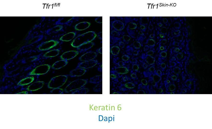
76 a component intermediate fiber found in epithelial cells, is expressed in the basal interfollicular skin epidermis as well as in the outer root sheaths of hair follicles (57). Keratin 14-Cre transgene expression was detected in the skin and dental epithelium by E14.5 (58). Tfr1 Skin-KO mice were born in Mendelian ratios and were indistinguishable from their wildtype littermates at birth. Starting at P4, the Tfr1 Skin-KO pups fail to thrive (Figure 17), resulting in their deaths between P7 to P9. Penetrance of the lethal phenotype was 96% in our experiments with a lone Tfr1 Skin-KO female mouse surviving into adulthood, possibly due to low recombination efficiency. The escaper mouse showed no overt signs of skin defects and was of normal body weight compared with wildtype littermates. As keratin 14 is also expressed in the oral ectoderm during development (59), the mouth was examined for any defects. Lack of any gross abnormalities in the mouth and tongue of the Tfr1 Skin-KO pups as well as the presence of milk in the stomach suggests that deletion of Tfr1 in the skin epidermis may be the main cause of the neonatal lethality. Gross epidermal architecture of skin appears unaffected in Tfr1 Skin-KO mice In order to assess the general architecture of the skin epithelium, hematoxylin and eosin (H&E) and Ki67 was performed on the dorsal skin epithelium of P5 wildtype and Tfr1 Skin-KO mice. H&E staining showed no breakage of the stratum corneum much like the wildtype (Figure 18). Hair follicles were present in Tfr1 Skin-KO mouse, however, the morphology of the hair follicles appeared abnormal. Most strikingly, a severe loss of 67
77 adipocytes was observed in Tfr1 Skin-KO mice at P5. Ki67 staining showed no loss of proliferating cells in the basal layer of the interfollicular Tfr1 Skin-KO skin (Figure 18). However, proliferation within the root sheaths of the hair follicles appeared abnormal. Wound healing is a property of skin epidermis which is initiated after barrier function has been compromised. Wound healing is characterized by the induction of keratin 6 at the wound edge ahead of the migration of epithelial cells into the wound site (60). To determine if the loss of Tfr1 in the skin caused a phenotype similar to wound healing, P5 dorsal skin epithelia of Tfr1 Skin-KO and wildtype pups were stained for keratin 6. Immunofluorescence for keratin 6 showed less staining in Tfr1 Skin-KO skin compared to wildtype, preliminarily suggesting that the loss of Tfr1 does not induce the wound healing response (Figure 19). Increased apoptosis in the skin epidermis of Tfr1 Skin-KO mice Loss of Tfr1 in cardiomyocytes resulted in increased apoptosis (Wenjing Xu, unpublished data). In order to assess whether the loss of Tfr1 in the skin epithelium shows a similar increase in apoptosis, TUNEL staining was performed in P5 dorsal skin epithelia of Tfr1 Skin-KO and wildtype pups. Tfr1 Skin-KO skin showed a marked increase in apoptosis in both the basal epithelium (white arrow) as well as within the root sheaths of hair follicles (red arrow) (Figure 20). Interestingly, increased apoptosis was also observed in a population of cells interspersed between hair follicles (yellow arrow), possibly corresponding to intradermal adipocytes. 68
78 4.4 Discussion Given the phenotype of the Tfr1 IEC-KO pups and the similarity in developmental hierarchy between intestinal and skin epithelial cells, initial efforts in characterizing the Tfr1 Skin-KO pups were focused on determining the effects of the loss of Tfr1 on skin epithelial barrier function and proliferation. Gross epidermal architecture appeared uncompromised as assessed through H&E staining. No signs of bleeding or infection were observed in regions of the skin subjected to high mechanical stress, such as between digits. Furthermore, wound healing response, a sign of compromised barrier function, did not appear to be initiated as the levels of keratin 6 was not elevated in the Tfr1 Skin-KO skin. It is possible that the increase in apoptotic cells observed in the Tfr1 Skin-KO is an intrinsic response to the loss of Tfr1 rather than a secondary response due to a barrier defect. The number of proliferative cells in the basal epidermis appears unaffected by the increase in apoptosis, possibly due to an increase in proliferation as a compensatory measure. However, more work is needed to fully quantify levels of proliferation. Future work will focus on characterizing the levels of Tfr1 at the mrna and protein levels in primary keratinocytes isolated from wildtype and Tfr1 Skin-KO pups. Because of the existence of the escaper Tfr1 Skin-KO mouse, it will be critical to determine the levels of residual Tfr1 in Tfr1 Skin-KO mice. H&E, Ki67, and TUNEL staining of the skin epithelium, coupled with RT-PCR of primary keratinocytes, at earlier time points will be essential to determine when the phenotype manifests itself and to correlate transcriptional changes after the loss of Tfr1 to early or late timepoints. The primary 69
79 cause of the neonatal lethality will also be the focus of future work. Hypothermia due to impaired adipose development could be a possibility. Tfr1 has been shown to have iron-independent roles in the intestinal epithelium. To determine if this is the case in the skin, Tfr1 Skin-KO ;Rosa26 Tfr1R654A mice will be generated and Tfr1 Skin-KO pups will be injected with iron-dextran in order to observe any extension of viability or rescue of lethality. Finally, the striking loss of adipocytes seen in the Tfr1 Skin-KO will be the focus of future work. A correlation between the growth of the hair follicle and adipocyte development has been observed in mice. During the anagen phase of hair follicle growth, the dermal adipocyte layer increases in thickness (61). Conversely, during the telogen phase of hair follicle growth, the adipose layer becomes thinner. In wildtype neonates, hair follicles undergo the anagen phase up to P15 with a corresponding thickening to the dermal adipose layer. The abnormal phenotype and increased apoptosis of the Tfr1 Skin-KO hair follicle coupled with the inability of the adipose layer to thicken during development suggests an impairment in this process. One possible candidate pathway that might explain this impairment in the relationship between hair follicle development and adipose thickening is Wnt signaling. The loss of Wnt signaling in the epidermis delays the ability of the hair follicle to undergo the anagen phase, while activation of Wnt signaling induces hair follicles to enter the anagen phase in mice (62). Furthermore, activation of Wnt signaling in the skin epidermis induces secretion of ligands which promotes adipocyte differentiation (63). The Tfr1 Skin-KO mouse model will be useful in determining 70
80 the relationship between Tfr1, Wnt signaling, and the interdependence of hair follicle growth with adipose development. 71

81 72
82 Figure 17. Tfr1 Skin-KO pups at fail to thrive. Body sizes of wildtype and Tfr1 Skin-KO pups at P5. 73

83 74
84 Figure 18. Gross architecture remains intact in Tfr1 Skin-KO skin epithelium, but lack of adipose layer and abnormal proliferation is observed in root sheaths. Sections of wildtype and Tfr1 Skin-KO dorsal skin at P5 were stained with H&E or Ki67 to evaluate overall architecture and proliferation, respectively. The architecture of Tfr1 Skin-KO dorsal skin is abnormal, with a prominent deficiency of adipose tissue. Overall, proliferation appeared to be similar in wildtype and mutant animals. 75
85 76
86 Figure 19. Levels of keratin 6 are not elevated in Tfr1 Skin-KO skin epithelium. Sections of wildtype and Tfr1 Skin-KO dorsal skin at P5 were stained for Keratin 6 (green) and DAPI (blue). 77

87 78
Week 3 The Pancreas: Pancreatic ph buffering:
 Week 3 The Pancreas: A gland with both endocrine (secretion of substances into the bloodstream) & exocrine (secretion of substances to the outside of the body or another surface within the body) functions
Genotype analysis by Southern blots of nine independent recombinated ES cell clones by
 Supplemental Figure 1 Selected ES cell clones show a correctly recombined conditional Ngn3 allele Genotype analysis by Southern blots of nine independent recombinated ES cell clones by hybridization with
The Small Intestine. The pyloric sphincter at the bottom of the stomach opens, squirting small amounts of food into your small intestine.
 The Small Intestine The pyloric sphincter at the bottom of the stomach opens, squirting small amounts of food into your small intestine. approximately six metres (the longest section of your digestive
Sphincters heartburn diaphragm The Stomach gastric glands pepsin, chyme The Small Intestine 1-Digestion Is Completed in the Small Intestine duodenum
 Sphincters are muscles that encircle tubes and act as valves. The tubes close when the sphincters contract and they open when the sphincters relax. When food or saliva is swallowed, the sphincter relaxes
18. PANCREATIC FUNCTION AND METABOLISM. Pancreatic secretions ISLETS OF LANGERHANS. Insulin
 18. PANCREATIC FUNCTION AND METABOLISM ISLETS OF LANGERHANS Some pancreatic functions have already been discussed in the digestion section. In this one, the emphasis will be placed on the endocrine function
SUPPLEMENTARY INFORMATION
 DOI: 10.1038/ncb2607 Figure S1 Elf5 loss promotes EMT in mammary epithelium while Elf5 overexpression inhibits TGFβ induced EMT. (a, c) Different confocal slices through the Z stack image. (b, d) 3D rendering
Physiology Unit 4 DIGESTIVE PHYSIOLOGY
 Physiology Unit 4 DIGESTIVE PHYSIOLOGY In Physiology Today Functions Motility Ingestion Mastication Deglutition Peristalsis Secretion 7 liters/day! Exocrine/endocrine Digestion Absorption Digestion of
The Digestive System. What is the advantage of a one-way gut? If you swallow something, is it really inside you?
 The Digestive System What is the advantage of a one-way gut?! If you swallow something, is it really inside you? Functions and Processes of the Digestive System: Move nutrients, water, electrolytes from
Soft palate elevates, closing off the nasopharynx. Hard palate Tongue Bolus Epiglottis. Glottis Larynx moves up and forward.
 The Cephalic Phase Chemical and mechanical digestion begins in the mouth Saliva is an exocrine secretion Salivary secretion is under autonomic control Softens and lubricates food Chemical digestion: salivary
Control of Glucose Metabolism
 Glucose Metabolism Control of Glucose Metabolism The pancreas is both an exocrine and endocrine gland. It secretes digestive enzymes into the duodenum (exocrine) and 3 specific hormones into the bloodstream
Molecular Cell Biology - Problem Drill 19: Cell Signaling Pathways and Gene Expression
 Molecular Cell Biology - Problem Drill 19: Cell Signaling Pathways and Gene Expression Question No. 1 of 10 1. Which statement about cell signaling is correct? Question #1 (A) Cell signaling involves receiving
(A) PCR primers (arrows) designed to distinguish wild type (P1+P2), targeted (P1+P2) and excised (P1+P3)14-
 PCR primers (arrows) designed to distinguish wild type (P1+P2), targeted (P1+P2) and excised (P1+P3)14-") 1 Supplemental Figure Legends Figure S1. Mammary tumors of ErbB2 KI mice with 14-3-3σ ablation have elevated ErbB2 transcript levels and cell proliferation (A) PCR primers (arrows) designed to distinguish
1 Supplemental Figure Legends Figure S1. Mammary tumors of ErbB2 KI mice with 14-3-3σ ablation have elevated ErbB2 transcript levels and cell proliferation (A) PCR primers (arrows) designed to distinguish
CHAPTER 6 SUMMARIZING DISCUSSION
 CHAPTER 6 SUMMARIZING DISCUSSION More than 20 years ago the founding member of the Wnt gene family, Wnt-1/Int1, was discovered as a proto-oncogene activated in mammary gland tumors by the mouse mammary
Supplemental Figure 1: Lrig1-Apple expression in small intestine. Lrig1-Apple is observed at the crypt base and in insterstial cells of Cajal, but is
 Supplemental Figure 1: Lrig1-Apple expression in small intestine. Lrig1-Apple is observed at the crypt base and in insterstial cells of Cajal, but is not co-expressed in DCLK1-positive tuft cells. Scale
Supplemental Data Tamoxifen administration to Vil-Scap- mice.
 Supplemental Data FIGURE S1. Tamoxifen administration to Vil-Scap - mice. In the experiments shown in Fig. 1 to Fig. 5, tamoxifen (2 mg per dose) was dissolved in corn oil and administered by orogastric
HLA and antigen presentation. Department of Immunology Charles University, 2nd Medical School University Hospital Motol
 HLA and antigen presentation Department of Immunology Charles University, 2nd Medical School University Hospital Motol MHC in adaptive immunity Characteristics Specificity Innate For structures shared
AN ANIMAL S DIET MUST SUPPLY CHEMICAL ENERGY, ORGANIC MOLECULES, AND ESSENTIAL NUTRIENTS
 1 ANIMAL NUTRITION 2 3 4 5 6 7 Food is taken in, taken apart, and taken up in the process of animal nutrition In general, animals fall into three categories: Herbivores eat mainly plants and algae Carnivores
Abdulrahman Alhanbali. Lojayn Salah. Mohammad Khatatbeh. 1 P a g e
 7 Abdulrahman Alhanbali Lojayn Salah Mohammad Khatatbeh 1 P a g e In this lecture we will talk about digestion and absorption of food in the alimentary tract. But first of all we have some important points
VIII Curso Internacional del PIRRECV. Some molecular mechanisms of cancer
 VIII Curso Internacional del PIRRECV Some molecular mechanisms of cancer Laboratorio de Comunicaciones Celulares, Centro FONDAP Estudios Moleculares de la Celula (CEMC), ICBM, Facultad de Medicina, Universidad
Generating Mouse Models of Pancreatic Cancer
 Generating Mouse Models of Pancreatic Cancer Aom Isbell http://www2.massgeneral.org/cancerresourceroom/types/gi/index.asp Spring/Summer 1, 2012 Alexandros Tzatsos, MD PhD Bardeesy Lab: Goals and Objectives
Postn MCM Smad2 fl/fl Postn MCM Smad3 fl/fl Postn MCM Smad2/3 fl/fl. Postn MCM. Tgfbr1/2 fl/fl TAC
 A Smad2 fl/fl Smad3 fl/fl Smad2/3 fl/fl Tgfbr1/2 fl/fl 1. mm B Tcf21 MCM Tcf21 MCM Smad3 fl/fl Tcf21 MCM Smad2/3 fl/fl Tcf21 MCM Tgfbr1/2 fl/fl αmhc MCM C 1. mm 1. mm D Smad2 fl/fl Smad3 fl/fl Smad2/3
Hormones and Homeostasis
 Hormones and Homeostasis The endocrine system is a system of organs that releases chemical message molecules, called hormones, into the blood. Unlike the nervous system whose action helps the body react
Digestion and Absorption
 Digestion and Absorption Digestion and Absorption Digestion is a process essential for the conversion of food into a small and simple form. Mechanical digestion by mastication and swallowing Chemical digestion
Station One: Nutrition
 Station One: Nutrition Name that thing! 1. Chemical substances, found in foods, which are used in the human body. 2. Nutrient in human diet where foods are the only possible source of the nutrient. 3.
Protein Trafficking in the Secretory and Endocytic Pathways
 Protein Trafficking in the Secretory and Endocytic Pathways The compartmentalization of eukaryotic cells has considerable functional advantages for the cell, but requires elaborate mechanisms to ensure
DIGESTIVE. CHAPTER 17 Lecture: Part 1 Part 2 BIO 212: ANATOMY & PHYSIOLOGY II
 BIO 212: ANATOMY & PHYSIOLOGY II CHAPTER 17 Lecture: DIGESTIVE Part 1 Part 2 Dr. Lawrence G. Altman www.lawrencegaltman.com Some illustrations are courtesy of McGraw-Hill. SMALL INTESTINE DUODENUM > JEJUNUM
Module 3 Lecture 7 Endocytosis and Exocytosis
 Module 3 Lecture 7 Endocytosis and Exocytosis Endocytosis: Endocytosis is the process by which cells absorb larger molecules and particles from the surrounding by engulfing them. It is used by most of
HLA and antigen presentation. Department of Immunology Charles University, 2nd Medical School University Hospital Motol
 HLA and antigen presentation Department of Immunology Charles University, 2nd Medical School University Hospital Motol MHC in adaptive immunity Characteristics Specificity Innate For structures shared
 SUMMARY Coeliac disease is a common food intolerance in Western populations, in which it has a prevalence of about 1%. In early infancy, when the transition is made to a gluten-containing diet (particularly
Hepatogenesis I Liver development
 Hepatogenesis I Liver development HB 308 George Yeoh Room 2.59 MCS Building yeoh@cyllene.uwa.edu.au Topics Early liver development Tissue interaction - role of morphogens and cytokines Liver enriched transcription
ANATOMY & PHYSIOLOGY ONLINE COURSE - SESSION 13 THE DIGESTIVE SYSTEM
 ANATOMY & PHYSIOLOGY ONLINE COURSE - SESSION 13 THE DIGESTIVE SYSTEM The digestive system also known as the alimentary canal or gastrointestinal tract consists of a series of hollow organs joined in a
10/27/2016. Processing in the Large Intestine. The colon of the large intestine is connected to the small intestine
 The hepatic portal vein carries nutrient-rich blood from the capillaries of the villi to the liver, then to the heart The liver regulates nutrient distribution, interconverts many organic molecules, and
Neoplasia 18 lecture 6. Dr Heyam Awad MD, FRCPath
 Neoplasia 18 lecture 6 Dr Heyam Awad MD, FRCPath ILOS 1. understand the role of TGF beta, contact inhibition and APC in tumorigenesis. 2. implement the above knowledge in understanding histopathology reports.
SUPPLEMENTARY INFORMATION
 doi:10.1038/nature12652 Supplementary Figure 1. PRDM16 interacts with endogenous EHMT1 in brown adipocytes. Immunoprecipitation of PRDM16 complex by flag antibody (M2) followed by Western blot analysis
BIO360 Fall 2013 Quiz 1
 BIO360 Fall 2013 Quiz 1 1. Examine the diagram below. There are two homologous copies of chromosome one and the allele of YFG carried on the light gray chromosome has undergone a loss-of-function mutation.
Understandings, Applications & Skills
 D.2 Digestion Understandings, Applications & Skills Statement D.2.U1 Nervous and hormonal mechanisms control the secretion of digestive juices. D.2.U2 Exocrine glands secrete to the surface of the body
Supplementary Figure 1:
 Supplementary Figure 1: (A) Whole aortic cross-sections stained with Hematoxylin and Eosin (H&E), 7 days after porcine-pancreatic-elastase (PPE)-induced AAA compared to untreated, healthy control aortas
Chapter 14: The Digestive System
 Chapter 14: The Digestive System Digestive system consists of Muscular tube (digestive tract) alimentary canal Accessory organs teeth, tongue, glandular organs 6 essential activities 1. 2. 3. 4. 5. 6.
Biology 12 - Digestion Notes
 Biology 12 - Digestion Notes Anatomy Physiology Functions of the Digestive System -------------------------------------------------------------------------------------- food (enzymes, bile, HCl) to assist
mirna Dr. S Hosseini-Asl
 mirna Dr. S Hosseini-Asl 1 2 MicroRNAs (mirnas) are small noncoding RNAs which enhance the cleavage or translational repression of specific mrna with recognition site(s) in the 3 - untranslated region
LIST OF ORGANS FOR HISTOPATHOLOGICAL ANALYSIS:!! Neural!!!!!!Respiratory:! Brain : Cerebrum,!!! Lungs and trachea! Olfactory, Cerebellum!!!!Other:!
 LIST OF ORGANS FOR HISTOPATHOLOGICAL ANALYSIS:!! Neural!!!!!!Respiratory:! Brain : Cerebrum,!!! Lungs and trachea! Olfactory, Cerebellum!!!!Other:! Spinal cord and peripheral nerves! Eyes, Inner ear, nasal
(b) Stomach s function 1. Dilution of food materials 2. Acidification of food (absorption of dietary Fe in small intestine) 3. Partial chemical digest
 Stomach s function 1. Dilution of food materials 2. Acidification of food (absorption of dietary Fe in small intestine) 3. Partial chemical digest") (1) General features a) Stomach is widened portion of gut-tube: between tubular and spherical; Note arranged of smooth muscle tissue in muscularis externa. 1 (b) Stomach s function 1. Dilution of food
(1) General features a) Stomach is widened portion of gut-tube: between tubular and spherical; Note arranged of smooth muscle tissue in muscularis externa. 1 (b) Stomach s function 1. Dilution of food
Journal Club. 03/04/2012 Lama Nazzal
 Journal Club 03/04/2012 Lama Nazzal NOTCH and the kidneys Is an evolutionarily conserved cell cell communication mechanism. Is a regulator of cell specification, differentiation, and tissue patterning.
What systems are involved in homeostatic regulation (give an example)?
?") 1 UNIVERSITY OF PNG SCHOOL OF MEDICINE AND HEALTH SCIENCES DIVISION OF BASIC MEDICAL SCIENCES DISCIPLINE OF BIOCHEMISTRY AND MOLECULAR BIOLOGY GLUCOSE HOMEOSTASIS (Diabetes Mellitus Part 1): An Overview
1 UNIVERSITY OF PNG SCHOOL OF MEDICINE AND HEALTH SCIENCES DIVISION OF BASIC MEDICAL SCIENCES DISCIPLINE OF BIOCHEMISTRY AND MOLECULAR BIOLOGY GLUCOSE HOMEOSTASIS (Diabetes Mellitus Part 1): An Overview
Chapter 15 Gastrointestinal System
 Chapter 15 Gastrointestinal System Dr. LL Wang E-mail: wanglinlin@zju.edu.cn Rm 608, Block B, Research Building, School of Medicine, Zijingang Campus Pancreatic Secretion The exocrine cells in the pancreas
Diseases of exocrine pancreas
 Diseases of exocrine pancreas The exocrine pancreas constitutes 80% to 85% of the organ and is composed of acinar cells that secrete enzymes needed for digestion. the accessory duct of Santorini, the main
T-cell activation T cells migrate to secondary lymphoid tissues where they interact with antigen, antigen-presenting cells, and other lymphocytes:
 Interactions between innate immunity & adaptive immunity What happens to T cells after they leave the thymus? Naïve T cells exit the thymus and enter the bloodstream. If they remain in the bloodstream,
Digestive System 7/15/2015. Outline Digestive System. Digestive System
 Digestive System Biology 105 Lecture 18 Chapter 15 Outline Digestive System I. Functions II. Layers of the GI tract III. Major parts: mouth, pharynx, esophagus, stomach, small intestine, large intestine,
Ch 7 Nutrition in humans
 Ch 7 Nutrition in humans Think about (Ch 7, p.2) 1. The stomach churns food into smaller pieces physically. The stomach wall secretes proteases to chemically digest proteins. It also releases hydrochloric
CHM333 LECTURE 34: 11/30 12/2/09 FALL 2009 Professor Christine Hrycyna
 Lipid Metabolism β-oxidation FA Acetyl-CoA Triacylglycerols (TAGs) and glycogen are the two major forms of stored energy in vertebrates Glycogen can supply ATP for muscle contraction for less than an hour
e. Undigested material is compacted and stored until the colon is full. When the colon is full, a signal to empty it is sent by sensors in the walls
 Digestive System 1. General a. Animals obtain energy by breaking food molecules into smaller pieces. b. The basic fuel molecules are amino acids, lipids and sugars c. Digestion is the chemical breakdown
Type of file: PDF Size of file: 0 KB Title of file for HTML: Supplementary Information Description: Supplementary Figures
 Type of file: PDF Size of file: 0 KB Title of file for HTML: Supplementary Information Description: Supplementary Figures Supplementary Figure 1 mir-128-3p is highly expressed in chemoresistant, metastatic
Intracellular Vesicular Traffic Chapter 13, Alberts et al.
 Intracellular Vesicular Traffic Chapter 13, Alberts et al. The endocytic and biosynthetic-secretory pathways The intracellular compartments of the eucaryotic ell involved in the biosynthetic-secretory
KRISHNA TEJA PHARMACY COLLEGE HUMAN ANATOMY AND PHYSIOLOGY. DIGESTIVE SYSTEM Dr.B.Jyothi
 KRISHNA TEJA PHARMACY COLLEGE HUMAN ANATOMY AND PHYSIOLOGY DIGESTIVE SYSTEM Dr.B.Jyothi Prof, Dept. Of Pharmacology KTPC The Digestive System Food undergoes six major processes: 1. Ingestion : process
UNIVERSITY OF PNG SCHOOL OF MEDICINE AND HEALTH SCIENCES DIVISION OF BASIC MEDICAL SCIENCES DISCIPLINE OF BIOCHEMISTRY AND MOLECULAR BIOLOGY
 1 UNIVERSITY OF PNG SCHOOL OF MEDICINE AND HEALTH SCIENCES DIVISION OF BASIC MEDICAL SCIENCES DISCIPLINE OF BIOCHEMISTRY AND MOLECULAR BIOLOGY GLUCOSE HOMEOSTASIS An Overview WHAT IS HOMEOSTASIS? Homeostasis
PBS Class #2 Introduction to the Immune System part II Suggested reading: Abbas, pgs , 27-30
 PBS 803 - Class #2 Introduction to the Immune System part II Suggested reading: Abbas, pgs. 15-25, 27-30 Learning Objectives Compare and contrast the maturation of B and T lymphocytes Compare and contrast
Cellular control of cholesterol. Peter Takizawa Department of Cell Biology
 Cellular control of cholesterol Peter Takizawa Department of Cell Biology Brief overview of cholesterol s biological role Regulation of cholesterol synthesis Dietary and cellular uptake of cholesterol
Cell Quality Control. Peter Takizawa Department of Cell Biology
 Cell Quality Control Peter Takizawa Department of Cell Biology Cellular quality control reduces production of defective proteins. Cells have many quality control systems to ensure that cell does not build
ulcer healing role 118 Bicarbonate, prostaglandins in duodenal cytoprotection 235, 236
 Subject Index Actin cellular forms 48, 49 epidermal growth factor, cytoskeletal change induction in mucosal repair 22, 23 wound repair 64, 65 polyamine effects on cytoskeleton 49 51 S-Adenosylmethionine
Pancreas Fox Chapter 18 part 2 (also Chapter 19.3 & 19.4)
") Vert Phys PCB3743 Pancreas Fox Chapter 18 part 2 (also Chapter 19.3 & 19.4) T. Houpt, Ph.D. Anatomy of Digestive System Peristalsis Stomach and Acid Secretion Liver and Bile Secretion Pancreas and pancreatic
Vert Phys PCB3743 Pancreas Fox Chapter 18 part 2 (also Chapter 19.3 & 19.4) T. Houpt, Ph.D. Anatomy of Digestive System Peristalsis Stomach and Acid Secretion Liver and Bile Secretion Pancreas and pancreatic
Signaling Vascular Morphogenesis and Maintenance
 Signaling Vascular Morphogenesis and Maintenance Douglas Hanahan Science 277: 48-50, in Perspectives (1997) Blood vessels are constructed by two processes: vasculogenesis, whereby a primitive vascular
Journal Club WS 2012/13 Stefanie Nickl
 Journal Club WS 2012/13 Stefanie Nickl Background Mesenchymal Stem Cells First isolation from bone marrow 30 ys ago Isolation from: spleen, heart, skeletal muscle, synovium, amniotic fluid, dental pulp,
10/23/2013 ANIMAL NUTRITION ANIMAL NUTRITION ESSENTIAL NUTRIENTS AN ANIMAL S DIET MUST STUPPLY: AMINO ACIDS
 ANIMAL NUTRITION Food is taken in, taken apart, and taken up in the process of animal nutrition In general, animals fall into three categories: Herbivores Carnivores Omnivores ANIMAL NUTRITION Chapter
VIRAL AGENTS CAUSING GASTROENTERITIS
 VIRAL AGENTS CAUSING GASTROENTERITIS VIRAL AGENTS CAUSING GASTROENTERITIS Pathogens discussed in our lectures 1. Rotavirus 2. Enteric adenoviruses 3. Caliciviruses 4. Astroviruses 5. Toroviruses Viruses
Healing & Repair. Tissue Regeneration
 Healing & Repair Dr. Srikumar Chakravarthi Repair & Healing: Are they same? Repair :Regeneration of injured cells by cells of same type, as with regeneration of skin/oral mucosa (requires basement membrane)
Biomarkers of GI tract diseases. By Dr. Gouse Mohiddin Shaik
 By Dr. Gouse Mohiddin Shaik Introduction The gastrointestinal (GI) tract is a complex system performing multiple biological functions which are anatomically distributed Site for food processing and absorption
Summary of chemical breakdown of food by hydrolytic enzymes (Protein enzymes).
.") Biology 12 Digestive System Digestion Overview: The digestive process can be divided into 4 phases: 1. ingestion - includes swallowing and peristalsis 2. digestion - the physical (by teeth) and chemical
Biology 12 Digestive System Digestion Overview: The digestive process can be divided into 4 phases: 1. ingestion - includes swallowing and peristalsis 2. digestion - the physical (by teeth) and chemical
Cells & Tissues. Chapter 3
 Cells & Tissues Chapter 3 Cell Theory Cell is structural and functional unit of life Activity of an organism is dependent upon its cells Principle of Complementarity functions of cells are dependent upon
renoprotection therapy goals 208, 209
 Subject Index Aldosterone, plasminogen activator inhibitor-1 induction 163, 164, 168 Aminopeptidases angiotensin II processing 64 66, 214 diabetic expression 214, 215 Angiotensin I intrarenal compartmentalization
GPR120 *** * * Liver BAT iwat ewat mwat Ileum Colon. UCP1 mrna ***
 a GPR120 GPR120 mrna/ppia mrna Arbitrary Units 150 100 50 Liver BAT iwat ewat mwat Ileum Colon b UCP1 mrna Fold induction 20 15 10 5 - camp camp SB202190 - - - H89 - - - - - GW7647 Supplementary Figure
Quantification of early stage lesions for loss of p53 should be shown in the main figures.
 Reviewer #1 (Remarks to the Author): Expert in prostate cancer The manuscript "Clonal dynamics following p53 loss of heterozygosity in Kras-driven cancers" uses a number of novel genetically engineered
Study Aid for D1: Nutrition
 Study Aid for D1: Nutrition Essential nutrient Food only source. Ex. Some Amino acids, minerals, calcium, vitamins, water, some fatty acids Non-essential nutrient Can be made in body. Ex. Glucose, starch.
Supplementary Figure 1
 Supplementary Figure 1 Control Pancreatitis Supplementary Figure 2 A Panc Liver SI Spleen H 2 O B EZH2 fl/fl C EZH2 fl/fl 37bp EZH2 ERK2 D E 5 EZH2 fl/fl Fasting Glucose (mg/dl) 2 18 16 14 12 1 8 6 4 2
Chapter 41. Lecture 14. Animal Hormones. Dr. Chris Faulkes
 Chapter 41 Lecture 14 Animal Hormones Dr. Chris Faulkes Animal Hormones Aims: To appreciate the variety and roles of hormones in the body To understand the basic types of hormones To understand how hormones
Nutrients. Chapter 25 Nutrition, Metabolism, Temperature Regulation
 Chapter 25 Nutrition, Metabolism, Temperature Regulation 25-1 Nutrients Chemicals used by body to produce energy, provide building blocks or function in other chemical reactions Classes Carbohydrates,
Summary and Concluding Remarks
 Summary and Concluding Remarks Chapter 6 The intestinal epithelium provides an excellent model system for investigating molecular mechanisms regulating cell lineage establishment, stem cell proliferation,
Problem Set 8 Key 1 of 8
 7.06 2003 Problem Set 8 Key 1 of 8 7.06 2003 Problem Set 8 Key 1. As a bright MD/PhD, you are interested in questions about the control of cell number in the body. Recently, you've seen three patients
Bio& 242 Unit 1 / Lecture 4
 Bio& 242 Unit 1 / Lecture 4 system: Gastric hormones GASTRIN: Secretion: By enteroendocrine (G) in gastric pits of the mucosa. Stimulus: Stomach distention and acid ph of chyme causes Gastrin. Action:
The gallbladder. Bile secretion:
 The gallbladder is a thin walled green muscular sac on the inferior surface of the liver. The gallbladder stores bile that is not immediately needed for digestion and concentrates it. When the muscular
Probe. Hind III Q,!?R'!! /0!!!!D1"?R'! vector. Homologous recombination
 Supple-Zhang Page 1 Wild-type locus Targeting construct Targeted allele Exon Exon3 Exon Probe P1 P P3 FRT FRT loxp loxp neo vector amh I Homologous recombination neo P1 P P3 FLPe recombination Q,!?R'!!
Islets of Langerhans consist mostly of insulin-producing β cells. They will appear to be densely labeled
 Are adult pancreatic beta cells formed by self-duplication or stem cell differentiation? Introduction Researchers have long been interested in how tissues produce and maintain the correct number of cells
Determination of the temporal pattern and importance of BALF1 expression in Epstein-Barr viral infection
 Determination of the temporal pattern and importance of BALF1 expression in Epstein-Barr viral infection Melissa Mihelidakis May 6, 2004 7.340 Research Proposal Introduction Apoptosis, or programmed cell
Digestive System. Part 3
 Digestive System Part 3 Digestion Ingested materials must be broken down for absorption Majority of absorption in small intestine Water and alcohol in stomach mucosa Some salts and vitamins in large intestine
L1, 2 : Biochemical Aspects of Digestion of Lipids, Proteins, and Carbohydrates
 L1, 2 : Biochemical Aspects of Digestion of Lipids, Proteins, and Carbohydrates OBJECTIVES: Understand the process of digestion of dietary lipids, protein and carbohydrates including, the organs involved,
Learning Targets. The Gastrointestinal (GI) Tract. Also known as the alimentary canal. Hollow series of organs that food passes through
 Tract. Also known as the alimentary canal. Hollow series of organs that food passes through") Digestion the multistep process of breaking down food into molecules the body can use Learning Targets Describe the path food takes through the digestive system. Identify the major organs of the digestive
Digestion the multistep process of breaking down food into molecules the body can use Learning Targets Describe the path food takes through the digestive system. Identify the major organs of the digestive
Series Editors: Daniel Kamin, MD and Christine Waasdorp Hurtado, MD
 NASPGHAN Physiology Lecture Series GI Physiology Module: Absorption of Water and Ions Jason Soden, MD Reviewers: George Fuchs MD: UAMS College of Medicine / Arkansas Children s Hospital Wayne Lencer MD:
Supplementary Figure 1: Hsp60 / IEC mice are embryonically lethal (A) Light microscopic pictures show mouse embryos at developmental stage E12.
 Light microscopic pictures show mouse embryos at developmental stage E12.") Supplementary Figure 1: Hsp60 / IEC mice are embryonically lethal (A) Light microscopic pictures show mouse embryos at developmental stage E12.5 and E13.5 prepared from uteri of dams and subsequently genotyped.
Supplementary Figure 1: Hsp60 / IEC mice are embryonically lethal (A) Light microscopic pictures show mouse embryos at developmental stage E12.5 and E13.5 prepared from uteri of dams and subsequently genotyped.
Exploring a Link Between Spy1 and Hepatocellular Carcinoma Progression
 University of Windsor Scholarship at UWindsor UWill Discover Undergraduate Conference UWill Discover 2016 Mar 29th, 4:00 PM - 5:00 PM Exploring a Link Between Spy1 and Hepatocellular Carcinoma Progression
History of Investigation
 Acini - Pancreatic juice (1º) (2º) Secretions- neuronal and hormonal mechanisms 1) Secretin - bicarbonate rich 2) Cholecystokinin - enzyme rich Islets of Langerhans (contain 4 cell types) Alpha cells (α)-
Anatomy of the liver and pancreas
 Anatomy of the liver and pancreas Prof. Abdulameer Al-Nuaimi E-mail: a.al-nuaimi@sheffield.ac.uk abdulameerh@yahoo.com Liver Aorta Pulm. Trunk Rt. At, Duct. Art. Lt. Ven. Rt. Ven. Internal Posterior
Expression of acid base transporters in the kidney collecting duct in Slc2a7 -/-
 Supplemental Material Results. Expression of acid base transporters in the kidney collecting duct in Slc2a7 -/- and Slc2a7 -/- mice. The expression of AE1 in the kidney was examined in Slc26a7 KO mice.
BMP6 treatment compensates for the molecular defect and ameliorates hemochromatosis in Hfe knockout mice
 SUPPLEMENTARY MATERIALS BMP6 treatment compensates for the molecular defect and ameliorates hemochromatosis in Hfe knockout mice Elena Corradini, Paul J. Schmidt, Delphine Meynard, Cinzia Garuti, Giuliana
Digestive System Module 6: Accessory Organs in Digestion: The Liver, Pancreas, and Gallbladder
 Connexions module: m49293 1 Digestive System Module 6: Accessory Organs in Digestion: The Liver, Pancreas, and Gallbladder Donna Browne Based on Accessory Organs in Digestion: The Liver, Pancreas, and
Digestive system L 4. Lecturer Dr. Firdous M. Jaafar Department of Anatomy/Histology section
 Digestive system L 4 Lecturer Dr. Firdous M. Jaafar Department of Anatomy/Histology section objectives 1-Describe the structure of liver. 2-Define liver lobule, and identify its zones. 3-Define portal
Enzyme-coupled Receptors. Cell-surface receptors 1. Ion-channel-coupled receptors 2. G-protein-coupled receptors 3. Enzyme-coupled receptors
 Enzyme-coupled Receptors Cell-surface receptors 1. Ion-channel-coupled receptors 2. G-protein-coupled receptors 3. Enzyme-coupled receptors Cell-surface receptors allow a flow of ions across the plasma
The Endocrine Pancreas (Chapter 10) *
 *") OpenStax-CNX module: m62118 1 The Endocrine Pancreas (Chapter 10) * Ildar Yakhin Based on The Endocrine Pancreas by OpenStax This work is produced by OpenStax-CNX and licensed under the Creative Commons
OpenStax-CNX module: m62118 1 The Endocrine Pancreas (Chapter 10) * Ildar Yakhin Based on The Endocrine Pancreas by OpenStax This work is produced by OpenStax-CNX and licensed under the Creative Commons
The Interaction of Alcohol and Iron-Overload in the in-vivo Regulation of Iron Responsive Genes
 Cantaurus, Vol. 5, -, May 7 McPherson College Division of Science and Technology The Interaction of Alcohol and Iron-Overload in the in-vivo Regulation of Iron Responsive Genes Callie Crist, Elizabeth
Figure S1. Generation of inducible PTEN deficient mice and the BMMCs (A) B6.129 Pten loxp/loxp mice were mated with B6.
 B6.129 Pten loxp/loxp mice were mated with B6.") Figure S1. Generation of inducible PTEN deficient mice and the BMMCs (A) B6.129 Pten loxp/loxp mice were mated with B6.129-Gt(ROSA)26Sor tm1(cre/ert2)tyj /J mice. To induce deletion of the Pten locus,
Figure S1. Generation of inducible PTEN deficient mice and the BMMCs (A) B6.129 Pten loxp/loxp mice were mated with B6.129-Gt(ROSA)26Sor tm1(cre/ert2)tyj /J mice. To induce deletion of the Pten locus,
Plasma lipoproteins & atherosclerosis by. Prof.Dr. Maha M. Sallam
 Biochemistry Department Plasma lipoproteins & atherosclerosis by Prof.Dr. Maha M. Sallam 1 1. Recognize structures,types and role of lipoproteins in blood (Chylomicrons, VLDL, LDL and HDL). 2. Explain