Supplementary Figure 1
|
|
|
- Gwendoline Campbell
- 6 years ago
- Views:
Transcription
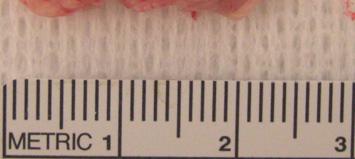
1 Supplementary Figure 1 Control Pancreatitis


 2 18")
2 Supplementary Figure 2 A Panc Liver SI Spleen H 2 O B EZH2 fl/fl C EZH2 fl/fl 37bp EZH2 ERK2 D E 5 EZH2 fl/fl Fasting Glucose (mg/dl) EZH2 fl/fl


3 Supplementary Figure 3 A -EZH2 EZH2 fl/fl EZH2 ERK2 EZH2 fl/fl B EZH2 fl/fl Ctl D3 D9 Ctl D3 D9 EZH1 ERK2
4 Supplementary Figure 4 EZH2 fl/fl


5 Supplementary Figure 5 A Metaplastic Intermediates Ductal cells Acinar cells Dedifferentiation 1-2 days Proliferation 3-5 days Redifferentiation 5-7 days C B e; e; Cr SET H H2 p ZH2 EZ EZ E H2 EZ l fl/f 48- Day 3 EZH2fl/fl r -C SET l fl/fp48 2 Control Amylase Vinculin Day 1 EZH2 SET Control Trichrome Control E Day 3 Control EZH2 SET EZH2 SET EZH2fl/fl Day 3 EZH2fl/fl D -CD45 3 -CK19 6 p=.31 5 CK19 + cells/fov CD45 + cells/fov p=.15 EZH2fl/fl EZH2 SET EZH2fl/fl EZH2 SET
6 Supplementary Figure 6 Day 1 Day 5 EZH2 fl/fl
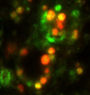
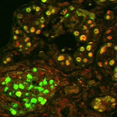
7 Supplementary Figure 7 A p48cre; EZH2 fl/fl Control Day 3 B EZH2 fl/fl PDX1/Ki-67 C Is PDX1/EZH2 Ki-67/DAPI
8 Supplementary Figure 8 A EZH2 fl/fl Control EZH2 fl/fl Cerulein Control Cerulein B EZH2 fl/fl p48cre; p48cre; ; p16 Ink4afl/fl Day 9 Day 3 Control Fraction Bound/Input IgG EZH2

9 Supplementary Figure 9 Kras G12D Kras G12D ; 1 month 3 month Pancreas weight (g) Kras G12D Kras G12D ; 1 month 3 month


10 Supplementary Figure 1 KrasG12D A KrasG12D; EZH2 SET 2 month 1 month Fraction of Total Ducts B KrasG12D KrasG12D;EZH2 SET N 1a 1b month N 1a 1b 2 month 2 3
11 Supplementary Figure 11 A B Kras G12D Kras G12D ; CD45 + Cells/2x FOV Kras G12D Kras G12D ; Side Scatter p48-cre Kras G12D Kras G12D ; % 2% 1% 16% 15% % 3% 2% 14% 16% % % % % % CD11c CD11b Gr-1 CD3 CD19 C Trichrome -SMA Kras G12D ; Kras G12D


12 Supplementary Figure 12 A PanIN/FOV Vehicle Nimesulide 2 Kras G12D Kras G12D ; B Vehicle Nimesulide Kras G12D
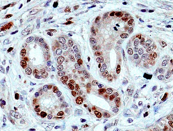
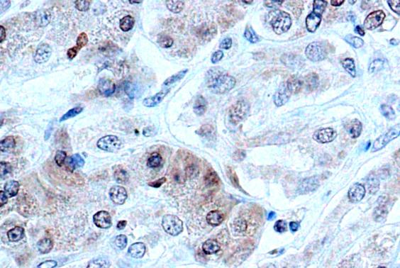
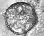
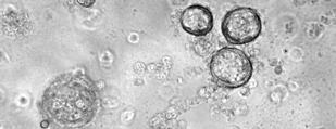
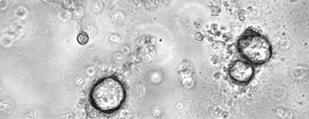
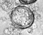
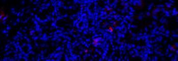
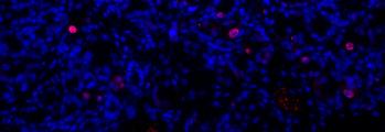
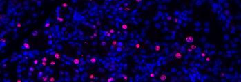
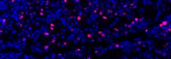
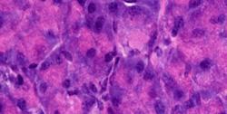
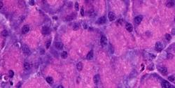
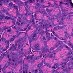
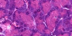
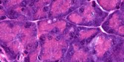
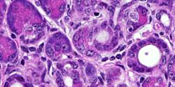
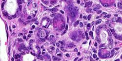
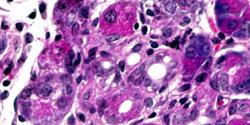
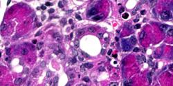
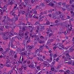
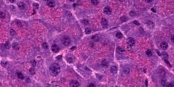
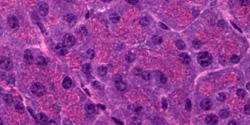
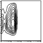
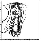
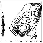
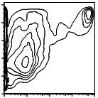
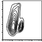
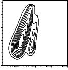
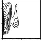
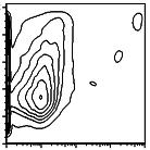
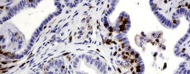
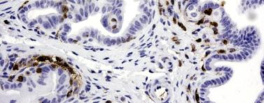
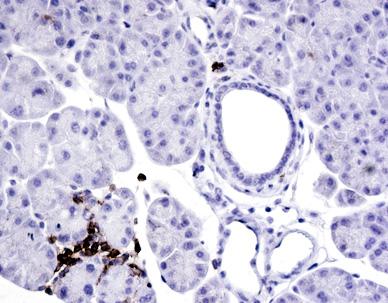
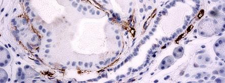
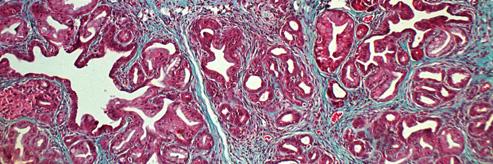
13 SUPPLEMENTARY FIGURE LEGENDS Supplementary Figure 1: EZH2 upregulation in human pancreatitis samples. Histological sections from human control and pancreatitis samples (obtained from the Tissue Acquistion and Banking Services of the NYU Experimental Pathology Core Facilities) were stained with EZH2 antibody. Results are representative of the staining pattern seen in 3 pancreatic patients. Scale bar, 5. Supplementary Figure 2: EZH2 deletion in EZH2 ΔSET animals. (A) PCR analysis of DNA from abdominal organs of EZH2 ΔSET mice. The 37bp fragment corresponding to the recombined allele is present only in the pancreas. Panc, pancreas; SI, small intestine. (B) Western blot for EZH2 expression in pancreatic lysates of EZH2 fl/fl and EZH2 ΔSET. ERK2 serves as a loading control. (C) General appearance of pancreata from EZH2 fl/fl and EZH2 ΔSET mice. (D) H&E stained sections from EZH2 fl/fl and EZH2 ΔSET mice. Scale bar, 5. (E) Fasting glucose levels (means +/- SD) measured in EZH2 fl/fl and EZH2 ΔSET mice. Data in all panels are from 2-month-old mice and are representative of 3 mice per genotype. Supplementary Figure 3: EZH1 and EZH2 expression during pancreatic regeneration. (A) EZH2 expression was analyzed from EZH2 fl/fl and EZH2 ΔSET mice on day 3 after final cerulein injection by immunohistochemical staining of pancreata (left) and western blot of pancreatic lysates (right). Scale bar, 5. ERK2 serves as a loading control. (B) Western blot shows no compensatory EZH1 upregulation in EZH2 fl/fl or p48-
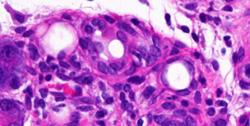
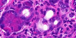
14 Cre;EZH2 ΔSET pancreata upon cerulein injection. ERK2 serves as loading control. Data in all panels are from 2-month-old mice and are representative of 3 mice per genotype. Supplementary Figure 4: Impaired regeneration of the exocrine pancreas in p48- Cre;EZH2 ΔSET mice. Representative images (n = 3 for each genotype) of H&E-stained sections from tissues harvested 21 days following final injection of cerulein. Scale bar, 5. Supplementary Figure 5: Pancreatic regeneration and loss of amylase during injury phase. (A) A schematic depiction of the key steps that mediate the process of pancreatic regeneration in response to injury. (B) Western blot analysis of amylase expression demonstrating an equivalent level of acinar cell loss in EZH2 fl/fl and EZH2 ΔSET mice on day 1 after final cerulein injection. Results are representative of data obtained from 3 animals per genotype. (C-E) Trichrome C staining (C), immunohistochemical staining for CD45 (D) and CK19 (E) from EZH2 fl/fl and EZH2 ΔSET pancreata on day 3 after final cerulein injection compared to control. For panel C, results are representative of data obtained from 3 animals per genotype. For panels D and E, quantifications were performed by counting CD45+ or CK19+ cells per four randomly selected 2X fields of view (FOV) (5 mice per genotype) on day 3 after final cerulein injection. Scale bars, 5 (C, D) and 2 (E).
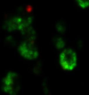
15 Supplementary Figure 6: Acinar cell cultures from EZH2 fl/fl and EZH2 ΔSET pancreata. Acinar cells from EZH2 fl/fl and EZH2 ΔSET mice display similar morphologic transition at days 1 and 5 in culture. Scale bar, 5. Supplementary Figure 7: EZH2 is required for the proliferative expansion of the metaplastic epithelium during regeneration. (A) Immunofluorescent detection of Ki67- positive cells at days and 3 after the final injection of cerulein shows enhanced proliferation in EZH2 fl/fl but not EZH2 ΔSET pancreata. Scale bar, 5. (B) Double immunofluorescence staining for PDX1 and Ki67 at day 3 after final cerulein injection demonstrates that the majority of Ki67+ proliferating cells are confined to PDX1- expressing metaplastic lesions (marked by asterisks) in EZH2 fl/fl but not p48- Cre;EZH2 ΔSET pancreata. Scale bar, 2. (C) Double immunofluorescence for PDX1 and EZH2 on day 3 after final cerulein injection shows that EZH2 is expressed in PDX1- positive metaplastic epithelium (marked by asterisk) of the regenerating pancreas. Is, islet cells. Scale bar, 2. Images shown in all panels are representative of data obtained from 3 animals per genotype. Supplementary Figure 8: EZH2 controls pancreatic regeneration through the suppression of p16 Ink4a expression. (A) ChIP analysis from pancreata harvested at day 3 after final cerulein injection shows recruitment of EZH2 to the p16 Ink4a locus in EZH2 fl/fl but not EZH2 ΔSET pancreata. Results represent the means +/- SD of three independent determinations. (B) Pancreata from EZH2 fl/fl, EZH2 ΔSET and p48- Cre;EZH2 ΔSET ;p16 Ink4afl/fl mice were harvested on days 3 and 9 following final cerulein
16 injection. H&E analysis of the sections showed that impaired regeneration was rescued in EZH2 ΔSET ;p16 Ink4afl/fl mice. Images shown are representative of data obtained from 3 animals per genotype. Scale bar, 5. Supplementary Figure 9: General appearance and weight comparison of p48- Cre;Kras G12D and Kras G12D ;EZH2 ΔSET pancreata. Pancreata from p48- Cre;Kras G12D ;EZH2 ΔSET mice are enlarged at 1 month and smaller at 3 months, relative to pancreata from Kras G12D mice assessed by appearance and weight. Data are representative of 3 mice of each genotype and age. Supplementary Figure 1: EZH2 deficiency accelerates PanIN progression. (A) Lower magnification H&E stained sections from 1- and 2-month-old Kras G12D and p48- Cre;Kras G12D ;EZH2 ΔSET mice. Arrowheads indicate early PanIN lesions, and asterisks indicate advanced lesions. Scale bar, 1. (B) Histological progression of PanINs in Kras G12D and Kras G12D ;EZH2 ΔSET pancreata at indicated ages (N, Normal; 1, 1a, 1b, 2, 3, PanIN Stage). Results represent the means +/- SD of lesions per section identified at 1 month (5 mice per genotype) and 2 months of age (5 mice per genotype). At least 4 sections were analyzed per animal. () p<.5. Supplementary Figure 11: EZH2 deficiency results in an exacerbated stromal response. (A) Immunohistochemical detection of pan-leukocyte marker CD45 reveals enhanced leukocyte infiltration in the pancreas of Kras G12D ;EZH2 ΔSET relative to p48- Cre;Kras G12D pancreata. Quantification was performed by counting CD45+ cells in ten
17 randomly selected 2X FOV in 4 animals. Scale bar, 5. (B) Flow cytometry profiles of pancreata from mice of the indicated genotypes demonstrating enhanced recruitment of dendritic cells (CD11c), neutrophils (CD11b), and macrophages (Gr-1), but not T (CD3) or B (CD19) cells to the pancreas of Kras G12D ;EZH2 ΔSET mice. Results are representative of 4 separate experiments with 3 mice per genotype. (C) Enhanced collagen deposition (Trichrome stain, scale bar, 1 ) and desmoplasia [smooth muscle actin ( -SMA) immunohistochemistry; scale bar, 5 ] in Kras G12D ;EZH2 ΔSET pancreata at 2 months of age. Images shown are representative of data obtained from 3 animals per genotype. Supplementary Figure 12: Nimesulide treatment and PanIN progression in p48- Cre;Kras G12D versus Kras G12D ;EZH2 ΔSET pancreata. (A) PanIN lesions per ten randomly selected 2X FOVs were counted in H&E stained pancreatic sections (5 mice per genotype) from vehicle and nimesulide treated 1 month old Kras G12D and Kras G12D ;EZH2 ΔSET mice. (B) H&E images of pancreata from vehicle and nimesulide treated, 1 month old Kras G12D mice show that nimesulide treatment does not effect Kras G12D pancreata. Arrowheads indicate early PanIN lesions. Images shown are representative of data obtained from 3 animals per genotype. Scale bar, 5.
18 SUPPLEMENTARY MATERIALS AND METHODS Immunofluorescence and immunohistochemistry Immunofluorescence was performed on 8μm thick frozen sections of pancreas. Slides were air dried for 15min and fixed with ice cold 4% paraformaldehyde for 1min. Sections were permeabilized with.2% Triton-X1 and blocked with 1% chicken serum for 1hr. Primary antibodies were diluted in 1% chicken serum,.5% Tween 2 and incubated overnight at 4 o C. Secondary antibodies raised in chicken (Invitrogen) were incubated on sections for 1hr. Immunohistochemistry was done on 5 μm sections from paraffin embedded blocks. Sections were deparaffinized and rehydrated, followed by quenching in 3% H 2 O 2 in methanol. Antigen retrieval was performed in 95 o C.1M citrate buffer (ph 6) with.5% Tween 2. Sections were blocked with 5% goat serum. Primary and secondary antibodies were diluted in 1% BSA/PBS. After secondary antibody incubation VectaStain Elite ABC Reagent (Vector Laboratories) was applied and the resulting antibody/conjugate was developed with diaminobenzamidine (Sigma). Sections were then counterstained with hematoxylin and mounted with Permount (Fisher). Hematoxylin and eosin staining was also performed on paraffin sections. Tissue Staining Antibodies used are rabbit anti-ezh1 (Margueron et al. 28), rabbit anti-ezh2 (Kuzmichev et al. 24), rabbit anti-h3k27me3 (Millipore, 7-449), goat anti-pdx1 (C.V. Wright, Vanderbilt University), rabbit anti-ki67 (Novacastra), goat anti-amylase (Santa Cruz), rabbit anti-amylase (Sigma), rabbit anti-sma (Abcam), rabbit anti-
19 p16 INK4A (Santa Cruz), rat anti-ck19 (TROMA-III-c, developed by R. Kemler, obtained from Developmental Studies Hybridoma Bank under the auspices of the NICHD and maintained by The University of Iowa, Department of Biology, Iowa City, IA 52242). Trichrome C staining was performed by the histopathology core at NYU School of Medicine. Quantitative immunofluorescence analysis for amylase Quantitative immunofluorescence analysis of amylase expression was done using ImageJ Vs The freehand outline tool was used to outline all areas staining for amylase in 8-1 random fields of view (4 2 /field of view) from the pancreata of two mice for each timepoint and condition. The measured area was used to determine a percentage of amylase positive cell staining / field of view. Protein and RNA extraction For proteins, pancreata were flash frozen and homogenized in lysis buffer containing 5mM Tris ph 7.4, 4mM NaCl,.5% NP-4, 5mM EDTA ph 8, and 5mM NaF with protease inhibitors (aprotinin, leupeptin, sodium orthovanadate, PMSF, DTT). For RNA, pancreata were flash frozen in liquid nitrogen and resuspended in pre-chilled RNAlater ICE (Ambion) to be incubated at least overnight at -2 o C. A small piece of frozen pancreas was ground with a mortar and pestle, and the RNEasy kit (Qiagen) was used to extract RNA from the ground tissue.
20 Quantitative PCR RNA was reverse-transcribed using Quantitect Reverse Transcription Kit (Qiagen). All primers were designed using Primer 3 software available from MIT. qpcr was performed using SYBR Green Master Mix (USB) and amplified using a Stratagene Mx35p. Results were analyzed using MxPRO software. Acinar cell isolation and culture Pancreata were harvested and acinar cells were isolated as described (Sawey et al. 27). Cells were plated and maintained on Matrigel using Waymouth media supplemented with 1% FBS, 4μg/mL dexamethasone,.4mg/ml trypsin inhibitor and penicillin/streptomycin. Chromatin immunoprecipitation Chromatin was isolated by weighing and mincing pancreatic tissue in PBS with protease inhibitors. Proteins were crosslinked with 1% formaldehyde for 15min at room temperature. Then glycine was added to a final concentration of.125m. Nuclei were then sonicated using a bioruptor for 15min twice to obtain chromatin. Crosslinked complexes were isolated by immunoprecipitation with the indicated antibodies. Crosslinking was reversed by incubation at 65 o C for 15min and 2μg RNase A and 4μg proteinase K were added to samples. DNA was isolated using phenol-chloroform extraction and ethanol precipitation and subjected to quantitative PCR.
21 Flow cytometry Pancreas was minced on ice with a razor blade and then digested with 1mg/ml collagenase for 1min at 37 o C. After lysing red blood cells, the sample was filtered through a 7μm mesh. Fc block was used before adding appropriate fluorescent conjugated antibodies. Fasting Glucose Measurements After overnight fasting, mice tails were cut and blood was applied to a Medisense Precision QID glucose monitor.
(A) PCR primers (arrows) designed to distinguish wild type (P1+P2), targeted (P1+P2) and excised (P1+P3)14-
 PCR primers (arrows) designed to distinguish wild type (P1+P2), targeted (P1+P2) and excised (P1+P3)14-") 1 Supplemental Figure Legends Figure S1. Mammary tumors of ErbB2 KI mice with 14-3-3σ ablation have elevated ErbB2 transcript levels and cell proliferation (A) PCR primers (arrows) designed to distinguish
1 Supplemental Figure Legends Figure S1. Mammary tumors of ErbB2 KI mice with 14-3-3σ ablation have elevated ErbB2 transcript levels and cell proliferation (A) PCR primers (arrows) designed to distinguish
Effec<ve Use of PI3K and MEK Inhibitors to Treat Mutant K Ras G12D and PIK3CA H1047R Murine Lung Cancers
 Effec
Effec
Antibodies: LB1 buffer For 50 ml For 10ml For 30 ml Final 1 M HEPES, ph 2.5 ml 0.5 ml 1.5 ml 50mM. 5 M NaCl 1.4 ml 280 µl 0.
 Experiment: Date: Tissue: Purpose: ChIP-Seq Antibodies: 11x cross-link buffer: Regent Stock Solution Final Vol for 10 ml of 11xstock concentration 5 M NaCl 0.1M 0.2 ml 0.5 M EDTA 1 mm 20 ul 0.5 M EGTA,
SUPPLEMENTARY INFORMATION
 SUPPLEMENTARY INFORMATION FOR Liver X Receptor α mediates hepatic triglyceride accumulation through upregulation of G0/G1 Switch Gene 2 (G0S2) expression I: SUPPLEMENTARY METHODS II: SUPPLEMENTARY FIGURES
Supplementary Information
 Supplementary Information Supplementary Figure 1. CD4 + T cell activation and lack of apoptosis after crosslinking with anti-cd3 + anti-cd28 + anti-cd160. (a) Flow cytometry of anti-cd160 (5D.10A11) binding
(Stratagene, La Jolla, CA) (Supplemental Fig. 1A). A 5.4-kb EcoRI fragment
 (Supplemental Fig. 1A). A 5.4-kb EcoRI fragment") SUPPLEMENTAL INFORMATION Supplemental Methods Generation of RyR2-S2808D Mice Murine genomic RyR2 clones were isolated from a 129/SvEvTacfBR λ-phage library (Stratagene, La Jolla, CA) (Supplemental Fig.
SUPPLEMENTAL INFORMATION Supplemental Methods Generation of RyR2-S2808D Mice Murine genomic RyR2 clones were isolated from a 129/SvEvTacfBR λ-phage library (Stratagene, La Jolla, CA) (Supplemental Fig.
Supporting Information
 Supporting Information Pang et al. 10.1073/pnas.1322009111 SI Materials and Methods ELISAs. These assays were performed as previously described (1). ELISA plates (MaxiSorp Nunc; Thermo Fisher Scientific)
Impact of hyper-o-glcnacylation on apoptosis and NF-κB activity SUPPLEMENTARY METHODS
 SUPPLEMENTARY METHODS 3D culture and cell proliferation- MiaPaCa-2 cell culture in 3D was performed as described previously (1). Briefly, 8-well glass chamber slides were evenly coated with 50 µl/well
Supplementary data Supplementary Figure 1 Supplementary Figure 2
 Supplementary data Supplementary Figure 1 SPHK1 sirna increases RANKL-induced osteoclastogenesis in RAW264.7 cell culture. (A) RAW264.7 cells were transfected with oligocassettes containing SPHK1 sirna
Supplementary Table 1. Primer sequences for conventional RT-PCR on mouse islets
 Supplementary Table 1. Primer sequences for conventional RT-PCR on mouse islets Gene 5 Forward 3 5 Reverse 3.T. Product (bp) ( C) mnox1 GTTCTTGGGCTGCCTTGG GCTGGGGCGGCGG 60 300 mnoxa1 GCTTTGCCGCGTGC GGTTCGGGTCCTTTGTGC
p = formed with HCI-001 p = Relative # of blood vessels that formed with HCI-002 Control Bevacizumab + 17AAG Bevacizumab 17AAG
 A.. Relative # of ECs associated with HCI-001 1.4 1.2 1.0 0.8 0.6 0.4 0.2 0.0 ol b p < 0.001 Relative # of blood vessels that formed with HCI-001 1.4 1.2 1.0 0.8 0.6 0.4 0.2 0.0 l b p = 0.002 Control IHC:
General Laboratory methods Plasma analysis: Gene Expression Analysis: Immunoblot analysis: Immunohistochemistry:
 General Laboratory methods Plasma analysis: Plasma insulin (Mercodia, Sweden), leptin (duoset, R&D Systems Europe, Abingdon, United Kingdom), IL-6, TNFα and adiponectin levels (Quantikine kits, R&D Systems
(A) RT-PCR for components of the Shh/Gli pathway in normal fetus cell (MRC-5) and a
 RT-PCR for components of the Shh/Gli pathway in normal fetus cell (MRC-5) and a") Supplementary figure legends Supplementary Figure 1. Expression of Shh signaling components in a panel of gastric cancer. (A) RT-PCR for components of the Shh/Gli pathway in normal fetus cell (MRC-5) and
Supplementary figure legends Supplementary Figure 1. Expression of Shh signaling components in a panel of gastric cancer. (A) RT-PCR for components of the Shh/Gli pathway in normal fetus cell (MRC-5) and
MTC-TT and TPC-1 cell lines were cultured in RPMI medium (Gibco, Breda, The Netherlands)
") Supplemental data Materials and Methods Cell culture MTC-TT and TPC-1 cell lines were cultured in RPMI medium (Gibco, Breda, The Netherlands) supplemented with 15% or 10% (for TPC-1) fetal bovine serum
Supplemental data Materials and Methods Cell culture MTC-TT and TPC-1 cell lines were cultured in RPMI medium (Gibco, Breda, The Netherlands) supplemented with 15% or 10% (for TPC-1) fetal bovine serum
Epithelial interleukin-25 is a key mediator in Th2-high, corticosteroid-responsive
 Online Data Supplement: Epithelial interleukin-25 is a key mediator in Th2-high, corticosteroid-responsive asthma Dan Cheng, Zheng Xue, Lingling Yi, Huimin Shi, Kan Zhang, Xiaorong Huo, Luke R. Bonser,
Chromatin IP (Isw2) Fix soln: 11% formaldehyde, 0.1 M NaCl, 1 mm EDTA, 50 mm Hepes-KOH ph 7.6. Freshly prepared. Do not store in glass bottles.
 Fix soln: 11% formaldehyde, 0.1 M NaCl, 1 mm EDTA, 50 mm Hepes-KOH ph 7.6. Freshly prepared. Do not store in glass bottles.") Chromatin IP (Isw2) 7/01 Toshi last update: 06/15 Reagents Fix soln: 11% formaldehyde, 0.1 M NaCl, 1 mm EDTA, 50 mm Hepes-KOH ph 7.6. Freshly prepared. Do not store in glass bottles. 2.5 M glycine. TBS:
Chromatin IP (Isw2) 7/01 Toshi last update: 06/15 Reagents Fix soln: 11% formaldehyde, 0.1 M NaCl, 1 mm EDTA, 50 mm Hepes-KOH ph 7.6. Freshly prepared. Do not store in glass bottles. 2.5 M glycine. TBS:
SUPPLEMENTARY INFORMATION. Supplementary Figures S1-S9. Supplementary Methods
 SUPPLEMENTARY INFORMATION SUMO1 modification of PTEN regulates tumorigenesis by controlling its association with the plasma membrane Jian Huang 1,2#, Jie Yan 1,2#, Jian Zhang 3#, Shiguo Zhu 1, Yanli Wang
Chromatin Immunoprecipitation (ChIPs) Protocol (Mirmira Lab)
 Protocol (Mirmira Lab)") Chromatin Immunoprecipitation (ChIPs) Protocol (Mirmira Lab) Updated 12/3/02 Reagents: ChIP sonication Buffer (1% Triton X-100, 0.1% Deoxycholate, 50 mm Tris 8.1, 150 mm NaCl, 5 mm EDTA): 10 ml 10 % Triton
Chromatin Immunoprecipitation (ChIPs) Protocol (Mirmira Lab) Updated 12/3/02 Reagents: ChIP sonication Buffer (1% Triton X-100, 0.1% Deoxycholate, 50 mm Tris 8.1, 150 mm NaCl, 5 mm EDTA): 10 ml 10 % Triton
Supplementary Figure 1 IMQ-Induced Mouse Model of Psoriasis. IMQ cream was
 Supplementary Figure 1 IMQ-Induced Mouse Model of Psoriasis. IMQ cream was painted on the shaved back skin of CBL/J and BALB/c mice for consecutive days. (a, b) Phenotypic presentation of mouse back skin
SUPPLEMENTARY INFORMATION
 Supplementary Figures Supplementary Figure S1. Binding of full-length OGT and deletion mutants to PIP strips (Echelon Biosciences). Supplementary Figure S2. Binding of the OGT (919-1036) fragments with
Supplementary Information Titles Journal: Nature Medicine
 Supplementary Information Titles Journal: Nature Medicine Article Title: Corresponding Author: Supplementary Item & Number Supplementary Fig.1 Fig.2 Fig.3 Fig.4 Fig.5 Fig.6 Fig.7 Fig.8 Fig.9 Fig. Fig.11
Supplemental figure 1. PDGFRα is expressed dominantly by stromal cells surrounding mammary ducts and alveoli. A) IHC staining of PDGFRα in
 IHC staining of PDGFRα in") Supplemental figure 1. PDGFRα is expressed dominantly by stromal cells surrounding mammary ducts and alveoli. A) IHC staining of PDGFRα in nulliparous (left panel) and InvD6 mouse mammary glands (right
Supplemental figure 1. PDGFRα is expressed dominantly by stromal cells surrounding mammary ducts and alveoli. A) IHC staining of PDGFRα in nulliparous (left panel) and InvD6 mouse mammary glands (right
Supplementary Materials. for Garmy-Susini, et al, Integrin 4 1 signaling is required for lymphangiogenesis and tumor metastasis
 Supplementary Materials for Garmy-Susini, et al, Integrin 4 1 signaling is required for lymphangiogenesis and tumor metastasis 1 Supplementary Figure Legends Supplementary Figure 1: Integrin expression
Supplementary Figure 1: Hsp60 / IEC mice are embryonically lethal (A) Light microscopic pictures show mouse embryos at developmental stage E12.
 Light microscopic pictures show mouse embryos at developmental stage E12.") Supplementary Figure 1: Hsp60 / IEC mice are embryonically lethal (A) Light microscopic pictures show mouse embryos at developmental stage E12.5 and E13.5 prepared from uteri of dams and subsequently genotyped.
Supplementary Figure 1: Hsp60 / IEC mice are embryonically lethal (A) Light microscopic pictures show mouse embryos at developmental stage E12.5 and E13.5 prepared from uteri of dams and subsequently genotyped.
Supplementary Table 1. The primers used for quantitative RT-PCR. Gene name Forward (5 > 3 ) Reverse (5 > 3 )
 Reverse (5 > 3 )") 770 771 Supplementary Table 1. The primers used for quantitative RT-PCR. Gene name Forward (5 > 3 ) Reverse (5 > 3 ) Human CXCL1 GCGCCCAAACCGAAGTCATA ATGGGGGATGCAGGATTGAG PF4 CCCCACTGCCCAACTGATAG TTCTTGTACAGCGGGGCTTG
770 771 Supplementary Table 1. The primers used for quantitative RT-PCR. Gene name Forward (5 > 3 ) Reverse (5 > 3 ) Human CXCL1 GCGCCCAAACCGAAGTCATA ATGGGGGATGCAGGATTGAG PF4 CCCCACTGCCCAACTGATAG TTCTTGTACAGCGGGGCTTG
Islet viability assay and Glucose Stimulated Insulin Secretion assay RT-PCR and Western Blot
 Islet viability assay and Glucose Stimulated Insulin Secretion assay Islet cell viability was determined by colorimetric (3-(4,5-dimethylthiazol-2-yl)-2,5- diphenyltetrazolium bromide assay using CellTiter
LIST OF ORGANS FOR HISTOPATHOLOGICAL ANALYSIS:!! Neural!!!!!!Respiratory:! Brain : Cerebrum,!!! Lungs and trachea! Olfactory, Cerebellum!!!!Other:!
 LIST OF ORGANS FOR HISTOPATHOLOGICAL ANALYSIS:!! Neural!!!!!!Respiratory:! Brain : Cerebrum,!!! Lungs and trachea! Olfactory, Cerebellum!!!!Other:! Spinal cord and peripheral nerves! Eyes, Inner ear, nasal
Supplementary Information POLO-LIKE KINASE 1 FACILITATES LOSS OF PTEN-INDUCED PROSTATE CANCER FORMATION
 Supplementary Information POLO-LIKE KINASE 1 FACILITATES LOSS OF PTEN-INDUCED PROSTATE CANCER FORMATION X. Shawn Liu 1, 3, Bing Song 2, 3, Bennett D. Elzey 3, 4, Timothy L. Ratliff 3, 4, Stephen F. Konieczny
SUPPLEMENTARY METHODS
 SUPPLEMENTARY METHODS Histological analysis. Colonic tissues were collected from 5 parts of the middle colon on day 7 after the start of DSS treatment, and then were cut into segments, fixed with 4% paraformaldehyde,
Protocol for Gene Transfection & Western Blotting
 The schedule and the manual of basic techniques for cell culture Advanced Protocol for Gene Transfection & Western Blotting Schedule Day 1 26/07/2008 Transfection Day 3 28/07/2008 Cell lysis Immunoprecipitation
Western Immunoblotting Preparation of Samples:
 Western Immunoblotting Preparation of Samples: Total Protein Extraction from Culture Cells: Take off the medium Wash culture with 1 x PBS 1 ml hot Cell-lysis Solution into T75 flask Scrap out the cells
Supplementary Figure 1.
 Supplementary Figure 1. Increased β cell mass and islet diameter in βtsc2 -/- mice up to 35 weeks A: Reconstruction of multiple anti-insulin immunofluorescence images showing differences in β cell mass
AP VP DLP H&E. p-akt DLP
 A B AP VP DLP H&E AP AP VP DLP p-akt wild-type prostate PTEN-null prostate Supplementary Fig. 1. Targeted deletion of PTEN in prostate epithelium resulted in HG-PIN in all three lobes. (A) The anatomy
Serum Amyloid A3 Gene Expression in Adipocytes is an Indicator. of the Interaction with Macrophages
 Serum Amyloid A3 Gene Expression in Adipocytes is an Indicator of the Interaction with Macrophages Yohei Sanada, Takafumi Yamamoto, Rika Satake, Akiko Yamashita, Sumire Kanai, Norihisa Kato, Fons AJ van
Impact of Sox9 Dosage and Hes1-mediated Notch Signaling in Controlling the Plasticity of Adult Pancreatic Duct Cells in Mice
 Impact of Sox9 Dosage and Hes1-mediated Notch Signaling in Controlling the Plasticity of Adult Pancreatic Duct Cells in Mice Shinichi Hosokawa 1,3,Kenichiro Furuyama 1,3, Masashi Horiguchi 1,3,Yoshiki
Supplementary Materials for
 immunology.sciencemag.org/cgi/content/full/2/16/eaan6049/dc1 Supplementary Materials for Enzymatic synthesis of core 2 O-glycans governs the tissue-trafficking potential of memory CD8 + T cells Jossef
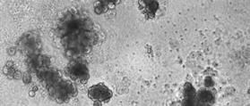
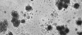
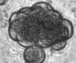
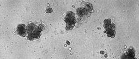
Supplementary Figure 1: Neuregulin 1 increases the growth of mammary organoids compared to EGF. (a) Mammary epithelial cells were freshly isolated,
 Mammary epithelial cells were freshly isolated,") 1 2 3 4 5 6 7 8 9 10 Supplementary Figure 1: Neuregulin 1 increases the growth of mammary organoids compared to EGF. (a) Mammary epithelial cells were freshly isolated, embedded in matrigel and exposed
1 2 3 4 5 6 7 8 9 10 Supplementary Figure 1: Neuregulin 1 increases the growth of mammary organoids compared to EGF. (a) Mammary epithelial cells were freshly isolated, embedded in matrigel and exposed
Supplementary Figure 1: si-craf but not si-braf sensitizes tumor cells to radiation.
 Supplementary Figure 1: si-craf but not si-braf sensitizes tumor cells to radiation. (a) Embryonic fibroblasts isolated from wildtype (WT), BRAF -/-, or CRAF -/- mice were irradiated (6 Gy) and DNA damage
Supplemental figures and figure legends (90517-INS-RG-RV-2) Supplemental Figure 1.
 Supplemental Figure 1.") Supplemental figures and figure legends (957-INS-RG-RV-) Supplemental Figure. A B.5.5 Interaction p=.89 Model p
Supplemental figures and figure legends (957-INS-RG-RV-) Supplemental Figure. A B.5.5 Interaction p=.89 Model p
Figure S1. Generation of inducible PTEN deficient mice and the BMMCs (A) B6.129 Pten loxp/loxp mice were mated with B6.
 B6.129 Pten loxp/loxp mice were mated with B6.") Figure S1. Generation of inducible PTEN deficient mice and the BMMCs (A) B6.129 Pten loxp/loxp mice were mated with B6.129-Gt(ROSA)26Sor tm1(cre/ert2)tyj /J mice. To induce deletion of the Pten locus,
Figure S1. Generation of inducible PTEN deficient mice and the BMMCs (A) B6.129 Pten loxp/loxp mice were mated with B6.129-Gt(ROSA)26Sor tm1(cre/ert2)tyj /J mice. To induce deletion of the Pten locus,
Supplemental Figure 1
 Supplemental Figure 1 A S100A4: SFLGKRTDEAAFQKLMSNLDSNRDNEVDFQEYCVFLSCIAMMCNEFFEGFPDK Overlap: SF G DE KLM LD N D VDFQEY VFL I M N FF G PD S100A2: SFVGEKVDEEGLKKLMGSLDENSDQQVDFQEYAVFLALITVMCNDFFQGCPDR
Cell Culture. The human thyroid follicular carcinoma cell lines FTC-238, FTC-236 and FTC-
 Supplemental material and methods Reagents. Hydralazine was purchased from Sigma-Aldrich. Cell Culture. The human thyroid follicular carcinoma cell lines FTC-238, FTC-236 and FTC- 133, human thyroid medullary
Supplementary Figure 1. EC-specific Deletion of Snail1 Does Not Affect EC Apoptosis. (a,b) Cryo-sections of WT (a) and Snail1 LOF (b) embryos at
 Cryo-sections of WT (a) and Snail1 LOF (b) embryos at") Supplementary Figure 1. EC-specific Deletion of Snail1 Does Not Affect EC Apoptosis. (a,b) Cryo-sections of WT (a) and Snail1 LOF (b) embryos at E10.5 were double-stained for TUNEL (red) and PECAM-1 (green).
Supplementary Figure 1. EC-specific Deletion of Snail1 Does Not Affect EC Apoptosis. (a,b) Cryo-sections of WT (a) and Snail1 LOF (b) embryos at E10.5 were double-stained for TUNEL (red) and PECAM-1 (green).
SUPPLEMENTARY FIGURES
 SUPPLEMENTARY FIGURES 1 Supplementary Figure 1, Adult hippocampal QNPs and TAPs uniformly express REST a-b) Confocal images of adult hippocampal mouse sections showing GFAP (green), Sox2 (red), and REST
Supplementary Information Supplementary Fig. 1. Elevated Usp9x in melanoma and NRAS mutant melanoma cells are dependent on NRAS for 3D growth.
 Supplementary Information Supplementary Fig. 1. Elevated Usp9x in melanoma and NRAS mutant melanoma cells are dependent on NRAS for 3D growth. a. Immunoblot for Usp9x protein in NRAS mutant melanoma cells
Supplementary Figure 1. Method development.
 Supplementary Figure 1 Method development. Titration experiments to determine standard antibody:lysate concentration. Lysates (~2 mg of total proteins) were prepared from cells expressing FLAG- tagged
for six pairs of mice. (b) Representative FACS analysis of absolute number of T cells (CD4 + and
 Representative FACS analysis of absolute number of T cells (CD4 + and") SUPPLEMENTARY DATA Supplementary Figure 1: Peripheral lymphoid organs of SMAR1 -/- mice have an effector memory phenotype. (a) Lymphocytes collected from MLNs and Peyer s patches (PPs) of WT and SMAR1
SUPPLEMENTARY DATA Supplementary Figure 1: Peripheral lymphoid organs of SMAR1 -/- mice have an effector memory phenotype. (a) Lymphocytes collected from MLNs and Peyer s patches (PPs) of WT and SMAR1
SUPPLEMENTARY DATA. Supplementary Table 2. Antibodies used for Immunofluoresence. Supplementary Table 3. Real-time PCR primer sequences.
 Supplementary Table 2. Antibodies used for Immunofluoresence. Antibody Dilution Source Goat anti-pdx1 1:100 R&D Systems Rabbit anti-hnf6 1:100 Santa Cruz Biotechnology Mouse anti-nkx6.1 1:200 Developmental
Supporting Information
 Supporting Information Franco et al. 10.1073/pnas.1015557108 SI Materials and Methods Drug Administration. PD352901 was dissolved in 0.5% (wt/vol) hydroxyl-propyl-methylcellulose, 0.2% (vol/vol) Tween
SUPPLEMENTARY INFORMATION. CXCR4 inhibitors could benefit to HER2 but not to Triple-Negative. breast cancer patients
 SUPPLEMENTARY INFORMATION CXCR4 inhibitors could benefit to HER2 but not to Triple-Negative breast cancer patients Lefort S. 1,2, Thuleau A. 3, Kieffer Y. 1,2, Sirven P. 1,2, Bieche I. 4, Marangoni E.
The Schedule and the Manual of Basic Techniques for Cell Culture
 The Schedule and the Manual of Basic Techniques for Cell Culture 1 Materials Calcium Phosphate Transfection Kit: Invitrogen Cat.No.K2780-01 Falcon tube (Cat No.35-2054:12 x 75 mm, 5 ml tube) Cell: 293
Programmed necrosis, not apoptosis, is a key mediator of cell loss and DAMP-mediated inflammation in dsrna-induced retinal degeneration
 Programmed necrosis, not apoptosis, is a key mediator of cell loss and DAMP-mediated inflammation in dsrna-induced retinal degeneration The Harvard community has made this article openly available. Please
Supporting Information
 Supporting Information Desnues et al. 10.1073/pnas.1314121111 SI Materials and Methods Mice. Toll-like receptor (TLR)8 / and TLR9 / mice were generated as described previously (1, 2). TLR9 / mice were
Soft Agar Assay. For each cell pool, 100,000 cells were resuspended in 0.35% (w/v)
") SUPPLEMENTARY MATERIAL AND METHODS Soft Agar Assay. For each cell pool, 100,000 cells were resuspended in 0.35% (w/v) top agar (LONZA, SeaKem LE Agarose cat.5004) and plated onto 0.5% (w/v) basal agar.
SUPPLEMENTARY MATERIAL AND METHODS Soft Agar Assay. For each cell pool, 100,000 cells were resuspended in 0.35% (w/v) top agar (LONZA, SeaKem LE Agarose cat.5004) and plated onto 0.5% (w/v) basal agar.
Immunostaining was performed on tumor biopsy samples arranged in a tissue-microarray format or on
 Supplemental Methods Immunohistochemical Analyses Immunostaining was performed on tumor biopsy samples arranged in a tissue-microarray format or on prostatectomy sections obtained post-study. Briefly,
MicroRNA sponges: competitive inhibitors of small RNAs in mammalian cells
 MicroRNA sponges: competitive inhibitors of small RNAs in mammalian cells Margaret S Ebert, Joel R Neilson & Phillip A Sharp Supplementary figures and text: Supplementary Figure 1. Effect of sponges on
SUPPLEMENTAL MATERIAL. Supplementary Methods
 SUPPLEMENTAL MATERIAL Supplementary Methods Culture of cardiomyocytes, fibroblasts and cardiac microvascular endothelial cells The isolation and culturing of neonatal rat ventricular cardiomyocytes was
Comparison of primary tumor sections from MMTV-PyMT or MTLn3-ErbB3-
 Supplemental Data Comparison of primary tumor sections from MMTV-PyMT or MTLn3-ErbB3- GFP tumors in mice either injected with control or clodronate-containing liposomes and stained for macrophages using
Erzsebet Kokovay, Susan Goderie, Yue Wang, Steve Lotz, Gang Lin, Yu Sun, Badrinath Roysam, Qin Shen,
 Cell Stem Cell, Volume 7 Supplemental Information Adult SVZ Lineage Cells Home to and Leave the Vascular Niche via Differential Responses to SDF1/CXCR4 Signaling Erzsebet Kokovay, Susan Goderie, Yue Wang,
Supplementary Figure 1
 Supplementary Figure 1 a Percent of body weight! (%) 4! 3! 1! Epididymal fat Subcutaneous fat Liver SD Percent of body weight! (%) ** 3! 1! SD Percent of body weight! (%) 6! 4! SD ** b Blood glucose (mg/dl)!
Nuclear Extraction Kit
 Nuclear Extraction Kit Catalog Number KA1346 50 assays Version: 07 Intended for research use only www.abnova.com Table of Contents Introduction... 3 Principle of the Assay... 3 General Information... 4
Online Data Supplement. Anti-aging Gene Klotho Enhances Glucose-induced Insulin Secretion by Upregulating Plasma Membrane Retention of TRPV2
 Online Data Supplement Anti-aging Gene Klotho Enhances Glucose-induced Insulin Secretion by Upregulating Plasma Membrane Retention of TRPV2 Yi Lin and Zhongjie Sun Department of physiology, college of
Atg5 flox/flox ; CAG-Cre, 19M brain heart lung. spleen stomach colon. Takamura_Fig. S1
 Takamura_Fig. S1 brain heart lung spleen stomach colon kidney SM Supplemental Figure 1 Histological findings of tg5 flox/flox ;CG-Cre mouse tissues. H&E staining of the brain, heart, lung, spleen, stomach,
SUPPLEMENT. Materials and methods
 SUPPLEMENT Materials and methods Cell culture and reagents Cell media and reagents were from Invitrogen unless otherwise indicated. Antibiotics and Tet-certified serum were from Clontech. In experiments
Supplementary Figure S1. Venn diagram analysis of mrna microarray data and mirna target analysis. (a) Western blot analysis of T lymphoblasts (CLS)
 Western blot analysis of T lymphoblasts (CLS)") Supplementary Figure S1. Venn diagram analysis of mrna microarray data and mirna target analysis. (a) Western blot analysis of T lymphoblasts (CLS) and their exosomes (EXO) in resting (REST) and activated
Supplementary Figure S1. Venn diagram analysis of mrna microarray data and mirna target analysis. (a) Western blot analysis of T lymphoblasts (CLS) and their exosomes (EXO) in resting (REST) and activated
Supplemental Figure 1. Intracranial transduction of a modified ptomo lentiviral vector in the mouse
 Supplemental figure legends Supplemental Figure 1. Intracranial transduction of a modified ptomo lentiviral vector in the mouse hippocampus targets GFAP-positive but not NeuN-positive cells. (A) Stereotaxic
SREBP-2 promotes stem cell-like properties and metastasis by transcriptional activation of c-myc in prostate cancer
 SREBP-2 promotes stem cell-like properties and metastasis by transcriptional activation of c-myc in prostate cancer Supplementary Material Supplementary Methods Supplementary References Supplementary Figure
Supplementary Fig. 1. Identification of acetylation of K68 of SOD2
 Supplementary Fig. 1. Identification of acetylation of K68 of SOD2 A B H. sapiens 54 KHHAAYVNNLNVTEEKYQEALAK 75 M. musculus 54 KHHAAYVNNLNATEEKYHEALAK 75 X. laevis 55 KHHATYVNNLNITEEKYAEALAK 77 D. rerio
hexahistidine tagged GRP78 devoid of the KDEL motif (GRP78-His) on SDS-PAGE. This
 on SDS-PAGE. This") SUPPLEMENTAL FIGURE LEGEND Fig. S1. Generation and characterization of. (A) Coomassie staining of soluble hexahistidine tagged GRP78 devoid of the KDEL motif (GRP78-His) on SDS-PAGE. This protein was expressed
SUPPLEMENTAL FIGURE LEGEND Fig. S1. Generation and characterization of. (A) Coomassie staining of soluble hexahistidine tagged GRP78 devoid of the KDEL motif (GRP78-His) on SDS-PAGE. This protein was expressed
(a) Significant biological processes (upper panel) and disease biomarkers (lower panel)
 Significant biological processes (upper panel) and disease biomarkers (lower panel)") Supplementary Figure 1. Functional enrichment analyses of secretomic proteins. (a) Significant biological processes (upper panel) and disease biomarkers (lower panel) 2 involved by hrab37-mediated secretory
Supplementary Figure 1. Functional enrichment analyses of secretomic proteins. (a) Significant biological processes (upper panel) and disease biomarkers (lower panel) 2 involved by hrab37-mediated secretory
The following protocol describes the isolation of nuclei from tissue. Item. Catalog No Manufacturer
 SOP: Nuclei isolation from tissue and DNaseI treatment Date modified: 090923 Modified by: P. Sabo. (UW) The following protocol describes the isolation of nuclei from tissue. Ordering Information Item.
PREPARATION OF IF- ENRICHED CYTOSKELETAL PROTEINS
 TMM,5-2011 PREPARATION OF IF- ENRICHED CYTOSKELETAL PROTEINS Ice-cold means cooled in ice water. In order to prevent proteolysis, make sure to perform all steps on ice. Pre-cool glass homogenizers, buffers
Supplementary Figures
 Supplementary Figures Supplementary Figure 1. Confirmation of Dnmt1 conditional knockout out mice. a, Representative images of sorted stem (Lin - CD49f high CD24 + ), luminal (Lin - CD49f low CD24 + )
Report on Pathology. Study: The effect of Compound X on pancreatic islets in rhesus macaques
 Report on Pathology Study: The effect of Compound X on pancreatic islets in rhesus macaques Prepared for: Client Name Client Address January 1, 2013 Prepared by: Charter Preclinical Services 21 Main St.,
PRODUCT INFORMATION & MANUAL
 PRODUCT INFORMATION & MANUAL Mitochondrial Extraction Kit NBP2-29448 Research use only. Not for diagnostic or therapeutic procedures www.novusbio.com P: 303.760.1950 P: 888.506.6887 F: 303.730.1966 technical@novusbio.com
Supplemental Information
 Supplemental Information Tobacco-specific Carcinogen Induces DNA Methyltransferases 1 Accumulation through AKT/GSK3β/βTrCP/hnRNP-U in Mice and Lung Cancer patients Ruo-Kai Lin, 1 Yi-Shuan Hsieh, 2 Pinpin
Evaluation of directed and random motility in microslides Assessment of leukocyte adhesion in flow chambers
 Evaluation of directed and random motility in microslides Motility experiments in IBIDI microslides, image acquisition and processing were performed as described. PMN, which ended up in an angle < 180
Procaspase-3. Cleaved caspase-3. actin. Cytochrome C (10 M) Z-VAD-fmk. Procaspase-3. Cleaved caspase-3. actin. Z-VAD-fmk
 Z-VAD-fmk. Procaspase-3. Cleaved caspase-3. actin. Z-VAD-fmk") A HeLa actin - + + - - + Cytochrome C (1 M) Z-VAD-fmk PMN - + + - - + actin Cytochrome C (1 M) Z-VAD-fmk Figure S1. (A) Pan-caspase inhibitor z-vad-fmk inhibits cytochrome c- mediated procaspase-3 cleavage.
A HeLa actin - + + - - + Cytochrome C (1 M) Z-VAD-fmk PMN - + + - - + actin Cytochrome C (1 M) Z-VAD-fmk Figure S1. (A) Pan-caspase inhibitor z-vad-fmk inhibits cytochrome c- mediated procaspase-3 cleavage.
SUPPLEMENTARY INFORMATION
 doi:10.1038/nature12652 Supplementary Figure 1. PRDM16 interacts with endogenous EHMT1 in brown adipocytes. Immunoprecipitation of PRDM16 complex by flag antibody (M2) followed by Western blot analysis
SUPPLEMENTARY MATERIAL. Sample preparation for light microscopy
 SUPPLEMENTARY MATERIAL Sample preparation for light microscopy To characterize the granulocytes and melanomacrophage centers, cross sections were prepared for light microscopy, as described in Material
Geneaid DNA Isolation Kit
 Instruction Manual Ver. 02.21.17 For Research Use Only Geneaid DNA Isolation Kit GEB100, GEB01K, GEB01K+ GEC150, GEC1.5K, GEC1.5K+ GET150, GET1.5K, GET1.5K+ GEE150, GEE1.5K, GEE1.5K+ Advantages Sample:
Proteomic profiling of small-molecule inhibitors reveals dispensability of MTH1 for cancer cell survival
 Supplementary Information for Proteomic profiling of small-molecule inhibitors reveals dispensability of MTH1 for cancer cell survival Tatsuro Kawamura 1, Makoto Kawatani 1, Makoto Muroi, Yasumitsu Kondoh,
Data Sheet TIGIT / NFAT Reporter - Jurkat Cell Line Catalog #60538
 Data Sheet TIGIT / NFAT Reporter - Jurkat Cell Line Catalog #60538 Background: TIGIT is a co-inhibitory receptor that is highly expressed in Natural Killer (NK) cells, activated CD4+, CD8+ and regulatory
Supporting Information
 Supporting Information Fujishita et al. 10.1073/pnas.0800041105 SI Text Polyp Scoring. Intestinal polyps were counted as described (1). Briefly, the small and large intestines were excised, washed with
Hopkins University, Howard Hughes Medical Institute, USA) (27). Cells were maintained in DMEM
 (27). Cells were maintained in DMEM") Supplementary Materials and Methods Cell Culture HCT116 (TP53 +/+ and TP53 -/- ) cells were provided by Dr. Bert Vogelstein (Johns Hopkins University, Howard Hughes Medical Institute, USA) (27). Cells
Supplementary Materials and Methods Cell Culture HCT116 (TP53 +/+ and TP53 -/- ) cells were provided by Dr. Bert Vogelstein (Johns Hopkins University, Howard Hughes Medical Institute, USA) (27). Cells
Dissected tissues were minced and lysed in lysis buffer (1x Tris buffered saline (TBS), 1% NP-40,
, 1% NP-40,") Data Supplement for Dincheva et al., Effect of Early-Life Fluoxetine on Anxiety-Like Behaviors in BDNF Val66Met Mice. Am J Psychiatry (doi: 10.1176/appi.ajp.2017.15121592) Contents Supplemental Methods
Data Supplement for Dincheva et al., Effect of Early-Life Fluoxetine on Anxiety-Like Behaviors in BDNF Val66Met Mice. Am J Psychiatry (doi: 10.1176/appi.ajp.2017.15121592) Contents Supplemental Methods
Supplementary Figure 1. Efficiency of Mll4 deletion and its effect on T cell populations in the periphery. Nature Immunology: doi: /ni.
 Supplementary Figure 1 Efficiency of Mll4 deletion and its effect on T cell populations in the periphery. Expression of Mll4 floxed alleles (16-19) in naive CD4 + T cells isolated from lymph nodes and
The toll-like receptor 4 ligands Mrp8 and Mrp14 play a critical role in the development of autoreactive CD8 + T cells
 1 SUPPLEMENTARY INFORMATION The toll-like receptor 4 ligands Mrp8 and Mrp14 play a critical role in the development of autoreactive CD8 + T cells Karin Loser 1,2,6, Thomas Vogl 2,3, Maik Voskort 1, Aloys
TSH Receptor Monoclonal Antibody (49) Catalog Number MA3-218 Product data sheet
 Catalog Number MA3-218 Product data sheet") Website: thermofisher.com Customer Service (US): 1 800 955 6288 ext. 1 Technical Support (US): 1 800 955 6288 ext. 441 TSH Receptor Monoclonal Antibody (49) Catalog Number MA3-218 Product data sheet Details
Website: thermofisher.com Customer Service (US): 1 800 955 6288 ext. 1 Technical Support (US): 1 800 955 6288 ext. 441 TSH Receptor Monoclonal Antibody (49) Catalog Number MA3-218 Product data sheet Details
Chemical Chaperones Mitigate Experimental Asthma By Attenuating Endoplasmic
 Chemical Chaperones Mitigate Experimental Asthma By Attenuating Endoplasmic Reticulum Stress Lokesh Makhija, BE, Veda Krishnan, MSc, Rakhshinda Rehman, MTech, Samarpana Chakraborty, MSc, Shuvadeep Maity,
Pretargeting and Bioorthogonal Click Chemistry-Mediated Endogenous Stem Cell Homing for Heart Repair
 Pretargeting and Bioorthogonal Click Chemistry-Mediated Endogenous Stem Cell Homing for Heart Repair Mouse Model of Myocardial Infarction (MI) All animal work was compliant with the Institutional Animal
Supplemental Materials for. Effects of sphingosine-1-phosphate receptor 1 phosphorylation in response to. FTY720 during neuroinflammation
 Supplemental Materials for Effects of sphingosine-1-phosphate receptor 1 phosphorylation in response to FTY7 during neuroinflammation This file includes: Supplemental Table 1. EAE clinical parameters of
TFEB-mediated increase in peripheral lysosomes regulates. Store Operated Calcium Entry
 TFEB-mediated increase in peripheral lysosomes regulates Store Operated Calcium Entry Luigi Sbano, Massimo Bonora, Saverio Marchi, Federica Baldassari, Diego L. Medina, Andrea Ballabio, Carlotta Giorgi
HCC1937 is the HCC1937-pcDNA3 cell line, which was derived from a breast cancer with a mutation
 SUPPLEMENTARY INFORMATION Materials and Methods Human cell lines and culture conditions HCC1937 is the HCC1937-pcDNA3 cell line, which was derived from a breast cancer with a mutation in exon 20 of BRCA1
Essential Medium, containing 10% fetal bovine serum, 100 U/ml penicillin and 100 µg/ml streptomycin. Huvec were cultured in
 Supplemental data Methods Cell culture media formulations A-431 and U-87 MG cells were maintained in Dulbecco s Modified Eagle s Medium. FaDu cells were cultured in Eagle's Minimum Essential Medium, containing
BMP6 treatment compensates for the molecular defect and ameliorates hemochromatosis in Hfe knockout mice
 SUPPLEMENTARY MATERIALS BMP6 treatment compensates for the molecular defect and ameliorates hemochromatosis in Hfe knockout mice Elena Corradini, Paul J. Schmidt, Delphine Meynard, Cinzia Garuti, Giuliana
Suppl Video: Tumor cells (green) and monocytes (white) are seeded on a confluent endothelial
 and monocytes (white) are seeded on a confluent endothelial") Supplementary Information Häuselmann et al. Monocyte induction of E-selectin-mediated endothelial activation releases VE-cadherin junctions to promote tumor cell extravasation in the metastasis cascade
Supplementary Information Häuselmann et al. Monocyte induction of E-selectin-mediated endothelial activation releases VE-cadherin junctions to promote tumor cell extravasation in the metastasis cascade
marker. DAPI labels nuclei. Flies were 20 days old. Scale bar is 5 µm. Ctrl is
 Supplementary Figure 1. (a) Nos is detected in glial cells in both control and GFAP R79H transgenic flies (arrows), but not in deletion mutant Nos Δ15 animals. Repo is a glial cell marker. DAPI labels
SUPPLEMENTARY MATERIAL
 SUPPLEMENTARY MATERIAL Table S1. Primers and fluorescent probes used for qrt-pcr analysis of relative expression levels of PPP family phosphatases. gene name forward primer, 5-3 probe, 5-3 reverse primer,
For the 5 GATC-overhang two-oligo adaptors set up the following reactions in 96-well plate format:
 Supplementary Protocol 1. Adaptor preparation: For the 5 GATC-overhang two-oligo adaptors set up the following reactions in 96-well plate format: Per reaction X96 10X NEBuffer 2 10 µl 10 µl x 96 5 -GATC