Differential Diagnosis of ONH Edema Beth A. Steele, OD, FAAO
|
|
|
- Augusta Adams
- 5 years ago
- Views:
Transcription
1 Differential Diagnosis of ONH Edema Beth A. Steele, OD, FAAO Please silence all mobile devices and remove items from chairs so others can sit. Unauthorized recording of this session is prohibited.
2 Disclosure Statement: Optos Advisory Board

3 Some things make you look twice. Worrisome findings. Elevation Pallor Discoloration NFL defects Vascular changes Tools you have Stereoscopic DFE! Swinging flashlight test Pupil cycle time Red free filter SVP VF OCT FAF B scan


4 Terminology Disc Edema Papilledema Bilateral papilledema Pseudopapilledema Pseudotumor Benign Idiopathic Intractranial Hypertension


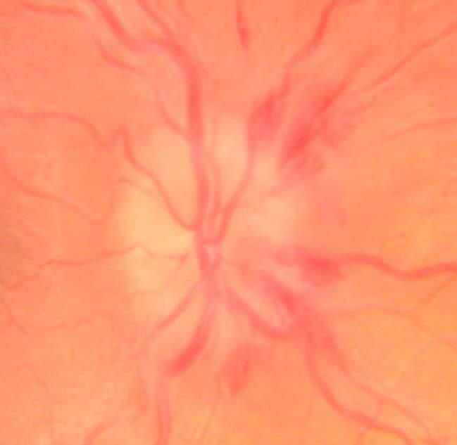
5 Disc characteristics: Tortuous vessels Blurred margins Elevated topography Disc hemorrhages Papilledema on a silver platter 5
6

7 Other causes of disc edema Unilateral Vascular Diabetic Papillopathy Bilateral Toxic Inflammatory Infectious Compressive Hydrocephalus
8 Is it? And if so when why is it? Pseudopapilledema ONH drusen Anatomically crowded discs Hyperopic disc Myopic / tilted / obliquely inserted disc


9 Myopic / tilted discs? Be careful not to hide behind a comfortable label
10 Exam VF Photography Clinical Tool Exam Papilledema Hyperemia, Dilated capillaries, Vessel obscuration Buried ONH Drusen Absence of physiologic cup Anomalous vessel branching Anatomically Crowded Disc Absence of cup, elevated appearance SVP Absent Present in 80% normals Present in 80% normals Visual Field Enlarged blind spot, Various defects (e.g. Normal central defect arcuate, central, etc) FAF Normal Increased FAF Normal

11 Spontaneous Venous Pulsation (SVP) Occurs when IOP > Central Retinal Vein pressure 80% of normals have an SVP What if it is absent? 20% of normals don t have one But if once present, and now gone consider CRV is subject to external compression where it crosses optic nerve sheath SVP is absent if CRV pressure elevated (due to ICP) Increased venous pressure upstream

12 FAF in ONH Drusen Hyper AF Buried harder to visualize hyper EDI superior to AF Loft et al. J Neurophthalmology Optos.com
13 Spectral Domain OCT Clinical Tool Papilledema Buried ONH Drusen Anatomically Crowded Disc OCT disc topography Smooth, continuous elevation Non uniform elevation Smooth, continuous elevation OCT RNFL scan Thicker RNFL, especially nasal Thicker hypo reflective space between retina and RPE ( lazy V ) Increased in acutely damaged tissue, may be decreased over time Normal OCT/MRI Globe convexity Flattened / pushed forward Normal/concave Normal/increased concavity for hyperopes


14

15


16 Key features disc edema on OCT: Smooth contour of elevation Nasal RNFL >86um 80% specificity Thick hyporeflective space adjacent to disc lazy V 90% specificity Johnson L. Archives of Ophth 2009.


17 Globe Convexity Increased ICP will push the globe anteriorly Easiest to appreciate with a 9mm scan With EDI, can see an anteriorly displaced Bruch s membrane
18 Ultrasonography B scan Clinical Tool Papilledema Buried ONH Drusen Anatomically Crowded Disc B scan reflectivity Normal Hyper reflectivity with reduced gain Normal B scan ON sheath Increased Normal Decreased diameter B scan crescent present absent absent shadow B scan 30 Test Positive Negative Negative


19
 but non specific (36.4%) Kimberly HH, et al. Acad Emerg Med.")
20 B scan Ultrasonography Review of Tools ONH Sheath Diameter <5mm Elevation of ONH <1mm Crescent sign 30 degree test (+) when >15% in nerve sheath width following a 30 degree eccentric gaze highly sensitive (100%) but non specific (36.4%) Kimberly HH, et al. Acad Emerg Med


21 43 AA Female 20/15 140/90 DFE: drance heme OD and disc elevated nasally Brightness comparison equal Pupil Cycle Time equal and normal +SVP


22


23

24 Optic Disc Drusen review and new Up to 2.4% of population Hereditary component 75% bilateral Benign? Retrobulbar axonal degeneration due to drusen! 24 87% have VF defects Most commonly nasal, arcuate or partial arcuate Worsen over time Degree of VF defect correlates with RNFL thinning around the disc Deficit and prognosis worse when lesions are more superficial Chan et al. Clin and Exp Ophth July Auw Haedrich C, et al. Surv Ophthalmol 2002 Malmqvist L et al. J Neuroophthalmol March, Duncan, et al. J AAOPS, Feb, Hamann et al. Acta Ophth 2018.


25 What about in kids? Around 20% have VF defects Up to 24% have CNVM associated! 50% with elevated discs due to IIHTN also had ONH Drusen! Gospe SM III. Br J Ophth Auw Haedrich C, et al. Surv Ophthalmol 2002 Malmqvist L et al. J Neuroophthalmol March, Duncan, et al. J AAOPS, Feb, 2016.


26 At what point do you lower IOP in patient with drusen? 71 year old with longstanding ONH drusen Progressive VF loss OD>OS IOP averages 21mmHg OU
27


28 Decrease in IOP : of retinal ganglion cell function Stabilizes RNFL thickness May delay the progression of optic neuropathy Podja Wilczek et al. Ophthalmic Res, 2017.


29 34 WF 20/20, pupils normal, CF normal 7.00 OU BMI 31 Denies H/A Elevated ONH.1/.1 CD +SVP


30 Normal RNFL thickness No subretinal cavitation


31 Globe convexity is normal Vs
32

 3 mm")

33 Measuring the ONH Sheath Diameter Axial images of the optic nerve (V and H) 3 mm behind the posterior eye wall Hyperechogenic borders
34 In summary ( ) hyperreflectivity consistent with drusen ( ) crescent sign Normal ONH sheath diameter Normal RNFL thickness +SVP Normal globe convexity Stability. Presumed crowded discs but careful follow up


35 32 AA FM Severe headaches, BMI 42


36 Disc characteristics: Tortuous vessels? Mildly indistinct margins Elevated topography Paton s folds? SVP absent (-) disc hemes
37
38 Papilledema Suspected? Now what Brain Imaging MRI rule out space occupying mass MRV rule out cerebral venous thrombosis Lumbar Puncture Higher than 25/30 cm H2O is abnormal Referral? Determine underlying cause/association if any Weight Associated medications

39 MRI normal This patient: opening pressure = 28cm H 2 0
40 Idiopathic Intracranial HTN 90 98% complain of headache Nausea/vomiting/dizziness 40% Pulsatile tinnitus 16 60% Visual disturbances 30% Mollan SP, et al. Pract Neurol 2018
41 Etiologies Idiopathic But what causes the papilledema? Axoplasmic flow stasis swollen nerve fibers compression of venules in the area and so venous stasis/leakage accumula on of extracellular fluid Associated Medications Oral contraceptives Steroids Tamoxifen Vitamin A Nitrofurantoin Tetracyclines
42 IIH Treatment Trial JAMA, April 2014 acetazolamide + a low sodium weight reduction diet vs. diet alone modest improvement in visual field function OCT Substudy of the IITT, Ophthalmology, Sept 2015 Better RNFL thickness, TRT, and ONH volume swelling measurements RNFL and Total Retinal Thickness (TRT) useful in following and monitoring response to treatment
43 Acetazolamide + Low Sodium/Wt Loss Diet The Idiopathic Intracranial Hypertension Treatment Trial. JAMA ,250mg tablets, and 500mg SR capsules (Diamox Sequels, Duramed Pharmaceuticals) Contraindications: Sulfa allergy? chemical structure different than antibiotics little evidence of cross sensitivity consider avoiding if hx of severe reaction Long term: liver, kidney disease, severe COPD Caution with sickle cell Caution with low potassium paresthesia metallic taste fatigue malaise gastrointestinal disturbances Common Side Effects decreased libido metabolic acidosis electrolyte imbalance (including hypokalemia) renal calculi blood dyscrasias Than T, Smith H. must have oral meds.
44 Treatment Goals Visual Fields OCT Labs Referral? Mollan SP, et al. Pract Neurol 2018


45 Following Tx Diamox 250mg BID x 2 years
46


47 Those tools can miss subtle elevation.. 3 years earlier was consistent with anatomical elevation; then lost to f/u x 3 years 3 years later patient decides to come back


48 21 AAF High BMI MRI clear Initial opening pressure of 52

49
")
50 After 1 month of 250mg Diamox BID still (+) H/A
 Denies H/A")
51 2 mos, then 5 mos (now TID Diamox) Denies H/A
52 Watching VF carefully H/A s improved RNFL thickness reduced Total volume reduced
53 Repeated Lumbar Puncture for Recalcitrant Cases? Not well reported Procedure causes anxiety, local discomfort, complications, headache LP induced reduction of ICP is only short lived Yiangou et al. Hoffman J. Cephalagia 2018.


54 4:30 on a Friday. 41 year old male has never had an eye exam LPE: 11 years ago No medications C/o: severe headache and blurred vision x 2 weeks BCVA: 20/30, 20/60 Pupils: appeared normal CFs: reduced OD, OS EOM: normal

55 Large meningioma at base of skull
56 Pre/post surgical RNFL scans can drive decisions for surgery (i.e. urgency) ing: become permanent Stable: may wait even if VF shows loss Comparison of pre vs. post surgical scans in neuro cases Meningioma can grow back, so useful for monitoring progression


57 26 WF, blurry vision Hx hydracephalous, multiple previous surgeries VA 20/40 OD, OS H/A developing with more intensity MRI all clear. ICP was borderline

58
59 77
60 Please remember to complete your session evaluations on the Academy.18 meeting app Tweet about this session using the official meeting hashtag #Academy18
Visual Fields A Road Map to Management.
 Visual Fields A Road Map to Management. Disclosures Dr. Beth Steele Company Position Received Optos Advisory Board Honorarium Med Op Consultant Honorarium Beth A. Steele, OD, FAAO bsteele@uab.edu FACTS
12/2/16. Ways to differentiate:
 Nate Lighthizer, O.D., F.A.A.O. Assistant Dean for Clinical Care Services Director of CE Chief of Specialty Care Clinics Chief of Electrodiagnostics Clinic Oklahoma College of Optometry lighthiz@nsuok.edu
Papilledema. Golnaz Javey, M.D. and Jeffrey J. Zuravleff, M.D.
 Papilledema Golnaz Javey, M.D. and Jeffrey J. Zuravleff, M.D. Papilledema specifically refers to optic nerve head swelling secondary to increased intracranial pressure (IICP). Optic nerve swelling from
Neuro-Ocular Grand Rounds
 Neuro-Ocular Grand Rounds Anthony B. Litwak,OD, FAAO VA Medical Center Baltimore, Maryland Dr. Litwak is on the speaker and advisory boards for Alcon and Zeiss Meditek COMMON OPTIC NEUROPATHIES THAT CAN
Alan G. Kabat, OD, FAAO (901)
") THE SWOLLEN OPTIC DISC: EMERGENCY OR ANOMALY? Alan G. Kabat, OD, FAAO (901) 252-3691 Memphis, Tennessee alan.kabat@alankabat.com Course description: The swollen disc presents a diagnostic dilemma. While
THE SWOLLEN OPTIC DISC: EMERGENCY OR ANOMALY? Alan G. Kabat, OD, FAAO (901) 252-3691 Memphis, Tennessee alan.kabat@alankabat.com Course description: The swollen disc presents a diagnostic dilemma. While
Neuro-Ocular Grand Rounds Anthony B. Litwak,OD, FAAO VA Medical Center Baltimore, Maryland
 Neuro-Ocular Grand Rounds Anthony B. Litwak,OD, FAAO VA Medical Center Baltimore, Maryland Dr. Litwak is on the speaker and advisory boards for Alcon and Zeiss Meditek COMMON OPTIC NEUROPATHIES THAT CAN
Typical idiopathic intracranial hypertension Optic nerve appearance and brain MRI findings. Jonathan A. Micieli, MD Valérie Biousse, MD
 Typical idiopathic intracranial hypertension Optic nerve appearance and brain MRI findings Jonathan A. Micieli, MD Valérie Biousse, MD A 24 year old African American woman is referred for bilateral optic
An Organized Approach to the Patient with Papilledema and IIH
 An Organized Approach to the Patient with Papilledema and IIH Leonard V. Messner, OD, FAAO James L. Fanelli, OD, FAAO Please silence all mobile devices and remove items from chairs so others can sit. Unauthorized
Pearls, Pitfalls and Advances in Neuro-Ophthalmology
 Pearls, Pitfalls and Advances in Neuro-Ophthalmology Nancy J. Newman, MD Emory University Atlanta, GA Consultant for Gensight Biologics, Santhera Data Safety Monitoring Board for Quark AION Study Medical-legal
OPTIC NERVE SWELLING IN CHILDHOOD
 OPTIC NERVE SWELLING IN CHILDHOOD Melissa W. Ko, MD, FAAN One of the main findings on a pediatric neurologic examination that can instill fear and lead to an urgent referral to neuro-ophthalmology is the
OCT in the Diagnosis and Follow-up of Glaucoma
 OCT in the Diagnosis and Follow-up of Glaucoma Karim A Raafat MD. Professor Of Ophthalmology Cairo University Hmmmm! Do I have Glaucoma or not?! 1 Visual Function 100% - N Gl Structure : - 5000 axon /
Optic Nerve Anomalies
 Optic Nerve Anomalies Raman Bhakhri, OD, FAAO Southern California College of Optometry Marshall B. Ketchum University Goals for today Review some of the optic nerve anomalies that can be seen in practice
Neuro Ocular Grand Rounds Anthony B. Litwak, OD, FAAO VA Medical Center Baltimore, MD
 Neuro Ocular Grand Rounds Anthony B. Litwak, OD, FAAO VA Medical Center Baltimore, MD 58 YOWM! C/O I think there is something wrong with my vision, but I m not sure what it is.! +PMH for HTN, atrial fibrillation,
University Hospital Basel. Optical Coherence Tomography Emerging Role in the Assessment of MS PD Dr. Konstantin Gugleta
 University Hospital Basel Optical Coherence Tomography Emerging Role in the Assessment of MS PD Dr. Konstantin Gugleta 15th State of the Art SMSS, Lucerne January 2013 Retinal Nerve Fiber Layer 1.200.000
The Glaucoma Suspect. Evaluating the Suspect Disk. Dr Michael Forrest. ! the usual suspects: ! is it glaucoma? ! is it swollen?
 Evaluating the Suspect Disk Dr Michael Forrest Senior Lecturer, The University of Queensland Northside Eye Specialists, Nundah Visiting Ophthalmologist, Mater Hospital, Brisbane Australian Vision Convention
Optic Nerve Disorders: Structure and Function and Causes
 Optic Nerve Disorders: Structure and Function and Causes Using Visual Fields, OCT and B-scan Ultrasound to Diagnose and Follow Optic Nerve Visual Losses Ohio Ophthalmological Society and Ophthalmic Tech
Question 1: Comment on the optic nerve appearance of each eye.
 Case 2 - Right Optic Nerve Head Drusen (ONHD) A 41 year old female was referred by her optometrist for a workup for unilateral optic disc drusen, OCT, and visual field changes. The patient was otherwise
Learn Connect Succeed. JCAHPO Regional Meetings 2015
 Learn Connect Succeed JCAHPO Regional Meetings 2015 OPTIC NEUROPATHY AS EASY AS 1,2,3,4 OPTIC NERVE ANATOMY M. Tariq Bhatti, MD Departments of Ophthalmology and Neurology Duke Eye Center and Duke University
Is it Papilloedema? John Ross Ainsworth Orthoptic staff Birmingham Children s Hospital Birmingham and Midland Eye Centre University of Birmingham
 Is it Papilloedema? John Ross Ainsworth Orthoptic staff Birmingham Children s Hospital Birmingham and Midland Eye Centre University of Birmingham Aims Children/young people A bit about hypoplasia / NFL
The Optic Nerve Head In Glaucoma. Clinical Pearl #1. Characteristics of Normal Disk 9/26/2017. Initial detectable damage Structure vs function
 The Optic Nerve Head In Glaucoma Clinical Pearl #1 Eric E. Schmidt, O.D., F.A.A.O. Omni Eye Specialists Wilmington,NC schmidtyvision@msn.com Glaucoma is an optic neuropathy Initial detectable damage Structure
Neuro-Ophthalmic Masqueraders
 Neuro-Ophthalmic Masqueraders Leonid Skorin, Jr., OD, DO, MS, FAAO, FAOCO Mayo Clinic Health System in Albert Lea Denise Goodwin, OD, FAAO Pacific University College of Optometry Please silence all mobile
THE SWOLLEN DISC. Valerie Biousse, MD Emory University School of Medicine Atlanta, GA
 THE SWOLLEN DISC Valerie Biousse, MD Emory University School of Medicine Atlanta, GA Updated from: Neuro-Ophthalmology Illustrated. Biousse V, Newman NJ. Thieme, New-York,NY. 2 nd Ed, 2016. Edema of the
Optic Disc: Anatomy, Variants, Unusual discs. Kathleen B. Digre, MD Professor Neurology, Ophthalmology
 Optic Disc: Anatomy, Variants, Unusual discs Kathleen B. Digre, MD Professor Neurology, Ophthalmology THE OPHTHALMOSCOPE DIRECT OPHTHALMOSCOPY Jan Purkinje 1823 Hermann von Helmholtz 1851 Hand held ophthalmoscope
NANOS Patient Brochure
 NANOS Patient Brochure Pseudotumor Cerebri Copyright 2016. North American Neuro-Ophthalmology Society. All rights reserved. These brochures are produced and made available as is without warranty and for
OPTOMETRY REVIEW. Key words: drusen of optic nerve head, multimodal imaging, optic nerve head, papilloedema, pseudopapilloedema
 C L I N I C A L A N D E X P E R I M E N T A L OPTOMETRY REVIEW The usefulness of multimodal imaging for differentiating pseudopapilloedema and true swelling of the optic nerve head: a review and case series
Dr/ Marwa Abdellah EOS /16/2018. Dr/ Marwa Abdellah EOS When do you ask Fluorescein angiography for optic disc diseases???
 When do you ask Fluorescein angiography for optic disc diseases??? 1 NORMAL OPTIC DISC The normal optic disc on fluorescein angiography is fluorescent due to filling of vessels arising from the posterior
Intro to Glaucoma/2006
 Intro to Glaucoma/2006 Managing Patients with Glaucoma is Exciting Interesting Challenging But can often be frustrating! Clinical Challenges To identify patients with risk factors for possible glaucoma.
Learn Connect Succeed. JCAHPO Regional Meetings 2017
 Learn Connect Succeed JCAHPO Regional Meetings 2017 NO FINANCIAL DISCLOSURES Technician s Role in Neuro-Ophthalmology Workup Beth Koch COT, ROUB Cleveland 9/16/2017 What Tests Are You Expected To Perform?
The Management of Infant Aphakia
 The Management of Infant Aphakia Christina Twardowski O.D., FAAO Please silence all mobile devices and remove items from chairs so others can sit. Unauthorized recording of this session is prohibited.
The Diagnostic Dilemma of Pseudopapilledema. Tiffenie Harris, OD, FAAO Associate Professor Western University College of Optometry
 The Diagnostic Dilemma of Pseudopapilledema Tiffenie Harris, OD, FAAO Associate Professor Western University College of Optometry Author s Bio Dr. Harris is a graduate of Indiana University School of Optometry.
11/10/2017. Headache and Increased Pressure: A tale of 2 cases. Kathleen Digre MD University of Utah TWO CASES. 23 yo medical practice manager
 Headache and Increased Pressure: A tale of 2 cases Kathleen Digre MD University of Utah TWO CASES 23 yo medical practice manager September 2016 began developing intense frontal headaches first intermittent
3/16/2018. Optic Nerve Examination. Hassan Eisa Swify FRCS Ed (Ophthalmology) Air Force Hospital
 Air Force Hospital") Optic Nerve Examination Hassan Eisa Swify FRCS Ed (Ophthalmology) Air Force Hospital 1 Examination Structure ( optic disc) Function Examination of the optic disc The only cranial nerve (brain tract) which
Optic Nerve Examination Hassan Eisa Swify FRCS Ed (Ophthalmology) Air Force Hospital 1 Examination Structure ( optic disc) Function Examination of the optic disc The only cranial nerve (brain tract) which
Is this glaucoma? Leo Semes, OD Michael Chaglasian, OD Danica Marrelli, OD. Optometry s Meeting 2015 Seattle, WA
 Is this glaucoma? Leo Semes, OD Michael Chaglasian, OD Danica Marrelli, OD Optometry s Meeting 2015 Seattle, WA Case 1. 54 WM Engineer is referred to UAB Eye Care as a glaucoma suspect. Mild myopic refractive
A Curious Case of Bilateral Optic Disc Edema Brittney Dautremont, DO, MPH
 A Curious Case of Bilateral Optic Disc Edema Brittney Dautremont, DO, MPH PGY2 Ophthalmology Resident Grandview Medical Center Dayton, OH CASE PRESENTATION 51 year old white female presenting with blurred
Chapter 2 Long Duration Flight Data
 Chapter 2 Long Duration Flight Data Astronaut s bodies suffer in microgravity. Without effective countermeasures, muscles atrophy, bones shed calcium, and eyesight deteriorates. We ve known about this
BMB Disclosures. Papilledema can be a. Neurological Emergency, Causing Preventable Blindness
 Reasonable Doubt: Can High Intracranial Pressure Occur Without Papilledema? 15 February 2013 Jonathan C. Horton hortonj@vision.ucsf.edu http://www.ucsf.edu/hortonlab BMB Disclosures Financial Disclosures
How Strongly Do You Feel That This Patient Has Glaucoma? % % % % %
 My Favorite Cases Anthony B. Litwak, OD, FAAO VA Medical Center Baltimore, Maryland Dr. Litwak is a speaker and on advisory boards for Alcon and Zeiss Meditek CASE CR 35 yohf Neg PMH +FOH mother and grandmother
My Favourite Cases Anthony B. Litwak, OD, FAAO VA Medical Center Baltimore, MD
 My Favourite Cases Anthony B. Litwak, OD, FAAO VA Medical Center Baltimore, MD Dr. Litwak is a speaker and on advisory boards for Alcon and Zeiss Meditek CASE CR! 35 YOHF! Neg PMH! +FOH mother and grandmother
What is IIH? Idiopathic Intracranial Hypertension (IIH)
") What is IIH? Idiopathic Intracranial Hypertension (IIH) What is Idiopathic Intracranial Hypertension? Idiopathic intracranial hypertension (IIH), also known as benign intracranial hypertension or pseudotumour
What is IIH? Idiopathic Intracranial Hypertension (IIH) What is Idiopathic Intracranial Hypertension? Idiopathic intracranial hypertension (IIH), also known as benign intracranial hypertension or pseudotumour
PITFALLS IN PAPILLOEDEMA
 PITFALLS IN PAPILLOEDEMA SRC 2013 Why care about papilloedema? Dr Neil Shuey FRACP MBBS(Hons) MScOptom St Vincent s Hospital, Melbourne Royal Victorian Eye & Ear Hospital Disclosures: Travel grants Biogen
Ocular Manifestations of Systemic Disease: Grand Rounds Kimberly K. Reed, O.D., FAAO
 Ocular Manifestations of Systemic Disease: Grand Rounds Kimberly K. Reed, O.D., FAAO Course description: This course describes several ocular presentations that result from a systemic disease or condition.
Accuracy of Diagnostic Imaging Modalities for Classifying Pediatric Eyes as Papilledema Versus Pseudopapilledema
 Accuracy of Diagnostic Imaging Modalities for Classifying Pediatric Eyes as Papilledema Versus Pseudopapilledema Melinda Y. Chang, MD, 1,2,3 Federico G. Velez, MD, 1,2,3,4 Joseph L. Demer, MD, PhD, 1,2,5,6,7
Incorporating OCT Angiography Into Patient Care
 Incorporating OCT Angiography Into Patient Care Beth A. Steele, OD, FAAO OCT A: Introduction Isolates microvascular circulation from OCT image data Axial resolution = 5 microns (i.e. fine capillaries visible)
Neuropathy (NAION) and Avastin. Clinical Assembly of the AOCOO-HNS Foundation May 9, 2013
 and Avastin. Clinical Assembly of the AOCOO-HNS Foundation May 9, 2013") Non Arteritic Ischemic Optic Neuropathy (NAION) and Avastin Shalom Kelman, MD Clinical Assembly of the AOCOO-HNS Foundation May 9, 2013 Anterior Ischemic Optic Neuropathy Acute, painless, visual loss,
Non Arteritic Ischemic Optic Neuropathy (NAION) and Avastin Shalom Kelman, MD Clinical Assembly of the AOCOO-HNS Foundation May 9, 2013 Anterior Ischemic Optic Neuropathy Acute, painless, visual loss,
Mark Dunbar: Disclosure
 Important Things to Understand About OCT Mark T. Dunbar, O.D., F.A.A.O. Bascom Palmer Eye Institute University of Miami, School of Medicine Mark Dunbar: Disclosure Optometry Advisory Board for: Allergan
COEXISTENCE OF OPTIC NERVE HEAD DRUSEN
 COEXISTENCE OF OPTIC NERVE HEAD DRUSEN AND COMBINED HAMARTOMA OF THE RETINA AND RETINAL PIGMENT EPITHELIUM IN A TAIWANESE MALE Yo-Chen Chang 1 and Rong-Kung Tsai 2,3 1 Department of Ophthalmology, Kaohsiung
My Doc told me I needed an eye exam because.. Bruce Onofrey, OD, RPh, FAAO Professor, U. Houston UEI
 My Doc told me I needed an eye exam because.. Bruce Onofrey, OD, RPh, FAAO eyedoc3@aol.com Professor, U. Houston UEI Drugs can blind you (the patient) Cataract Glaucoma Uveitis Dry eye Macular disease
CHAPTER 13 CLINICAL CASES INTRODUCTION
 2 CHAPTER 3 CLINICAL CASES INTRODUCTION The previous chapters of this book have systematically presented various aspects of visual field testing and is now put into a clinical context. In this chapter,
Optical coherence tomography of the retinal nerve fibre layer in mild papilloedema and pseudopapilloedema
 294 SCIENTIFIC REPORT Optical coherence tomography of the retinal nerve fibre layer in mild papilloedema and pseudopapilloedema E Z Karam, T R Hedges... Aims: To determine the degree to which optical coherence
Evaluation of ONH Pallor in Glaucoma Patients and Suspects. Leticia Rousso, O.D. Joseph Sowka, O.D
 Evaluation of ONH Pallor in Glaucoma Patients and Suspects Leticia Rousso, O.D Joseph Sowka, O.D I. Abstract This case report will evaluate a young glaucoma suspect with unilateral sectoral optic nerve
Michelle L. Ischayek D.O. Emergency Medicine Resident Aria Health
 Michelle L. Ischayek D.O. Emergency Medicine Resident Aria Health History 15 year old African female with CC of Headache. Onset: 2 weeks ago Location: Frontal Character: Sharp & Throbbing Radiation: None
Advances in OCT Murray Fingeret, OD
 Disclosures Advances in OCT Murray Fingeret, OD Consultant Alcon, Allergan, Bausch & Lomb, Carl Zeiss Meditec, Diopsys, Heidelberg Engineering, Reichert, Topcon Currently Approved OCT Devices OCT Devices
Idiopathic Intracranial Hypertension (Pseudotumor Cerebri) David I. Kaufman, D.O. Michigan State University Department of Neurology and Ophthalmology
 David I. Kaufman, D.O. Michigan State University Department of Neurology and Ophthalmology") Idiopathic Intracranial Hypertension (Pseudotumor Cerebri) David I. Kaufman, D.O. Michigan State University Department of Neurology and Ophthalmology 26 year old 5 3, 300 pound female with papilledema,
Idiopathic Intracranial Hypertension (Pseudotumor Cerebri) David I. Kaufman, D.O. Michigan State University Department of Neurology and Ophthalmology 26 year old 5 3, 300 pound female with papilledema,
Sequential non-arteritic anterior ischemic optic neuropathy (NAION) Jonathan A. Micieli, MD Valérie Biousse, MD
 Jonathan A. Micieli, MD Valérie Biousse, MD") Sequential non-arteritic anterior ischemic optic neuropathy (NAION) Jonathan A. Micieli, MD Valérie Biousse, MD A 68 year old white woman had a new onset of floaters in her right eye and was found to have
Sequential non-arteritic anterior ischemic optic neuropathy (NAION) Jonathan A. Micieli, MD Valérie Biousse, MD A 68 year old white woman had a new onset of floaters in her right eye and was found to have
Divakar Gupta Glaucoma Fellow, Duke Eye Center 5/14/16
 Divakar Gupta Glaucoma Fellow, Duke Eye Center 5/14/16 Pathophysiology of glaucoma Consider risk factors of glaucoma Understand the side effects of glaucoma medications Diagnostic testing Leading cause
ACTIVATED OR NOT? RETINAL CASE PRESENTATION Shorye Payne, MD Medical Retinal Specialist Robley Rex VA Eye Clinic
 ACTIVATED OR NOT? RETINAL CASE PRESENTATION Shorye Payne, MD Medical Retinal Specialist Robley Rex VA Eye Clinic C We anticipate that the future management of posterior uveal melanoma (PUM) will focus
A Case of Carotid-Cavernous Fistula
 A Case of Carotid-Cavernous Fistula By : Mohamed Elkhawaga 2 nd Year Resident of Ophthalmology Alexandria University A 19 year old male patient came to our outpatient clinic, complaining of : -Severe conjunctival
Intrapapillary hemorrhage with concurrent peripapillary and vitreous hemorrhage in two healthy young patients
 Moon et al. BMC Ophthalmology (2018) 18:172 https://doi.org/10.1186/s12886-018-0833-z CASE REPORT Open Access Intrapapillary hemorrhage with concurrent peripapillary and vitreous hemorrhage in two healthy
Case #1: 68 M with floaters OS
 Case #1: 68 M with floaters OS Point-of-Care Ocular Sonography for the Emergency Department Nate Teismann MD Dept of Emergency Medicine, UCSF Topics in EM 2012 Acute onset of dark spots in L eye 2 days
Picture of patient with apparent lid retraction and poor elevation. Shows you Orbital CT-Scan with muscle involvement including IR and asks is this
 NEUROLOGY Q: MENINGIOMAS AND SKULL (*2) Real skull is given, and you are asked to point to tuberculum sella What kind of meningioma occurs at this location? Where is the anterior clinoid process? Where
Scleral Lenses: How do you know what is best
 Scleral Lenses: How do you know what is best Alan Kwok, OD, FAAO, FSLS Tar Vaz, OD, FAAO Please silence all mobile devices and remove items from chairs so others can sit. Unauthorized recording of this
8/6/17. Disclosures Aerie Pharmaceuticals Alcon BioTissue Diopsys Optovue Shire
 Nathan Lighthizer, O.D., F.A.A.O. Associate Professor Assistant Dean for Clinical Care Director of Continuing Education Chief of Specialty Care Clinics Oklahoma College of Optometry Tahlequah, OK lighthiz@nsuok.edu
DISCLOSURE: What to do? 2/22/2016
 DISCLOSURE: Dr. Joseph Sowka is a member of the Speakers Bureau for Alcon Laboratories, and Carl Zeiss Meditec. He is on the advisory boards for Alcon, Zeiss, and Allergan. He is a consultant for Alcon.
Non-arteritic anterior ischemic optic neuropathy (NAION) with segmental optic disc edema. Jonathan A. Micieli, MD Valérie Biousse, MD
 with segmental optic disc edema. Jonathan A. Micieli, MD Valérie Biousse, MD") Non-arteritic anterior ischemic optic neuropathy (NAION) with segmental optic disc edema Jonathan A. Micieli, MD Valérie Biousse, MD A 75 year old white woman lost vision in the inferior part of her visual
Non-arteritic anterior ischemic optic neuropathy (NAION) with segmental optic disc edema Jonathan A. Micieli, MD Valérie Biousse, MD A 75 year old white woman lost vision in the inferior part of her visual
Grand Rounds Clinical Cases from Alex D. Gibberman, O.D. Harpers Point Eye Associates
 Grand Rounds Clinical Cases from 2016 Alex D. Gibberman, O.D. Harpers Point Eye Associates Relevant Financial Interests -none Case 1: 54 year old African American Female CC: Noticed a green line in
Early Detection Of Glaucoma Clinical Clues. Points To Live By. Glaucoma Risk Factors 10/3/2014
 Early Detection Of Glaucoma Clinical Clues Eric E. Schmidt, O.D. Omni Eye Specialists Wilmington, NC schmidtyvision@msn.com Points To Live By 25% of G pxs NEVER have IOP >21mm 50% of G pxs have trough
Intracranial hypertension and headache. Daniel Tibussek, MD
 Intracranial hypertension and headache. Daniel Tibussek, MD none Disclosures Overview Case Clinical presentation of pediatric PTC Nomenclature, Definition What is intracranial hypertension? Diagnostic
Clinical Study Optic Nerve Sonography in the Diagnostic Evaluation of Pseudopapilledema and Raised Intracranial Pressure: A Cross-Sectional Study
 Neurology Research International Volume 2015, Article ID 146059, 4 pages http://dx.doi.org/10.1155/2015/146059 Clinical Study Optic Nerve Sonography in the Diagnostic Evaluation of Pseudopapilledema and
Optic Disc Evaluation: Is the Optic Disc Glaucomatous and Has it Progressed?
 Optic Disc Evaluation: Is the Optic Disc Glaucomatous and Has it Progressed? Jody Piltz-Seymour, M.D. Clinical Professor Perelman School of Medicine University of Pennsylvania Wills Glaucoma Service Valley
Glaucoma: a disease of the macula?
 Glaucoma: a disease of the macula? Derek MacDonald, OD, FAAO Waterloo, Ontario, Canada Please silence all mobile devices and remove items from chairs so others can sit. Unauthorized recording of this session
OCCLUSIVE VASCULAR DISORDERS OF THE RETINA
 OCCLUSIVE VASCULAR DISORDERS OF THE RETINA Learning outcomes By the end of this lecture the students would be able to Classify occlusive vascular disorders (OVD) of the retina. Correlate the clinical features
Retina Conference. Janelle Fassbender, MD, PhD University of Louisville Department of Ophthalmology and Visual Sciences 09/04/2014
 Retina Conference Janelle Fassbender, MD, PhD University of Louisville Department of Ophthalmology and Visual Sciences 09/04/2014 Subjective CC/HPI: 64 year old Caucasian female referred by outside ophthalmologist
Anterior Ischemic Optic Neuropathy (AION)
") Anterior Ischemic Optic Neuropathy (AION) Your doctor thinks you have suffered an episode of anterior ischemic optic neuropathy (AION). This is the most common cause of sudden decreased vision in patients
Anterior Ischemic Optic Neuropathy (AION) Your doctor thinks you have suffered an episode of anterior ischemic optic neuropathy (AION). This is the most common cause of sudden decreased vision in patients
Course # Getting to Know Your OCT
 Course # 140 Getting to Know Your OCT Course Title: Lecturer: Getting to Know Your OCT Brad Sutton, OD, FAAO IU School of Optometry Financial Disclosures No financial disclosures Optical Coherence Tomography-OCT
NASA s Visual Impairment & Intracranial Pressure Risk: Utilizing the ISS for Risk Reduction
 NASA s Visual Impairment & Intracranial Pressure Risk: Utilizing the ISS for Risk Reduction Christian Otto, M.D. Lead Scientist, NASA VIIP Project Page No. 1 1 St Annual ISS Research & Development Conference
Point-of-care Ocular Ultrasound to Detect Optic Disc Swelling
 ORIGINAL RESEARCH CONTRIBUTION Point-of-care Ocular Ultrasound to Detect Optic Disc Swelling Nathan Teismann, MD, Patrick Lenaghan, MD, Rachel Nolan, John Stein, MD, and Ari Green, MD Abstract Objectives:
Case Series. The efficacy of optic nerve ultrasonography for differentiating papilloedema from pseudopapilloedema in eyes with swollen optic discs
 Case Series The efficacy of optic nerve ultrasonography for differentiating papilloedema from pseudopapilloedema in eyes with swollen optic discs Meira Neudorfer, Maytal Siegman Ben-Haim, Igal Leibovitch
Glaucoma Diagnosis. Definition of Glaucoma. Diagnosing Glaucoma. Vision Institute Annual Fall Conference
 Glaucoma Diagnosis Vision Institute Annual Fall Conference Mitchell W. Dul, OD, MS, FAAO mdul@sunyopt.edu Richard J. Madonna, MA, OD, FAAO rmadonna@sunyopt.edu Definition of Glaucoma Glaucoma can be regarded
3/16/2018. Optic nerve axons of retinal ganglion cells. 1.2 million nerve fibers. ON sheath: continuous with the meninges dura arachnoid and pia mater
 Optic nerve axons of retinal ganglion cells 1.2 million nerve fibers. ON sheath: continuous with the meninges dura arachnoid and pia mater 1 1.Visual Acuity 2.Color Vision 3.Pupil 4.Contrast sensitivity
Optical Coherence Tomography: Pearls for the Anterior Segment Surgeon Basic Science Michael Stewart, M.D.
 Optical Coherence Tomography: Pearls for the Anterior Segment Surgeon Basic Science Michael Stewart, M.D. Disclosure OCT Optical Coherence Tomography No relevant financial relationships I will refer to
Dr. Litwak is on the speaker bureau and advisory panel for Alcon and Zeiss Meditek
 My Favorite Cases Anthony B. Litwak,OD, FAAO VA Medical Center Baltimore, Maryland Dr. Litwak is on the speaker bureau and advisory panel for Alcon and Zeiss Meditek Case LA 62 yobf +HTN, + DM POH told
Spaceflight Associated Neuro-ocular Syndrome (SANS): Current Clinical Insight & Questions of Interest
: Current Clinical Insight & Questions of Interest") Spaceflight Associated Neuro-ocular Syndrome (SANS): Current Clinical Insight & Questions of Interest Tyson Brunstetter, OD, PhD, MBA, FAAO, FAsMA Captain, Medical Service Corps, U.S. Navy Deputy SANS
Spaceflight Associated Neuro-ocular Syndrome (SANS): Current Clinical Insight & Questions of Interest Tyson Brunstetter, OD, PhD, MBA, FAAO, FAsMA Captain, Medical Service Corps, U.S. Navy Deputy SANS
Comparison of management options for scleral buckle exposure
 Comparison of management options for scleral buckle exposure Abstract: Scleral buckling is a technique used for repair of rhegmatogenous retinal detachment in eyes with retinal breaks. This report demonstrates
Probe Selection A high frequency (7-12 MHz) linear array transducer should be used to visualize superficial structures (Image 1).
 linear array transducer should be used to visualize superficial structures (Image 1).") ! Teresa S. Wu, MD, FACEP Director, Emergency Ultrasound Program & Fellowships Co-Director, Women s Imaging Fellowship Maricopa Medical Center Associate Professor, Emergency Medicine Director, Simulation
! Teresa S. Wu, MD, FACEP Director, Emergency Ultrasound Program & Fellowships Co-Director, Women s Imaging Fellowship Maricopa Medical Center Associate Professor, Emergency Medicine Director, Simulation
What Is O.C.T. and Why Should I Give A Rip? OCT & Me How Optical Coherence Tomography Changed the Life of a Small Town Optometrist 5/19/2014
 OCT & Me How Optical Coherence Tomography Changed the Life of a Small Town Optometrist Email: myoder@wcoil.com Mark A. Yoder, O.D. 107 N. Main Street PO Box 123 Bluffton, OH 45817 @yoderod 115.02 Histoplasma
IDIOPATHIC INTRACRANIAL HYPERTENSION
 IDIOPATHIC INTRACRANIAL HYPERTENSION ASSESSMENT OF VISUAL FUNCTION AND PROGNOSIS FOR VISUAL OUTCOME Doctor of Philosophy thesis Anglia Ruskin University, Cambridge Fiona J. Rowe Department of Orthoptics,
ZEISS AngioPlex OCT Angiography. Clinical Case Reports
 Clinical Case Reports Proliferative Diabetic Retinopathy (PDR) Case Report 969 PROLIFERATIVE DIABETIC RETINOPATHY 1 1-year-old diabetic female presents for follow-up of proliferative diabetic retinopathy
A Review Of Risk Factors. Early Detection Of Glaucoma Clinical Clues. A risk factor analysis is critical. Points To Live By
 A Review Of Risk Factors Early Detection Of Glaucoma Clinical Clues Eric E. Schmidt, O.D. Omni Eye Specialists Wilmington, NC schmidtyvision@msn.com FINDACAR Family history IOP Nearsightedness Diabetes/Vascular
Course # Flashes and Floaters and Curtains, Oh My!
 Course # 132 Flashes and Floaters and Curtains, Oh My! FLASHES and FLOATERS and CURTAINS, OH MY!!! FLASHES OF LIGHT Vitreous is the villain Retinal traction Retinal hole Retinal tear Migraine Classic migraine
Course # Flashes and Floaters and Curtains, Oh My!
 Course # 132 Flashes and Floaters and Curtains, Oh My! FLASHES and FLOATERS and CURTAINS, OH MY!!! FLASHES OF LIGHT Vitreous is the villain Retinal traction Retinal hole Retinal tear Migraine Classic migraine
Beyond the C/D Ratio: Evaluating a Glaucomatous Optic Nerve. Marcus Gonzales, OD, FAAO Cedar Springs Eye Clinic COPE ID#: GL
 Beyond the C/D Ratio: Evaluating a Glaucomatous Optic Nerve Marcus Gonzales, OD, FAAO Cedar Springs Eye Clinic COPE ID#: 27809-GL Points to Remember Glaucoma affects the ONH in characteristic patterns
OCT Angiography The Next Frontier
 Choroid Retina avascular 5/13/2017 OCT Angiography The Next Frontier Pierce Kenworthy OD, FAAO June 9, 2017 OCT Angiography (OCTA) 2016 Non-invasive, motion contrast imaging Represents erythrocyte movement
The Role of the RNFL in the Diagnosis of Glaucoma
 Chapter 1. The Role of the RNFL in the Diagnosis of Glaucoma Introduction Glaucoma is an optic neuropathy characterized by a loss of of retinal ganglion cells and their axons, the Retinal Nerve Fiber Layer
The Prevalence of diabetic optic neuropathy in type 2 diabetes mellitus
 The Prevalence of diabetic optic neuropathy in type 2 diabetes mellitus Received: 25/4/2016 Accepted: 8/12/2016 Introduction Diabetic papillopathy is an atypical form of non-arteritic anterior ischemic
Two Pits in a Pod: Using EDI-OCT to evaluate the lamina cribrosa in a patient with openangle glaucoma and multiple optic pits
 Two Pits in a Pod: Using EDI-OCT to evaluate the lamina cribrosa in a patient with openangle glaucoma and multiple optic pits Abstract: EDI-OCT imaging is used to evaluate glaucoma by examining the thinning
CONFESSIONS OF A PSEUDOTUMOR CEREBRIST
 CONFESSIONS OF A PSEUDOTUMOR CEREBRIST Jean B Kassem, M.D. Neuro-Ophthalmology, Orbital Surgery, Oculoplastics Bellingham Eye Physicians Bellingham, WA Goals Understand Intracranial Hypertension and its
Advanced Examination of the Retina: Scleral Indentation & Retinal 3-Mirror
 Advanced Examination of the Retina: Scleral Indentation & Retinal 3-Mirror Meredith Whiteside, OD, FAAO Nimesh Patel, OD, FAAO John Shan, OD, FAAO Please silence all mobile devices. Unauthorized recording
Professor Helen Danesh-Meyer. Eye Institute Auckland
 Professor Helen Danesh-Meyer Eye Institute Auckland Bitten by Ophthalmology Emergencies Helen Danesh-Meyer, MBChB, MD, FRANZCO Sir William and Lady Stevenson Professor of Ophthalmology Head of Glaucoma
Idiopathic Intracranial Hypertension
 Idiopathic Intracranial Hypertension Dr. Mar'n Su+onBrown MD. FRCPC Neuro-Ophthalmology, Neurology Div of Neurology, Island Health Clinical Assistant Professor, Div of Neurology, UBC Stroke Rapid Assessment
Comparison of cross sectional optical coherence tomography images of elevated optic nerve heads across acquisition devices and scan protocols
 Patel et al. Eye and Vision (2018) 5:17 https://doi.org/10.1186/s40662-018-0112-3 RESEARCH Open Access Comparison of cross sectional optical coherence tomography images of elevated optic nerve heads across