CONFESSIONS OF A PSEUDOTUMOR CEREBRIST
|
|
|
- Bertram Raymond Beasley
- 5 years ago
- Views:
Transcription


1 CONFESSIONS OF A PSEUDOTUMOR CEREBRIST Jean B Kassem, M.D. Neuro-Ophthalmology, Orbital Surgery, Oculoplastics Bellingham Eye Physicians Bellingham, WA
2 Goals Understand Intracranial Hypertension and its Effects on Vision History Taking Role of Office Procedures Role of Imaging Treatment
3 Overview Terminology Differential for Optic Disc Edema/Elevated Intracanial Pressure Diagnostic Criteria for Idiopathic Intracranial Hypertension Office Procedures Imaging Lumbar Puncture Treatment - IIH Medical Surgical
4 TERMINOLOGY Papilledema Optic disc edema Papillitis Pseudopapilledema Optic neuropathy
5 TERMINOLOGY Papilledema Due to elevated cerebrospinal fluid (CSF) pressure Optic disc edema Generic term for optic disc swelling Papillitis Inflammation of optic disc Pseudopapilledema Drusen Optic neuropathy Generic term for optic nerve damage
6 TERMINOLOGY Papilledema Due to elevated cerebrospinal fluid (CSF) pressure Optic disc edema Generic term for optic disc swelling Papillitis Inflammation of optic disc Pseudopapilledema Drusen Optic neuropathy Generic term for optic nerve damage
7 TERMINOLOGY Pseudotumor Cerebri Idiopathic Intracranial Hypertension Hydrocephalus
8 TERMINOLOGY Idiopathic Intracranial Hypertension primary intracranial hypertension with no cause found (Modified Dandy Criteria) Pseudotumor Cerebri older, now generic term for intracranial hypertension I use this for the secondary forms Hydrocephalus infantile form, geriatric form (normotensive), obstructive form

9 CSF HOMEOSTASIS


10 CSF HOMEOSTASIS
11 CSF HOMEOSTASIS
12 OPTIC DISC EDEMA WHAT NOW?
13 OFFICE TESTING Is it edema? Is there vision loss (optic neuropathy)? OUTSIDE TESTING What is the cause of the elevated intracranial pressure? How high is the opening pressure?
14 OFFICE Is it edema? History Sensorimotor Exam Funduscopy IVFA/Autofluorescence OCT
15 OFFICE Is it edema? History signs and symptoms of elevated intracranial pressure Sensorimotor Exam Funduscopy IVFA/Autofluorescence OCT
16 HEADACHE most frequent symptom generally holocranial or retrobulbar relatively constant aching or throbbing variable intensity, often worse supine may be associated with nausea or lightheadedness
17 TRANSIENT VISUAL OBSCURATIONS unilateral or bilateral blurring, dimming or loss of vision lasting 2 or 3 seconds secondary to optic disc swelling often with positional changes, head turn or eye movements
18 DIPLOPIA Horizontal, Binocular 6th Nerve Palsy
19 OFFICE Is it edema? History Sensorimotor Exam Funduscopy IVFA/Autofluorescence OCT

20
21 OFFICE Is it edema? History Sensorimotor Exam Funduscopy IVFA/Autofluorescence OCT


22 Diagnosis and Grading of Papilledema in Patients With Raised Intracranial Pressure Using Optical Coherence Tomography vs Clinical Expert Assessment Using a Clinical Staging Scale Arch Ophthalmol. 2010;128(6): doi: /archophthalmol Copyright 2012 American Medical Association. All rights reserved.

23 OFFICE Is it edema? History Sensorimotor Exam Funduscopy IVFA/Autofluorescence OCT


24 High Definition Images: HD 5 Line Raster OD OS Scan Angle: 0 Spacing: 0.25 mm Length: 6 mm OFFICE Is it edema? History Sensorimotor Exam Funduscopy IVFA/Autofluorescence OCT Name: Kassem, Jean ID: CZMI Exam Date: 3/11/2016 DOB: 2/8/1976 Exam Time: 3:42 PM Comments Doctor's Signature Gender: Male Serial Number: Technician: Operator, Cirrus Signal Strength: 8/10 High Definition Images: HD 5 Line Raster Scan Angle: 0 Spacing: 0.25 mm CZMI Length: 6 mm OD SW Ver: Copyright 2015 Carl Zeiss Meditec, Inc All Rights Reserved Page 1 of 1 OS

25 OPTIC DISC DRUSEN (PSEUDOPAPILLEDEMA) 1% of the population More frequent in caucasians Bilateral in 75% May be inherited as an AD trait with incomplete penetrance or may be spontaneous Usually not visible at birth Rarely visible < age 10 buried

26 OPTIC DISC DRUSEN Calcify with age, become more prominent Often asymptomatic, found incidentally Often mistaken for papilledema Optic discs often congenitally anomalous Crowded Loss of physiologic cup Tri-branching vessels Situs inversus
27 VISUAL FIELD LOSS Internal compressive optic neuropathy 70% develop some visual field loss Gamut of visual field deficits, mimics glaucoma Increased risk NA-ION, BRVO, CRVO
28 OPTIC DISC DRUSEN: TREATMENT No proven treatment Monitor for choroidal neovascularization Monitor HVF Consider topical therapy to lower IOP Neuroprotective agent-> Brimonidine Radial optic neurotomy Manual removal of drusen with vitrectomy
29 FLUORESCEIN ANGIOGRAM differentiate optic disc drusen from true papilledema autofluorescence of disc drusen on initial redfree photographs may show late staining with optic disc drusen true leakage in papilledema

30 B SCAN ULTRASOUND


31 PSEUDOPAPILLEDEMA
32 OFFICE Is There Vision Loss? HVF Vision Pupils
33 OFFICE HVF 30-2 Enlarged BS
34 OFFICE Visual Field in Papilledema Enlarged blind spot and generalized constriction are most common nasal step and arcuate scotomas possible cecocentral scotoma less likely
35 OFFICE Is There Vision Loss? HVF Vision Pupils - ALWAYS CHECK FOR APD
36 OPTIC DISC EDEMA WHAT NOW? > Diagnostics Imaging Intracranial Pressure
37 DIFFERENTIAL DIAGNOSIS Intracranial mass lesion with obstructive hydrocephalus Ischemic (AION) Hypertensive Urgency Papillitis (infection, inflammation) atypical optic neuritis meningitis neuroretinitis Infiltrative (leukemia, sarcoid) Pseudopapilledema IIH or Secondary Pseudotumor
38 SECONDARY PSEUDOTUMOR CEREBRI medications nalidixic acid fluoroquinolones tetracycline doxycycline minocycline Acutane growth hormone hypervitaminosis A lithium carbonate prolonged steroid use venous occlusive disease dural sinus thrombosis infiltrative disease meningeal carcinomatosis sarcoidosis systemic disease Systemic lupus erythematosis Behcet s disease Acromegaly infectious disease post streptococcal post viral
39 MODIFIED DANDY CRITERIA IIH SIGNS AND SYMPTOMS OF ELEVATED INTRACRANIAL PRESSURE ABSENCE OF LOCALIZING FINDINGS ON NEUROLOGIC EXAM NORMAL NEURO IMAGING, EXCEPT EMPTY SELLA AND CSF SPACE AROUND NERVES (MRI AND MRV) NORMAL CSF COMPOSITION AWAKE AND ALERT WITH NO OTHE RCAUSE FOR ELEV CSF CSF PRESSURE >25CM H 2 0 OR >20 IF: PULSATILE TINNITUS FRISEN GRADE 2 PAPILLEDEMA B SCAN NEGATIVE FOR DRUSEN PARTIALLY EMPTY SELLA WITH CSF SPACE NEXT TO GLOBE ON MRI

40 IMAGING MRI Brain MRV Brain - Dural Sinus Thrombosis
41 RADIOLOGIC IMAGING STUDIES normal to small sized ventricles no evidence of a mass lesion empty sella in up to 70% clear differentiation between the optic nerve and sheath enlarged, elongated subarachnoid space flattening of posterior aspect of the globe MRI better to rule out infiltrative dz, VST

42 opening pressure >20 cm H 2 O falsely high reading: position, valsalva falsely low reading: multiple puncture R/O infection, inflammation radiologic guidance if poor landmarks
43 LUMPAR PUNCTURE BLIND (BEDSIDE) FLUOROSCOPIC GUIDED (X Ray) CT GUIDED
 between July and October 2013.")
44 LUMPAR PUNCTURE Methods SonoSite Model M-Turbo 10 IIH patients underwent 11 ultrasound guided lumbar punctures with a low frequency curvilinear probe (Model M-Turbo, Manufactured by SonoSite, Bothell, WA) between July and October 2013.


45 Methods 4" 24 gauge Pencan pencil-point needle or a ", 24 or 22 gauge Sprotte pencil-point needle

46 Results Left Parasagittal Ultrasound View, Lumbar Region L4 L5 S1
47 Results In 10/11 procedures, only one attempt at puncture OP was obtained, as was sufficient CSF for multiple studies No subject had a post-puncture headache 2 post-procedure complications serous drainage at puncture site (1) - resolved spont. paresthesia (1) - resolved after a dose of dexamethasone They loved it
48 EPIDEMIOLOGY: IIH F:M ratio of 8:1 in the adult population peak incidence 3rd decade (infancy to old age) incidence in general population 1:100,000 women 20-44, >10% over IBW 13:100,000 women 20-44, >20% over IBW 19.3:100,000 men 20-44, >20% over IBW 1.5:100,000

49 IIH in Men Men with IIH have more symptoms associated with testosterone deficiency OSA OSA -- cause vs. chance association Bruce et al. J Neuro Sci 290;2010, 86 89
50 PERMANENT VISUAL LOSS compressive optic nerve damage optic disc infarction choroidal folds subretinal hemorrhage
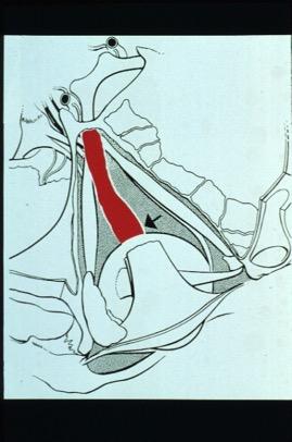
51 VISUAL FIELD LOSS Enlarged blind spot and generalized constriction are most common nasal step and arcuate scotomas possible cecocentral scotoma less likely
52 OBSERVED SIGNS papilledema may be unilateral sine papilledema relative afferent pupillary defect cranial nerve VI palsy
53 MANAGEMENT suspect exogenous agents should be discontinued LP done initially for dx may be therapeutic weight loss is the most effective treatment may get off Rx and avoid surgery consider dietician
54 CARBONIC ANHYDRASE INHIBITORS Neptazane 50 mg bid to qid Diamox 250 mg bid to 500 mg qid common adverse effects tingling and numbness in fingers/toes metallic taste K+ wasting relative contraindication 1st 4 months pregnancy sulfa based - can be used if allergy is abs aplastic anemia is a rare idiosyncratic rxn
55 ALTERNATIVE DRUGS Lasix 20 mg qd to 40 mg qid corticosteroids generally not indicated may be useful if inflammatory mechanism (I.e., SLE) further weight gain and fluid retention rebound on withdrawal iv solumedrol may be useful acutely Octreotide Topamax Beta blockers?


56 SURGICAL MEASURES - CSF SHUNTS Ventriculoperitoneal Lumboperitoneal
57 LUMBOPERITONEAL SHUNT more likely to reduce ICP, relieve H/A than ONSF Contraindicated (relative) in Chiari Malformation average replacement every 2-3 shunt years migration, closure, infection over or under-filtration
58 VENTRICULOPERITONEAL SHUNT more likely to reduce ICP, relieve H/A average replacement every 4 shunt years migration, closure, infection over or under-filtration NEUROSURGEON DECIDES WHICH TYPE TO USE
59 OPTIC NERVE SHEATH FENESTRATION window or multiple longitudinal slits made in the anterior dural covering of the optic nerve immediate decompression of optic nerve tip less likely to maintain lower ICP, relieve H/A neuroprotective


60 OPTIC NERVE SHEATH FENESTRATION: MEDIAL APPROACH


61 OPTIC NERVE SHEATH FENESTRATION: MEDIAL APPROACH
62 Visual Outcomes following Optic Nerve Sheath Fenestration via the Medial Transconjunctival Approach Steven E Katz, MD 1 et al 207 eyes of 104 patients from Outcomes: MD on HVF and Papilledema Grade Followed for 6 months Edema resolved completely in 76% in 1 week, 71% at 6 months MD at 1 week, at 6 months Conclusion: Safe and effective treatment for disc edema
63 CASE PRESENTATION 22 y.o. obese wf presents with severe holocranial progressive H/A and loss of peripheral vision over 6 months hx IIH s/p R ONSF 18 months prior left relative afferent pupillary defect generalized visual field constriction L>R on diamox 500 mg sequels p.o. qid LP with opening pressure 32 cm H 2 O

64


65 PRESENTING VISUAL FIELDS

66 1 WEEK AFTER LEFT ONSF
67 1WEEK AFTER RIGHT ONSF
68 PATIENT MONITORING papilledema may not resolve completely with tx may not recur with ICP optic nerve appearance alone is not adequate to assess for recurrent elevation of ICP! subjective symptoms and visual field progression may be more reliable visual fields patient education and participation are essential
69 IIH ACCORDING TO JEAN Initial OP < 35 cm H 2 O Initial OP cm H 2 O Initial OP > 46 cm H 2 O Low threshold for ONSF Profound papilledema and ONSF

70 IDIOPATHIC INTRACRANIAL HYPERTENSION TREATMENT TRIAL (IIHTT)
71 IIHTT The IIHTT is a prospective clinical treatment trial on idiopathic intracranial hypertension that includes a genetic association study in search of single polymorphisms (SNPs) to identify metabolic and hormonal factors that differentiate between obese women who have IIH and obese women who do not - the first NORDIC multicenter study.
72 IIHTT: SPECIFIC AIM ONE IIH patients with Mild Visual Loss (-2 to -5 db baseline PMD) will be recrutied to participate in this randomized, double-masked, placebocontrolled trial to determine the additional benefit of acetazolamide (up to 4 gm a day) added to a low sodium, weight reduction diet. Hypothesis: Acetazolamide + diet is superior to diet alone in restoring vision or preventing visual loss in IIH patients with mild visual loss.
73 IIHTT: SPECIFIC AIM TWO (a) To identify proteomic and genetic risk factors for IIH by screening a large cohort of IIH patients and controls, (b) To determine the serum and CSF levels of potential mediators of IIH suggested by the genetic analysis, and (c) To conduct an association study in search of single nucleotide polymorphisms (SNPs) that confer risk for developing IIH. A cohort of 154 IIH patients and 154 controls matched on body-mass index, ethnicity and gender will be genotyped at SNPs contained within genes encoding molecules likely to be involved in the etiology of IIH using the SNPlex genotyping system. Specifically, genes associated with obesity will be profiled. (d) To test the hypotheses IIH is associated with abnormal metabolism of leptin or vitamin A or both, leptin levels, vitamin A and related factors will be measured at baseline and six months.

74 IIHTT: RESULTS

75 IIHTT: RESULTS
76 IIH: TAKE HOME POINTS History - Signs of Elevated ICP Check for APD! HVF 30-2 OCT 5 line raster may be helpful If scheduling LP - OPENING PRESSURE Vision loss is preventable/treatable
77 IT S NOT ALWAYS PSEUDOTUMOR. MANY OTHER CAUSES FOR BILATERAL DISC EDEMA


78 PSEUDO PSEUDOTUMOR (TUMOR)

79 HYPERTENSIVE CRISIS Hypertensive crisis Generally bilateral Blurred vision Headache Dizziness encephalopathy

80 IINFECTION Meningitis Stiff neck Headache Fever Skin rash Obtundation Considerations HIV+ Cryptococcus Tertiary syphylis May be associated with raised ICP

81 INFECTION/INFLAMMATION Neuroretinitis Infectious/immune-mediated Ddx Cat scratch disease Post-viral: HSV, hepatitis B, mumps Spirochetes: syphylis, Lyme, leptospirosis Possible: toxoplasmosis, toxocariasis, histoplasmosis Tuberculosis Leber s idiopathic stellate neuroretinitis Treatment Antibiotics: doxycycline, erythromycin, azithromycin, ciprofloxacin, rifampin Corticosteroids


82 INFILTRATIVE: SARCOID Uveitis Papillitis Infiltrative, compressive optic neuropathy Systemic manifestations: lungs, skin 5/100,000 caucasians 40/100,000 African Americans ACE, lysozyme serum CSF ACE MRI brain with gad Tissue diagnosis
83 INFILTRATIVE: LEUKEMIA Papillitis Uveitis Hyphema
84 INFILTRATIVE: LARGE CELL LYMPHOMA Refractory uveitis Papillitis Choroidal infiltrates Usually known CNS involvement Vitrectomy with cell cytology can be diagnostic

85 THANK YOU!
Neuro-Ocular Grand Rounds
 Neuro-Ocular Grand Rounds Anthony B. Litwak,OD, FAAO VA Medical Center Baltimore, Maryland Dr. Litwak is on the speaker and advisory boards for Alcon and Zeiss Meditek COMMON OPTIC NEUROPATHIES THAT CAN
Neuro-Ocular Grand Rounds Anthony B. Litwak,OD, FAAO VA Medical Center Baltimore, Maryland
 Neuro-Ocular Grand Rounds Anthony B. Litwak,OD, FAAO VA Medical Center Baltimore, Maryland Dr. Litwak is on the speaker and advisory boards for Alcon and Zeiss Meditek COMMON OPTIC NEUROPATHIES THAT CAN
THE SWOLLEN DISC. Valerie Biousse, MD Emory University School of Medicine Atlanta, GA
 THE SWOLLEN DISC Valerie Biousse, MD Emory University School of Medicine Atlanta, GA Updated from: Neuro-Ophthalmology Illustrated. Biousse V, Newman NJ. Thieme, New-York,NY. 2 nd Ed, 2016. Edema of the
Papilledema. Golnaz Javey, M.D. and Jeffrey J. Zuravleff, M.D.
 Papilledema Golnaz Javey, M.D. and Jeffrey J. Zuravleff, M.D. Papilledema specifically refers to optic nerve head swelling secondary to increased intracranial pressure (IICP). Optic nerve swelling from
12/2/16. Ways to differentiate:
 Nate Lighthizer, O.D., F.A.A.O. Assistant Dean for Clinical Care Services Director of CE Chief of Specialty Care Clinics Chief of Electrodiagnostics Clinic Oklahoma College of Optometry lighthiz@nsuok.edu
NANOS Patient Brochure
 NANOS Patient Brochure Pseudotumor Cerebri Copyright 2016. North American Neuro-Ophthalmology Society. All rights reserved. These brochures are produced and made available as is without warranty and for
Alan G. Kabat, OD, FAAO (901)
") THE SWOLLEN OPTIC DISC: EMERGENCY OR ANOMALY? Alan G. Kabat, OD, FAAO (901) 252-3691 Memphis, Tennessee alan.kabat@alankabat.com Course description: The swollen disc presents a diagnostic dilemma. While
THE SWOLLEN OPTIC DISC: EMERGENCY OR ANOMALY? Alan G. Kabat, OD, FAAO (901) 252-3691 Memphis, Tennessee alan.kabat@alankabat.com Course description: The swollen disc presents a diagnostic dilemma. While
Typical idiopathic intracranial hypertension Optic nerve appearance and brain MRI findings. Jonathan A. Micieli, MD Valérie Biousse, MD
 Typical idiopathic intracranial hypertension Optic nerve appearance and brain MRI findings Jonathan A. Micieli, MD Valérie Biousse, MD A 24 year old African American woman is referred for bilateral optic
Khalil Zahra, M.D Neuro-interventional radiology
 Khalil Zahra, M.D Neuro-interventional radiology 1 Disclosure None 2 Outline Etiology and pathogensis Imaging techniques and Features Literature review Treatment modalities Endovascular techniques Long
Neuro Ocular Grand Rounds Anthony B. Litwak, OD, FAAO VA Medical Center Baltimore, MD
 Neuro Ocular Grand Rounds Anthony B. Litwak, OD, FAAO VA Medical Center Baltimore, MD 58 YOWM! C/O I think there is something wrong with my vision, but I m not sure what it is.! +PMH for HTN, atrial fibrillation,
OPTIC NERVE SWELLING IN CHILDHOOD
 OPTIC NERVE SWELLING IN CHILDHOOD Melissa W. Ko, MD, FAAN One of the main findings on a pediatric neurologic examination that can instill fear and lead to an urgent referral to neuro-ophthalmology is the
Pearls, Pitfalls and Advances in Neuro-Ophthalmology
 Pearls, Pitfalls and Advances in Neuro-Ophthalmology Nancy J. Newman, MD Emory University Atlanta, GA Consultant for Gensight Biologics, Santhera Data Safety Monitoring Board for Quark AION Study Medical-legal
Idiopathic Intracranial Hypertension (Pseudotumor Cerebri) David I. Kaufman, D.O. Michigan State University Department of Neurology and Ophthalmology
 David I. Kaufman, D.O. Michigan State University Department of Neurology and Ophthalmology") Idiopathic Intracranial Hypertension (Pseudotumor Cerebri) David I. Kaufman, D.O. Michigan State University Department of Neurology and Ophthalmology 26 year old 5 3, 300 pound female with papilledema,
Idiopathic Intracranial Hypertension (Pseudotumor Cerebri) David I. Kaufman, D.O. Michigan State University Department of Neurology and Ophthalmology 26 year old 5 3, 300 pound female with papilledema,
11/10/2017. Headache and Increased Pressure: A tale of 2 cases. Kathleen Digre MD University of Utah TWO CASES. 23 yo medical practice manager
 Headache and Increased Pressure: A tale of 2 cases Kathleen Digre MD University of Utah TWO CASES 23 yo medical practice manager September 2016 began developing intense frontal headaches first intermittent
An Organized Approach to the Patient with Papilledema and IIH
 An Organized Approach to the Patient with Papilledema and IIH Leonard V. Messner, OD, FAAO James L. Fanelli, OD, FAAO Please silence all mobile devices and remove items from chairs so others can sit. Unauthorized
Learn Connect Succeed. JCAHPO Regional Meetings 2015
 Learn Connect Succeed JCAHPO Regional Meetings 2015 OPTIC NEUROPATHY AS EASY AS 1,2,3,4 OPTIC NERVE ANATOMY M. Tariq Bhatti, MD Departments of Ophthalmology and Neurology Duke Eye Center and Duke University
Optic Nerve Disorders: Structure and Function and Causes
 Optic Nerve Disorders: Structure and Function and Causes Using Visual Fields, OCT and B-scan Ultrasound to Diagnose and Follow Optic Nerve Visual Losses Ohio Ophthalmological Society and Ophthalmic Tech
Optic Nerve Anomalies
 Optic Nerve Anomalies Raman Bhakhri, OD, FAAO Southern California College of Optometry Marshall B. Ketchum University Goals for today Review some of the optic nerve anomalies that can be seen in practice
Michelle L. Ischayek D.O. Emergency Medicine Resident Aria Health
 Michelle L. Ischayek D.O. Emergency Medicine Resident Aria Health History 15 year old African female with CC of Headache. Onset: 2 weeks ago Location: Frontal Character: Sharp & Throbbing Radiation: None
What is IIH? Idiopathic Intracranial Hypertension (IIH)
") What is IIH? Idiopathic Intracranial Hypertension (IIH) What is Idiopathic Intracranial Hypertension? Idiopathic intracranial hypertension (IIH), also known as benign intracranial hypertension or pseudotumour
What is IIH? Idiopathic Intracranial Hypertension (IIH) What is Idiopathic Intracranial Hypertension? Idiopathic intracranial hypertension (IIH), also known as benign intracranial hypertension or pseudotumour
Management of Pseudo Tumor Cerebri by Frequent Tapping VS lumboperitoneal Shunt
 The Egyptian Journal of Hospital Medicine (July 2018) Vol. 72 (5), Page 4556-4560 Management of Pseudo Tumor Cerebri by Frequent Tapping VS lumboperitoneal Shunt Ali K. Ali, Maamoun M. Abo Shousha, Mohammed
Idiopathic Intracranial Hypertension
 Idiopathic Intracranial Hypertension Dr. Mar'n Su+onBrown MD. FRCPC Neuro-Ophthalmology, Neurology Div of Neurology, Island Health Clinical Assistant Professor, Div of Neurology, UBC Stroke Rapid Assessment
Headache Assessment In Primary Eye Care
 Headache Assessment In Primary Eye Care Spencer Johnson, O.D., F.A.A.O. Northeastern State University Oklahoma College of Optometry johns137@nsuok.edu Course Objectives Review headache classification Understand
MOHAMED LOTFY, M.D.*; MOATAZ A. EL-AWADY, M.D.**; ASHRAF E. ZAGHLOUL, M.D.** and TAREK NEHAD, M.D.***
 Med. J. Cairo Univ., Vol. 84, No. 2, December: 301-306, 2016 www.medicaljournalofcairouniversity.net Effect of Therapeutic Lumbar Puncture on the Visual Outcome and the Further Need for Surgery in Patients
OCCLUSIVE VASCULAR DISORDERS OF THE RETINA
 OCCLUSIVE VASCULAR DISORDERS OF THE RETINA Learning outcomes By the end of this lecture the students would be able to Classify occlusive vascular disorders (OVD) of the retina. Correlate the clinical features
Case #1: 68 M with floaters OS
 Case #1: 68 M with floaters OS Point-of-Care Ocular Sonography for the Emergency Department Nate Teismann MD Dept of Emergency Medicine, UCSF Topics in EM 2012 Acute onset of dark spots in L eye 2 days
BMB Disclosures. Papilledema can be a. Neurological Emergency, Causing Preventable Blindness
 Reasonable Doubt: Can High Intracranial Pressure Occur Without Papilledema? 15 February 2013 Jonathan C. Horton hortonj@vision.ucsf.edu http://www.ucsf.edu/hortonlab BMB Disclosures Financial Disclosures
Intracranial hypertension and headache. Daniel Tibussek, MD
 Intracranial hypertension and headache. Daniel Tibussek, MD none Disclosures Overview Case Clinical presentation of pediatric PTC Nomenclature, Definition What is intracranial hypertension? Diagnostic
Five steps: Overview
 Optic atrophy is not a diagnosis Andrew G. Lee, MD Professor of Ophthalmology, Neurology and Neurosurgery, Weill Cornell Medical College Chair, Department of Ophthalmology, Houston Methodist Hospital,
Prevalence of venous sinus stenosis in Pseudotumor cerebri(ptc) using digital subtraction angiography (DSA)
 using digital subtraction angiography (DSA)") Prevalence of venous sinus stenosis in Pseudotumor cerebri(ptc) using digital subtraction angiography (DSA) Dr.Mohamed hamdy ibrahim MBBC,MSc,MD, PhD Neurology Degree Kings lake university (USA). Fellow
Prevalence of venous sinus stenosis in Pseudotumor cerebri(ptc) using digital subtraction angiography (DSA) Dr.Mohamed hamdy ibrahim MBBC,MSc,MD, PhD Neurology Degree Kings lake university (USA). Fellow
Divakar Gupta Glaucoma Fellow, Duke Eye Center 5/14/16
 Divakar Gupta Glaucoma Fellow, Duke Eye Center 5/14/16 Pathophysiology of glaucoma Consider risk factors of glaucoma Understand the side effects of glaucoma medications Diagnostic testing Leading cause
Headache Syndrome. Karen Alvarez, D.O Nemours Children s Specialty Care Jacksonville, FL
 Headache Syndrome Karen Alvarez, D.O Nemours Children s Specialty Care Jacksonville, FL What is a headache? A headache or cephalgia is defined as pain anywhere in the region of head or neck Where does
A Curious Case of Bilateral Optic Disc Edema Brittney Dautremont, DO, MPH
 A Curious Case of Bilateral Optic Disc Edema Brittney Dautremont, DO, MPH PGY2 Ophthalmology Resident Grandview Medical Center Dayton, OH CASE PRESENTATION 51 year old white female presenting with blurred
Question 1: Comment on the optic nerve appearance of each eye.
 Case 2 - Right Optic Nerve Head Drusen (ONHD) A 41 year old female was referred by her optometrist for a workup for unilateral optic disc drusen, OCT, and visual field changes. The patient was otherwise
IDIOPATHIC INTRACRANIAL HYPERTENSION
 IDIOPATHIC INTRACRANIAL HYPERTENSION ASSESSMENT OF VISUAL FUNCTION AND PROGNOSIS FOR VISUAL OUTCOME Doctor of Philosophy thesis Anglia Ruskin University, Cambridge Fiona J. Rowe Department of Orthoptics,
Moncef Khairallah, MD
 Moncef Khairallah, MD Department of Ophthalmology, Fattouma Bourguiba University Hospital Faculty of Medicine, University of Monastir Monastir, Tunisia INTRODUCTION IU: anatomic form of uveitis involving
5/2/2016 EYE EMERGENCIES. Nathaniel Pelsor, O.D., FAAO Talley Medical-Surgical Eye Care Associates. Anatomy. Tools
 EYE EMERGENCIES Nathaniel Pelsor, O.D., FAAO Talley Medical-Surgical Eye Care Associates Anatomy Tools 1 Contact dermatitis Blepharitis HSV Preseptal Cellulitis Anterior Chamber Subconjunctival hemorrhage
DISORDERS OF THE NERVOUS SYSTEM
 DISORDERS OF THE NERVOUS SYSTEM Bell Work What s your reaction time? Go to this website and check it out: https://www.justpark.com/creative/reaction-timetest/ Read the following brief article and summarize
Differential Diagnosis of ONH Edema Beth A. Steele, OD, FAAO
 Differential Diagnosis of ONH Edema Beth A. Steele, OD, FAAO bsteele@uab.edu Please silence all mobile devices and remove items from chairs so others can sit. Unauthorized recording of this session is
Medical treatment and Monitoring in IIH
 Medical treatment and Monitoring in IIH Introduction When you have Idiopathic Intracranial Hypertension it is important that your vision, symptoms and medication are monitored on a regular basis. This
A Case of Carotid-Cavernous Fistula
 A Case of Carotid-Cavernous Fistula By : Mohamed Elkhawaga 2 nd Year Resident of Ophthalmology Alexandria University A 19 year old male patient came to our outpatient clinic, complaining of : -Severe conjunctival
Learn Connect Succeed. JCAHPO Regional Meetings 2017
 Learn Connect Succeed JCAHPO Regional Meetings 2017 NO FINANCIAL DISCLOSURES Technician s Role in Neuro-Ophthalmology Workup Beth Koch COT, ROUB Cleveland 9/16/2017 What Tests Are You Expected To Perform?
Index. Note: Page numbers of article titles are in boldface type.
 Index Note: Page numbers of article titles are in boldface type. A Acetazolamide, in idiopathic intracranial hypertension, 49 52, 60 Angiography, computed tomography, in cranial nerve palsy, 103 107 digital
The headache profile of idiopathic intracranial hypertension
 The headache profile of idiopathic intracranial hypertension Michael Wall CEPHALALGIA Wall M. The headache profile of idiopathic intracranial hypertension. Cephalalgia 1990;10:331-5. Oslo. ISSN 0333-1024
Is it Papilloedema? John Ross Ainsworth Orthoptic staff Birmingham Children s Hospital Birmingham and Midland Eye Centre University of Birmingham
 Is it Papilloedema? John Ross Ainsworth Orthoptic staff Birmingham Children s Hospital Birmingham and Midland Eye Centre University of Birmingham Aims Children/young people A bit about hypoplasia / NFL
Carotid Cavernous Fistula
 Chief Complaint: Double vision. Carotid Cavernous Fistula Alex W. Cohen, MD, PhD; Richard Allen, MD, PhD May 14, 2010 History of Present Illness: A 46 year old female patient presented to the Oculoplastics
Differential diagnosis of posterior uveitis
 Differential diagnosis of posterior uveitis Diagnostic approach 45-year old male. Floaters and decreased vision since 1 week Fever, lymphadenopathy, myalgias, night sweats, two months ago Oral ulcer sporadically
I have nothing to disclose but I
 OPTIC NEUROPATHIES Robert L. Tomsak MD PhD Professor of Ophthalmology and Neurology Wayne State t University it Sh School of Mdii Medicine I have nothing to disclose but I wish I did. dd Road map for this
Lumbar puncture. Invasive procedure: diagnostic or therapeutic. The subarachnoid space 4-13 ys: ml Replenished: 4-6 h Routine LP (3-5 ml): <1h
: <1h") Lumbar puncture Lumbar puncture Invasive procedure: diagnostic or therapeutic. The subarachnoid space 4-13 ys: 65-150ml Replenished: 4-6 h Routine LP (3-5 ml):
Lumbar puncture Lumbar puncture Invasive procedure: diagnostic or therapeutic. The subarachnoid space 4-13 ys: 65-150ml Replenished: 4-6 h Routine LP (3-5 ml):
Pseudotumor Cerebri Secondary to Minocycline Intake
 Pseudotumor Cerebri Secondary to Minocycline Intake Earl Robert G. Ang, MD, J. C. Chava Zimmerman, MD, and Elissa Malkin, DO, MPH Background: Pseudotumor cerebri, or idiopathic intracranial hypertension,
OPTIC NEUROPATHIES Optic Neuritis vs AION. Jacqueline M.S. Winterkorn, Ph.D., M.D.
 OPTIC NEUROPATHIES Optic Neuritis vs AION Jacqueline M.S. Winterkorn, Ph.D., M.D. OPTIC NEUROPATHIES Inflammatory Optic Neuritis Ischemic Optic Neuropathy Compressive Optic Neuropathy Traumatic Optic
Professor Helen Danesh-Meyer. Eye Institute Auckland
 Professor Helen Danesh-Meyer Eye Institute Auckland Bitten by Ophthalmology Emergencies Helen Danesh-Meyer, MBChB, MD, FRANZCO Sir William and Lady Stevenson Professor of Ophthalmology Head of Glaucoma
Role Of Various Factors In The Treatment Of Optic Neuritis----A Study Abstract Aim: Materials & Methods Discussion: Conclusion: Key words
 IOSR Journal of Dental and Medical Sciences (IOSR-JDMS) e-issn: 2279-0853, p-issn: 2279-0861.Volume 15, Issue 9 Ver. X (September). 2016), PP 51-57 www.iosrjournals.org Role Of Various Factors In The Treatment
53 year old woman attends your practice for routine exam. She has no past medical history or family history of note.
 Case 1 Normal Tension Glaucoma 53 year old woman attends your practice for routine exam. She has no past medical history or family history of note. Table 1. Right Eye Left Eye Visual acuity 6/6 6/6 Ishihara
Sequential non-arteritic anterior ischemic optic neuropathy (NAION) Jonathan A. Micieli, MD Valérie Biousse, MD
 Jonathan A. Micieli, MD Valérie Biousse, MD") Sequential non-arteritic anterior ischemic optic neuropathy (NAION) Jonathan A. Micieli, MD Valérie Biousse, MD A 68 year old white woman had a new onset of floaters in her right eye and was found to have
Sequential non-arteritic anterior ischemic optic neuropathy (NAION) Jonathan A. Micieli, MD Valérie Biousse, MD A 68 year old white woman had a new onset of floaters in her right eye and was found to have
Cases of visual impairment caused by cerebral venous sinus occlusion-induced intracranial hypertension in the absence of headache
 Zhao et al. BMC Neurology (2018) 18:159 https://doi.org/10.1186/s12883-018-1156-7 CASE REPORT Open Access Cases of visual impairment caused by cerebral venous sinus occlusion-induced intracranial hypertension
Coexistence of migraine and idiopathic intracranial hypertension without papilledema
 / GX'~C1~.. Coexistence of migraine and idiopathic intracranial hypertension without papilledema Ninan T. Mathew, MD; K. Ravishankar, MD; and Luis C. Sanin, MD!
Rebound Intracranial Hypertension Following Treatment of Spinal CSF Leaks
 Rebound Intracranial Hypertension Following Treatment of Spinal CSF Leaks Deborah I. Friedman, MD, MPH University of Texas Southwestern Medical Center Dallas, Texas Disclosures (past 2 years): Role Advisory
Pediatric Ocular Sonography
 Pediatric Ocular Sonography Cicero J Torres A Silva, MD Associate Professor of Radiology 2016 SPR Pediatric Ultrasound Course Yale University School of Medicine None Disclosures Objectives of Presentation
Objectives. Unexplained Vision Loss: Where Do I Go From Here. History. History. Drug Induced Vision Loss
 Objectives Unexplained Vision Loss: Where Do I Go From Here Denise Goodwin, OD, FAAO Coordinator, Neuro-ophthalmic Disease Clinic Pacific University College of Optometry goodwin@pacificu.edu Know the importance
Cryptococcal Meningitis
 Cryptococcal Meningitis Dr N Thumbiran Infectious Diseases Department UKZN Index patient 27 year old female Presented to King Edward Hospital on 17/07/2005 with: Severe headaches Vomiting Photophobia X
Case Follow Up. Sepi Jooniani PGY-1
 Case Follow Up Sepi Jooniani PGY-1 Triage 54 year old M Pt presents to prelim states noticed today he had reddness to eyes, states worse in R eye. Pt denies any pain or itching. No further complaints.
Overview INTRODUCTION 3/15/2018. Headache Emergencies. Other way to differentiate between them? Is there an easy way to differentiate between them?
 Overview Headache Emergencies Primary versus Secondary headache disorder Red flags 4 cases of unusual headache emergencies Disclaimer: we will not talk about brain bleed as patients usually go the ED.
Cerebral Toxoplasmosis in HIV-Infected Patients. Ahmed Saad,MD,FACP
 Cerebral Toxoplasmosis in HIV-Infected Patients Ahmed Saad,MD,FACP Introduction Toxoplasmosis: Caused by the intracellular protozoan, Toxoplasma gondii. Immunocompetent persons with primary infection
Chapter 2 Long Duration Flight Data
 Chapter 2 Long Duration Flight Data Astronaut s bodies suffer in microgravity. Without effective countermeasures, muscles atrophy, bones shed calcium, and eyesight deteriorates. We ve known about this
DEFINITION: DISCLOSURE: 7/25/2017 OPTIC NERVE GRAND ROUNDS: YOU VE GOT SOME NERVE. Joseph Sowka, OD, FAAO, Diplomate 28 YOF 28 YOF
 DISCLOSURE: OPTIC NERVE GRAND ROUNDS: YOU VE GOT SOME NERVE Dr. Joseph Sowka is a member of the Advisory Board for Novartis, Zeiss, B&L, and Allergan. He is a consultant for Novartis. Dr. Sowka has no
Case Series. The efficacy of optic nerve ultrasonography for differentiating papilloedema from pseudopapilloedema in eyes with swollen optic discs
 Case Series The efficacy of optic nerve ultrasonography for differentiating papilloedema from pseudopapilloedema in eyes with swollen optic discs Meira Neudorfer, Maytal Siegman Ben-Haim, Igal Leibovitch
10/27/2013. Optic Red Herrings
 Optic Red Herrings 1 Optic neuropathy Compressive Inflammatory Toxic Glaucomatous Ischemic Post traumatic GLAUCOMATOUS OPTIC NEUROPATHY Glaucoma: Traditionally defined as a progressive optic neuropathy
Neuro-ophthalmologyophthalmology. Marek Michalec, MD.
 Neuro-ophthalmologyophthalmology Marek Michalec, MD. Neuro-ophthalmology Study integrating ophthalmology and neurology Disorders affecting parts of CNS devoted to vision or eye: Afferent system (visual
Learn Connect Succeed. JCAHPO Regional Meetings 2017
 Learn Connect Succeed JCAHPO Regional Meetings 2017 You have some Nerve Asking Me to Work Up that Patient! What I Need to know about the Neuro- Ophthalmology Patient Financial Disclosures No relevant financial
Optic Disc: Anatomy, Variants, Unusual discs. Kathleen B. Digre, MD Professor Neurology, Ophthalmology
 Optic Disc: Anatomy, Variants, Unusual discs Kathleen B. Digre, MD Professor Neurology, Ophthalmology THE OPHTHALMOSCOPE DIRECT OPHTHALMOSCOPY Jan Purkinje 1823 Hermann von Helmholtz 1851 Hand held ophthalmoscope
Glossary. Working to relieve the pressure!
 A Acetazolamide - see Diamox Amitriptyline - a drug which is used in low dosage in IIH to treat some of the symptoms of the condition. Analgesic - a drug used to relieve pain, often referred to as painkillers.
Neuro-Ophthalmic Masqueraders
 Neuro-Ophthalmic Masqueraders Leonid Skorin, Jr., OD, DO, MS, FAAO, FAOCO Mayo Clinic Health System in Albert Lea Denise Goodwin, OD, FAAO Pacific University College of Optometry Please silence all mobile
Chronicity. Narrow Minded. Course Outline. Acute angle closure. Subacute angle closure. Classification of Angle Closure 5/19/2014
 Chronicity Narrow Minded The management of narrow angles in the optometric practice Acute Subacute Chronic Aaron McNulty, OD, FAAO Course Outline Classification of Angle Closure Evaluation of narrow angles
Clinician s Guide To Ordering NeuroImaging Studies
 Clinician s Guide To Ordering NeuroImaging Studies MRI CT South Jersey Radiology Associates The purpose of this general guide is to assist you in choosing the appropriate imaging test to best help your
Ophthalmology. Juliette Stenz, MD
 Ophthalmology Juliette Stenz, MD Required Slide Disclosures NO SIGNIFICANT FINANCIAL, GENERAL, OR OBLIGATION INTERESTS TO REPORT Required Slide At the end of this session, students will be able to: 1.
3/16/2018. Optic Nerve Examination. Hassan Eisa Swify FRCS Ed (Ophthalmology) Air Force Hospital
 Air Force Hospital") Optic Nerve Examination Hassan Eisa Swify FRCS Ed (Ophthalmology) Air Force Hospital 1 Examination Structure ( optic disc) Function Examination of the optic disc The only cranial nerve (brain tract) which
Optic Nerve Examination Hassan Eisa Swify FRCS Ed (Ophthalmology) Air Force Hospital 1 Examination Structure ( optic disc) Function Examination of the optic disc The only cranial nerve (brain tract) which
PITFALLS IN PAPILLOEDEMA
 PITFALLS IN PAPILLOEDEMA SRC 2013 Why care about papilloedema? Dr Neil Shuey FRACP MBBS(Hons) MScOptom St Vincent s Hospital, Melbourne Royal Victorian Eye & Ear Hospital Disclosures: Travel grants Biogen
Dr/ Marwa Abdellah EOS /16/2018. Dr/ Marwa Abdellah EOS When do you ask Fluorescein angiography for optic disc diseases???
 When do you ask Fluorescein angiography for optic disc diseases??? 1 NORMAL OPTIC DISC The normal optic disc on fluorescein angiography is fluorescent due to filling of vessels arising from the posterior
OPTIC NERVE DISORDERS
 OPTIC NERVE DISORDERS OPTIC NEUROPATHIES INFLAMMATORY OPTIC NEUROPATHIES Cat scratch disease. Lyme disease. Viral infections of childhood (measles, mumps, chicken pox) with or without encephalitis Immun-
Non-Traumatic Neuro Emergencies
 Department of Radiology University of California San Diego Non-Traumatic Neuro Emergencies John R. Hesselink, M.D. Nontraumatic Neuroemergencies 1. Acute focal neurological deficit 2. Worst headache of
Picture of patient with apparent lid retraction and poor elevation. Shows you Orbital CT-Scan with muscle involvement including IR and asks is this
 NEUROLOGY Q: MENINGIOMAS AND SKULL (*2) Real skull is given, and you are asked to point to tuberculum sella What kind of meningioma occurs at this location? Where is the anterior clinoid process? Where
Clinical Study Optic Nerve Sonography in the Diagnostic Evaluation of Pseudopapilledema and Raised Intracranial Pressure: A Cross-Sectional Study
 Neurology Research International Volume 2015, Article ID 146059, 4 pages http://dx.doi.org/10.1155/2015/146059 Clinical Study Optic Nerve Sonography in the Diagnostic Evaluation of Pseudopapilledema and
Complex Hydrocephalus
 2012 Hydrocephalus Association Conference Washington, DC - June 27-July1, 2012 Complex Hydrocephalus Marion L. Walker, MD Professor of Neurosurgery & Pediatrics Primary Children s Medical Center University
Case Report Atypical Presentation of Idiopathic Bilateral Optic Perineuritis in a Young Patient
 Case Reports in Ophthalmological Medicine Volume 2016, Article ID 6741925, 4 pages http://dx.doi.org/10.1155/2016/6741925 Case Report Atypical Presentation of Idiopathic Bilateral Optic Perineuritis in
Neuro-imaging for the Ophthalmologist. Karl C. Golnik, MD, MEd University of Cincinnati & The Cincinnati Eye Institute
 Neuro-imaging for the Ophthalmologist Karl C. Golnik, MD, MEd University of Cincinnati & The Cincinnati Eye Institute Neuro-ophthalmology is that subspecialty where the diagnosis is made upon reinterpretation
Disclosures. Visual Pathways. Visual Pathways. Visual Loss Understanding the Patterns. I have no financial disclosures. Tabby A.
 Visual oss Understanding the Patterns Tabby A. Kennedy, MD University of Wisconsin Department of adiology I have no financial disclosures Acknowledgements: indell Gentry Greg Avey JP Yu Judy Chen Disclosures
Misdiagnosed Vogt-Koyanagi-Harada (VKH) disease and atypical central serous chorioretinopathy (CSC)
 disease and atypical central serous chorioretinopathy (CSC)") HPTER 12 Misdiagnosed Vogt-Koyanagi-Harada (VKH) disease and atypical central serous chorioretinopathy (S) linical Features VKH disease is a bilateral granulomatous panuveitis often associated with exudative
HPTER 12 Misdiagnosed Vogt-Koyanagi-Harada (VKH) disease and atypical central serous chorioretinopathy (S) linical Features VKH disease is a bilateral granulomatous panuveitis often associated with exudative
Dr. Litwak is on the speaker bureau and advisory panel for Alcon and Zeiss Meditek
 My Favorite Cases Anthony B. Litwak,OD, FAAO VA Medical Center Baltimore, Maryland Dr. Litwak is on the speaker bureau and advisory panel for Alcon and Zeiss Meditek Case LA 62 yobf +HTN, + DM POH told
Cryptogenic Enlargement Of Bilateral Superior Ophthalmic Veins
 ISPUB.COM The Internet Journal of Radiology Volume 18 Number 1 Cryptogenic Enlargement Of Bilateral Superior Ophthalmic Veins K Kragha Citation K Kragha. Cryptogenic Enlargement Of Bilateral Superior Ophthalmic
Delayed Correction of Hypotony Maculopathy in a Patient with Glaucoma and Thyroid-Related Orbitopathy
 Published online: October 14, 2015 2015 The Author(s) Published by S. Karger AG, Basel 1663 2699/15/0063 0356$39.50/0 This article is licensed under the Creative Commons Attribution-NonCommercial 4.0 International
CHAPTER 13 CLINICAL CASES INTRODUCTION
 2 CHAPTER 3 CLINICAL CASES INTRODUCTION The previous chapters of this book have systematically presented various aspects of visual field testing and is now put into a clinical context. In this chapter,
Idiopathic Intracranial Hypertension in Pregnant Women
 Azza A. Ghali et al. Idiopathic Intracranial Hypertension in Pregnant Women Azza Abass Ghali, Ehab El-Seidy, Tarek Ragaiey Hussein 2, Manal Mostfa 3 Departments of Neuropsychiatry, Ophthalmology 2, Obstetrics
National Hospital for Neurology and Neurosurgery
 National Hospital for Neurology and Neurosurgery Venous sinus stents (for the treatment of venous sinus stenosis and idiopathic intracranial hypertension) Lysholm Department of Neuroradiology If you would
Ophthalmology Grand Rounds. Frank Tsai, MD February 16, 2012
 Ophthalmology Grand Rounds Frank Tsai, MD February 16, 2012 Case Presentation CC: seeing a dark spot HPI: 46 yo AA male Hx HTN c/o dark spot in vision OD, x3-4 weeks, constant 1 month Hx bilateral periorbital
Optic nerve sheath fenestration for Idiopathic Intracranial Hypertension. Optic nerve sheath fenestration
 Optic nerve sheath fenestration for Idiopathic Intracranial Hypertension Optic nerve sheath fenestration What is Idiopathic Intracranial Hypertension? Idiopathic Intracranial Hypertension (IIH) is a condition
Measures have been taken, by the Utah Department of Health, Bureau of Health Promotions, to ensure no conflict of interest in this activity.
 Measures have been taken, by the Utah Department of Health, Bureau of Health Promotions, to ensure no conflict of interest in this activity. CNE/CPE/CEU s are available for this live webinar. You must
MRI masterfile Part 5 WM Heme Strokes.ppt 1
 Ocular and Orbital Trauma Eye Trauma: Incidence 1.3 million eye injuries in the US per year. 40,000 of these injuries lead to blindness in the US. Patrick Sibony, MD March 23, 2013 Ophthalmic Emergencies
Spontaneous Intracranial Hypotension Diagnosis and Treatment
 Spontaneous Intracranial Hypotension Diagnosis and Treatment John W. Engstrom MD, Philip R. Weinstein MD, and William P. Dillon M.D. University of California, San Francisco Spontaneous Intracranial Hypotension