PHYSIOLOHY OF BRAIN STEM
|
|
|
- Beverly Mills
- 5 years ago
- Views:
Transcription

1

2 PHYSIOLOHY OF BRAIN STEM


3 Learning Objectives

4 The brain stem is the lower part of the brain. It is adjoining and structurally continuous with the spinal cord.

5 1 Mid Brain 2 Pons 3 Medulla Oblongata

6 The midbrain, pons and medulla connect to the cerebellum via the superior, middle and inferior peduncles respectively.
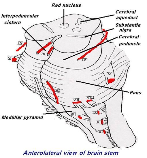
7 1 2 3 The midbrain is divided into three parts: The Tectum The Tegmentum Cerebral Peduncles

8 1- The Tectum ("roof" in latin) includes: a) The superior colliculus It constitutes center for visual reflexes It sends its superior brachium to the lateral geniculate body of the thalamus.
9 b) The inferior colliculus It is associated with auditory pathway It sends its inferior brachium to the medial geniculate body of the thalamus. The cerebral aqueduct runs through the midbrain, beneath the colliculi.
10 Ventral to the cerebral aqueduct. Several nuclei, tracts and the reticular formation is contained here. The ventral side is comprised of paired These transmit axons of UMN.

11 Periaqueductal Gray: Around the cerebral aqueduct, contains neurons involved in the pain desensitization pathway.
12 Occulomotor Nerve (CN III) nucleus. Trochlear Nerve (CN IV) nucleus. Red Nucleus This is a motor nucleus that sends a descending tract to the lower motor neurons.

13 Substantia Nigra: A concentration of neurons in the ventral portion of midbrain It is involved in motor function.

14 Central Tegmental Tract: Directly anterior to the floor of the 4th ventricle. It is a pathway by which many tracts project up to the cortex and down to the spinal cord.
15 Reticular Formation: A large area that is involved in various important functions of the midbrain: It contains LMN It is involved in the pain desensitization pathway It is involved in the arousal and consciousness systems It contains the locus coeruleus, which is involved in intensive alertness modulation and in autonomic reflexes.
, 7 (facial) & 8 (vestibulocochlear)")
16 o At the level of the midpons, trigeminal nerve (CN V) emerges. o Between the basal pons, cranial nerve 6 (abducens), 7 (facial) & 8 (vestibulocochlear) emerge (medial to lateral).
 tract as they head inferiorly to synapse on lower motor neuronal cell bodies within the")
17 The most medial part of the medulla is the anterior median fissure. Moving laterally on each side are the pyramids. They contain the fibers of the corticospinal (pyramidal) tract as they head inferiorly to synapse on lower motor neuronal cell bodies within the ventral horn of the spinal cord.
.")
18 The anterolateral sulcus is lateral to the pyramids. Emerging from the anterolateral sulci are the hypoglossal nerve (CN XII) rootlets. Lateral to the anterolateral sulci are the olives containing underlying inferior olivary nuclei and afferent fibers). Lateral (and dorsal) to the olives are the rootlets for glossopharyngeal (IX) & vagus (X) cranial nerves.

19 The most medial part of the medulla is the posterior median fissure. Moving laterally on each side is the fasciculus gracilis. Lateral to that is the fasciculus cuneatus. Superior to each of these, are the gracile and cuneate tubercles, respectively. Underlying these are their respective nuclei.

20 In the midline is the vagal trigone and superior to that is the hypoglossal trigone. Underlying each of these are motor nuclei for the respective cranial nerves.
. 3.")
21 1. Conduct functions. 2. Provides the origin of the cranial nerves (CN III-XII). 3. Conjugate eye movement. 4. Integrative functions.
22 1. Conduct functions All information related from the body to the cerebrum and cerebellum and vice versa, must traverse the brain stem.

23 a) The ascending sensory pathways coming from the body to the brain includes: The spinothalamin tract for pain and temperature sensation. The dorsal column, fasciculus gracilis, and cuneatus for touch, proprioceptive and pressure sensation.
24 b) Descending tracts Corticospinal tract (UMN): runs through crus cerebri (the anterior portion of the cerebral peduncle), basal part of pons and medullary pyramids; % of fibers cross in pyramidal decussation to form the lateral corticospinal tract, synapse on LMN in ventral horn of spinal cord. Upper motor neurons that originate in brain stem's vestibular, red, and reticular nuclei, which also descend and synapse in the spinal cord.
25 2. The brain stem provides the main motor and sensory innervation to the face and neck via the cranial nerves (CN III-XII). The fibers of cranial nerve nuclei except for olfactory & optic nerve either originating from, or terminating in the cranial nerve nuclei in brain stem.
26 CN III (oculomotor) CN IV (trochlear) Both moves eyes; CN III constricts the pupils, accommodates.
27 CN V (trigeminal): Chews and feels front of the head. CN VI (abducens): Moves eyes. CN VII (facial): Moves the face, tastes, salivates, cries. CN VIII (acoustic): Hears, regulates balance.
28 CN IX (glossopharyngeal): Tastes, salivates, swallows, monitors carotid body and sinus. CN X (vagus): Tastes, swallows, lifts palate, talks, communication to and from thoracoabdominal viscera. CN XI (accessory): Turns head, lifts shoulder. CN XII (hypoglossal): Moves tongue.
29 Sensory CN I CN II CN VIII Motor CN III CN IV CN VI CN XI CN XII Mixed CN V CN VII CN IX CN X
30
. From there, projections directly innervate the lateral rectus (contralateral to FEF) and the medial rectus muscle (ipsilateral to FEF).")
31 It refers to motor coordination of the eyes that allows for bilateral fixation on a single object The frontal eye field (FEF) projects to the opposite side at the midbrain-pontine junction, and then innervates the paramedian pontine reticular formation (PPRF). From there, projections directly innervate the lateral rectus (contralateral to FEF) and the medial rectus muscle (ipsilateral to FEF). The left FEF command to trigger conjugate eye movements to the right.
32 4. Integrative functions It controls consciousness & sleep cycle (alertness and arousal) through reticular formation. It has got center for cardiovascular, respiratory & autonomic nervous system. It has centers for cough, gag, swallow, and vomit. Sense of body balance (Vestibular functions)
33 osubstantia which is a part of the basal ganglia is present in midbrain and is involved in control of movement. omidbrain also contain red nucleus which regulate the motor activity through cerebellum.
34 o Inferior and superior colliculi are situated on the dorsal surface of the midbrain and is involved in auditory & visual processing required for head movements. o Pain sensitivity control: Periaqueductal grey matter of mesencephalon is an area which is rich in endogenous opioid and is important in modulation of painful stimuli.

35 oventral layer of brainstem is motor in function. omiddle layer is sensory in function & contains medial lemniscus which conveys sensory information from dorsal column.
![Function of Midbrain Nerve pathway to cerebral hemispheres CN III - Oculomotor [motor].](/docs-images/96/128197380/images/36-3.jpg "(Related to eye movement) CN IV - Trochlear [motor].")
36 Function of Midbrain Nerve pathway to cerebral hemispheres CN III - Oculomotor [motor]. (Related to eye movement) CN IV - Trochlear [motor]. (Superior oblique muscle of the eye which rotates the eye down and out) Auditory and Visual reflex centers
 deficits: Ipsilateral CN III, CN IV palsy and ptosis (drooping). Respiratory: Hyperventilating.")
37 Symptoms and signs of midbrain lesion Pupils: Midposition to dilated. Reactivity is sluggish to fixed. Movement: Abnormal extensor. Cranial Nerve (CN) deficits: Ipsilateral CN III, CN IV palsy and ptosis (drooping). Respiratory: Hyperventilating. Level of consciousness (LOC): Varies
38 Function of Pons CN V - Trigeminal [motor and sensory]. (Skin of face, tongue, teeth; muscle of mastication) Respiratory Center CN VI - Abducens [motor]. (Lateral rectus muscle of eye which rotates eye outward) CN VIII - Acoustic [sensory]. (He aring) CN VII - Facial [motor and sensory]. (Muscles of facial expression).
. -Hyperventilation. LOC: Semi-coma")
39 Symptoms and signs of lesion in pons Pupils size: Pinpoint Movement: Abnormal extensor CN Deficits: CN V, CN VI, CN VII, CN VIII Respiratory: -Apneustic (Abnormal respiration marked by sustained inhalation). -Hyperventilation. LOC: Semi-coma
40 Function of Medulla Oblongata Crossing of motor tracts. Cardiac Center Respiratory Center Vasomotor Center Centers for cough, gag, swallow, and vomit.
41 Function of Medulla Oblongata (Cont.) CN IX - Glossopharyneal )[mixed]. (Muscles & mucous membranes of pharynx, the constricted openings from the mouth & the oral pharynx and the posterior third of tongue). CN X - Vagus [mixed]. (Pharynx, larynx, heart, lungs, stomach) CN XI - Accessory [motor]. (Rotation of the head and shoulder) CN XII - Hypoglossal [motor]. (Intrinsic muscles of the tongue)
42 Symptoms and signs of lesion in Medulla Pupils: Dilated. Reactivity is Fixed. Movement: Ipsilateral paralysis. CN Palsies: Inability to control movement. Absent cough, gag. Respiratory: Abnormal breathing patterns LOC: Comatose.
43 To test reticular formation Alertness, Consciousness & Sleep.. Corticospinal tract Motor power, reflexes Pain response Facial grimacing on firm pressure over the supra orbital ridge. To test respiratory center look for the normal pattern of respiration
44 To test cardiovascular center Look for normal circulatory function. To test brainstem reflexes: Pupillary and corneal reflexes. Vestibulo-ocular reflex: Injection of iced water into the ear will produce eyes movement. Oculo-cephalic reflex: Eyes will be fixed when head is moved in one or another directions. Gag reflex. Cough reflex

45
PHYSIOLOGY OF THE BRAIN STEM
 PHYSIOLOGY OF THE BRAIN STEM Dr Syed Shahid Habib Professor & Consultant Clinical Neurophysiology Dept. of Physiology College of Medicine & KKUH King Saud University OBJECTIVES At the end of this lecture
DEVELOPMENT OF BRAIN
 Ahmed Fathalla OBJECTIVES At the end of the lecture, students should: List the components of brain stem. Describe the site of brain stem. Describe the relations between components of brain stem & their
Unit VIII Problem 3 Neuroanatomy: Brain Stem, Cranial Nerves and Scalp
 Unit VIII Problem 3 Neuroanatomy: Brain Stem, Cranial Nerves and Scalp - Brain stem: It is connected to the cerebellum and cerebral hemispheres. Rostral end of brain stem: diencephalon is the area which
By Dr. Saeed Vohra & Dr. Sanaa Alshaarawy
 By Dr. Saeed Vohra & Dr. Sanaa Alshaarawy 1 By the end of the lecture, students will be able to : Distinguish the internal structure of the components of the brain stem in different levels and the specific
Brainstem. By Dr. Bhushan R. Kavimandan
 Brainstem By Dr. Bhushan R. Kavimandan Development Ventricles in brainstem Mesencephalon cerebral aqueduct Metencephalon 4 th ventricle Mylencephalon 4 th ventricle Corpus callosum Posterior commissure
Lecture 4 The BRAINSTEM Medulla Oblongata
 Lecture 4 The BRAINSTEM Medulla Oblongata Introduction to brainstem 1- Medulla oblongata 2- Pons 3- Midbrain - - - occupies the posterior cranial fossa of the skull. connects the narrow spinal cord
Done by : Areej Al-Hadidi
 Brainstem &diencephalon Done by : Areej Al-Hadidi Brainstem Functions Ascending and descending tracts Reflex centers Cardiovascular and respiratory centers Coughing, sneezing, swallowing Nuclei of the
Brainstem. Amadi O. Ihunwo, PhD School of Anatomical Sciences
 Brainstem Amadi O. Ihunwo, PhD School of Anatomical Sciences Lecture Outline Constituents Basic general internal features of brainstem External and Internal features of Midbrain Pons Medulla Constituents
Brainstem. Telencephalon Diencephalon Cerebellum Brain stem
 Brainstem Brainstem 脑 脊髓 Brainstem Telencephalon Diencephalon Cerebellum Brain stem Ventral view Lateral view 10 pairs of the cranial nerves are attached to the brain stem The brainstem Midbrain Pons Medulla
b. The groove between the two crests is called 2. The neural folds move toward each other & the fuse to create a
 Chapter 13: Brain and Cranial Nerves I. Development of the CNS A. The CNS begins as a flat plate called the B. The process proceeds as: 1. The lateral sides of the become elevated as waves called a. The
Brainstem. Steven McLoon Department of Neuroscience University of Minnesota
 Brainstem Steven McLoon Department of Neuroscience University of Minnesota 1 Course News Change in Lab Sequence Week of Oct 2 Lab 5 Week of Oct 9 Lab 4 2 Goal Today Know the regions of the brainstem. Know
Brain and spinal nerve. By: shirin Kashfi
 Brain and spinal nerve By: shirin Kashfi Nervous system: central nervous system (CNS) peripheral nervous system (PNS) Brain (cranial) nerves Spinal nerves Ganglions (dorsal root ganglions, sympathetic
The Nervous System: Sensory and Motor Tracts of the Spinal Cord
 15 The Nervous System: Sensory and Motor Tracts of the Spinal Cord PowerPoint Lecture Presentations prepared by Steven Bassett Southeast Community College Lincoln, Nebraska Introduction Millions of sensory
Brain and Cranial Nerves (Ch. 15) Human Anatomy lecture. caudal = toward the spinal cord)
 Human Anatomy lecture. caudal = toward the spinal cord)") Insight: Some cranial nerve disorders Brain and Cranial Nerves (Ch. 15) Human Anatomy lecture I. Overview (Directional terms: rostral = toward the forehead caudal = toward the spinal cord) A. 3 Major parts
Insight: Some cranial nerve disorders Brain and Cranial Nerves (Ch. 15) Human Anatomy lecture I. Overview (Directional terms: rostral = toward the forehead caudal = toward the spinal cord) A. 3 Major parts
Internal Organisation of the Brainstem
 Internal Organisation of the Brainstem Major tracts and nuclei of the brainstem (Notes) The brainstem is the major pathway for tracts and houses major nuclei, that contain sensory, motor and autonomics
Non-cranial nerve nuclei
 Brainstem Non-cranial nerve nuclei Nucleus Gracile nucleus Cuneate nucleus Infeiro olivary nucleus Pontine nucleus inferior colliculus superior colliculus Red nucleus Substantia nigra Pretectal area Site
 Spinal Cord Tracts DESCENDING SPINAL TRACTS: Are concerned with somatic motor function, modification of ms. tone, visceral innervation, segmental reflexes. Main tracts arise form cerebral cortex and others
Principles of Anatomy and Physiology
 Principles of Anatomy and Physiology 14 th Edition CHAPTER 14 The Brain and Cranial Nerves Introduction The purpose of the chapter is to: 1. Understand how the brain is organized, protected, and supplied
Omar Sami. Aseel Abdeen. Muhammad Al-Salem. 1 P a g e
 Omar Sami Aseel Abdeen Muhammad Al-Salem 1 P a g e Using only section 2 record, I wrote this sheet; as the video is not ready yet. Despite pointing the structures, I ve tried to include all the scientific
Doctor Osama Asa ad Khader. Mohammad Alsalem
 6 Doctor 2015 Osama Asa ad Khader Mohammad Alsalem A quick revision for the spinal cord blood supply: Arterial Blood supply of spinal cord The spinal cord got its arterial supply by two ways: Longitudinal
Cranial Nerves. Steven McLoon Department of Neuroscience University of Minnesota
 Cranial Nerves Steven McLoon Department of Neuroscience University of Minnesota 1 Course News Change in Lab Sequence Week of Oct 2 Lab 5 Week of Oct 9 Lab 4 2 Sensory and Motor Systems Sensory Systems:
Review or skim Ch 12 on the vascular supply of the brain. Just look at pictures and legends for the clinical part at the end.
 Dental Neuroanatomy January 20 and 27, 10-12, 2011 Suzanne S. Stensaas, Ph.D. Dear Students: Please print these notes and bring them with you. My style is to use a Tablet PC and I draw on either a Word
M555 Medical Neuroscience Lab 1: Gross Anatomy of Brain, Crainal Nerves and Cerebral Blood Vessels
 M555 Medical Neuroscience Lab 1: Gross Anatomy of Brain, Crainal Nerves and Cerebral Blood Vessels Anatomical Directions Terms like dorsal, ventral, and posterior provide a means of locating structures
I: To describe the pyramidal and extrapyramidal tracts. II: To discuss the functions of the descending tracts.
 Descending Tracts I: To describe the pyramidal and extrapyramidal tracts. II: To discuss the functions of the descending tracts. III: To define the upper and the lower motor neurons. 1. The corticonuclear
Cranial Nerves and Spinal Cord Flashcards
 1. Name the cranial nerves and their Roman numeral. 2. What is Cranial Nerve I called, and what does it 3. Scientists who are trying to find a way to make neurons divide to heal nerve injuries often study
Blood supply to the brain Blood brain barrier isolates neural tissue from general circulation
 The Brain and Cranial Nerves Objectives Name the major regions of the brain and describe their functions. Discuss the formation, circulation, and functions of the CSF. List the main components of the medulla
Stanley Pruisinger 1980's
 Neuroanatomy Prion disease cerebellum chapter b/c cerebellar ataxia here as a warning for obvious reasons. Creutzfeldt - Jakob Disease (CJD) "Spongiform" (brain turns to sponge) Jews in Lybia who ate
C h a p t e r PowerPoint Lecture Slides prepared by Jason LaPres North Harris College Houston, Texas
 C h a p t e r 15 The Nervous System: The Brain and Cranial Nerves PowerPoint Lecture Slides prepared by Jason LaPres North Harris College Houston, Texas Copyright 2009 Pearson Education, Inc., publishing
Motor tracts Both pyramidal tracts and extrapyramidal both starts from cortex: Area 4 Area 6 Area 312 Pyramidal: mainly from area 4 Extrapyramidal:
 Motor tracts Both pyramidal tracts and extrapyramidal both starts from cortex: Area 4 Area 6 Area 312 Pyramidal: mainly from area 4 Extrapyramidal: mainly from area 6 area 6 Premotorarea: uses external
PTA 106 Unit 1 Lecture 1B
 PTA 106 Unit 1 Lecture 1B Medulla Oblongata Cardiovascular Center: Regulates the rate and force of the heartbeat and the diameter of blood vessels Medullary Rhythmicity Area: adjusts the basic rhythm of
General Sensory Pathways of the Face Area, Taste Pathways and Hearing Pathways
 General Sensory Pathways of the Face Area, Taste Pathways and Hearing Pathways Lecture Objectives Describe pathways for general sensations (pain, temperature, touch and proprioception) from the face area.
Lecturer. Prof. Dr. Ali K. Al-Shalchy MBChB/ FIBMS/ MRCS/ FRCS 2014
 Lecturer Prof. Dr. Ali K. Al-Shalchy MBChB/ FIBMS/ MRCS/ FRCS 2014 Dorsal root: The dorsal root carries both myelinated and unmyelinated afferent fibers to the spinal cord. Posterior gray column: Long
ACTIVITY 7: NERVOUS SYSTEM HISTOLOGY, BRAIN, CRANIAL NERVES
 ACTIVITY 7: NERVOUS SYSTEM HISTOLOGY, BRAIN, CRANIAL NERVES LABORATORY OBJECTIVES: 1. Histology: Identify structures indicated on three different slides or images of nervous system tissue. These images
Unit VIII Problem 4 Physiology lab: Brain Stem Lesions
 Unit VIII Problem 4 Physiology lab: Brain Stem Lesions - Motor and sensory somatotopy: Pre-central gyrus: is the motor area. Post-central gyrus: is the sensory area. Somatotopy: there is a map of thee
The Nervous System PART B
 7 The Nervous System PART B PowerPoint Lecture Slide Presentation by Jerry L. Cook, Sam Houston University ESSENTIALS OF HUMAN ANATOMY & PHYSIOLOGY EIGHTH EDITION ELAINE N. MARIEB Central Nervous System
Auditory and Vestibular Systems
 Auditory and Vestibular Systems Objective To learn the functional organization of the auditory and vestibular systems To understand how one can use changes in auditory function following injury to localize
Brainstem: Midbrain. 1. Midbrain gross external anatomy 2. Internal structure of the midbrain:
 Brainstem: Midbrain 1. Midbrain gross external anatomy 2. Internal structure of the midbrain: cerebral peduncles tegmentum tectum (guadrigeminal plate) Midbrain Midbrain general features location between
Cranial Nerve VIII (The Vestibulo-Cochlear Nerve)
") Cranial Nerve VIII (The Vestibulo-Cochlear Nerve) Please view our Editing File before studying this lecture to check for any changes. Color Code Important Doctors Notes Notes/Extra explanation Objectives
Cranial Nerve VIII (The Vestibulo-Cochlear Nerve) Please view our Editing File before studying this lecture to check for any changes. Color Code Important Doctors Notes Notes/Extra explanation Objectives
The Nervous System: Central Nervous System
 The Nervous System: Central Nervous System I. Anatomy of the nervous system A. The CNS & the body by: 1. monitoring of the body 2. & information between parts of the body 3. acting as a to gather, store,
Course: Physical Assessment II Date: October 17, 2008 Doc: Practice Quiz 1
 Course: Physical Assessment II Date: October 17, 2008 Doc: Practice Quiz 1 This is the practice quiz we did in Class 4. The answers are at the end of the quiz should you wish to test yourself. Complete
Bellringer: The central nervous system is comprised of: What is the name of the outermost layer of the brain? a. Brain. b.
 Bellringer: The central is comprised of: a. Brain b. Spinal cord c. Sensory receptors d. Both a and b What is the name of the outermost layer of the brain? a. Pia mater b. Dura mater c. Arachnoid d. Pons
Brainstem: Medulla oblongata and pons
 Brainstem: Medulla oblongata and pons 1. Overview of the brainstem subdivisions 2. Embryonic development of the brainstem 3. Medulla oblongata external features 4. Internal structure of the medulla oblongata
Chapter 14: The Brain and Cranial Nerves. Copyright 2009, John Wiley & Sons, Inc.
 Chapter 14: The Brain and Cranial Nerves Development of the Brain Three to four-week embryo: prosencephalon, mesencephalon and rhombencephalon. Five-week embryo: telencephalon (cerebrum), diencephalon
Nervous System. The Peripheral Nervous System Agenda Review of CNS v. PNS PNS Basics Cranial Nerves Spinal Nerves Reflexes Pathways
 Nervous System Agenda Review of CNS v. PNS PNS Basics Cranial Nerves Spinal Nerves Sensory Motor Review of CNS v. PNS Central nervous system (CNS) Brain Spinal cord Peripheral nervous system (PNS) All
THE CENTRAL NERVOUS SYSTE M
 THE CENTRAL NERVOUS SYSTE M Structure and Functio n THIRD EDITIO N PER BRODAL A Brief Survey, x i Studying the Structures and Function of the Nervous System, xii i Animal Experiments Crucial for Progress,
Human Brain and Senses October 13, 2008 Page 1. Examination of the Human Brain
 Human Brain and Senses October 13, 2008 Page 1 Examination of the Human Brain With only a few hours today we can only begin to scratch the surface of a complex subject like neuroanatomy. The purpose of
THE BACK. Dr. Ali Mohsin. Spinal Cord
 Spinal Cord THE BACK Dr. Ali Mohsin The spinal cord is the elongated caudal part of the CNS. It starts as the inferior continuation of the medulla oblongata at the level of foramen magnum, & ends as an
Nervous System. Student Learning Objectives:
 Nervous System Student Learning Objectives: Identify the primary parts of the neuron Identify the major structures of the central nervous system Identify the major structures of the peripheral nervous
THE BRAINSTEM. Raymond S. Price, MD University of Pennsylvania
 THE BRAINSTEM Raymond S. Price, MD University of Pennsylvania Overview of Brainstem Functions The brainstem serves numerous crucial neurologic functions. The most clinically relevant functions include:
Lab 16: PNS: Nerves and Autonomic NS Hamilton Answers to Pre- Lab Assignments
 Lab 16: PNS: Nerves and Autonomic NS Hamilton Answers to Pre- Lab Assignments Pre-Lab Activity 1: 1. a. olfactory nerve b. optic nerve c. oculomotor nerve d. abducens nerve e. trochlear nerve f. trigeminal
PERIPHERAL NERVOUS SYSTEM
 CHAPTER 13 PERIPHERAL NERVOUS SYSTEM Functional division of nervous system = afferent info to the CNS ascending spinal cord = efferent info from CNS descending spinal cord somatic skin, muscles visceral
Ch 13: Central Nervous System Part 1: The Brain p 374
 Ch 13: Central Nervous System Part 1: The Brain p 374 Discuss the organization of the brain, including the major structures and how they relate to one another! Review the meninges of the spinal cord and
Embryonic Brain Development
 Chapter 14 The Brain and Cranial Nerves Largest organ in the body? Brain functions in sensations, memory, emotions, decision making, behavior 19-1 19-2 Embryonic Brain Development Principal Parts of the
The neurvous system senses, interprets, and responds to changes in the environment. Two types of cells makes this possible:
 NERVOUS SYSTEM The neurvous system senses, interprets, and responds to changes in the environment. Two types of cells makes this possible: the neuron and the supporting cells ("glial cells"). Neuron Neurons
CRANIAL NERVES. Dr. Amani A. Elfaki Associate Professor Department of Anatomy
 CRANIAL NERVES Dr. Amani A. Elfaki Associate Professor Department of Anatomy LEARNING OBJECTIVES Named the cranial nerves Identify the funcunal component of each cranial nerve Identify the effect of each
PSY 302: CHAPTER 3 NOTES THE BRAIN (PART II) - 9/5/17. By: Joseline
 - 9/5/17. By: Joseline") PSY 302: CHAPTER 3 NOTES THE BRAIN (PART II) - 9/5/17 By: Joseline Left 3 MAJOR FISSURES : 2HEMISPHERES Right Lateral Ventricle Central Fissure Third Ventricle Sulcus Lateral Fissure Gyros Fissure- Fissures
PSY 302: CHAPTER 3 NOTES THE BRAIN (PART II) - 9/5/17 By: Joseline Left 3 MAJOR FISSURES : 2HEMISPHERES Right Lateral Ventricle Central Fissure Third Ventricle Sulcus Lateral Fissure Gyros Fissure- Fissures
Chapter 3. Structure and Function of the Nervous System. Copyright (c) Allyn and Bacon 2004
 Allyn and Bacon 2004") Chapter 3 Structure and Function of the Nervous System 1 Basic Features of the Nervous System Neuraxis: An imaginary line drawn through the center of the length of the central nervous system, from the
Chapter 3 Structure and Function of the Nervous System 1 Basic Features of the Nervous System Neuraxis: An imaginary line drawn through the center of the length of the central nervous system, from the
Chapter 13 Brain and Cranial Nerves
 Chapter 13 Brain and Cranial Nerves 13-1 Brain and Cranial Nerves Brain Part of CNS contained in cranial cavity Control center for many of body s functions Much like a complex computer but more Parts of
This lab activity is aligned with Visible Body s Human Anatomy Atlas app.
 1 This lab activity is aligned with Visible Body s Human Anatomy Atlas app. Learn more at visiblebody.com/professors We've split our Cranial Nerves lab activity into two parts. Part 1 is pre-lab exercises
Medical Neuroscience Tutorial
 Pain Pathways Medical Neuroscience Tutorial Pain Pathways MAP TO NEUROSCIENCE CORE CONCEPTS 1 NCC1. The brain is the body's most complex organ. NCC3. Genetically determined circuits are the foundation
Examination and Diseases of Cranial Nerves
 Cranial nerve evaluation is an important part of a neurologic exam. There are some differences in the assessment of cranial nerves with different species but the general principles are the same. Going
Brain Stem. 1. Midbrain 2. Pons 3. Medulla Oblongata
 Brain Stem 1. Midbrain 2. Pons 3. Medulla Oblongata 1 Ext. features Medulla Oblongata *Direct continuation of Spinal Cord *Extend from foramen magnum to lower Pons *More than 2.5 cm in length. *Lower part
SENSORY (ASCENDING) SPINAL TRACTS
 SPINAL TRACTS") SENSORY (ASCENDING) SPINAL TRACTS Dr. Jamila El-Medany Dr. Essam Eldin Salama OBJECTIVES By the end of the lecture, the student will be able to: Define the meaning of a tract. Distinguish between the different
SENSORY (ASCENDING) SPINAL TRACTS Dr. Jamila El-Medany Dr. Essam Eldin Salama OBJECTIVES By the end of the lecture, the student will be able to: Define the meaning of a tract. Distinguish between the different
Human Nervous System:
 OLLI Brain: Making Sense of Our World: Lecture 3 Human Nervous System: The Motor & Sensory Divisions Copyright 2004 Pearson Education, Inc., publishing as Benjamin Cummings Organization of the Nervous
Anatomy and Physiology (Bio 220) The Brain Chapter 14 and select portions of Chapter 16
 The Brain Chapter 14 and select portions of Chapter 16") Anatomy and Physiology (Bio 220) The Brain Chapter 14 and select portions of Chapter 16 I. Introduction A. Appearance 1. physical 2. weight 3. relative weight B. Major parts of the brain 1. cerebrum 2.
Anatomy and Physiology (Bio 220) The Brain Chapter 14 and select portions of Chapter 16 I. Introduction A. Appearance 1. physical 2. weight 3. relative weight B. Major parts of the brain 1. cerebrum 2.
Cranial Nerve VII & VIII
 Cranial Nerve VII & VIII Lecture Objectives Follow up the course of facial nerve from its point of central connections, exit and down to its target areas. Follow up the central connections of the facial
Neural Integration I: Sensory Pathways and the Somatic Nervous System
 15 Neural Integration I: Sensory Pathways and the Somatic Nervous System PowerPoint Lecture Presentations prepared by Jason LaPres Lone Star College North Harris An Introduction to Sensory Pathways and
Biology 218 Human Anatomy
 Chapter 21 Adapted form Tortora 10 th ed. LECTURE OUTLINE A. Overview of Sensations (p. 652) 1. Sensation is the conscious or subconscious awareness of external or internal stimuli. 2. For a sensation
General Sensory Pathways of the Trunk and Limbs
 General Sensory Pathways of the Trunk and Limbs Lecture Objectives Describe gracile and cuneate tracts and pathways for conscious proprioception, touch, pressure and vibration from the limbs and trunk.
Protection of the Brain. Overview of the Brain. Visual Anatomy & Physiology First Edition. Martini & Ober. Chapter 13. Lecture 20
 Visual Anatomy & Physiology First Edition Martini & Ober Chapter 13 Brain and Cranial Nerves Lecture 20 1 Overview of the Brain Functions Major Parts regulates visceral activities cerebrum (two hemispheres)
lecture #2 Done by : Tyma'a Al-zaben
 lecture #2 Done by : Tyma'a Al-zaben ** Hello SERTONIN! note:: the slide included within the sheet but make sure back to slide for pictures in the previous lecture we talk about ascending tract and its
Chapter 13 Lecture Outline *
 Anatomy and Physiology, Seventh Edition Rod R. Seeley Idaho State University Trent D. Stephens Idaho State University Philip Tate Phoenix College Chapter 13 Lecture Outline * *See PowerPoint Image Slides
Fig. 1.4 (A B). The brain stem. Anterior view. Bar: 5 mm.
. The brain stem. Anterior view. Bar: 5 mm.") Section I A Fig. 1.4 (A B). The brain stem. Anterior view. Bar: 5 mm. Medulla The boundaries between the spinal cord and medulla are noted on Fig. 1.30. The pontomedullary sulcus (1) separates the medulla
Section I A Fig. 1.4 (A B). The brain stem. Anterior view. Bar: 5 mm. Medulla The boundaries between the spinal cord and medulla are noted on Fig. 1.30. The pontomedullary sulcus (1) separates the medulla
Cranial nerves.
 Cranial nerves eaglezhyxzy@163.com Key Points of Learning Name Components Passing through Peripheral distribution Central connection Function Cranial nerves Ⅰ olfactory Ⅱ optic Ⅲ occulomotor Ⅳ trochlear
Sheet lab 3. Page 8B Section1 of medulla at pyramidal {motor} decussation:
 Sheet lab 3 Page 8B Section1 of medulla at pyramidal {motor} decussation: This section is at lower third of medulla and is the most close part to spinal cord and it has some characteristic of spinal cord
Chapter 10 The Nervous System: The Brain and Cranial Nerves
 Chapter 10 The Nervous System: The Brain and Cranial Nerves Copyright 2015 Wolters Kluwer Health Lippincott Williams & Wilkins Overview Key Terms aphasia corpus callosum meninges basal nuclei diencephalon
A&P 1 Brain & Cranial Nerves Guide #1 - Pre-Lab Exercises
 A&P 1 Brain & Cranial Nerves Guide #1 - Pre-Lab Exercises In this "Pre-lab Guide", we will be looking at the brain & cranial nerves. This should be done before lab, so we don't waste time in lab! This
The Brain and Cranial Nerves Pg. 129
 The Brain and Cranial Nerves Pg. 129 Three Main Regions of the Brain Forebrain Cerbral hemispheres Diencephalon Midbrain Brain stem Hindbrain Pons Cerebellum Medulla oblongata Forebrain Interprets sensory
Biological Bases of Behavior. 3: Structure of the Nervous System
 Biological Bases of Behavior 3: Structure of the Nervous System Neuroanatomy Terms The neuraxis is an imaginary line drawn through the spinal cord up to the front of the brain Anatomical directions are
Nervous System The Brain and Spinal Cord Unit 7b
 Nervous System The Brain and Spinal Cord Unit 7b Chetek High School Mrs. Michaelsen 9.12 Meninges A. Meninges 1. The organs of the CNS are covered by membranes a. The meninges are divided into 3 layers:
BRAINSTEM SYNDROMES OF NEURO-OPHTHALMOLOGICAL INTEREST
 BRAINSTEM SYNDROMES OF NEURO-OPHTHALMOLOGICAL INTEREST Steven L. Galetta, MD NYU Langone Medical Center New York, NY I. Anatomical Considerations The brain stem is about the size of a fat forefinger and
Unit VIII Problem 5 Physiology: Cerebellum
 Unit VIII Problem 5 Physiology: Cerebellum - The word cerebellum means: the small brain. Note that the cerebellum is not completely separated into 2 hemispheres (they are not clearly demarcated) the vermis
ACTIVITY 7: NERVOUS SYSTEM HISTOLOGY, BRAIN, CRANIAL NERVES NERVOUS SYSTEM TISSUES: HISTOLOGY SLIDES
 ACTIVITY 7: NERVOUS SYSTEM HISTOLOGY, BRAIN, CRANIAL NERVES OBJECTIVES: 1) How to get ready: Read Chapter 14 & 15 McKinley et al., Human Anatomy, 4e. All text references are for this textbook. Read dissection
The Spinal Cord. The Nervous System. The Spinal Cord. The Spinal Cord 1/2/2016. Continuation of CNS inferior to foramen magnum.
 The Nervous System Spinal Cord Continuation of CNS inferior to foramen magnum Simpler than the brain Conducts impulses to and from brain Two way conduction pathway Reflex actions Passes through vertebral
Peripheral Nervous System
 Peripheral Nervous System Sensory Receptors Motor Endings Cranial Nerves The Four Plexuses Extremities Review of Reflexes Fast, preprogrammed, inborn, automatic responses Occur in the CNS at the spinal
The Brain and Cranial Nerves Pg Three Main Regions of the Brain. Forebrain
 The Brain and Cranial Nerves Pg. 129 Three Main Regions of the Brain Forebrain Cerbral hemispheres Diencephalon Midbrain Brain stem Hindbrain Pons Cerebellum Medulla oblongata Interprets sensory inputs
Peripheral Nervous System Dr. Gary Mumaugh
 Peripheral Nervous System Dr. Gary Mumaugh Spinal Nerves Overview Thirty-one pairs of spinal nerves are connected to the spinal cord No special names; numbered by level of vertebral column at which they
Brain Stem. Nervous System (Part A-3) Module 8 -Chapter 14
 Module 8 -Chapter 14") Nervous System (Part A-3) Module 8 -Chapter 14 Overview Susie Turner, M.D. 1/9/13 Cellular structure of the nervous system Neurons Neuroglia Nervous System Divisions Central nervous system Peripheral nervous
Nervous System (Part A-3) Module 8 -Chapter 14 Overview Susie Turner, M.D. 1/9/13 Cellular structure of the nervous system Neurons Neuroglia Nervous System Divisions Central nervous system Peripheral nervous
Anatomy & Physiology Central Nervous System Worksheet
 1. What are the two parts of the CNS? 2. What are the four functions of the CNS Anatomy & Physiology Central Nervous System Worksheet 3. What are the four functions of the meninges? (p430) 4. Starting
Gross Morphology of the Brain
 Gross Morphology of the Brain Done by : Marah Marahleh & Razan Krishan *slides in bold Principal Parts of the Brain Cerebrum : largest part of the brain Diencephalon Thalamus & hypothalamus Cerebellum
Nervous System: An Introduction. HAP Susan Chabot Lemon Bay High School
 Nervous System: An Introduction HAP Susan Chabot Lemon Bay High School Function of the Nervous System 3 overlapping functions SENSORY INPUT - Monitor changes inside and outside of the body; these changes
Neural Integration I: Sensory Pathways and the Somatic Nervous System
 C h a p t e r 15 Neural Integration I: Sensory Pathways and the Somatic Nervous System PowerPoint Lecture Slides prepared by Jason LaPres Lone Star College - North Harris Copyright 2009 Pearson Education,
The Central Nervous System I. Chapter 12
 The Central Nervous System I Chapter 12 The Central Nervous System The Brain and Spinal Cord Contained within the Axial Skeleton Brain Regions and Organization Medical Scheme (4 regions) 1. Cerebral Hemispheres
Laboratory Manual for Comparative Anatomy and Physiology Figure 15.1 Transparency Master 114
 Neuron Capillary Astrocyte Microglial cell Neuron Fluid-filled cavity Process of oligodendrocyte Ependymal cells Brain or spinal cord tissue Myelin sheath Nerve fibers Figure 15.1 Transparency Master 114
Cranial Nerves VII to XII
 Cranial Nerves VII to XII MSTN121 - Neurophysiology Session 13 Department of Myotherapy Cranial Nerve VIII: Vestibulocochlear Sensory nerve with two distinct branches. Vestibular branch transmits information
Model 3-50B or 3-88 III VIII. Olfactory Nerve. Optic Nerve. Oculomotor Nerve. Trochlear Nerve. Trigeminal Nerve. Abducens Nerve.
 Model 3-50B or 3-88 I Olfactory Nerve II Optic Nerve Oculomotor Nerve III IV Trochlear Nerve Trigeminal Nerve V VI Abducens Nerve Glossopharyngeal Nerve IX VII Facial Nerve VIII Vestibocochlear Nerve or
The Brain and Cranial Nerves Student Objectives
 The Brain and Cranial Nerves Student Objectives Chapter 14 Textbook and Laboratory Manual Name the major regions of the brain and describe their functions Name the ventricles of the brain and describe
The Nervous System PART C. PowerPoint Lecture Slide Presentation by Patty Bostwick-Taylor, Florence-Darlington Technical College
 PowerPoint Lecture Slide Presentation by Patty Bostwick-Taylor, Florence-Darlington Technical College The Nervous System 7 PART C Protection of the Central Nervous System Scalp and skin Skull and vertebral
Lecture - Chapter 13: Central Nervous System
 Lecture - Chapter 13: Central Nervous System 1. Describe the following structures of the brain, what is the general function of each: a. Cerebrum b. Diencephalon c. Brain Stem d. Cerebellum 2. What structures
stored information, making decisions, and taking action. 1. It is also the center for intellect, emotions, behavior, and memory.
 Chapter 14 - Outline I. INTRODUCTION A. The brain is the center for registering sensations, correlating them with one another and with stored information, making decisions, and taking action. 1. It is