Brain Stem. 1. Midbrain 2. Pons 3. Medulla Oblongata
|
|
|
- Emery Watson
- 5 years ago
- Views:
Transcription


1 Brain Stem 1. Midbrain 2. Pons 3. Medulla Oblongata 1
2 Ext. features Medulla Oblongata *Direct continuation of Spinal Cord *Extend from foramen magnum to lower Pons *More than 2.5 cm in length. *Lower part is closed & resemble spinal cord. * Upper part is open & forms part of the floor of 4 th. Ventricle. 2
3 3
 Med.")
4 White Matter *(Pyramidal Fibers) Cortico-Spinal Fibers Cortico-Nuclear Fibers *Medial Longitudinal Fasciculus *Spinal tract of Trigeminal Nerve *Post. & Ant. Spino-Cerebellar Tracts *Spinal Lemniscus (Spino-Thalamic Tracts) Med. Lemniscus Fibers *Internal Arcate Fibers Olivo-Cerebellar Fibers Reticulo-Cerebellar Fibers *Descending Tracts (Extra-Pyramidal) 4
5 Internal structure Main Nuclei: 1. Gracile & Cuneate Nuclei 2. Accessory Cuneate Nuclei 3. Olivary Nuclei (main-pricipal) 4. Accessory olivary Nuclei 5. Arcuate Nuclei 6. Lat. Reticular Nuclei 7. Spinal Nucleus & Tract Trigeminal Nerve 8. Hypoglossal nerve Nuclei 9. Cranial Accessory Nerve Nuclei 10. Vagus Nerve Nuclei 11. Glossopharyngeal Nerve Nuclei 12. Solitory Nucleus & Tract 13. Ambiguus Nucleus 14. Inf. Salivatory nucleus 5
6 Nuclei of The Medulla Oblongata A. Medullary-Cerebellar Relay Nuclei 1. Olivary nuclei (Main, Principle) 2. Accessory Olivary nuclei 3. Lat. Reticular nucleus 4. Arcuate nucleus 5. Accessory cuneate nucleus B. Crainial Nerve Nuclei 1. Spinal Trigeminal nerve nucleus 2. Hypoglossal nerve nucleus 3. Cranial Accessory nerve nucleus 4. Vagus nerve nuclei 5. Glossophryngeal nerve nuclei C. Other Nuclei 1. Gracilis & Cunate nuclei 6

7 7
8 Secondary Reflux Fibs of Trigeminal: To various motor nuclei of cranial nerves (largely uncrossed) Provide connections for many reflexes due to stimulation of areas innervated by V 1.Corneal Reflex (to motor nucl. VII) 2.Lacrimal // (to sup. Salivatory nucl. Of NI) 3.Sneezing // (to RF, N.Ambig, Phrenic N., IC muscles nuclei in spinal cord) 4.Vomitting Reflex (to DMN of X, N.Ambig, Solitary N.) Eff. Fibs. Spinal Nucl. V: 1. Trigemino-thalamic --- (most fibs., cross & join Med. Lemn.) 2.Trigemino-Reticular --- (some fibs.) 3.Trigemino-cerebellar --- (Inf cerebellar ped. (crossed & uncrossed) 4.Trigeminal (for reflexus) --- (Asc. & Dsc. on same side connecting with motor nuclei of cranial nerves, eg. Trigemino- 8 Facial)
9 Lesions V Nerve Whole Nerve produce: 1- Anesthesia of corresponding Ant. Half of scalp, face, cornea, conjunctiva, mucous membranes (nose, mouth, pre-sulcal tongue) 2- Paralysis & atrophy in muscles supplied by V * Divisions produce: 1- Limited sensory loss & if lingual nerve is affected = loss of taste in Ant. 2/3 of tongue 9
10 Hypoglossal nuclei (somatic motor) Efferent fibers: To muscles of tongue except Palatoglosses Afferent fibers : *Receive fibers (crossed & uncrossed) & collaterals from reticular neurons (some of these fibs. Constitute the terminal part of a corticobulbar system effecting voluntary movements of tongue * Receive fibers which are 2ndary Glossopharyngeal, Vagal & Trigeminal which mediate reflex tongue movements in response to stimuli from lingual oral, and pharyngeal mucous membrane (ie. Taste, touch, thermal & pain 10
11 Lesions of Hypoglossal Nerve 1- LMN paralysis of ipsilat. ½ of tongue with loss of move ment, tone & atrophy of muscles affected PN: Since genioglossus effects protrusion of tongue to opposite side, the tongue, when protruded, will deviate to side of injury. 2- The close proximity of the emerging roots of the XII nerve & the pyramidal tracts forms the basis of the Inferior (or Hypoglossal) alternating hemiplegia which results from ventral lessions of this area. = a. LMN paralysis of ipsilateral ½ of the tongue b. A contralateral hemiplegia 11
12 Desc. Fibs from: MIDLINE 1. Cerebral Cortex (cortico-olivary) 2. Red Nucl. (rubro-olivary) 3. Periaqueductal grey mater Par-olivo-Cerebellar Fibs Dorsal Accessory Olivary Nucl. Olivo-Cerebellar Fibs. Parolivo-Cerebellar Fibs Med. Accessory Olivary Nucl. Principal Olivary Nucl. Asc. Fibs from: Spino-Olivary Tract 12
13 Vagus Nerve Functional components GSA (general somatic afferents) (cell bodies in superior ganglia of vagus ---relay on Spinal Nucl. Trigeminal N.) GVA (general visceral afferents) ( cell bodies in inferior (Nodose) ganglia of vagus --- relay on dorsal vagal sensory nucl. (Med.Solitary Nucl.)) SVA (special visceral afferents) ( cell bodies in inferior (Nodose) ganglia of vagus --- relay on lat. Solitary nucl.) GVE (general visceral efferents) Cell bodies in: Inf. Salivatory nucl glands Dorsal vagal motor nucl. --- SVE (special visceral efferents) ( cell bodies smooth in muscle nucl. Ambiguus)
14 Spinal Nucl. Trigeminal (GSA) Lat. Solitary nucl. (SVA) (Gustatory Nucl.) Med. Solitary Nucl. (GVA) (Dorsal Vagal sensory nucl.) Dorsal Vagal motor nucl. (GVE) Nucleus Ambiguus (SVE) 14
15 Secondary Fibers X Nerve *From sensory nuclei X & IX go to various motor nuclei of cranial & spinal nerves e.g.: to hypoglossal & salivatory nuclei for lingual & secretory reflexus. * From pharyngeal, respiratory & alimentary mucus membranes pass to nucl. Ambiguus and are involved in pharyngeal & laryngeal reflexes * Other nerve impulses go to dorsal motor nucl. Of X, Phrenic nucl. & nuclei of intercostal muscles in spinal cord which are involved in coughing, vomiting & respiratory reflexes 15
16 Lesions of X nerve * Bilat. Destruction of X (fatal) (أختناق ) Asphyxia 1- Paralysis of larynx = 2- Paralysis of esophagus & stomach = pain & sever vomiting 3- Loss of vagal reflexes e.g: respiratory reflex = Dyspnea ) صعوبة التنفس )& cardiac acceleration * Unilat. Destruction of X Produces Ipsilat. Paralysis of : Soft palate = Hoarseness ) بحة ) of voice (خدر ) anesthesia, )صعوبة البلع ) Dysphagia Pharynx = Larynx = Dyspnea (therefore ipsilat. Loss of cough & palatal reflexes) * Destruction of visceral motor fibers = ipsilat. Loss of Carotid Sinus reflexes 16
17 Glossopharyngeal Nerve Functional components GSA (general somatic afferents) (cell bodies in superior ganglia of IX --- relay on Spinal Nucl. Trigeminal N.) GVA (general visceral afferents) (cell bodies in inf. Ganglia (Petrous gang.) of IX--- relay on dorsal vagal sensory nucl.(med.solitary Nucl.)) SVA (special visceral afferents) (cell bodies in inf. Ganglia (Petrous gang.) of IX--- relay on Lat. Solitary Nucl. ) GVE (general visceral efferents) (Inf. Salivatory Nucl.) SVE (special visceral efferents) (Nucl. Ambiguus) 17
18 Lesions of IX Nerve Major symptoms include: 1- Loss of pharyngeal (gag) rflex 2- Loss of Carotid Sinus reflex 3- Loss of taste in Post. 1/3 rd. Of tongue 18
19 Nucleus ambiguus (SVE) * Composed of typical LMN whose axons innervate Larynx & Pharynx * Lie ventrally between Hypoglossal & Dorsal vagal nuclei. * Fibers from upper end of this nucl. Travel in Glossopharyngeal nerve (to Stylopharyngeus muscle) * Fibers from lower end travel in Vagus & Cranial accessory nerves * It supply striated muscles of soft palate, Pharynx & Larynx. * Inferiorly it is continues with spinal nucl. of Accessory nerve. Efferents: * To striated muscles of Larynx & pharynx. Afferents: * Terminals from cortico-nuclear tracts (both crossed & uncrossed) for the voluntary control of swallowing & phonation. * Receives impulses from pharyngeal & Laryngeal muscles for tonic control. * Receives impulses from 2ndary. X, XI and V which convey impulses from oral, pharyngeal, & respiratory mucosa that mediate various reflexes (e.g.. Coughing, vomiting, pharyngeal and laryngeal reflexes) 19
20 Lateral Medullary Syndrome (Of Wallenberg) Symptoms & Signs Damage 1. Dysphagia,Dysarthria Nucl.Ambiguus 2. Ipsilat. Analgesia & Thermoanaesthesia of face Spinal Nucl.&Tract Trigeminal 3.Contralat. Pain & temp. loss of body & neck Spinal lemniscus 4.Ipsilat. Limb & gait ataxia. Inf. Cerebellar peduncle 5.Ipsilat. Horner's syndrome Desc. Sympathetic fibers 6.Vertigo, Nystagmus, Nausea & Vomiting Vestibular Nuclei (Caused by thrombosis of post. Inf. Cerebellar art.) 20
21 Medial Medullary Syndrome Symptoms & Signs Damage 1.Contralateral Hemiparesis Pyramidal Tracts 2. II impairment of position sense. Med. Lemniscus 3.Ipsilateral paralysis of tongue Hypoglossal Nucl. (with deviation to paralyzed side when protruded) (Caused by thrombosis of medullary branch of Vertebral art.) 21
General Sensory Pathways of the Face Area, Taste Pathways and Hearing Pathways
 General Sensory Pathways of the Face Area, Taste Pathways and Hearing Pathways Lecture Objectives Describe pathways for general sensations (pain, temperature, touch and proprioception) from the face area.
Sheet lab 3. Page 8B Section1 of medulla at pyramidal {motor} decussation:
 Sheet lab 3 Page 8B Section1 of medulla at pyramidal {motor} decussation: This section is at lower third of medulla and is the most close part to spinal cord and it has some characteristic of spinal cord
By Dr. Saeed Vohra & Dr. Sanaa Alshaarawy
 By Dr. Saeed Vohra & Dr. Sanaa Alshaarawy 1 By the end of the lecture, students will be able to : Distinguish the internal structure of the components of the brain stem in different levels and the specific
Done by : Areej Al-Hadidi
 Brainstem &diencephalon Done by : Areej Al-Hadidi Brainstem Functions Ascending and descending tracts Reflex centers Cardiovascular and respiratory centers Coughing, sneezing, swallowing Nuclei of the
PHYSIOLOHY OF BRAIN STEM
 PHYSIOLOHY OF BRAIN STEM Learning Objectives The brain stem is the lower part of the brain. It is adjoining and structurally continuous with the spinal cord. 1 Mid Brain 2 Pons 3 Medulla Oblongata The
PHYSIOLOGY OF THE BRAIN STEM
 PHYSIOLOGY OF THE BRAIN STEM Dr Syed Shahid Habib Professor & Consultant Clinical Neurophysiology Dept. of Physiology College of Medicine & KKUH King Saud University OBJECTIVES At the end of this lecture
Cranial Nerve VII & VIII
 Cranial Nerve VII & VIII Lecture Objectives Follow up the course of facial nerve from its point of central connections, exit and down to its target areas. Follow up the central connections of the facial
Brainstem. Telencephalon Diencephalon Cerebellum Brain stem
 Brainstem Brainstem 脑 脊髓 Brainstem Telencephalon Diencephalon Cerebellum Brain stem Ventral view Lateral view 10 pairs of the cranial nerves are attached to the brain stem The brainstem Midbrain Pons Medulla
Cranial Nerves IX-X (Glossopharyngeal & Vagus Nerves)
") Cranial Nerves IX-X (Glossopharyngeal & Vagus Nerves) Please view our Editing File before studying this lecture to check for any changes. Color Code Important Doctors Notes Notes/Extra explanation Objectives
Cranial Nerves IX-X (Glossopharyngeal & Vagus Nerves) Please view our Editing File before studying this lecture to check for any changes. Color Code Important Doctors Notes Notes/Extra explanation Objectives
Non-cranial nerve nuclei
 Brainstem Non-cranial nerve nuclei Nucleus Gracile nucleus Cuneate nucleus Infeiro olivary nucleus Pontine nucleus inferior colliculus superior colliculus Red nucleus Substantia nigra Pretectal area Site
lecture #2 Done by : Tyma'a Al-zaben
 lecture #2 Done by : Tyma'a Al-zaben ** Hello SERTONIN! note:: the slide included within the sheet but make sure back to slide for pictures in the previous lecture we talk about ascending tract and its
DEVELOPMENT OF BRAIN
 Ahmed Fathalla OBJECTIVES At the end of the lecture, students should: List the components of brain stem. Describe the site of brain stem. Describe the relations between components of brain stem & their
Unit VIII Problem 3 Neuroanatomy: Brain Stem, Cranial Nerves and Scalp
 Unit VIII Problem 3 Neuroanatomy: Brain Stem, Cranial Nerves and Scalp - Brain stem: It is connected to the cerebellum and cerebral hemispheres. Rostral end of brain stem: diencephalon is the area which
Lecture 4 The BRAINSTEM Medulla Oblongata
 Lecture 4 The BRAINSTEM Medulla Oblongata Introduction to brainstem 1- Medulla oblongata 2- Pons 3- Midbrain - - - occupies the posterior cranial fossa of the skull. connects the narrow spinal cord
Autonomic Nervous System, Visceral Sensation and Visceral Reflexes Jeff Dupree, Ph.D.
 Autonomic Nervous System, Visceral Sensation and Visceral Reflexes Jeff Dupree, Ph.D. OBJECTIVES After studying the material of this lecture, the student should know the: 1. basic divisions of the autonomic
Doctor Osama Asa ad Khader. Mohammad Alsalem
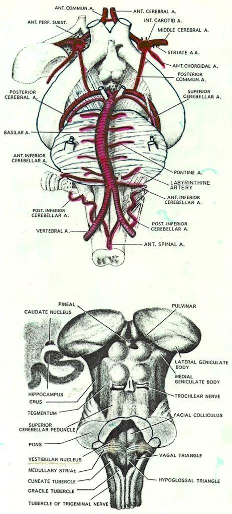
 6 Doctor 2015 Osama Asa ad Khader Mohammad Alsalem A quick revision for the spinal cord blood supply: Arterial Blood supply of spinal cord The spinal cord got its arterial supply by two ways: Longitudinal
 Spinal Cord Tracts DESCENDING SPINAL TRACTS: Are concerned with somatic motor function, modification of ms. tone, visceral innervation, segmental reflexes. Main tracts arise form cerebral cortex and others
SENSORY (ASCENDING) SPINAL TRACTS
 SPINAL TRACTS") SENSORY (ASCENDING) SPINAL TRACTS Dr. Jamila El-Medany Dr. Essam Eldin Salama OBJECTIVES By the end of the lecture, the student will be able to: Define the meaning of a tract. Distinguish between the different
SENSORY (ASCENDING) SPINAL TRACTS Dr. Jamila El-Medany Dr. Essam Eldin Salama OBJECTIVES By the end of the lecture, the student will be able to: Define the meaning of a tract. Distinguish between the different
Laith Sorour. Facial nerve (vii):
:") Laith Sorour Cranial nerves 7 & 8 Hello, there are edited slides please go back to them to see pictures, they are not that much important in this lecture but still, and yes slides are included :p Let s
Laith Sorour Cranial nerves 7 & 8 Hello, there are edited slides please go back to them to see pictures, they are not that much important in this lecture but still, and yes slides are included :p Let s
Functional components
 Facial Nerve VII cranial nerve Emerges from Pons Two roots Functional components: 1. GSA (general somatic afferent) 2. SA (Somatic afferent) 3. GVE (general visceral efferent) 4. BE (Special visceral/branchial
The Nervous System: Sensory and Motor Tracts of the Spinal Cord
 15 The Nervous System: Sensory and Motor Tracts of the Spinal Cord PowerPoint Lecture Presentations prepared by Steven Bassett Southeast Community College Lincoln, Nebraska Introduction Millions of sensory
Brainstem. By Dr. Bhushan R. Kavimandan
 Brainstem By Dr. Bhushan R. Kavimandan Development Ventricles in brainstem Mesencephalon cerebral aqueduct Metencephalon 4 th ventricle Mylencephalon 4 th ventricle Corpus callosum Posterior commissure
Cranial Nerves. Steven McLoon Department of Neuroscience University of Minnesota
 Cranial Nerves Steven McLoon Department of Neuroscience University of Minnesota 1 Course News Change in Lab Sequence Week of Oct 2 Lab 5 Week of Oct 9 Lab 4 2 Sensory and Motor Systems Sensory Systems:
Functional Distinctions
 Functional Distinctions FUNCTION COMPONENT DEFICITS Start Basal Ganglia Spontaneous Movements Move UMN/LMN Cerebral Cortex Brainstem, Spinal cord Roots/peripheral nerves Plan Cerebellum Ataxia Adjust Cerebellum
Cranial Nerves VII to XII
 Cranial Nerves VII to XII MSTN121 - Neurophysiology Session 13 Department of Myotherapy Cranial Nerve VIII: Vestibulocochlear Sensory nerve with two distinct branches. Vestibular branch transmits information
Anatomy #9. Rashed AL-Jomared. The Cranial Nerves IX. Amneh Hazaimeh & Alanood Bostanji
 Anatomy #9 The Cranial Nerves IX Rashed AL-Jomared Amneh Hazaimeh & Alanood Bostanji السالم عليكم This lecture talks about the cranial nerves IX & X:: *Glossopharyngeal nerve : The nerve gets out of the
I: To describe the pyramidal and extrapyramidal tracts. II: To discuss the functions of the descending tracts.
 Descending Tracts I: To describe the pyramidal and extrapyramidal tracts. II: To discuss the functions of the descending tracts. III: To define the upper and the lower motor neurons. 1. The corticonuclear
cardiac plexus is continuous with the coronary and no named branches pain from the heart and lungs
 Nerves of the Thoracic Region Nerve Source Branches Motor Sensory Notes cardiac plexus cardiac brs. of the vagus n. and cervical ; thoracic l nn. the heart and lungs cardiac, cervical cardiac, vagal vagus
Brain and spinal nerve. By: shirin Kashfi
 Brain and spinal nerve By: shirin Kashfi Nervous system: central nervous system (CNS) peripheral nervous system (PNS) Brain (cranial) nerves Spinal nerves Ganglions (dorsal root ganglions, sympathetic
Internal Organisation of the Brainstem
 Internal Organisation of the Brainstem Major tracts and nuclei of the brainstem (Notes) The brainstem is the major pathway for tracts and houses major nuclei, that contain sensory, motor and autonomics
CRANIAL NERVES. Dr. Amani A. Elfaki Associate Professor Department of Anatomy
 CRANIAL NERVES Dr. Amani A. Elfaki Associate Professor Department of Anatomy LEARNING OBJECTIVES Named the cranial nerves Identify the funcunal component of each cranial nerve Identify the effect of each
Motor tracts Both pyramidal tracts and extrapyramidal both starts from cortex: Area 4 Area 6 Area 312 Pyramidal: mainly from area 4 Extrapyramidal:
 Motor tracts Both pyramidal tracts and extrapyramidal both starts from cortex: Area 4 Area 6 Area 312 Pyramidal: mainly from area 4 Extrapyramidal: mainly from area 6 area 6 Premotorarea: uses external
Upper and Lower Motoneurons for the Head Objectives
 Upper and Lower Motoneurons for the Head Objectives Know the locations of cranial nerve motor nuclei Describe the effects of motor cranial nerve lesions Describe how the corticobulbar tract innervates
Unit VIII Problem 5 Physiology: Cerebellum
 Unit VIII Problem 5 Physiology: Cerebellum - The word cerebellum means: the small brain. Note that the cerebellum is not completely separated into 2 hemispheres (they are not clearly demarcated) the vermis
b. The groove between the two crests is called 2. The neural folds move toward each other & the fuse to create a
 Chapter 13: Brain and Cranial Nerves I. Development of the CNS A. The CNS begins as a flat plate called the B. The process proceeds as: 1. The lateral sides of the become elevated as waves called a. The
Principles of Anatomy and Physiology
 Principles of Anatomy and Physiology 14 th Edition CHAPTER 14 The Brain and Cranial Nerves Introduction The purpose of the chapter is to: 1. Understand how the brain is organized, protected, and supplied
Cranial Nerves and Spinal Cord Flashcards
 1. Name the cranial nerves and their Roman numeral. 2. What is Cranial Nerve I called, and what does it 3. Scientists who are trying to find a way to make neurons divide to heal nerve injuries often study
Organization of The Nervous System PROF. SAEED ABUEL MAKAREM
 Organization of The Nervous System PROF. SAEED ABUEL MAKAREM Objectives By the end of the lecture, you should be able to: List the parts of the nervous system. List the function of the nervous system.
Review or skim Ch 12 on the vascular supply of the brain. Just look at pictures and legends for the clinical part at the end.
 Dental Neuroanatomy January 20 and 27, 10-12, 2011 Suzanne S. Stensaas, Ph.D. Dear Students: Please print these notes and bring them with you. My style is to use a Tablet PC and I draw on either a Word
PERIPHERAL NERVOUS SYSTEM
 CHAPTER 13 PERIPHERAL NERVOUS SYSTEM Functional division of nervous system = afferent info to the CNS ascending spinal cord = efferent info from CNS descending spinal cord somatic skin, muscles visceral
THE CENTRAL NERVOUS SYSTE M
 THE CENTRAL NERVOUS SYSTE M Structure and Functio n THIRD EDITIO N PER BRODAL A Brief Survey, x i Studying the Structures and Function of the Nervous System, xii i Animal Experiments Crucial for Progress,
Auditory and Vestibular Systems
 Auditory and Vestibular Systems Objective To learn the functional organization of the auditory and vestibular systems To understand how one can use changes in auditory function following injury to localize
THE BACK. Dr. Ali Mohsin. Spinal Cord
 Spinal Cord THE BACK Dr. Ali Mohsin The spinal cord is the elongated caudal part of the CNS. It starts as the inferior continuation of the medulla oblongata at the level of foramen magnum, & ends as an
Brainstem. Amadi O. Ihunwo, PhD School of Anatomical Sciences
 Brainstem Amadi O. Ihunwo, PhD School of Anatomical Sciences Lecture Outline Constituents Basic general internal features of brainstem External and Internal features of Midbrain Pons Medulla Constituents
Neural Integration I: Sensory Pathways and the Somatic Nervous System
 15 Neural Integration I: Sensory Pathways and the Somatic Nervous System PowerPoint Lecture Presentations prepared by Jason LaPres Lone Star College North Harris An Introduction to Sensory Pathways and
Organization of The Nervous System PROF. MOUSAED ALFAYEZ & DR. SANAA ALSHAARAWY
 Organization of The Nervous System PROF. MOUSAED ALFAYEZ & DR. SANAA ALSHAARAWY Objectives At the end of the lecture, the students should be able to: List the parts of the nervous system. List the function
General Sensory Pathways of the Trunk and Limbs
 General Sensory Pathways of the Trunk and Limbs Lecture Objectives Describe gracile and cuneate tracts and pathways for conscious proprioception, touch, pressure and vibration from the limbs and trunk.
Medical Neuroscience Tutorial Notes
 Medical Neuroscience Tutorial Notes Cranial Nerve Nuclei MAP TO NEUROSCIENCE CORE CONCEPTS 1 NCC1. The brain is the body's most complex organ. LEARNING OBJECTIVES After study of the assigned learning materials,
Brainstem and Cranial Nerves II. Nerves covered in other lectures. A reminder about embryology. Prof. Stuart Bunt
 Brainstem and Cranial Nerves II Prof. Stuart Bunt Nerves covered in other lectures 1 Olfactory 2 Optic 3,4,6 Extraocular eye muscles 8 Vestibulo-cochlear 5 Motor and Sensory to the face and muscles of
Brainstem. Steven McLoon Department of Neuroscience University of Minnesota
 Brainstem Steven McLoon Department of Neuroscience University of Minnesota 1 Course News Change in Lab Sequence Week of Oct 2 Lab 5 Week of Oct 9 Lab 4 2 Goal Today Know the regions of the brainstem. Know
Unit VIII Problem 4 Physiology lab: Brain Stem Lesions
 Unit VIII Problem 4 Physiology lab: Brain Stem Lesions - Motor and sensory somatotopy: Pre-central gyrus: is the motor area. Post-central gyrus: is the sensory area. Somatotopy: there is a map of thee
By : Prof Saeed Abuel Makarem & Dr.Sanaa Alshaarawi
 By : Prof Saeed Abuel Makarem & Dr.Sanaa Alshaarawi OBJECTIVES By the end of the lecture, students shouldbe able to: List the nuclei of the deep origin of the trigeminal and facial nerves in the brain
Cranial Nerve VII - Facial Nerve. The facial nerve has 3 main components with distinct functions
 Cranial Nerve VII - Facial Nerve The facial nerve has 3 main components with distinct functions Somatic motor efferent Supplies the muscles of facial expression; posterior belly of digastric muscle; stylohyoid,
The NIHSS score is 4 (considering 2 pts for the ataxia involving upper and lower limbs.
 Neuroscience case 5 1. Speech comprehension, ability to speak, and word use were normal in Mr. Washburn, indicating that aphasia (cortical language problem) was not involved. However, he did have a problem
CN V! touch! pain! Touch! P/T!
 CN V! touch! pain! Touch! P/T! Visual Pathways! L! R! B! A! C! D! LT! E! F! RT! G! hypothalamospinal! and! ALS! Vestibular Pathways! 1. Posture/Balance!!falling! 2. Head Position! 3. Eye-Head Movements
Lecturer. Prof. Dr. Ali K. Al-Shalchy MBChB/ FIBMS/ MRCS/ FRCS 2014
 Lecturer Prof. Dr. Ali K. Al-Shalchy MBChB/ FIBMS/ MRCS/ FRCS 2014 Dorsal root: The dorsal root carries both myelinated and unmyelinated afferent fibers to the spinal cord. Posterior gray column: Long
Omar Sami. Aseel Abdeen. Muhammad Al-Salem. 1 P a g e
 Omar Sami Aseel Abdeen Muhammad Al-Salem 1 P a g e Using only section 2 record, I wrote this sheet; as the video is not ready yet. Despite pointing the structures, I ve tried to include all the scientific
Introduction to Head and Neck Anatomy
 Introduction to Head and Neck Anatomy Nervous Tissue Controls and integrates all body activities within limits that maintain life Three basic functions 1. sensing changes with sensory receptors 2. interpreting
Brainstem: Medulla oblongata and pons
 Brainstem: Medulla oblongata and pons 1. Overview of the brainstem subdivisions 2. Embryonic development of the brainstem 3. Medulla oblongata external features 4. Internal structure of the medulla oblongata
Faculty of Dental Medicine and Surgery. Sem 4 Cranial Nerves Dr. Abbas Garib Alla
 Faculty of Dental Medicine and Surgery Sem 4 Cranial Nerves Dr. Abbas Garib Alla Cranial Nerves I through XII FUNCTIPONAL CLSSIFICATION OF THE CN parasympathetic nerves 1973 PHARYNGEAL ARCHES nerves 1975
Spinal Cord Organization. January 12, 2011
 Spinal Cord Organization January 12, 2011 Spinal Cord 31 segments terminates at L1-L2 special components - conus medullaris - cauda equina no input from the face Spinal Cord, Roots & Nerves Dorsal root
Brain Stem. Nervous System (Part A-3) Module 8 -Chapter 14
 Module 8 -Chapter 14") Nervous System (Part A-3) Module 8 -Chapter 14 Overview Susie Turner, M.D. 1/9/13 Cellular structure of the nervous system Neurons Neuroglia Nervous System Divisions Central nervous system Peripheral nervous
Nervous System (Part A-3) Module 8 -Chapter 14 Overview Susie Turner, M.D. 1/9/13 Cellular structure of the nervous system Neurons Neuroglia Nervous System Divisions Central nervous system Peripheral nervous
C h a p t e r PowerPoint Lecture Slides prepared by Jason LaPres North Harris College Houston, Texas
 C h a p t e r 15 The Nervous System: The Brain and Cranial Nerves PowerPoint Lecture Slides prepared by Jason LaPres North Harris College Houston, Texas Copyright 2009 Pearson Education, Inc., publishing
Cranial nerves.
 Cranial nerves eaglezhyxzy@163.com Key Points of Learning Name Components Passing through Peripheral distribution Central connection Function Cranial nerves Ⅰ olfactory Ⅱ optic Ⅲ occulomotor Ⅳ trochlear
INTRODUCTION: ANATOMY UNDERLYING CLINICAL TESTS OF CRANIAL NERVES
 INTRODUCTION: ANATOMY UNDERLYING CLINICAL TESTS OF CRANIAL NERVES CRANIAL NERVE I - OLFACTORY I - OLFACTORY NERVE - SMELL TEST: SMELL ODORS (note: not ammonia; pain in nasal cavity CN5 DAMAGE: LOSS OF
Abdullah AlZibdeh. Dr. Maha ElBeltagy. Maha ElBeltagy
 19 Abdullah AlZibdeh Dr. Maha ElBeltagy Maha ElBeltagy Introduction In this sheet, we discuss the cerebellum; its lobes, fissures and deep nuclei. We also go into the tracts and connections in which the
THE BRAINSTEM. Raymond S. Price, MD University of Pennsylvania
 THE BRAINSTEM Raymond S. Price, MD University of Pennsylvania Overview of Brainstem Functions The brainstem serves numerous crucial neurologic functions. The most clinically relevant functions include:
Cranial nerve Dept. of Anatomy Zhou Hong Ying
 Cranial nerve Dept. of Anatomy Zhou Hong Ying Key Points of Learning Name Components Passing through Peripheral distribution Central connection Function Cranial nerve Ⅰ olfactory Ⅱ optic Ⅲ occulomotor
Blood supply to the brain Blood brain barrier isolates neural tissue from general circulation
 The Brain and Cranial Nerves Objectives Name the major regions of the brain and describe their functions. Discuss the formation, circulation, and functions of the CSF. List the main components of the medulla
Biology 218 Human Anatomy
 Chapter 21 Adapted form Tortora 10 th ed. LECTURE OUTLINE A. Overview of Sensations (p. 652) 1. Sensation is the conscious or subconscious awareness of external or internal stimuli. 2. For a sensation
ParasymPathetic Nervous system. Done by : Zaid Al-Ghnaneem
 ParasymPathetic Nervous system Done by : Zaid Al-Ghnaneem In this lecture we are going to discuss Parasympathetic, in the last lecture we took sympathetic and one of the objectives of last lecture was
Ms. K. GOWRI. M.Pharm., Lecturer.
 Ms. K. GOWRI. M.Pharm., Lecturer. CENTRAL NERVOUS SYSTEM (CNS) central nervous system consists of brain and spinal cord membrane covering the brain and spinal cord are surrounded by three membrane Meninges
The Nervous System PART C. PowerPoint Lecture Slide Presentation by Patty Bostwick-Taylor, Florence-Darlington Technical College
 PowerPoint Lecture Slide Presentation by Patty Bostwick-Taylor, Florence-Darlington Technical College The Nervous System 7 PART C Protection of the Central Nervous System Scalp and skin Skull and vertebral
The Nervous System PART B
 7 The Nervous System PART B PowerPoint Lecture Slide Presentation by Jerry L. Cook, Sam Houston University ESSENTIALS OF HUMAN ANATOMY & PHYSIOLOGY EIGHTH EDITION ELAINE N. MARIEB Central Nervous System
Jolanta Marszalek CN 1,9-12
 Jolanta Marszalek CN 1,9-12 Olfactory SVA, special visceral afferent, which carries the modality of smell Shortest of the CNs Does not emanate from brainstem (CN II) The afferent nerve fibers of the olfactory
Peripheral Nervous System Dr. Gary Mumaugh
 Peripheral Nervous System Dr. Gary Mumaugh Spinal Nerves Overview Thirty-one pairs of spinal nerves are connected to the spinal cord No special names; numbered by level of vertebral column at which they
Cerebellum John T. Povlishock, Ph.D.
 Cerebellum John T. Povlishock, Ph.D. OBJECTIVES 1. To identify the major sources of afferent inputs to the cerebellum 2. To define the pre-cerebellar nuclei from which the mossy and climbing fiber systems
Examination and Diseases of Cranial Nerves
 Cranial nerve evaluation is an important part of a neurologic exam. There are some differences in the assessment of cranial nerves with different species but the general principles are the same. Going
Lec#10 Part 2. Dawood Alatefi. Mariam Hassouneh. Dr.Mohammed Al-Salem
 Lec#10 Part 2 Dawood Alatefi Mariam Hassouneh Dr.Mohammed Al-Salem This part s record is found on the Batch s channel as LabA.3, starting from 0.00min until 26.20min. Hope this ll be an easy part on you;
Introduction and Basic structural organization of the nervous system
 Introduction and Basic structural organization of the nervous system **the slides are in bold and the book is in red Done by : razan krishan & marah marahleh INTRODUCTION The nervous system, along with
Medical Neuroscience Tutorial
 Pain Pathways Medical Neuroscience Tutorial Pain Pathways MAP TO NEUROSCIENCE CORE CONCEPTS 1 NCC1. The brain is the body's most complex organ. NCC3. Genetically determined circuits are the foundation
The neurvous system senses, interprets, and responds to changes in the environment. Two types of cells makes this possible:
 NERVOUS SYSTEM The neurvous system senses, interprets, and responds to changes in the environment. Two types of cells makes this possible: the neuron and the supporting cells ("glial cells"). Neuron Neurons
Neural Integration I: Sensory Pathways and the Somatic Nervous System
 C h a p t e r 15 Neural Integration I: Sensory Pathways and the Somatic Nervous System PowerPoint Lecture Slides prepared by Jason LaPres Lone Star College - North Harris Copyright 2009 Pearson Education,
The Spinal Cord. The Nervous System. The Spinal Cord. The Spinal Cord 1/2/2016. Continuation of CNS inferior to foramen magnum.
 The Nervous System Spinal Cord Continuation of CNS inferior to foramen magnum Simpler than the brain Conducts impulses to and from brain Two way conduction pathway Reflex actions Passes through vertebral
SOMATOSENSORY SYSTEMS: Pain and Temperature Kimberle Jacobs, Ph.D.
 SOMATOSENSORY SYSTEMS: Pain and Temperature Kimberle Jacobs, Ph.D. Sensory systems are afferent, meaning that they are carrying information from the periphery TOWARD the central nervous system. The somatosensory
Brain and Cranial Nerves (Ch. 15) Human Anatomy lecture. caudal = toward the spinal cord)
 Human Anatomy lecture. caudal = toward the spinal cord)") Insight: Some cranial nerve disorders Brain and Cranial Nerves (Ch. 15) Human Anatomy lecture I. Overview (Directional terms: rostral = toward the forehead caudal = toward the spinal cord) A. 3 Major parts
Insight: Some cranial nerve disorders Brain and Cranial Nerves (Ch. 15) Human Anatomy lecture I. Overview (Directional terms: rostral = toward the forehead caudal = toward the spinal cord) A. 3 Major parts
PTA 106 Unit 1 Lecture 1B
 PTA 106 Unit 1 Lecture 1B Medulla Oblongata Cardiovascular Center: Regulates the rate and force of the heartbeat and the diameter of blood vessels Medullary Rhythmicity Area: adjusts the basic rhythm of
CN modalities Sensory: SSA (Vision) Mixed: GSE, proprioceptive. Mixed: GSE, proprioceptive
 Mixed: GSE, proprioceptive. Mixed: GSE, proprioceptive") CN 2 3 4 6 modalities Sensory: SSA (Vision) course Rods and cones of the retina bipolar neurons gangli on cells Optic nerve optic foramen Optic chiasm Optic tracts Sup colliculi LGN Optic radiation cortex
CN 2 3 4 6 modalities Sensory: SSA (Vision) course Rods and cones of the retina bipolar neurons gangli on cells Optic nerve optic foramen Optic chiasm Optic tracts Sup colliculi LGN Optic radiation cortex
Cranial Nerve VIII (The Vestibulo-Cochlear Nerve)
") Cranial Nerve VIII (The Vestibulo-Cochlear Nerve) Please view our Editing File before studying this lecture to check for any changes. Color Code Important Doctors Notes Notes/Extra explanation Objectives
Cranial Nerve VIII (The Vestibulo-Cochlear Nerve) Please view our Editing File before studying this lecture to check for any changes. Color Code Important Doctors Notes Notes/Extra explanation Objectives
Copy Right- Hongqi ZHANG-Department of Anatomy-Fudan University. Systematic Anatomy. Nervous system Cerebellum. Dr.Hongqi Zhang ( 张红旗 )
") Systematic Anatomy Nervous system Cerebellum Dr.Hongqi Zhang ( 张红旗 ) Email: zhanghq58@126.com 1 The Cerebellum Cerebellum evolved and developed with the complication of animal movement. Key points about
Systematic Anatomy Nervous system Cerebellum Dr.Hongqi Zhang ( 张红旗 ) Email: zhanghq58@126.com 1 The Cerebellum Cerebellum evolved and developed with the complication of animal movement. Key points about
Posterior White Column-Medial Lemniscal Pathway
 Posterior White Column-Medial Lemniscal Pathway Modality: Discriminative Touch Sensation (include Vibration) and Conscious Proprioception Receptor: Most receptors except free nerve endings Ist Neuron:
Nervous System. The Peripheral Nervous System Agenda Review of CNS v. PNS PNS Basics Cranial Nerves Spinal Nerves Reflexes Pathways
 Nervous System Agenda Review of CNS v. PNS PNS Basics Cranial Nerves Spinal Nerves Sensory Motor Review of CNS v. PNS Central nervous system (CNS) Brain Spinal cord Peripheral nervous system (PNS) All
Sensory systems. Taste/gustatory
 Sensory systems Taste/gustatory Sensory systems basic concepts Modality of Sensation Receptor Sensory Tract primary neuron secondary neuron tertiary neuron termination Receptors of sensory systems - primary
Oral cavity : consist of two parts: the oral vestibule and the oral cavity proper. Oral vestibule : is slit like space between.
 Oral cavity Oral cavity : consist of two parts: the oral vestibule and the oral cavity proper Oral vestibule : is slit like space between the teeth, buccal gingiva, lips, and cheeks 1 Oral cavity Oral
Functional neuroanatomy of the neurological examination: Cranial nerves
 Functional neuroanatomy of the neurological examination: Cranial nerves Chris Thomson BVSc(Hons), Dip ACVIM (Neurol), Dip ECVN, PhD Associate Professor Neurobiology, Dept. of Vet. Med., University of Alaska,
Spinal cord. We have extension of the pia mater below L1-L2 called filum terminale
 Spinal cord Part of the CNS extend from foramen magnum to the level of L1-L2 (it is shorter than the vertebral column) it is covered by spinal meninges. It is cylindrical in shape. It s lower end become
Stanley Pruisinger 1980's
 Neuroanatomy Prion disease cerebellum chapter b/c cerebellar ataxia here as a warning for obvious reasons. Creutzfeldt - Jakob Disease (CJD) "Spongiform" (brain turns to sponge) Jews in Lybia who ate
Lab 16: PNS: Nerves and Autonomic NS Hamilton Answers to Pre- Lab Assignments
 Lab 16: PNS: Nerves and Autonomic NS Hamilton Answers to Pre- Lab Assignments Pre-Lab Activity 1: 1. a. olfactory nerve b. optic nerve c. oculomotor nerve d. abducens nerve e. trochlear nerve f. trigeminal
Nervous System. Student Learning Objectives:
 Nervous System Student Learning Objectives: Identify the primary parts of the neuron Identify the major structures of the central nervous system Identify the major structures of the peripheral nervous