CKET GUIDE STROKE ASSESSMENT POCKET GUIDE S
|
|
|
- Leslie Fitzgerald
- 6 years ago
- Views:
Transcription
1 E ASSESS POCKET GUIDE GUIDE STROKE ASSESSM OKE ASSESSMENT POCKET GUIDE ENT POCKET GUIDE STROKE ASSESSMENT KET GUIDE STROKE ASSESSMENT POCKET GUIDE STROKE ASSESSMENT POCKET GUIDE STROKE ASSESSMENT STROKE ASSESSMENT POCKET GUIDE SMENT POCKET GUIDE STROKE ASSESSMENT POCKET GUIDE STRO POCKET GUIDE STROKE ASSESSMENT POCKET GUIDE STROKE ASSESSM E STROKE ASSESSMENT POCKET GUIDE STROKE ASSESSMENT POCKE SSESSMENT POCKET GUIDE STROKE ASSESSMENT POCKET GUIDE STRO POCKET GUIDE STROKE ASSESSMENT POCKET GUIDE STROKE ASSE OKE ASSESSMENT POCKET GUIDE STROKE ASSESSMENT POC CKET GUIDE STROKE ASSESSMENT POCKET GUIDE S OKE ASSESSMENT POCKET GUIDE STROKE A CKET GUIDE STROKE ASSESSMENT ASSESSMENT POCKET GUID KET GUIDE STRO ASSESSME G
2 STROKE ASSESSMENT POCKET GUIDE
3 TABLE OF CONTENTS i. Common Signs and Symptoms of Stroke Syndromes...1 ii. Common Symptoms in Stroke Patients...2 iii. Glossary of Frequently used Terms in Stroke...3 iv. Cranial Nerve Function and Testing...4 v. Types of Stroke...5 vi. Functions of the Brain and Their Relation to Stroke...7 vii. Canadian Neurological Scale...11 viii. NIH Stroke Scale...13 iv. FAST Signs of Stroke...26
4 COMMON SIGNS AND SYMPTOMS OF STROKE SYNDROMES Anterior Cerebral Stroke Middle Cerebral Stroke Posterior Cerebral Stroke Vertebro Basilar Stroke Thalamic Stroke Lacunar Stroke Four Types Contralateral sensorimotor deficit: foot and leg Arm paresis Gait ataxia Bladder incontinence Personality and behaviour changes Flat affect, distractible Perservation and amnesia Contralateral sensorimotor deficit: face, arm, leg Contralateral homonymous hemianopsia Contralateral hemispatial neglect or inattention (usually in Right Hemispheric Strokes) Aphasia, alexia, agraphia (usually in Left Hemispheric Stroke or dominant hemisphere) Gaze deviation towards affected hemisphere Dysarthria Pure homonymous hemianopsia Nausea Vomiting Ataxia Vertigo Weakness Sensory loss Dysarthria Vertigo Limb and gait ataxia Cranial nerve dysfunction Coma at onset Diplopia Cross sensory loss Bilateral motor deficits Isolated field defect Pure motor/ sensory loss Dysarthria Dysphagia Alteration in senses (except smell) Alteration in pain, crude touch (loss) Alteration in temperature Contralateral hemiplegia Hyper-sensitivity to stimulus Vertical and lateral gaze deficits Short-term memory loss Pure motor hemiparesis Contralateral hemiparesis of face, arm and leg Ataxic Hemiparesis Ipsilateral paresis of leg Arm and leg ataxia Dysarthria and Clumsy Hand Syndrome Dysarthia Weakness of hand Impaired manual dexterity Pure Sensory Stroke Impairments in pain, temperature, touch, position and vibration
5 COMMON SYMPTOMS IN STROKE PATIENTS The effects of a stroke depend on several factors including the location of the obstruction and how much brain tissue is affected. However, because one side of the brain controls the opposite side of the body, a stroke affecting one side will result in neurological complications on the opposite side. Right Hemispheric Stroke Left Hemispheric Stroke If the stroke occurs in the brain s right side, the left side of the body will be affected, which could produce any or all of the following: Contralateral face, arm and leg weakness or hemiparesis Contralateral arm and/or leg sensory loss or extinction Hemispatial neglect or inattention Deficit and/or neglect of left visual field Right gaze preference Impulsive or overestimation of abilities (risk for injury) If the stroke occurs in the left side of the brain, the right side of the body will be affected, producing some or all of the following: Contralateral face, arm and leg weakness or hemiparesis Contralateral face, arm and/or leg sensory loss Aphasia, alexia, agraphia Slow and cautious behaviour Deficits in right visual field Left gaze preference 2
6 GLOSSARY OF FREQUENTLY USED TERMS IN STROKE Alexia: Inability to read in the presence of normal vision. Agnosia: Difficulty recognizing familiar objects through the senses; perception without meaning. Agraphia: Loss of the ability to write. Aphasia: Expressive Aphasia: loss of the ability to express one s thoughts in speech and/or writing. Receptive Aphasia: inability to comprehend spoken and/or written language. Global Aphasia: inability to comprehend and/or use language skills. Apraxia: Impaired ability to perform learned, complex motor tasks in the absence of weakness, sensory impairment or aphasia. Aprosody: Loss of the understanding or expression of the normal musicality, intonation and gesture involved in normal communication. This often results in misunderstanding of humour or sarcasm. Ataxia: Poor coordination of muscular movements such as walking or reaching for objects. Cognition: Thinking and processing information: perceiving, remembering, imagining, judging, reasoning, conceiving. Contralateral: Pertaining to the side of the body opposite the reference point. Dysarthria: Dysphagia: Impaired articulation that may be caused by a motor deficit of the tongue or speech muscles. Impaired ability to swallow. Hemiparesis: Muscular weakness affecting one half of the body. Hemispatial: Loss or reduced attention directed toward the contralateral side. Neglect May be minimal such that the person does not recognize double simultaneous stimuli (e.g. touching both arms at the same time; the patient is only aware of the examiner touching the right arm) or severe (the patient does not recognize their own left arm or leg). Hemianopsia: Blindness in one half of the visual field. Differentiating left hemianopsia from left visual neglect can be difficult. Paralysis: Vertigo: Neurologic muscular weakness to the extent of immobility. A false sense of rotation or movement; often described as the world is spinning around me or like being on a boat on the ocean.
7 CRANIAL NERVE FUNCTION AND TESTING Nerve Function Testing I Olfactory hemispheric II Optic hemispheric smell visual acuity visual fields assess fundi pinch 1 nostril and get patient to smell objects, test on both sides opthalmoscope close 1 eye, look at nose and cheek peripheral vision x 4 Nerve Function Testing VII Facial pons VIII Acoustic pons taste (salivary glands) facial expressions proprioception cochlea (hearing) vestibular (balance) orientation is space smile wrinkle forehead puff out cheeks rub fingers nest to ear stand with feet together III Oculomotor originates midbrain, emerges pons IV Trochlear midbrain V Trigeminal originates in 4th ventricle, emerges pons VI Abducens pons eyeball, eyelid movement proprioception eyeball movement proprioception chewing, opening jaw 3 branches, ophthalmic, maxillary, mandibular sensory: pain, touch, temperature lateral movements of the eyeball follows finger in star pattern (nystagnus) pupillary response assessed with III & VI cotton wool or soft touch from finger to 3 areas on both sides of face (sensory) bite down on tongue depressor-both sides (motor) assessed with III & IV IX Glossopharyngeal medulla X Vagus medulla XI Spinal Accessory medulla XII Hypoglossal medulla controls swallowing controls secretion of saliva gag reflex,cough taste muscles of speech sensory: impulses received from throat, esophagus, heart, lung, stomach, small intestine affects HR and BP shoulder and neck strength innervates tongue assess swallowing say ah, watch for upward movement in the back of the throatquality & volume of speech gag reflex, assess both sides of throat ask pt to shrug shoulders (symmetry) rotate head observe for symmetry when tongue is out 4

8 TYPES OF STROKE Stroke or brain attack is a sudden problem affecting the blood vessels of the brain. There are several types of stroke, and each type has different causes. Ischemic Stroke The most common type of stroke, accounting for almost 80% of all strokes, is caused by a clot or other blockage within an artery leading to the brain. Two types: 1) embolic and 2) thrombotic stroke. An embolic stroke is also caused by a clot within an artery (emboli) that was formed somewhere other than in the brain itself. Often from the heart, these emboli will travel the bloodstream until they become lodged and cannot travel any further. A thrombotic stroke occurs when diseased or damaged cerebral arteries become blocked by the formation of a blood clot within the brain. Referred to as cerebral thrombosis or cerebral infarction, this type of event is responsible for almost 50% of all strokes. Large-vessel thrombosis is the term used when the blockage is in one of the brain s larger blood-supplying arteries such as the carotid or middle cerebral. Small-vessel thrombosis involves one (or more) of the brain s smaller, yet deeper penetrating arteries. This latter type of stroke is also called a Lacunar stroke.

9 Hemorrhagic Stroke Intracerebral hemorrhage occurs when a diseased blood vessel within the brain bursts, allowing blood to leak inside the brain. The sudden increase in pressure within the brain can cause damage to the brain cells surrounding the blood. Often occurs in selected parts of the brain, including the basal ganglia, cerebellum, brainstem, or cortex. Subarachnoid hemorrhage occurs when a blood vessel just outside the brain ruptures. The area of the skull surrounding the brain (the subarachnoid space) rapidly fills with blood. A patient with subarachnoid hemorrhage may have a sudden, intense headache, neck pain, and nausea or vomiting. 6
10 FUNCTIONS OF THE BRAIN AND THEIR RELATION TO STROKE Structure/ Circulation Frontal Lobe (emotions, motor, cognition, expressive language) Anterior Cerebral Artery (ACA) Middle Cerebral Artery (MCA) Parietal Lobe (Sensation and Perception, Integration of Sensory Input) Key Functions Associated Dysfunction Voluntary motor function Memory for habits and motor activities Controls expressive language, articulating speech (Broca s Area) Assigns meaning to words we choose Behavioural spontaneity Controls emotional responses Executive Functions: task initiation, motivation, planning and self-monitoring Concentration/reasoning Judgment/problem solving Bladder control (micturation center) Visual attention Touch perception Goal directed voluntary movements Manipulation of objects Integration of different sensory input Paralysis/paresis: of the face, arm and leg (MCA) or leg and foot (ACA) Inability to express language (Broca s Aphasia) Emotional lability, mood changes Impulsivity of thought, affect and action Lack of spontaneity in interacting with others Inability to attend to task Inability to plan a sequence of complex tasks, i.e. making coffee Impaired judgment, problem-solving Change in personality, sexual and social behaviour Incontinence Difficulty focusing visual attention or attending to more than one object at a time Loss of sensation Difficulty with hand/eye coordination; distinguishing left and right
11 Anterior Cerebral Artery Middle Cerebral Artery Posterior Cerebral Artery The ability to sense the position, location, orientation and movement of the body and its parts (Proprioception) Inability to perceive objects normally (Agnosia) Neglecting part of the body or space (contralateral neglect/ difficulties with ADLs) Difficulty reading, writing (Agraphia), drawing, constructing, naming objects, calculating Denial of deficits (Anosagnosia) Temporal Lobe (Auditory Sensation and Perception, Memory, Language Comprehension, Affect) Middle Cerebral Artery Posterior Cerebral Artery Hearing ability Receptive language (Wernicke s Area) Integration of visual, auditory, somatic information Memory (storage, retrieval of words, experiences) Emotions Impaired auditory sensation and perception Difficulty recognizing faces Difficulty selectively attending to auditory and visual input Disturbed language comprehension, word recognition (Wernicke s Aphasia) Difficulty organizing verbal information Short-term memory loss Disturbance of long-term memory Altered personality, emotional behaviour, sexual behaviour Impulsiveness, aggressiveness, indifference, depression Persistant talking Occipital Lobe (Vision) Posterior Cerebral Artery Middle Cerebral Artery Vision Spatial organization and interpretation of visual information Visual reflexes Defects in vision: visual field cuts, diplopia (Hemianopia) Inability to recognize familiar objects, words, colours, or movement of an object (Agnosias) Difficulty with reading and writing 8
12 FUNCTIONS OF THE BRAIN AND THEIR RELATION TO STROKE (CON D) Structure/ Circulation Key Functions Associated Dysfunction Brainstem (Body Functions and Movements) (Midbrain, Pons, Medulla) Basilar Artery Vertebral Artery Houses Cranial Nerves III-XII 1) Receives information from cranial structures and controls muscles of the head (Cranial Nerves) 2) Contains neural circuits that transmit information from the spinal cord up to brain structures and from brain down to spinal cord 3) Brainstem structures work together to regulate arousal (reticular activating system) 4) Individually they subserve specific sensory and motor functions Midbrain (Visual/auditory reflexes) Unable to move eye up, down or in Inappropriate responses to visual or auditory stimuli Pons (Blood pressure and respiratory regulation) Altered respiratory function Impaired chewing and facial sensation Unable to move the eye out Altered taste Abnormal facial expression Problems with equilibrium and hearing Medulla (Blood pressure and respiratory regulation) Altered respiratory, cardiac and blood pressure function Altered sensation and limb weakness Difficulty maintaining posture control Swallowing problems Unable to move head and shoulder, tongue Altered salivation
13 Diencephalon (Thalamus, Hypothalamus) Posterior Cerebral Artery Thalamus Transmits information to cerebral hemispheres for sensation and movement Hypothalamus Integrates the function of the autonomic nervous system (maintains blood pressure, heart rate, respiratory rate, temperature, fluid balance, hormone synthesis, sleepwakefulness) Contralateral weakness Contralateral sensory loss Vertical and lateral gaze deficits Hypersensitivity response to stimulus Alteration in temperature regulation Diabetes insipidus Abnormal heart and respiratory patterns Impaired blood sugar levels Cerebellum (Motor Control) Posterior Cerebral Artery Basilar Artery Vertebral Artery Regulates movements of eyes and limbs; helps maintain posture and balance Coordinates voluntary movement, muscle tone, balance and equilibrium Control of fine motor movements Limb and gait ataxia; impaired ability to walk Difficulty judging distance, when to stop Difficulty performing rapid alternating movements Vertigo Tremors Loss of balance and coordination Poor coordination of fine motor movements, weak muscles Basal Ganglia Middle Cerebral Artery Production of dopamine and coordination of muscle movement and posture Loss of postural control Tremor, rigidity, involuntary movements 10
14 CANADIAN NEUROLOGICAL SCALE Robert Côté. Used with permission. Assess: Vital Signs and Pupils Vital Signs: BP, Temp, Pulse, Respirations, Oximetry Pupils: Size and reaction to light Section A: MENTATION: LOC, Orientation, Speech LEVEL OF CONSCIOUSNESS: CNS (Alert, Drowsy) GCS (Stuporous, Comatose) ORIENTATION: Place (city or hospital), Time (month and year) *Patient can speak, write, or gesture their responses. SPEECH: SCORE: Patient is Oriented, score 1.0, if they correctly state both place and correct month and year. If dysarthric, speech must be intelligible. If patient cannot state both, Disoriented, score 0.0 RECEPTIVE: Ask patient the following separately (do not prompt by gesturing): 1. Close your eyes 2. Does a stone sink in water? 3. Point to the ceiling SCORE: If patient is unable to do all three, Receptive Deficit, score 0.0, go to A2. Section A1: MOTOR FUNCTION NO RECEPTIVE DEFICIT FACE: None 1.5 Mild 1.0 Significant 0.5 Total 0.0 Ask patient to smile/grin, note weakness in mouth or nasal/labial folds. SCORE: None/no weakness = 0.5 or Present/ weakness = 0.0 Test both limbs and always record the side with the WORST deficit and indicate side by entering a R/L. no weakness present mild weakness present, full ROM, cannot withstand resistance moderate weakness, some movement, not full ROM complete loss of movement; total weakness SCORE: Arm: Proximal Ask patient to lift arm degrees. Apply resistance between shoulder and elbow. Arm: Distal Ask patient to make fist and flex wrist backwards, apply resistance between wrist and knuckles.
15 EXPRESSIVE: 1. Show patient 3 items separately (pencil, watch, key) and ask patient to name each object. 2. Ask patient what each object is used for while holding each up again, i.e. What do you do with a pencil? SCORE: If patient is able to state the name and use of all 3 objects, Normal Speech, score 1.0. If patient is unable to state the name and use of all 3 objects, Expressive Deficit, score 0.5. *If patient answers all questions correctly but speech is slurred and intelligible, score Normal Speech and record SL along with the score. Leg: Proximal In supine, ask patient to flex hip to 90 degrees, apply pressure to mid thigh. Leg: Distal Ask patient to dorsiflex foot, apply resistance to top of foot. Section A2: MOTOR RESPONSE RECEPTIVE DEFICIT PRESENT FACE: ARMS: Have patient mimic your smile. If unable, note facial expression while applying sternal pressure. SCORE: Symmetrical 0.5 Asymmetrical 0.0 Demonstrate or lift patient s arms to 90 degrees, score ability to maintain equal levels (>5 secs). If unable to maintain raised arms, apply nail bed pressure to assess reflex response. SCORE: Score: Equal 1.5 Unequal 0.0 LEGS: Lift patient s hip to 90 degrees, score ability to maintain equal levels (>5 secs). If unable to maintain raised position, apply nail bed pressure to assess reflex response. SCORE: Equal 1.5 Unequal
16 NIH STROKE SCALE (NIHSS) 1a. Level of Consciousness (LOC)* 0 = Alert (keenly responsive) 1 = Not alert but arousable by minor stimulation 2 = Not alert: requires repeated stimulation to attend, or is obtunded and requires strong or painful stimulation to make movements 3 = Responds only with reflex motor or autonomic effects or totally unresponsive, flaccid, and flexic * The investigator should choose a response even in patients with endotracheal tubes, language barrier, orotracheal trauma/ bandages, etc. A score of 3 is only given if the patient fails to respond (other than reflexive posturing) after noxious stimulation. 1b. LOC Questions* Ask the patient: What month is it? How old are you? 0 = Answers both correctly 1 = Answers one correctly 2 = Answers neither correctly * Score only the initial answer (there is no credit for being close). Patients unable to speak due to intubation, orotracheal trauma, severe dysarthria, language barrier, etc., are scored 1. Aphasic and stuporous patients are scored Best Gaze* Establish eye contact and ask the patient to: Follow my finger. 0 = Normal 1 = Partial gaze palsy 2 = Forced deviation or total gaze paresis * Appropriate for aphasic patients. Forced deviation or total gaze paresis is not overcome by oculocephalic maneuver. Score voluntary or reflexive, horizontal eye movements (do not do caloric test). Test patients with ocular trauma, bandages, preexisting blindness, etc., for reflexive movement and a choice is made by the investigator. Patients with conjugate deviation of the eyes (overcome by voluntary or reflexive activity) and those with isolated peripheral nerve paresis (CN III, IV or VI) are scored Visual Fields* Use confrontation, finger counting, or visual threat. Confront upper/lower quadrants of visual field. 0 = No visual loss 1 = Partial hemianopsia 2 = Complete hemianopsia 3 = Bilateral hemianopsia
17 1c. LOC Commands* Command the patient to: Open and close your eyes. Grip and release your hand. 0 = Performs both correctly 1 = Performs one correctly 2 = Performs neither correctly * Make sure the patient is asked to use the unaffected nonparetic hand. Substitute another command if the hands cannot be used. Score only the first attempt. Patients too weak to complete the command can be scored if they ve made an unequivocal attempt to follow the command. If the patient is unresponsive, the task should be demonstrated. * Test patients with unilateral blindness or enucleation in remaining eye. Patients with clear-cut asymmetry, including quadrantanopia, are scored 1. Blind patients are scored 3. Test again using double simultaneous stimulation. Score 1 for extinction and record under item Facial Palsy* By words or pantomime, encourage the patient to: Show me your teeth. Raise your eyebrows. Close your eyes. 0 = Normal symmetrical movement 1 = Minor paralysis (flattened nasolabial fold, asymmetry on smiling) 2 = Partial paralysis (lower face) 3 = Complete paralysis * If possible, remove facial bandages, orotracheal tube, tape, etc., before testing. In poorly responsive patients, score symmetry of grimace in response to noxious stimuli. Healthcare professionals using the NIH Stroke Scale as a diagnostic tool on patients must show and document proper competency on the use of the tool. Before using this pocket guide, make sure that you are following the standards for training and certification program, which is located at and distributed FREE of charge to healthcare professionals by If you have any additional questions please strokebestpractices@hsf.ca. 14
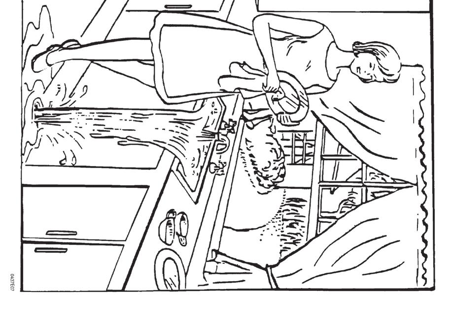
18 NIH STROKE SCALE (NIHSS) CONT D 5. Arm Motor* Alternately position patient s arms. Extend each arm with palms down (90 if sitting, 45 if supine). 0 = No drift 1 = Drift 2 = Some effort vs gravity 3 = No effort vs gravity 4 = No movement UN = Amputation or joint fusion * Test each arm in turn (nonparetic arm first). Drift is scored if arm falls before 10 seconds. Score untestable (UN) only for patients with amputations or joint fusions of shoulder. 6. Leg Motor* Alternately position patient s legs. Extend each leg (30, always while supine). 0 = No drift 1 = Drift 2 = Some effort vs gravity 3 = No effort vs gravity 4 = No movement UN = Amputation or joint fusion * Test each leg in turn (nonparetic leg first). Drift is scored if leg falls before 5 seconds. Score UN only for patients with amputations or joint fusions of hip. 9. Best Language* Using pictures and a sentence list (see reverse), ask the patient to: Describe what you see in this picture. Name the items in this picture. Read these sentences. 0 = No aphasia 1 = Mild-to-moderate aphasia 2 = Severe aphasia 3 = Mute, global aphasia * Patients with visual loss can be asked to identify and describe objects placed in the hand. Intubated patients should be asked to write their answers. The examiner must choose a score for stuporous or uncooperative patients. Comatose patients (item 1a = 3) are scored 3. A score of 3 is only given if the patient is mute and unable to follow one-step commands. 10. Dysarthria* Using a simple word list (see reverse), ask the patient to: Read these words or Repeat these words. 0 = Normal articulation 1 = Mild-to-moderate dysarthria 2 = Severe dysarthria UN = Intubated or other physical barrier * Patients with severe aphasia can be scored based on the clarity of articulation of their spontaneous speech. Score UN only for patients who are intubated or have other physical barriers to speech. Do not tell patients why they are being tested.
19 7. Limb Ataxia* Ask patient (eyes open) to: Touch your finger to your nose. Touch your heel to your shin. 0 = Absent 1 = Present in one limb 2 = Present in two or more limbs UN = Amputation or joint fusion * Perform finger-nose and heel-shin tests on both sides to determine unilateral cerebellar lesion. Score 0 for patients who are paralyzed or cannot understand. Score 1 or 2 only if ataxia is disproportionate to weakness. Score UN only for patients with amputations or joint fusions. 8. Sensory* Test as many body parts as possible (arms [not hands], legs, trunk, face) for sensation using pinprick or noxious stimulus (in the obtunded or aphasic patient). 0 = Normal 1 = Mild-to-moderate sensory loss 2 = Severe-to-total sensory loss * Score sensory loss due to stroke only. Stuporous and aphasic patients are scored 0 or 1. Patients with brainstem stroke and bilateral sensory loss, quadriplegic patients who do not respond, and comatose patients (item 1a = 3) are scored 2. A score of 2 is only given when severe or total sensory loss is demonstrated. 11. Extinction and Inattention* Sufficient information to determine these scores may have been obtained during the prior testing. 0 = No abnormality 1 = Visual, tactile, auditory, spatial, or personal inattention 2 = Profound hemi-inattention or extinction to more than one modality * Lack of patient response and inattention may already be evident from the previous items. Score 0 if the patient has a severe visual loss preventing visual double simultaneous stimulation, but the response to cutaneous stimuli is normal, or if the patient has aphasia but does not appear to attend to both sides. The presence of visual spatial attention or anosognosia may also be evidence of abnormality. 16
20
21 18
22
23 20
24 You know how. Down to earth. I got home from work. Near the table in the dining room. They heard him speak on the radio last night.
25 MAMA TIP-TOP FIFTY-FIFTY THANKS HUCKLEBERRY BASEBALL PLAYER 22
26 LEARN THE SIGNS OF STROKE is it drooping? can you raise both? is it slurred or jumbled? to call right away. ACT BECAUSE THE QUICKE R YOU ACT, THE MORE OF THE PERSON YOU SAVE. Heart and Stroke Foundation of Canada, 2014
27 To learn more about the Signs of Stroke visit heartandstroke.ca/fast 24
28 The information contained in this document supports the Canadian Stroke Best Practice Recommendations For detailed recommendations and updates visit: strokebestpractices.ca HP6311E V3.4
what do the numbers really mean? NIHSS Timothy Hehr, RN MA Stroke Program Outreach Coordinator Allina Health
 what do the numbers really mean? NIHSS Timothy Hehr, RN MA Stroke Program Outreach Coordinator Allina Health NIHSS The National Institutes of Health Stroke Scale (NIHSS) is a tool used to objectively quantify
SARASOTA MEMORIAL HOSPITAL. NURSING PROCEDURE NATIONAL INSTITUTE OF HEALTH STROKE SCALE (neu04) Nursing
 Nursing") SARASOTA MEMORIAL HOSPITAL TITLE: ISSUED FOR: NURSING PROCEDURE NATIONAL INSTITUTE OF HEALTH Nursing DATE: REVIEWED: PAGES: PS1094 7/01 3/18 1 of 5 RESPONSIBILITY: RN, LPN PURPOSE: OBJECTIVE: DEFINITION:
SARASOTA MEMORIAL HOSPITAL TITLE: ISSUED FOR: NURSING PROCEDURE NATIONAL INSTITUTE OF HEALTH Nursing DATE: REVIEWED: PAGES: PS1094 7/01 3/18 1 of 5 RESPONSIBILITY: RN, LPN PURPOSE: OBJECTIVE: DEFINITION:
Assessing the Stroke Patient. Arlene Boudreaux, MSN, RN, CCRN, CNRN
 Assessing the Stroke Patient Arlene Boudreaux, MSN, RN, CCRN, CNRN Cincinnati Pre-Hospital Stroke Scale May be done by EMS o One of many o F facial droop on one side o A arm drift (hold a pizza box, close
Inside Your Patient s Brain Michelle Peterson, APRN, CNP Centracare Stroke and Vascular Neurology
 Inside Your Patient s Brain Michelle Peterson, APRN, CNP Centracare Stroke and Vascular Neurology Activity Everyone stand up, raise your right hand, tell your neighbors your name 1 What part of the brain
Stroke School for Internists Part 1
 Stroke School for Internists Part 1 November 4, 2017 Dr. Albert Jin Dr. Gurpreet Jaswal Disclosures I receive a stipend for my role as Medical Director of the Stroke Network of SEO I have no commercial
CVA. Alison Atwater PA-C
 CVA Alison Atwater PA-C Types of CVAs Ischemic strokes 80% of strokes 2/3 are thrombotic 1/3 are embolic emboli from the heart or arteries feeding the brain such as carotids, vertebral and basilar etc
NIHSS. Category Scale Definition Date/Time Date/Time Date/Time. Score Initial. Drip & Ship Protocol. Initials: Signature: Initials: Signature:
 NIHSS 1a. Level of Consciousness (Alert, drowsy, etc.) Category Scale Definition Date/Time Date/Time Date/Time 1b. LOC Question (Month, age) 1c. LOC Commands (Open, close eyes, make fist, let go) 2. Best
Course: Physical Assessment II Date: October 17, 2008 Doc: Practice Quiz 1
 Course: Physical Assessment II Date: October 17, 2008 Doc: Practice Quiz 1 This is the practice quiz we did in Class 4. The answers are at the end of the quiz should you wish to test yourself. Complete
Neurological Assessment
 Neurological Assessment Name: Age: Gender: Date: History Review of history related to neurological system YES/NO If YES, provide details: General Neurological Mental Illness Neurological disease Severe
Thrombolysis Assessment
 Thrombolysis Assessment Brief Clinical Summary of symptom onset of arrival of patient of assessment BP GCS BM If BM
Neurological Assessment. Lecture 8
 Neurological Assessment Lecture 8 Nervous System Central Nervous System Brain Spinal cord Peripheral Nervous System Cranial nerves Spinal nerves Central Nervous System-Brain Central Nervous System-Spinal
Stroke Transfer Checklist
 Stroke Transfer Checklist When preparing to transfer an acute stroke patient to the UF Health Shands Comprehensive Stroke Center, please make every attempt to include the following information: Results
The Neurologic Examination: High-Yield Strategies
 The Neurologic Examination: High-Yield Strategies S. Andrew Josephson, MD Assistant Professor, Department of Neurology Divisions of Neurovascular and Behavioral Neurology University of California San Francisco
Lab Activity 19 & 20. Cranial Nerves General Senses. Portland Community College BI 232
 Lab Activity 19 & 20 Cranial Nerves General Senses Portland Community College BI 232 Cranial Nerves Nerves that originate from the brain rather than the spinal cord Part of the peripheral nervous system
Examination and Diseases of Cranial Nerves
 Cranial nerve evaluation is an important part of a neurologic exam. There are some differences in the assessment of cranial nerves with different species but the general principles are the same. Going
Unit VIII Problem 3 Neuroanatomy: Brain Stem, Cranial Nerves and Scalp
 Unit VIII Problem 3 Neuroanatomy: Brain Stem, Cranial Nerves and Scalp - Brain stem: It is connected to the cerebellum and cerebral hemispheres. Rostral end of brain stem: diencephalon is the area which
Upper and Lower Motoneurons for the Head Objectives
 Upper and Lower Motoneurons for the Head Objectives Know the locations of cranial nerve motor nuclei Describe the effects of motor cranial nerve lesions Describe how the corticobulbar tract innervates
Bellringer: The central nervous system is comprised of: What is the name of the outermost layer of the brain? a. Brain. b.
 Bellringer: The central is comprised of: a. Brain b. Spinal cord c. Sensory receptors d. Both a and b What is the name of the outermost layer of the brain? a. Pia mater b. Dura mater c. Arachnoid d. Pons
Cranial Nerves Exam. 1. To learn how to examine the functions of the 12 pairs of cranial nerves.
 Cranial Nerves Exam [Purpose] 1. To learn how to examine the functions of the 12 pairs of cranial nerves. 2. To understand the function of the 12 pairs of cranial nerves. [Principle] The cranial nerves
I. Review of Cerebral Anatomy A. Meninges - Coverings of layers of tissue within the cranium. 1. Dura Mater - outer covering Tough Mother 2.
 1 I. Review of Cerebral Anatomy A. Meninges - Coverings of layers of tissue within the cranium. 1. Dura Mater - outer covering Tough Mother 2. Arachnoid Mater - middle covering 3. Pia mater - inner covering
WHAT ARE the COMPONENTS OF THE NERVOUS SYSTEM?
 The Nervous System WHAT ARE the COMPONENTS OF THE NERVOUS SYSTEM? The nervous system is made of: the brain & the spinal cord the nerves the senses There are lots of proteins and chemicals in your body
The Neurological System. Neurological Exam 5 Components. Mental Status Examination
 The Neurological System 1 Neurological Exam 5 Components Mental status Cranial nerves Reflexes Motor- includes Cerebellar function Sensory 2 Mental Status Examination Examination - ABCT Appearance Behavior
The Nervous System: Central Nervous System
 The Nervous System: Central Nervous System I. Anatomy of the nervous system A. The CNS & the body by: 1. monitoring of the body 2. & information between parts of the body 3. acting as a to gather, store,
Brain and Cranial Nerves (Ch. 15) Human Anatomy lecture. caudal = toward the spinal cord)
 Human Anatomy lecture. caudal = toward the spinal cord)") Insight: Some cranial nerve disorders Brain and Cranial Nerves (Ch. 15) Human Anatomy lecture I. Overview (Directional terms: rostral = toward the forehead caudal = toward the spinal cord) A. 3 Major parts
Insight: Some cranial nerve disorders Brain and Cranial Nerves (Ch. 15) Human Anatomy lecture I. Overview (Directional terms: rostral = toward the forehead caudal = toward the spinal cord) A. 3 Major parts
The Neurologic Examination: High-Yield Strategies
 The Neurologic Examination: High-Yield Strategies S. Andrew Josephson, MD Examination Approach Two types of neurologic examinations 1. Screening Examination 2. Testing Hypotheses Select high-yield tests
Stroke: Every Minute Counts! Primary Stroke Center, Ingalls Memorial Hospital
 Stroke: Every Minute Counts! Primary Stroke Center, Ingalls Memorial Hospital Objectives Describe the A & P of the nervous system Outline pathophysiological changes in the nervous system that may alter
Higher Cortical Function
 Emilie O Neill, class of 2016 Higher Cortical Function Objectives Describe the association cortical areas processing sensory, motor, executive, language, and emotion/memory information (know general location
b. The groove between the two crests is called 2. The neural folds move toward each other & the fuse to create a
 Chapter 13: Brain and Cranial Nerves I. Development of the CNS A. The CNS begins as a flat plate called the B. The process proceeds as: 1. The lateral sides of the become elevated as waves called a. The
CNS composed of: Grey matter Unmyelinated axons Dendrites and cell bodies White matter Myelinated axon tracts
 CNS composed of: Grey matter Unmyelinated axons Dendrites and cell bodies White matter Myelinated axon tracts The Brain: A Quick Tour Frontal Lobe Control of skeletal muscles Personality Concentration
Stroke: clinical presentations, symptoms and signs
 Stroke: clinical presentations, symptoms and signs Professor Peter Sandercock University of Edinburgh EAN teaching course Burkina Faso 8 th November 2017 Clinical diagnosis is important to Ensure stroke
Brain and spinal nerve. By: shirin Kashfi
 Brain and spinal nerve By: shirin Kashfi Nervous system: central nervous system (CNS) peripheral nervous system (PNS) Brain (cranial) nerves Spinal nerves Ganglions (dorsal root ganglions, sympathetic
CRANIAL NERVE TESTING FOR THE PRIMARY CARE OPTOMETRIST
 CRANIAL NERVE TESTING FOR THE PRIMARY CARE OPTOMETRIST Hannah Shinoda, OD Caroline Ooley, OD, FAAO Assistant Professors Pacific University College of Optometry The authors have no financial interest in
C h a p t e r PowerPoint Lecture Slides prepared by Jason LaPres North Harris College Houston, Texas
 C h a p t e r 15 The Nervous System: The Brain and Cranial Nerves PowerPoint Lecture Slides prepared by Jason LaPres North Harris College Houston, Texas Copyright 2009 Pearson Education, Inc., publishing
Nervous System: An Introduction. HAP Susan Chabot Lemon Bay High School
 Nervous System: An Introduction HAP Susan Chabot Lemon Bay High School Function of the Nervous System 3 overlapping functions SENSORY INPUT - Monitor changes inside and outside of the body; these changes
CRANIAL NERVES. Dr. Amani A. Elfaki Associate Professor Department of Anatomy
 CRANIAL NERVES Dr. Amani A. Elfaki Associate Professor Department of Anatomy LEARNING OBJECTIVES Named the cranial nerves Identify the funcunal component of each cranial nerve Identify the effect of each
Neurological Assessment Part 1
 Neurological Assessment Part 1 MOTOR EXAMINATION: Look at bulk, contour and symmetry of individual muscles: muscles of face upper arm arm thigh lower leg Look for atrophy--may help to localize the site
Chapter 10 The Nervous System: The Brain and Cranial Nerves
 Chapter 10 The Nervous System: The Brain and Cranial Nerves Copyright 2015 Wolters Kluwer Health Lippincott Williams & Wilkins Overview Key Terms aphasia corpus callosum meninges basal nuclei diencephalon
PHYSIOLOHY OF BRAIN STEM
 PHYSIOLOHY OF BRAIN STEM Learning Objectives The brain stem is the lower part of the brain. It is adjoining and structurally continuous with the spinal cord. 1 Mid Brain 2 Pons 3 Medulla Oblongata The
Peripheral Nervous System Dr. Gary Mumaugh
 Peripheral Nervous System Dr. Gary Mumaugh Spinal Nerves Overview Thirty-one pairs of spinal nerves are connected to the spinal cord No special names; numbered by level of vertebral column at which they
Cranial Nerves and Spinal Cord Flashcards
 1. Name the cranial nerves and their Roman numeral. 2. What is Cranial Nerve I called, and what does it 3. Scientists who are trying to find a way to make neurons divide to heal nerve injuries often study
Cranial Nerves VII to XII
 Cranial Nerves VII to XII MSTN121 - Neurophysiology Session 13 Department of Myotherapy Cranial Nerve VIII: Vestibulocochlear Sensory nerve with two distinct branches. Vestibular branch transmits information
Principles of Anatomy and Physiology
 Principles of Anatomy and Physiology 14 th Edition CHAPTER 14 The Brain and Cranial Nerves Introduction The purpose of the chapter is to: 1. Understand how the brain is organized, protected, and supplied
PHYSIOLOGY OF THE BRAIN STEM
 PHYSIOLOGY OF THE BRAIN STEM Dr Syed Shahid Habib Professor & Consultant Clinical Neurophysiology Dept. of Physiology College of Medicine & KKUH King Saud University OBJECTIVES At the end of this lecture
Nervous System. Student Learning Objectives:
 Nervous System Student Learning Objectives: Identify the primary parts of the neuron Identify the major structures of the central nervous system Identify the major structures of the peripheral nervous
Cerebrum. Impulses transmitted by:
 Sharon Niggemeier RN MSN Victoria Siegel RN, MSN, EdD Myung-Hee Pak, RN, MSN, CCRN Nervous system- divided into 2 structural parts Central Nervous System (CNS)- brain & spinal cord Peripheral Nervous System
A&P 1 Brain & Cranial Nerves Guide #1 - Pre-Lab Exercises
 A&P 1 Brain & Cranial Nerves Guide #1 - Pre-Lab Exercises In this "Pre-lab Guide", we will be looking at the brain & cranial nerves. This should be done before lab, so we don't waste time in lab! This
The NIHSS score is 4 (considering 2 pts for the ataxia involving upper and lower limbs.
 Neuroscience case 5 1. Speech comprehension, ability to speak, and word use were normal in Mr. Washburn, indicating that aphasia (cortical language problem) was not involved. However, he did have a problem
The Brain and Cranial Nerves Pg. 129
 The Brain and Cranial Nerves Pg. 129 Three Main Regions of the Brain Forebrain Cerbral hemispheres Diencephalon Midbrain Brain stem Hindbrain Pons Cerebellum Medulla oblongata Forebrain Interprets sensory
IMPAIRMENT OF THE NERVOUS SYSTEM
 IMPAIRMENT OF THE NERVOUS SYSTEM The following information provides criteria for the evaluation of permanent impairment resulting from dysfunction brain, spinal cord and cranial nerves and certain peripheral
/ / / / / / Hospital Abstraction: Stroke/TIA. Participant ID: Hospital Code: Multi-Ethnic Study of Atherosclerosis
 Multi-Ethnic Study of Atherosclerosis Participant ID: Hospital Code: Hospital Abstraction: Stroke/TIA History and Hospital Record 1. Was the participant hospitalized as an immediate consequence of this
Lab 16: PNS: Nerves and Autonomic NS Hamilton Answers to Pre- Lab Assignments
 Lab 16: PNS: Nerves and Autonomic NS Hamilton Answers to Pre- Lab Assignments Pre-Lab Activity 1: 1. a. olfactory nerve b. optic nerve c. oculomotor nerve d. abducens nerve e. trochlear nerve f. trigeminal
1. Processes nutrients and provides energy for the neuron to function; contains the cell's nucleus; also called the soma.
 1. Base of brainstem; controls heartbeat and breathing 2. tissue destruction; a brain lesion is a naturally or experimentally caused destruction of brain tissue 3. A thick band of axons that connects the
GLOSSARY. Active assisted movement: movement where the actions are assisted by an outside force.
 GLOSSARY The technical words used in this guide are listed here in alphabetic order. The first time one of these words is used in the guide, it is written in italics. Sometimes there is reference to a
Cranial Nerves. Steven McLoon Department of Neuroscience University of Minnesota
 Cranial Nerves Steven McLoon Department of Neuroscience University of Minnesota 1 Course News Change in Lab Sequence Week of Oct 2 Lab 5 Week of Oct 9 Lab 4 2 Sensory and Motor Systems Sensory Systems:
CN V! touch! pain! Touch! P/T!
 CN V! touch! pain! Touch! P/T! Visual Pathways! L! R! B! A! C! D! LT! E! F! RT! G! hypothalamospinal! and! ALS! Vestibular Pathways! 1. Posture/Balance!!falling! 2. Head Position! 3. Eye-Head Movements
The Brain and Cranial Nerves Pg Three Main Regions of the Brain. Forebrain
 The Brain and Cranial Nerves Pg. 129 Three Main Regions of the Brain Forebrain Cerbral hemispheres Diencephalon Midbrain Brain stem Hindbrain Pons Cerebellum Medulla oblongata Interprets sensory inputs
14 - Central Nervous System. The Brain Taft College Human Physiology
 14 - Central Nervous System The Brain Taft College Human Physiology Development of the Brain The brain begins as a simple tube, a neural tube. The tube or chamber (ventricle) is filled with cerebrospinal
The Nervous System. Divisions of the Nervous System. Branches of the Autonomic Nervous System. Central versus Peripheral
 The Nervous System Divisions of the Nervous System Central versus Peripheral Central Brain and spinal cord Peripheral Everything else Somatic versus Autonomic Somatic Nerves serving conscious sensations
The High-Yield Neurologic Examination
 The High-Yield Neurologic Examination S. Andrew Josephson MD Carmen Castro Franceschi and Gladyne K. Mitchell Neurohospitalist Distinguished Professor Chair, Department of Neurology Director, Neurohospitalist
Basic Brain Structure
 The Human Brain Basic Brain Structure Composed of 100 billion cells Makes up 2% of bodies weight Contains 15% of bodies blood supply Uses 20% of bodies oxygen and glucose Brain Protection Surrounded by
The Nervous System PART B
 7 The Nervous System PART B PowerPoint Lecture Slide Presentation by Jerry L. Cook, Sam Houston University ESSENTIALS OF HUMAN ANATOMY & PHYSIOLOGY EIGHTH EDITION ELAINE N. MARIEB Central Nervous System
Nervous System: Part IV The Central Nervous System The Brain
 Nervous System: Part IV The Central Nervous System The Brain Can you survive when part of your brain is destroyed? 2 Essential Knowledge 3.D.2 2. Cells communicate with each other through direct contact
Nervous System The Brain and Spinal Cord Unit 7b
 Nervous System The Brain and Spinal Cord Unit 7b Chetek High School Mrs. Michaelsen 9.12 Meninges A. Meninges 1. The organs of the CNS are covered by membranes a. The meninges are divided into 3 layers:
Chapter 18: The Brain & Cranial Nerves. Origin of the Brain
 Chapter 18: The Brain & Cranial Nerves BIO 218 Fall 2015 Origin of the Brain The brain originates from a structure called the neural tube, which arises during a developmental stage called neurulation.
Parts of the Brain. Hindbrain. Controls autonomic functions Breathing, Heartbeat, Blood pressure, Swallowing, Vomiting, etc. Upper part of hindbrain
 Parts of the Brain The human brain is made up of three main parts: 1) Hindbrain (or brainstem) Which is made up of: Myelencephalon Metencephalon 2) Midbrain Which is made up of: Mesencephalon 3) Forebrain
3/20/13. :: Slide 1 :: :: Slide 39 :: How Is the Nervous System Organized? Central Nervous System Peripheral Nervous System and Endocrine System
 :: Slide 1 :: :: Slide 39 :: How Is the Nervous System Organized? Central Nervous System Peripheral Nervous System and Endocrine System The nervous system is organized into several major branches, each
Nervous System. The Peripheral Nervous System Agenda Review of CNS v. PNS PNS Basics Cranial Nerves Spinal Nerves Reflexes Pathways
 Nervous System Agenda Review of CNS v. PNS PNS Basics Cranial Nerves Spinal Nerves Sensory Motor Review of CNS v. PNS Central nervous system (CNS) Brain Spinal cord Peripheral nervous system (PNS) All
The Deconstructed Neurological Examination
 The Deconstructed Neurological Examination Marguerite Knipe, DVM, Diplomate ACVIM (Neurology) I. MENTATION: Normal, Quiet, Obtunded (mild, moderate, severe), Stuporous, Comatose Define stuporous and comatose.
Functional Distinctions
 Functional Distinctions FUNCTION COMPONENT DEFICITS Start Basal Ganglia Spontaneous Movements Move UMN/LMN Cerebral Cortex Brainstem, Spinal cord Roots/peripheral nerves Plan Cerebellum Ataxia Adjust Cerebellum
10/13/2017. AllinaHealthSystems. Stroke Recognition Sandra K Hanson, MD Medical Director United Hospital Stroke Program
 Agenda Stroke Recognition Sandra K Hanson, MD Medical Director United Hospital Stroke Program Vascular anatomy NIH Stroke Scale Recognizing the hard to recognize Distinguishing stroke mimics 1 Symptoms
ID # COMPLETED: YES 1 DATE NO 2
 ID # COMPLETED: YES 1 DATE NO 2 NEUROLOGICAL EXAM "Normal, Abnormal, Other, Can't execute or Missing for each question. If you circle "Abnormal" or Other, also check the appropriate reason why or explain
Lecture 35 Association Cortices and Hemispheric Asymmetries -- M. Goldberg
 Lecture 35 Association Cortices and Hemispheric Asymmetries -- M. Goldberg The concept that different parts of the brain did different things started with Spurzheim and Gall, whose phrenology became quite
Spinal Cord: Clinical Applications. Dr. Stuart Inglis
 Spinal Cord: Clinical Applications Dr. Stuart Inglis Tabes dorsalis, also known as syphilitic myelopathy, is a slow degeneration (specifically, demyelination) of the nerves in the dorsal funiculus of the
The Human Brain: Anatomy, Functions, and Injury
 The Human Brain: Anatomy, Functions, and Injury Main Menu Brain Anatomy Brain Functions Injury Mechanisms Brain Anatomy Menu Skull Anatomy Interior Skull Surface Blood Vessels of the Brain Arteries of
Neuro Exam Explained
 Neuro Exam Explained Michael Nelson M.D. Providence Neurological Specialties East Primary Care Conference October 26 rd, 2017 Michael Nelson M.D. Medical School: University of Missouri-Columbia Residency:
INTRODUCTION: ANATOMY UNDERLYING CLINICAL TESTS OF CRANIAL NERVES
 INTRODUCTION: ANATOMY UNDERLYING CLINICAL TESTS OF CRANIAL NERVES CRANIAL NERVE I - OLFACTORY I - OLFACTORY NERVE - SMELL TEST: SMELL ODORS (note: not ammonia; pain in nasal cavity CN5 DAMAGE: LOSS OF
Neurological Examination
 Neurological Examination Charles University in Prague 1st Medical Faculty and General University Hospital Neurological examination: Why important? clinical history taking and bedside examination: classical
ACCESS CENTER:
 ACCESS CENTER: 1-877-367-8855 Emergency Specialty Services: BRAIN ATTACK Criteria: Stroke symptom onset time less than 6 hours Referring Emergency Department Patient Information Data: Time last known normal:
Anatomy of Nervous System. Neurological Assessment. Brain. Brain. Spinal Cord. Spinal Cord 03/23/2010. Central. Peripheral
 Anatomy of Nervous System Neurological Assessment Central Brain Spinal cord Peripheral Spinal Nerves Brain Cerebral cortex ( rind ) gray matter Frontal Parietal Temporal Occipital Wernicke s area receptive
Classification of Neurons
 HASPI Medical Anatomy & Physiology 11b Lab Activity Name(s): Period: Date: Neurons The neuron is the cell of the nervous system capable of conducting and sending nerve impulses throughout the body. All
Neuroanatomy of a Stroke. Joni Clark, MD Professor of Neurology Barrow Neurologic Institute
 Neuroanatomy of a Stroke Joni Clark, MD Professor of Neurology Barrow Neurologic Institute No disclosures Stroke case presentations Review signs and symptoms Review pertinent exam findings Identify the
NEUROLOGY CLERKSHIP CORE CURRICULUM GUIDELINES
 NEUROLOGY CLERKSHIP CORE CURRICULUM GUIDELINES Endorsed by the following organizations - October 2000: American Academy of Neurology Association of University Professors of Neurology American Neurological
Learning Objectives.
 Emilie O Neill, class of 2016 Learning Objectives 1. Describe the types of deficits that occur with lesions in association areas including: prosopagnosia, neglect, aphasias, agnosia, apraxia 2. Discuss
Structure, function and assessments of cranial nerves: Part 1 (CN 1-7) MSTN121 - Neurophysiology Session 12 Department of Myotherapy
 MSTN121 - Neurophysiology Session 12 Department of Myotherapy") Structure, function and assessments of cranial nerves: Part 1 (CN 1-7) MSTN121 - Neurophysiology Session 12 Department of Myotherapy Session objectives List the four functions of the cranial nerves (CNs).
Structure, function and assessments of cranial nerves: Part 1 (CN 1-7) MSTN121 - Neurophysiology Session 12 Department of Myotherapy Session objectives List the four functions of the cranial nerves (CNs).
Neural Basis of Motor Control
 Neural Basis of Motor Control Central Nervous System Skeletal muscles are controlled by the CNS which consists of the brain and spinal cord. Determines which muscles will contract When How fast To what
Clinical Learning Exercise #1
 Clinical Learning Exercise #1 Exercise: We are going to assume nothing is wrong with the peripheral nervous system and attempt to identify the central nervous system anatomical location for the following
Homework Week 2. PreLab 2 HW #2 Synapses (Page 1 in the HW Section)
") Homework Week 2 Due in Lab PreLab 2 HW #2 Synapses (Page 1 in the HW Section) Reminders No class next Monday Quiz 1 is @ 5:30pm on Tuesday, 1/22/13 Study guide posted under Study Aids section of website
Homework Week 2 Due in Lab PreLab 2 HW #2 Synapses (Page 1 in the HW Section) Reminders No class next Monday Quiz 1 is @ 5:30pm on Tuesday, 1/22/13 Study guide posted under Study Aids section of website
THE CENTRAL NERVOUS SYSTEM. The Brain & Spinal Cord
 THE CENTRAL NERVOUS SYSTEM The Brain & Spinal Cord Review: Nervous System Parallel Distributed Processing Composition of the CNS Nuclei: Clusters of neurons in the CNS ( neighborhoods ) Fiber Tracts/Pathways:
HISTORY TAKING ON NERVOUS SYSTEM. Dr. Amitesh Aggarwal
 HISTORY TAKING ON NERVOUS SYSTEM Dr. Amitesh Aggarwal General points History of neurological symptoms should also be taken from patient and close relative or friend Memory loss, intoxication, aphasia Patient
Lesson 14. The Nervous System. Introduction to Life Processes - SCI 102 1
 Lesson 14 The Nervous System Introduction to Life Processes - SCI 102 1 Structures and Functions of Nerve Cells The nervous system has two principal cell types: Neurons (nerve cells) Glia The functions
M555 Medical Neuroscience Lab 1: Gross Anatomy of Brain, Crainal Nerves and Cerebral Blood Vessels
 M555 Medical Neuroscience Lab 1: Gross Anatomy of Brain, Crainal Nerves and Cerebral Blood Vessels Anatomical Directions Terms like dorsal, ventral, and posterior provide a means of locating structures
Examination Approach. Examination Approach. Case 1: Mental Status. The Neurological Exam In the ICU: High Yield Techniques 5/8/2015
 The Neurological Exam In the ICU: High Yield Techniques Examination Approach Two types of neurologic examinations 1. Screening Examination 2. Testing Hypotheses Select high-yield tests and techniques S.
Cranial Nerve: eyelid and eyeball movement innervates superior oblique turns eye downward and laterally chewing face & mouth touch & pain
 Cranial Nerves Cranial Nerve: I Olfactory II Optic III Oculomotor IV Trochlear V Trigeminal VI Abducens VII Facial VIII Vestibulocochlear (auditory) IX Glossopharyngeal X Vagus XI Spinal Accessory XII
4 main parts 1) Cerebrum 2) Diencephalon 3) Brain stem 4) Cerebellum
 Cerebrum 2) Diencephalon 3) Brain stem 4) Cerebellum") 4 main parts 1) Cerebrum 2) Diencephalon 3) Brain stem 4) Cerebellum White Matter = myelinated tracts or nerves Gray Matter = unmyelinated tracts or nerves Brain: gray matter on outside, white matter inside
4 main parts 1) Cerebrum 2) Diencephalon 3) Brain stem 4) Cerebellum White Matter = myelinated tracts or nerves Gray Matter = unmyelinated tracts or nerves Brain: gray matter on outside, white matter inside
PERIPHERAL NERVOUS SYSTEM
 CHAPTER 13 PERIPHERAL NERVOUS SYSTEM Functional division of nervous system = afferent info to the CNS ascending spinal cord = efferent info from CNS descending spinal cord somatic skin, muscles visceral
Neural Integration I: Sensory Pathways and the Somatic Nervous System
 15 Neural Integration I: Sensory Pathways and the Somatic Nervous System PowerPoint Lecture Presentations prepared by Jason LaPres Lone Star College North Harris An Introduction to Sensory Pathways and
BRAIN STEM CASE HISTORIES CASE HISTORY VII
 463 Brain stem Case history BRAIN STEM CASE HISTORIES CASE HISTORY VII A 60 year old man with hypertension wakes one morning with trouble walking. He is feeling dizzy and is sick to his stomach. His wife
Objectives. Stroke Facts 2/27/2015. EMS in Stroke Care: A Critical Partnership
 EMS in Stroke Care: A Critical Partnership Spokane County EMS Objectives Identify the types and time limitations for acute ischemic stroke treatment options Identify the importance of early identification
Review on Nervous System, Senses and Musculoskeletal System
 Review on Nervous System, Senses and Musculoskeletal System Looking for answers? Visit http://msjadah.weebly.com 1. The Nervous System a) What is the function of nervous system? The nervous system receives,