NEUROVASCULAR ANATOMY
|
|
|
- Emil Russell
- 5 years ago
- Views:
Transcription

1 E. Michael Harned, M.D. Assistant Professor of Clinical Radiology ndiana University School of Medicine have nothing to disclose Neurovascular Magnetic Resonance Angiography And Magnetic Resonance Venography ACKNOWLEDGEMENTS 1. Daroff RB, Fenichel GM, Jankovic J, Mazziotta JC: Bradley s Neurology in Clinical Practice, 6 th ed. Saunders 2. Anderson CM, Edelman RR, Turski PA: Clinical Magnetic Resonance Angiography. Raven Press For neurovascular imaging, MRA has largely been replaced by CTA due to its higher spatial resolution and relatively fewer artifacts. However, MRA may still be desired in certain situations: Patients with a contraindication to iodinated contrast administration: history of anaphylactoid reaction, severe asthma, renal insufficiency, multiple myeloma Lack of venous access Patients having an MR of the brain, neck, or cervical spine where it is desired to get vascular imaging done at the same time- killing 2 birds with 1 stone NEUROVASCULAR ANATOMY 1


")

 -.")


2 nt. carot id Ant. communicat ing Ant. cerebral ATterial eircle Posttrior inferihr cc1 ehellar Extemal acoustic meatus Superior bulb of JV nferior petrosal smus Occtprtal vein - ) nternal jugular vein (JV - Factal vein - Lingual vein mal jugular vein (EJV) -. nferior bulb and valve of JV \ - Ant erior jugular vetn - Sternoclavtcular JOint Right brachrocephahc vem...;.,. Superior vena cava 2


 are able to generate signal May be performed")


3 wpt!- t lo t\ ' " ' "..._. 4/17/2014 _,jll 1 e " ' " h,...ton MRA Time-Of-Flight (TOF) Relies on repeated saturation pulses to a slice or volume so that background tissue is suppressed and only new spins (flow) are able to generate signal May be performed as a 2D or 3D technique.,.. C'l lij liu 0 C:n«fniOB TECHNQUES 2D examines a single slice at a time and then stacks them 3D examines a slab and uses phase-encoding along the Z-axis 3D TOF allows better spatial resolution but is more susceptible to signal loss due to in-slab flow 0 h :,.,...'N il " O>'t EL SEV ER. NC - NETERMAGES.COM RF Pulse Z Gradient,,-,., ' ',,' ' \, \ CJ \._. SATURATON NCREASES WTH: Shorter TR Larger flip angle Y Gradient 0 Gradient r ' Multiple repetitions Longer T1 c 3


4 -----tu _tion 18 nd nflow of Magnetized Blood into 3D Slab Projected Plane Acquir d Slice a a/sine 4


 points of k-space are filled first, at time of")
5 Phase-Contrast Relies on bipolar gradient pulse which causes phase shift for flowing spins but no phase shift for stationary spins time Excellent background suppression No need for saturation band Also provides information on flow direction May be performed as a 2D or 3D technique Statiollary a p l aeaamulale pit... lobe and tiled wrap l t d arauad _..._ MJM.aamuladoa durln1111e LDM loije.,.._ r 3D Phase-Contrast allows better spatial resolution and provides flow direction information along all 3 axes Contrast-Enhanced MRA Really a form of 3D TOF MRA but uses gadolinium to increase intravascular signal and overcome problem of signal loss due to in-slab flow mages acquired quickly while a bolus of gadolinium is in the vessels of interest Different physics- uses elliptical-centric k-space filling: Central (most important) points of k-space are filled first, at time of greatest intravascular gadolinium concentration May acquire preinjection source images as mask for subtraction- Digital Subtraction MRA 5


6 ATHEROSCLEROTC STENOTC DSEASE WHY T MATTERS (all numbers approximate and for United States) 795,000 people suffer new or recurrent stroke each year 1 stroke every 40 seconds Stroke kills more than 137,000 people per year. 1 stroke death every 4 minutes #4 cause of death 1 in every 18 deaths Stroke death rates per year per 100,000 population: 41.7 for white males 41.1 for white females 67.7 for black males 57.0 for black females Americans pay about $73.7 billion per year for stroke-related medical costs and disability Likely Histories/ndications: WHAT THE RADOLOGST NEEDS FROM YOU 3D TOF MRA circle of Willis- source images Stroke, CVA, or TA Specific neurologic deficit- hemiparesis, aphasia, facial numbness, etc. Carotid bruit CA stenosis on carotid ultrasound 3D TOF MRA of the circle of Willis Contrast-enhanced MRA of the arteries of the neck (Often a good idea to do a 2D TOF MRA of the neck first in case there is a problem with the gadolinium bolus- There is no going back and doing this once contrast has been injected) Please send both sets of source images as well as rotating segmented MPs of right carotid, left carotid, and vertebrobasilar for circle of Willis and for neck 6


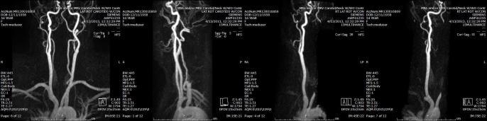


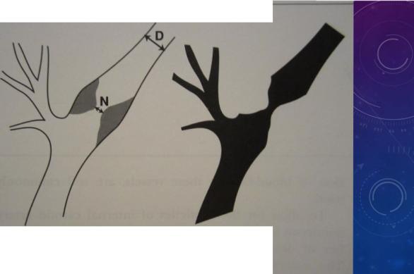
7 3D TOF MRA circle of Willis- MPs Contrast-enhanced MRA neck- source images Contrast-Enhanced MRA neck- MPs 2D TOF MRA neck- source images 2D TOF MRA neck- MPs Carotid stenosis%= (1 Nto) x 100 7


 and confirmed by")


8 B Figure 1. A 58-year-old man with truncus bicaroticus as normal variant that is displayed both on MRA and DSA (a and b; arrow heads), and 90% diameter stenosis of the right CA diagnosed on MRA (c; bold arrow) and confirmed by DSA (d; bold arrow). Willinek W A et al. Stroke. 2005;36:38-43 Copyright American Heart Association, nc. All rights reserved. 8
 and DSA image (b)")
 of the right")


9 Figure 2. Enlarged MP image from CE MRA (a) and DSA image (b) of the circle of Willis of a 44- year-old man: 50% stenosis in the M1 segment (arrow heads) of the right MCA was concordantly diagnosed on MRA and DSA. NON-ATHEROSCLEROTC STENOSS Willinek W A et al. Stroke. 2005;36:38-43 Likely Histories/ndications: Trauma Neck pain Specific neurologic deficit Drug abuse Protocol for evaluation same as for atherosclerotic stenosis 9
, but DSA revealed a thrombus causing a 55% diameter stenosis (b; arrow).")


10 Figure 4. A 61-year-old man patient with subacute right MCA territory infarction. 75% stenosis of right CA was diagnosed on CE MRA (a; arrow), but DSA revealed a thrombus causing a 55% diameter stenosis (b; arrow). From: Cocaine-nduced Cerebral Vasoconstriction Detected in Humans With Magnetic Resonance Angiography JAMA. 1998;279(5): doi: /jama Moyamoya Disease Disease where vessels at the base of the brain become constricted- Most commonly distal internal carotid arteries but may also involve proximal middle and anterior cerebral arteries Collateral circulation develops around the blocked vessels, but the collateral vessels are small, weak, and prone to hemorrhage On angiography, the collateral vessels resemble a puff of smoke - Moyamoya in Japanese Patients present with TAs, strokes, headaches, and seizures Most common in women in their 20s and 30s Willinek W A et al. Stroke. 2005;36:38-43 Copyright American Heart Association, nc. All rights reserved. Figure Legend: Axial maximum intensity projection images at baseline (left) and20 minutes following intravenous cocaine (0.4 mg/kg) administration (right).cocaine induced a signal loss at distal segments of the middle cerebral arteries(upper arrowheads) and in the posterior cerebral arteries (lower arrowheads),indicative of vasoconstriction. A indicates anterior; P, posterior; L, left;and R, right. Scale bar = 1 cm. Copyright 2012 American Medical Date of download: 12/22/2013 Association. All rights reserved. Association with Down s syndrome, neurofibromatosis type 1, sickle cell disease, prior brain radiation Figure 3. A 45-year-old woman with Moyamoya disease. NTRACRANAL ANEURYSMS Willinek W A et al. Stroke. 2005;36:38-43 Copyright American Heart Association, nc. All rights reserved. 10


11 Likely Histories/ndications: Subarachnoid hemorrhage Headaches WHAT THE RADOLOGST NEEDS FROM YOU 3D TOF MRA of the circle of Willis No need for gadolinium 0 lrehijiiun ttl t:111 ( t W c 1nh :al n " on hrltttcq '" l :.l r Family history of intracranial aneurysms Autosomal dominant polycystic kidney disease No need to image the neck lll'lltttl ''f'rlm n 101 trr htll.u l'u'lrt N t,- ;, ii Ị 00. ELSEVER. NC - NETERMAGES.COM 11


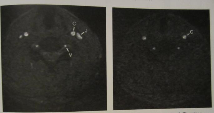
12 VENOUS PATHOLOGY Likely Histories/ndications: Possible venous sinus thrombosis- pregnancy, oral contraceptives, cancer, other hypercoagulable states Cavernous sinus syndrome Hyperdense dural venous sinus or intracranial vein on CT Venous infarct on CT or MR 12


13 2D TOF MRV head- source images 2D TOF MRV head- MPs WHAT THE RADOLOGST NEEDS FROM YOU 2D TOF of the head No need for gadolinium No need to image the neck 13
14 Thanks! Questions? 14
Essentials of Clinical MR, 2 nd edition. 99. MRA Principles and Carotid MRA
 99. MRA Principles and Carotid MRA As described in Chapter 12, time of flight (TOF) magnetic resonance angiography (MRA) is commonly utilized in the evaluation of the circle of Willis. TOF MRA allows depiction
Department of Radiology University of California San Diego. MR Angiography. Techniques & Applications. John R. Hesselink, M.D.
 Department of Radiology University of California San Diego MR Angiography Techniques & Applications John R. Hesselink, M.D. Vascular Imaging Arterial flow void Flow enhancement Gadolinium enhancement Vascular
Magnetic Resonance Angiography
 Magnetic Resonance Angiography 1 Magnetic Resonance Angiography exploits flow enhancement of GR sequences saturation of venous flow allows arterial visualization saturation of arterial flow allows venous
Non Contrast MRA. Mayil Krishnam. Director, Cardiovascular and Thoracic Imaging University of California, Irvine
 Non Contrast MRA Mayil Krishnam Director, Cardiovascular and Thoracic Imaging University of California, Irvine No disclosures Non contrast MRA-Why? Limitations of CTA Radiation exposure Iodinated contrast
MR Advance Techniques. Vascular Imaging. Class II
 MR Advance Techniques Vascular Imaging Class II 1 Vascular Imaging There are several methods that can be used to evaluate the cardiovascular systems with the use of MRI. MRI will aloud to evaluate morphology
Magnetic Resonance Imaging. Basics of MRI in practice. Generation of MR signal. Generation of MR signal. Spin echo imaging. Generation of MR signal
 Magnetic Resonance Imaging Protons aligned with B0 magnetic filed Longitudinal magnetization - T1 relaxation Transverse magnetization - T2 relaxation Signal measured in the transverse plane Basics of MRI
1Pulse sequences for non CE MRA
 MRI: Principles and Applications, Friday, 8.30 9.20 am Pulse sequences for non CE MRA S. I. Gonçalves, PhD Radiology Department University Hospital Coimbra Autumn Semester, 2011 1 Magnetic resonance angiography
Anatomic Evaluation of the Circle of Willis: MR Angiography versus Intraarterial Digital Subtraction Angiography
 Anatomic Evaluation of the Circle of Willis: MR Angiography versus Intraarterial Digital Subtraction Angiography K. W. Stock, S. Wetzel, E. Kirsch, G. Bongartz, W. Steinbrich, and E. W. Radue PURPOSE:
Imaging Acute Stroke and Cerebral Ischemia
 Department of Radiology University of California San Diego Imaging Acute Stroke and Cerebral Ischemia John R. Hesselink, M.D. Causes of Stroke Arterial stenosis Thrombosis Embolism Dissection Hypotension
Methods. Yahya Paksoy, Bülent Oğuz Genç, and Emine Genç. AJNR Am J Neuroradiol 24: , August 2003
 AJNR Am J Neuroradiol 24:1364 1368, August 2003 Retrograde Flow in the Left Inferior Petrosal Sinus and Blood Steal of the Cavernous Sinus Associated with Central Vein Stenosis: MR Angiographic Findings
MR Angiography in the evaluation of Lower Extremity Arterial Disease
 March 2001 MR Angiography in the evaluation of Lower Extremity Arterial Disease Ted Mau, Harvard Medical School Year III Objectives We will cover: Indications for Magnetic Resonance Angiography (MRA) Basic
Cerebral MR Venography: Normal Anatomy and Potential Diagnostic Pitfalls
 AJNR Am J Neuroradiol 21:74 78, January 2000 Cerebral MR Venography: Normal Anatomy and Potential Diagnostic Pitfalls R. H. Ayanzen, C. R. Bird, P. J. Keller, F. J. McCully, M. R. Theobald, and J. E. Heiserman
PTA 106 Unit 1 Lecture 3
 PTA 106 Unit 1 Lecture 3 The Basics Arteries: Carry blood away from the heart toward tissues. They typically have thicker vessels walls to handle increased pressure. Contain internal and external elastic
Advanced Vascular Imaging: Pulsatile Tinnitus. Disclosures. Pulsatile Tinnitus: Differential Diagnosis. Pulsatile Tinnitus
 Advanced Vascular Imaging: Pulsatile Tinnitus Patrick Turski MD, Zach Clark MD, Tabby Kennedy MD The Objectives of this presentation are to: Review the differential diagnosis of pulsatile tinnitus Discuss
41 year old female with headache. Elena G. Violari MD and Leo Wolansky MD
 41 year old female with headache Elena G. Violari MD and Leo Wolansky MD ? Dural Venous Sinus Thrombosis with Hemorrhagic Venous Infarct Acute intraparenchymal hematoma measuring ~3 cm in diameter centered
CT Versus MR for the Runoff
 CT Versus MR for the Runoff Robert R. Edelman, M.D. Dept. of Radiology NorthShore University HealthSystem Feinberg School of Medicine, Northwestern University Magnetic Resonance Computed Tomography Radio
Case Report 1. CTA head. (c) Tele3D Advantage, LLC
 Tele3D Advantage, LLC") Case Report 1 CTA head 1 History 82 YEAR OLD woman with signs and symptoms of increased intra cranial pressure in setting of SAH. CT Brain was performed followed by CT Angiography of head. 2 CT brain Extensive
Case Report 1 CTA head 1 History 82 YEAR OLD woman with signs and symptoms of increased intra cranial pressure in setting of SAH. CT Brain was performed followed by CT Angiography of head. 2 CT brain Extensive
Applicable Neuroradiology
 For the Clinical Neurology Clerkship LSU Medical School New Orleans Amy W Voigt, MD Clerkship Director Introduction The field of Radiology first developed following the discovery of X-Rays by Wilhelm Roentgen
Neuroradiology MR Protocols
 Neuroradiology MR Protocols Brain protocols N 1: Brain MRI without contrast N 2: Pre- and post-contrast brain MRI N 3 is deleted N 4: Brain MRI without or pre-/post-contrast (seizure protocol) N 5: Pre-
Comparison of Five Major Recent Endovascular Treatment Trials
 Comparison of Five Major Recent Endovascular Treatment Trials Sample size 500 # sites 70 (100 planned) 316 (500 planned) 196 (833 estimated) 206 (690 planned) 16 10 22 39 4 Treatment contrasts Baseline
Overview of imaging modalities for cerebral aneurysms
 Overview of imaging modalities for cerebral aneurysms Soroush Zaghi BIDMC PCE: Radiology August 2008 (Images from BIDMC, PACS.) Our Patient: Presentation Our patient is a 57 y/o woman who reports blowing
Field Strength. Regional Perfusion Imaging (RPI) matches cerebral arteries to flow territories
 matches cerebral arteries to flow territories") Field Strength Changing how the world looks at MR. Regional Perfusion Imaging (RPI) matches cerebral arteries to flow territories Research groups in Utrecht, Baltimore and Singapore collaborate on this
Field Strength Changing how the world looks at MR. Regional Perfusion Imaging (RPI) matches cerebral arteries to flow territories Research groups in Utrecht, Baltimore and Singapore collaborate on this
Flow Measurement Techniques from Medical Modalities for Computational Fluid Dynamics. Fukasaku K, Negoro M*
 Flow Measurement Techniques from Medical Modalities for Computational Fluid Dynamics # ## Fukasaku K, Negoro M* Riken, Department of Neurosurgery Motojima General Hospital Department of Neurosurgery Fujita
MR Imaging with the CCSVI or Haacke protocol
 MR Imaging with the CCSVI or Haacke protocol Reports from the Haacke protocol are often made available to the patients. The report consists of four major components: 1. anatomical images of major neck
Surface Appearance of the Vertebrobasilar Artery Revealed on Basiparallel Anatomic Scanning (BPAS) MR Imaging: Its Role for Brain MR Examination
 MR Imaging: Its Role for Brain MR Examination") AJNR Am J Neuroradiol 26:2508 2513, November/December 2005 Surface Appearance of the Vertebrobasilar Artery Revealed on Basiparallel Anatomic Scanning (BPAS) MR Imaging: Its Role for Brain MR Examination
AJNR Am J Neuroradiol 26:2508 2513, November/December 2005 Surface Appearance of the Vertebrobasilar Artery Revealed on Basiparallel Anatomic Scanning (BPAS) MR Imaging: Its Role for Brain MR Examination
HEAD AND NECK IMAGING. James Chen (MS IV)
") HEAD AND NECK IMAGING James Chen (MS IV) Anatomy Course Johns Hopkins School of Medicine Sept. 27, 2011 OBJECTIVES Introduce cross sectional imaging of head and neck Computed tomography (CT) Review head
HEAD AND NECK IMAGING James Chen (MS IV) Anatomy Course Johns Hopkins School of Medicine Sept. 27, 2011 OBJECTIVES Introduce cross sectional imaging of head and neck Computed tomography (CT) Review head
Brain AVM with Accompanying Venous Aneurysm with Intracerebral and Intraventricular Hemorrhage
 Cronicon OPEN ACCESS EC PAEDIATRICS Case Report Brain AVM with Accompanying Venous Aneurysm with Intracerebral and Intraventricular Hemorrhage Dimitrios Panagopoulos* Neurosurgical Department, University
Carotid Artery Disease and What s Pertinent JOSEPH A PAULISIN DO
 Carotid Artery Disease and What s Pertinent JOSEPH A PAULISIN DO Goal of treatment of carotid disease Identify those at risk of developing symptoms Prevent patients at risk from developing symptoms Prevent
Learning Objectives for Rotations in Vascular Surgery Year 3 Basic Clerkship
 Learning Objectives for Rotations in Vascular Surgery Year 3 Basic Clerkship CLINICAL PROBLEMS IN VASCULAR SURGERY 1. ABDOMINAL AORTIC ANEURYSM A 70 year old man presents in the emergency department with
NEURORADIOLOGY Part I
 NEURORADIOLOGY Part I Vörös Erika University of Szeged Department of Radiology SZEGED BRAIN IMAGING METHODS Plain film radiography Ultrasonography (US) Computer tomography (CT) Magnetic resonance imaging
Imaging of Cerebrovascular Disease
 Imaging of Cerebrovascular Disease A Practical Guide Val M. Runge, MD Editor-in-Chief of Investigative Radiology Institute for Diagnostic, Interventional, and Pediatric Radiology Inselspital, University
Imaging for Peripheral Vascular Disease
 Imaging for Peripheral Vascular Disease James G. Jollis, MD Director, Rex Hospital Cardiovascular Imaging Imaging for Peripheral Vascular Disease 54 year old male with exertional calf pain in his right
非對比劑與對比劑增強 MRA. 血管攝影與對比劑 A Course of MRI. 本週課程內容 -MR Angiography (MRA) Unenhanced MRA
 Unenhanced MRA") 本週課程內容 -MR Angiography (MRA) 血管攝影與對比劑 A Course of MRI 盧家鋒助理教授國立陽明大學物理治療暨輔助科技學系 alvin4016@ym.edu.tw 非對比劑增強 MRA(Unenhanced MRA) Time-of-flight (TOF) angiography Phase-contrast (PC) angiography 對比劑增強 MRA(Contrast-enhanced
本週課程內容 -MR Angiography (MRA) 血管攝影與對比劑 A Course of MRI 盧家鋒助理教授國立陽明大學物理治療暨輔助科技學系 alvin4016@ym.edu.tw 非對比劑增強 MRA(Unenhanced MRA) Time-of-flight (TOF) angiography Phase-contrast (PC) angiography 對比劑增強 MRA(Contrast-enhanced
Internal Carotid Artery Dissection
 May 2011 Internal Carotid Artery Dissection Carolyn April, HMS IV Agenda Presentation of a clinical case Discussion of the clinical features of ICA dissection Discussion of the imaging modalities used
Background. Recommendations for Imaging of Acute Ischemic Stroke: A Scientific Statement From the American Heart Association
 for Imaging of Acute Ischemic Stroke: A Scientific Statement From the American Heart Association An Scientific Statement from the Stroke Council, American Heart Association and American Stroke Association
Evaluation of Intracranial Vasculatures in Healthy Subjects with Arterial-Spin-Labeling-Based 4D-MR Angiography at 3T
 Magn Reson Med Sci, Vol. 15, No. 3, pp. 335 339, 2016 doi:10.2463/mrms.tn.2015-0081 TECHNICAL NOTE Evaluation of Intracranial Vasculatures in Healthy Subjects with Arterial-Spin-Labeling-Based 4D-MR Angiography
MRI Imaging of GP Medicare Eligible Conditions
 MRI Imaging of GP Medicare Eligible Conditions By Dr. Andrew Stuart Radiologist Sydney Adventist Hospital 0562/SAH/1112/SAH Learning Objectives Indications for GP referred Medicare eligible MRI scans MRI
HEAD/NECK VESSELS. Objectives
 Objectives Arterial Supply to Head and Neck Arteries to Head Surrounding Brain Common carotid arteries Arteries to Head Surrounding Brain External carotid arteries Arteries to Head Surrounding Brain External
NEURORADIOLOGY DIL part 4
 NEURORADIOLOGY DIL part 4 Strokes and infarcts K. Agyem MD, G. Hall MD, D. Palathinkal MD, Alexandre Menard March/April 2015 OVERVIEW Introduction to Neuroimaging - DIL part 1 Basic Brain Anatomy - DIL
[(PHY-3a) Initials of MD reviewing films] [(PHY-3b) Initials of 2 nd opinion MD]
![[(PHY-3a) Initials of MD reviewing films] [(PHY-3b) Initials of 2 nd opinion MD]](/thumbs/89/98619893.jpg "[(PHY-3a) Initials of MD reviewing films] [(PHY-3b) Initials of 2 nd opinion MD]") 2015 PHYSICIAN SIGN-OFF (1) STUDY NO (PHY-1) CASE, PER PHYSICIAN REVIEW 1=yes 2=no [strictly meets case definition] (PHY-1a) CASE, IN PHYSICIAN S OPINION 1=yes 2=no (PHY-2) (PHY-3) [based on all available
2015 PHYSICIAN SIGN-OFF (1) STUDY NO (PHY-1) CASE, PER PHYSICIAN REVIEW 1=yes 2=no [strictly meets case definition] (PHY-1a) CASE, IN PHYSICIAN S OPINION 1=yes 2=no (PHY-2) (PHY-3) [based on all available
CT INTERPRETATION COURSE
 CT INTERPRETATION COURSE Refresher Course ASTRACAT October 2012 Stroke is a Clinical Diagnosis A clinical syndrome characterised by rapidly developing clinical symptoms and/or signs of focal loss of cerebral
High Field MR of the Spine
 Department of Radiology University of California San Diego 3T for MR Applications Advantages High Field MR of the Spine Increased signal-to-noise Better fat suppression Increased enhancement with gadolinium
Carotid Artery Stenting
 Carotid Artery Stenting JESSICA MITCHELL, ACNP CENTRAL ILLINOIS RADIOLOGICAL ASSOCIATES External Carotid Artery (ECA) can easily be identified from Internal Carotid Artery (ICA) by noticing the branches.
OBJECTIVES. At the end of the lecture, students should be able to: List the cerebral arteries.
 DR JAMILA EL MEDANY OBJECTIVES At the end of the lecture, students should be able to: List the cerebral arteries. Describe the cerebral arterial supply regarding the origin, distribution and branches.
Evaluation of Carotid Vessels and Vertebral Artery in Stroke Patients with Color Doppler Ultrasound and MR Angiography
 Evaluation of Carotid Vessels and Vertebral Artery in Stroke Patients with Color Doppler Ultrasound and MR Angiography Dr. Pramod Shaha 1, Dr. Vinay Raj R 2, Dr. (Brig) K. Sahoo 3 Abstract: Aim & Objectives:
Dynamic 3D MR Angiography of Intra- and Extracranial Vascular Malformations at 3T: A Technical Note
 AJNR Am J Neuroradiol 26:630 634, March 2005 Technical Note Dynamic 3D MR Angiography of Intra- and Extracranial Vascular Malformations at 3T: A Technical Note S. Ziyeh, R. Strecker, A. Berlis, J. Weber,
Cerebrovascular Disorders. Blood, Brain, and Energy. Blood Supply to the Brain 2/14/11
 Cerebrovascular Disorders Blood, Brain, and Energy 20% of body s oxygen usage No oxygen/glucose reserves Hypoxia - reduced oxygen Anoxia - Absence of oxygen supply Cell death can occur in as little as
Clinician s Guide To Ordering NeuroImaging Studies
 Clinician s Guide To Ordering NeuroImaging Studies MRI CT South Jersey Radiology Associates The purpose of this general guide is to assist you in choosing the appropriate imaging test to best help your
Imaging of Moya Moya Disease
 Abstract Imaging of Moya Moya Disease Pages with reference to book, From 181 To 185 Rashid Ahmed, Hurnera Ahsan ( Liaquat National Hospital, Karachi. ) Moya Moya disease is a rare disease causing occlusion
IMAGING IN ACUTE ISCHEMIC STROKE
 IMAGING IN ACUTE ISCHEMIC STROKE Timo Krings MD, PhD, FRCP (C) Professor of Radiology & Surgery Braley Chair of Neuroradiology, Chief and Program Director of Diagnostic and Interventional Neuroradiology;
Cryptogenic Enlargement Of Bilateral Superior Ophthalmic Veins
 ISPUB.COM The Internet Journal of Radiology Volume 18 Number 1 Cryptogenic Enlargement Of Bilateral Superior Ophthalmic Veins K Kragha Citation K Kragha. Cryptogenic Enlargement Of Bilateral Superior Ophthalmic
Moyamoya Disease A Vasculopathy and an Uncommon Cause of Recurrent Cerebrovascular Accidents
 Moyamoya Disease A Vasculopathy and an Uncommon Cause of Recurrent Cerebrovascular Accidents Yasmin S. Hamirani, Md 1 *, Mohammad Valikhani, Md 2, Allison Sweney, Ms Iii 2, Hafsa Khan, Md 2, Mohammad Pathan,
Magnetic resonance techniques to measure distribution of cerebral blood flow
 212 M. Günther Magnetic resonance techniques to measure distribution of cerebral blood flow M. Günther 1,2 1 mediri GmbH, Heidelberg, Germany; 2 Neurologische Klinik, Universitätsklinikum Mannheim, Universität
Recent Advances in Neurology Difficult Cases
 Patient X: History Part 1 Recent Advances in Neurology Difficult Cases Heather J. Fullerton, MD, MAS Professor of Neurology & Pediatrics Director, Pediatric Brain Center Previously healthy 14-year old
Role of the Radiologist
 Diagnosis and Treatment of Blunt Cerebrovascular Injuries NORDTER Consensus Conference October 22-24, 2007 Clint W. Sliker, M.D. University of Maryland Medical Center R Adams Cowley Shock Trauma Center
UPDATES IN INTRACRANIAL INTERVENTION Jordan Taylor DO Metro Health Neurology 2015
 UPDATES IN INTRACRANIAL INTERVENTION Jordan Taylor DO Metro Health Neurology 2015 NEW STUDIES FOR 2015 MR CLEAN ESCAPE EXTEND-IA REVASCAT SWIFT PRIME RECOGNIZED LIMITATIONS IV Alteplase proven benefit
Endovascular treatment of intracranial aneurysms with detachable
 ORIGINAL RESEARCH L. Pierot C. Delcourt F. Bouquigny D. Breidt B. Feuillet O. Lanoix S. Gallas Follow-Up of Intracranial Aneurysms Selectively Treated with Coils: Prospective Evaluation of Contrast-Enhanced
Distal anterior cerebral artery (DACA) aneurysms are. Case Report
 aneurysms are. Case Report") 248 Formos J Surg 2010;43:248-252 Distal Anterior Cerebral Artery Aneurysm: an Infrequent Cause of Transient Ischemic Attack Followed by Diffuse Subarachnoid Hemorrhage: Report of a Case Che-Chuan Wang
248 Formos J Surg 2010;43:248-252 Distal Anterior Cerebral Artery Aneurysm: an Infrequent Cause of Transient Ischemic Attack Followed by Diffuse Subarachnoid Hemorrhage: Report of a Case Che-Chuan Wang
Case 37 Clinical Presentation
 Case 37 73 Clinical Presentation The patient is a 62-year-old woman with gastrointestinal (GI) bleeding. 74 RadCases Interventional Radiology Imaging Findings () Image from a selective digital subtraction
Pre-and Post Procedure Non-Invasive Evaluation of the Patient with Carotid Disease
 Pre-and Post Procedure Non-Invasive Evaluation of the Patient with Carotid Disease Michael R. Jaff, D.O., F.A.C.P., F.A.C.C. Assistant Professor of Medicine Harvard Medical School Director, Vascular Medicine
Ruptured aberrant internal carotid artery pseudoaneurysm presenting with spontaneous massive ear bleeding following a single sneeze: a case report
 Case eport JNET 7:312-316, 2013 uptured aberrant internal carotid artery pseudoaneurysm presenting with spontaneous massive ear bleeding following a single sneeze: a case report Seiichiro HIONO 1) Eiichi
Cerebral Vascular Diseases. Nabila Hamdi MD, PhD
 Cerebral Vascular Diseases Nabila Hamdi MD, PhD Outline I. Stroke statistics II. Cerebral circulation III. Clinical symptoms of stroke IV. Pathogenesis of cerebral infarcts (Stroke) 1. Ischemic - Thrombotic
Index. aneurysm, 92 carotid occlusion, 94 ICA stenosis, 95 intracranial, 92 MCA, 94
 A ADC. See Apparent diffusion coefficient (ADC) Aneurysm cerebral artery aneurysm, 93 CT scan, 93 gadolinium, 93 Angiography, 13 Anoxic brain injury, 25 Apparent diffusion coefficient (ADC), 7 Arachnoid
Spontaneous Recanalization after Complete Occlusion of the Common Carotid Artery with Subsequent Embolic Ischemic Stroke
 Original Contribution Spontaneous Recanalization after Complete Occlusion of the Common Carotid Artery with Subsequent Embolic Ischemic Stroke Abstract Introduction: Acute carotid artery occlusion carries
Moyamoya syndrome associated with cocaine abuse Case report
 Neurosurg Focus 5 (5):Article 7, 1998 Moyamoya syndrome associated with cocaine abuse Case report Marc S. Schwartz, M.D., and R. Michael Scott, M.D. Division of Neurosurgery, Albany Medical College, Albany,
Subclavian steal syndrome: an underdiagnosed disease
 Subclavian steal syndrome: an underdiagnosed disease Poster No.: C-0753 Congress: ECR 2017 Type: Educational Exhibit Authors: R. O. Martins 1, M. C. Calegari 2, M. Lopes 3, L. Santos 3, L. Cruz 3, R. Vasconcelos
Index. average stress 146. see ACIS
 Index ACIS (autonomous catheter insertion system) 156, 237 39, 241 49 acute stroke treatment 59, 69, 71 anatomical model 88 aneurismal clipping treatment 106, 110 aneurysm 2 3, 26, 47 50, 52 55, 67 68,
TCD AND VASOSPASM SAH
 CURRENT TREATMENT FOR CEREBRAL ANEURYSMS TCD AND VASOSPASM SAH Michigan Sonographers Society 2 Nd Annual Fall Vascular Conference Larry N. Raber RVT-RDMS Clinical Manager General Ultrasound-Neurovascular
INSTITUTE OF NEUROSURGERY & DEPARTMENT OF PICU
 CEREBRAL BYPASS An Innovative Treatment for Arteritis INSTITUTE OF NEUROSURGERY & DEPARTMENT OF PICU CASE 1 q 1 year old girl -recurrent seizure, right side limb weakness, excessive cry and irritability.
C. Douglas Phillips, MD FACR Director of Head and Neck Imaging Weill Cornell Medical College NewYork-Presbyterian Hospital
 C. Douglas Phillips, MD FACR Director of Head and Neck Imaging Weill Cornell Medical College NewYork-Presbyterian Hospital I have no financial disclosures Understand range of pathology that may present
/ / / / / / Hospital Abstraction: Stroke/TIA. Participant ID: Hospital Code: Multi-Ethnic Study of Atherosclerosis
 Multi-Ethnic Study of Atherosclerosis Participant ID: Hospital Code: Hospital Abstraction: Stroke/TIA History and Hospital Record 1. Was the participant hospitalized as an immediate consequence of this
On Call Guide to CT Perfusion. Updated: March 2011
 On Call Guide to CT Perfusion Updated: March 2011 CT Stroke Protocol 1. Non contrast CT brain 2. CT perfusion: contrast 40cc bolus dynamic imaging at 8 slice levels ~ 60 sec creates perfusion color maps
CARDIAC MRI. Cardiovascular Disease. Cardiovascular Disease. Cardiovascular Disease. Overview
 CARDIAC MRI Dr Yang Faridah A. Aziz Department of Biomedical Imaging University of Malaya Medical Centre Cardiovascular Disease Diseases of the circulatory system, also called cardiovascular disease (CVD),
Case Conference: Neuroradiology. Case 1: Tumor Case 1: 22yo F w/ HA and prior Seizures
 Case Conference: Neuroradiology Case 1: 22yo F w/ HA and prior Seizures David E. Rex, MD, PhD Stanford University Hospital Department of Radiology Case 1: Tumor Most likely gangiloglioma, oligodendroglioma,
Magnetic resonance angiography (MRA) can
 can") 774 Current Concepts of Cerebrovascular Disease and Stroke Magnetic Resonance Angiography Cerebrovascular Applications Paul M. Ruggieri, MD; Thomas J. Masaryk, MD; and Jeffrey S. Ross, MD Magnetic resonance
774 Current Concepts of Cerebrovascular Disease and Stroke Magnetic Resonance Angiography Cerebrovascular Applications Paul M. Ruggieri, MD; Thomas J. Masaryk, MD; and Jeffrey S. Ross, MD Magnetic resonance
Multidetector CTA for Diagnosing Blunt Cerebrovascular Injuries
 Multidetector CTA for Diagnosing Blunt Cerebrovascular Injuries 4 th Nordic Trauma Course 2006 Stuart E. Mirvis, M.D., FACR Department of Diagnostic Radiology and Nuclear Medicine, University of Maryland
Optimized phase contrast MRV technique outperforms timeof-flight in the diagnosis of cerebral venous thrombosis
 Optimized phase contrast MRV technique outperforms timeof-flight in the diagnosis of cerebral venous thrombosis Poster No.: C-3377 Congress: ECR 2010 Type: Topic: Authors: Keywords: DOI: Scientific Exhibit
Cerebral aneurysms A case study
 August 2001 Cerebral aneurysms A case study Heather L. Hinds, Harvard Medical School Year III Our Patient 57yr old woman History of migraines Presents with persistent headache several months duration different
Unit #3: Dry Lab A. David A. Morton, Ph.D.
 Unit #3: Dry Lab A David A. Morton, Ph.D. Skull Intracranial Hemorrhage Pg. 26 Epidural Hematoma Pg. 26 Skull Pg. 26 Subdural Hematoma Pg. 26 Subdural Hematoma Pg. 26 Subarachnoid Hemorrhage Pg. 26 Subarachnoid
TIA: Updates and Management 2008
 TIA: Updates and Management 2008 S. Andrew Josephson, MD Department of Neurology, Neurovascular Division University of California San Francisco Commonly Held TIA Misconceptions TIA is easy to diagnose
Normal Variations and Artifacts in MR Venography that may cause Pitfalls in the Diagnosis of Cerebral Venous Sinus Thrombosis.
 Normal Variations and Artifacts in MR Venography that may cause Pitfalls in the Diagnosis of Cerebral Venous Sinus Thrombosis. Poster No.: R-0005 Congress: 2015 ASM Type: Scientific Exhibit Authors: A.
Carotid Endarterectomy for Symptomatic Complete Occlusion of the Internal Carotid Artery
 2011 65 4 239 245 Carotid Endarterectomy for Symptomatic Complete Occlusion of the Internal Carotid Artery a* a b a a a b 240 65 4 2011 241 9 1 60 10 2 62 17 3 67 2 4 64 7 5 69 5 6 71 1 7 55 13 8 73 1
Physician s Vascular Interpretation Examination Content Outline
 Physician s Vascular Interpretation Examination Content Outline (Outline Summary) # Domain Subdomain Percentage 1 2 3 4 5 6 Cerebrovascular Abdominal Peripheral Arterial - Duplex Imaging Peripheral Arterial
Diagnosis of Subarachnoid Hemorrhage (SAH) and Non- Aneurysmal Causes
 and Non- Aneurysmal Causes") Diagnosis of Subarachnoid Hemorrhage (SAH) and Non- Aneurysmal Causes By Sheila Smith, MD Swedish Medical Center 1 Disclosures I have no disclosures 2 Course Objectives Review significance and differential
Diagnosis of Subarachnoid Hemorrhage (SAH) and Non- Aneurysmal Causes By Sheila Smith, MD Swedish Medical Center 1 Disclosures I have no disclosures 2 Course Objectives Review significance and differential
l' ".'"` va" Fig. 1 Patient 1. Precontrast computed tomographic scans demonstrating areas of increased attenuation
 136 -. i 'sit'' -k tz#. / e, = r + -e l' ".'"` va" "t 'hua th ;] fteqhiv.r'" ' Fig. 1 Patient 1. Precontrast computed tomographic scans demonstrating areas of increased attenuation in the region of the
Neurosurgical decision making in structural lesions causing stroke. Dr Rakesh Ranjan MS, MCh, Dip NB (Neurosurgery)
") Neurosurgical decision making in structural lesions causing stroke Dr Rakesh Ranjan MS, MCh, Dip NB (Neurosurgery) Subarachnoid Hemorrhage Every year, an estimated 30,000 people in the United States experience
Neurosurgical decision making in structural lesions causing stroke Dr Rakesh Ranjan MS, MCh, Dip NB (Neurosurgery) Subarachnoid Hemorrhage Every year, an estimated 30,000 people in the United States experience
Pearls and Pitfalls in Neuroradiology of Cerebrovascular Disease The Essentials with MR and CT
 Pearls and Pitfalls in Neuroradiology of Cerebrovascular Disease The Essentials with MR and CT Val M. Runge, MD Wendy R. K. Smoker, MD Anton Valavanis, MD Control # 823 Purpose The focus of this educational
SWISS SOCIETY OF NEONATOLOGY. Neonatal cerebral infarction
 SWISS SOCIETY OF NEONATOLOGY Neonatal cerebral infarction May 2002 2 Mann C, Neonatal and Pediatric Intensive Care Unit, Landeskrankenhaus und Akademisches Lehrkrankenhaus Feldkirch, Austria Swiss Society
How to Evaluate Patients with Cryptogenic Stroke
 How to Evaluate Patients with Cryptogenic Stroke Jeffrey Saver, MD Professor of Neurology Director, How to Evaluate Patients with Cryptogenic Stroke Disclosure Scientific Consultant, Unpaid Trialist: AGA
Neuroradiology. of Stroke and Headaches
 Neuroradiology of Stroke and Headaches Learning Objec:ves 1. Iden:fy T1 and T2 sequences 2. Recall the normal anatomy of the intracranial circula:on 3. Apply appropriate CT and MR imaging of the brain
Acute Ischemic Stroke Imaging. Ronald L. Wolf, MD, PhD Associate Professor of Radiology
 Acute Ischemic Stroke Imaging Ronald L. Wolf, MD, PhD Associate Professor of Radiology Title of First Slide of Substance An Illustrative Case 2 Disclosures No financial disclosures Off-label uses of some
Keep Imaging Simple: An Introduction To Neuroimaging
 Keep Imaging Simple: An Introduction To Neuroimaging Meghan Elkins, OD, FAAO Please silence all mobile devices and remove items from chairs so others can sit. Unauthorized recording of this session is
Moyamoya Syndrome with contra lateral DACA aneurysm: First Case report with review of literature
 Romanian Neurosurgery Volume XXXI Number 3 2017 July-September Article Moyamoya Syndrome with contra lateral DACA aneurysm: First Case report with review of literature Ashish Kumar Dwivedi, Pradeep Kumar,
AEROMEDICAL DECISION MAKING IN CEREBRAL ANEURYSMS. Pooshan Navāthé Michael Drane Peter Clem David Fitzgerald
 AEROMEDICAL DECISION MAKING IN CEREBRAL ANEURYSMS Pooshan Navāthé Michael Drane Peter Clem David Fitzgerald Disclaimer I receive a salary from the Commonwealth of Australia. I have no financial relationships
CT INTERPRETATION COURSE
 CT INTERPRETATION COURSE Introductory Lecture on Basic Principles ASTRACAT 2012 Part One Stroke is a Clinical Diagnosis A clinical syndrome characterised by rapidly developing clinical symptoms and/or
Medical Review Guidelines Magnetic Resonance Angiography
 Medical Review Guidelines Magnetic Resonance Angiography Medical Guideline Number: MRG2001-05 Effective Date: 2/13/01 Revised Date: 2/14/2006 OHCA Reference OAC 317:30-5-24. Radiology. (f) Magnetic Resonance
Non-Contrast MRA. How and When 1996! Why Non-Contrast MRA? Angiography: What are our goals? Inflow Techniques Differences in excitation hx
 A major teaching hospital of Harvard Medical School Angiography: What are our goals? Non-Contrast MRA: How and When Neil M. Rofsky, M.D. Professor of Radiology, Harvard Medical School Director of MRI &
Index. cardiology.theclinics.com. Note: Page numbers of article titles are in boldface type.
 Index Note: Page numbers of article titles are in boldface type. A ABI. See Ankle-brachial index (ABI). Afterload, deconstructing of, in ventricular vascular interaction in heart failure, 449 Air plethysmography
Principles Arteries & Veins of the CNS LO14
 Principles Arteries & Veins of the CNS LO14 14. Identify (on cadaver specimens, models and diagrams) and name the principal arteries and veins of the CNS: Why is it important to understand blood supply
acute-onset, severe, recurrent reversible vasoconstriction
 1 RCVS A clinical and radiologic syndrome Characterized by acute-onset, severe, recurrent headache and reversible vasoconstriction of the cerebral arteries RCVS accounts for most benign thunderclap headaches"