Department of Radiology University of California San Diego. MR Angiography. Techniques & Applications. John R. Hesselink, M.D.
|
|
|
- Clinton Davidson
- 5 years ago
- Views:
Transcription

1 Department of Radiology University of California San Diego MR Angiography Techniques & Applications John R. Hesselink, M.D.

2 Vascular Imaging Arterial flow void Flow enhancement Gadolinium enhancement

3 Vascular Flow Voids

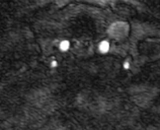
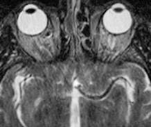
4 History: 35-year-old male with intermittent diplopia due to a sixth nerve palsy

5 Vascular Encasement


6 The Absent Flow Void
7 90 RF 180 RF Signal MR Flow Void Arterial Flow TE Voxel Flow Voxel Signal No flow Flow = 1/3 voxel Flow = 2/3 voxel Flow = 1 voxel


8 Flow Enhancement
9 90 RF 180 RF Signal RF Flow Enhancement Venous Flow: Short TR-TE Spin-echo Short TR Voxel Flow Voxel Signal No flow Flow = 1/3 voxel / TR Flow = 2/3 voxel / TR Flow = 1 voxel / TR


10 Flow Enhancement Arterial Flow: Gradient echo RF RF TR
11 MR Angiography Pulse Sequences 2D time of flight 3D time of flight 2D phase contrast 3D phase contrast
12 2D Time of Flight TR msec, TE 4.0 msec Flip angle degrees 1.5 mm sections Moving SAT band MIP algorithm

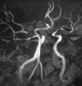
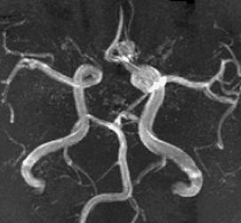
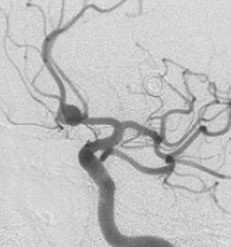
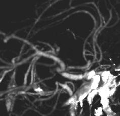
13 Normal 2D-TOF MRA mra


14 Segmented MIP Q-205-6


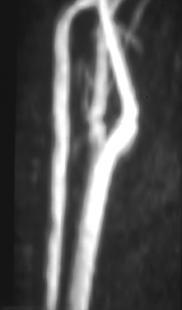
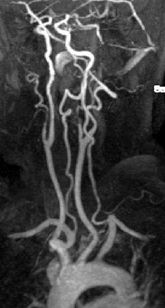
15 History: 23 y/o female with neck pain & vertigo following a snowboard accident 457

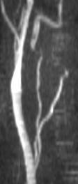

16 {Page 2}


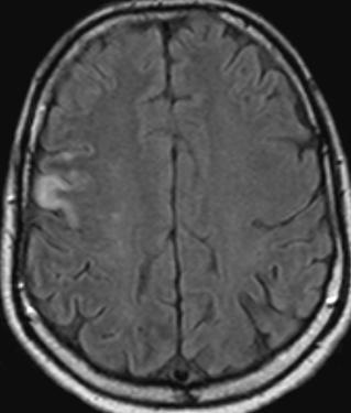
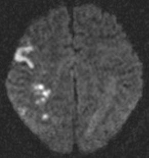
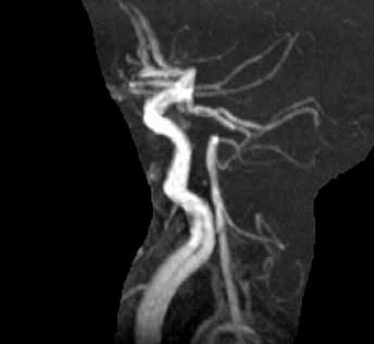
17 {Page 3} 4 months later Dx: Bilateral vertebral artery dissection
18 3D Time of Flight TR 9.0 msec, TE 2.0 msec Flip angle degrees 6-8 cm volume, phase encode in 2 directions mm sections Superior SAT band MIP algorithm


19 Normal 3D-TOF MRA {Video clip} {Video clip}
20 3D vs. 2D Time of Flight Higher signal-to-noise Shorter imaging times Less intravoxel dephasing Smoother vessel contours More saturation effects


21 3D TOF at 3T


22 History: 47 y/o man with von-hippel-lindau disease & new left arm weakness 486


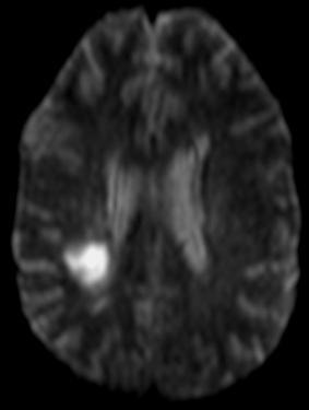

23 {Page 2} Dx: Infarct & MCA stenosis

24 History: 40 y/o man with episodes of left sided weakness 506

25 {Page 2}


26 {Page 3} {Video clip} Dx: Cerebral arteritis
27 2D Phase Contrast TR 25 msec, TE 6-7 msec Flip angle degrees 2-4 cm slab VENC factor Single projection image
28 Phase Contrast MR Angiography Velocity Encoding Gradient 1st Excitation Time Gradient T 2nd Excitation Time

29 3D Phase Contrast TR 25 msec, TE 6-7 msec Flip angle degrees 6-8 cm volume mm section VENC factor MIP algorithm
30 PC vs. TOF Angiography Less saturation effects Not affected by thrombus Velocity / flow information Long imaging times Arterial VENC misses slow flow


31 Phase Contrast Flow Direction Q-197-8


32 TOF vs. PC 2D-TOF 3D-PC Q-170-2


33 PC: Pseudoocclusion 3D-PC TOF Q
34 Imaging Slow Flow Thin-section 2D TOF Longer TR Lower flip angle Lower VENC factor Gd enhancement


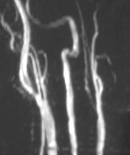
35 Intravoxel Dephasing & Signal Loss Q
36 Intravoxel Spin Dispersion Strategies To Minimize Smaller voxel size Shorter TE Flow compensation Gd-enhanced MRA
37 Gd-Enhanced MRA 3D gradient-echo sequence TR 4.5 msec, TE 1.5 msec High bandwidth & fractional echo Flip angle 25 degrees Matrix 173 x 256, FOV 187 x 250 Coronal volume 7 cm with 64 partitions 20 ml Gd 2ml/sec Acquisition time 21 sec


38 Timing Bolus Acquisition

39 Normal Gd-enhanced MRA {Click for video} mra


40 History: 83 y/o woman with slurred speech {Video clip} 503


41 {Page 2} Dx: Gd-MRA, multiple stenoses {Video clip}


42 History: 49 y/o man with neck pain following an MVA 523


43 {Page 2} {Video clip} Dx: Vertebral dissection


44 History: 51 y/o man with right-sided facial dysesthesias & ataxia 565


45 {Page 2} {Video clip} Dx: Lateral medullary syndrome & right vertebral occlusion
46 Vulnerable or High-Risk Atherosclerotic Plaque Imaging Criteria Thin fibrous cap Large lipid-necrotic core Fissured plaque Stenosis > 90 % Intraplaque hemorrhage Other factors: Inflammation, endothelial dysfunction, platelet aggregation


47 History: 48 y/o women with trauma to the neck Dx: Lipid core plaque 742
48 Venous / Sinus Occlusion Causes Hypercoagulable state Pregnancy Sepsis Dehydration Paranasal sinus infection Neoplastic invasion

49 2D TOF MR Venography TR 21 msec TE 4-6 msec Flip angle 60 o 1.5 mm sagittal or coronal sections Axial SAT band below skull base MIP algorithm {Video clip}


50 History: Newborn girl with seizures & decreased alertness


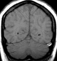
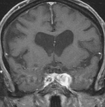
51 {Page 2}


52 Day 7 {Page 3} Dx: Hemorrhagic infarct with thrombosis of deep venous system, straight sinus & right transverse sinus 2 cm to right


53 History: 40 y/o woman with polycystic kidneys, renal failure, acute visual deficits & seizures 359

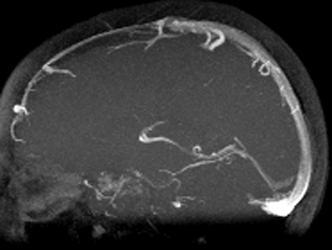

54 {Page 2} 2D PC Slabs Dx: Hypertensive encephalopathy


55 History: 32 y/o woman with headache, confusion & weakness 355


56 {Page 2}


57 Confusion & weakness {Page 3} Dx: Thrombosis of superior sagittal sinus, left transverse sinus & deep venous system


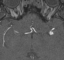
58 Meningioma


59 History: 60 y/o female with ataxia following trauma and concussion 387


60 {Page 2}

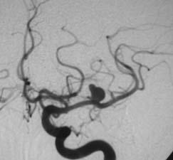

61 {Page 3} Dx: MCA aneurysm


62 History: 66 y/o male with dizziness and possible basilar insufficiency 542


63 {Video clip} {Page 2}


64 Dx: ACA aneurysm {Page 3}


65 History: A 62 y/o female with headaches for 3 weeks & a new 3 rd nerve palsy 3T 632


66 {Page 2} Dx: Acom & Pcom Aneurysms


67 History: 37 y/o woman with migraine headaches for many years 581


68 {Page 2} Dx: 2 mm AComm aneurysm
69 MR Angiography Aneurysms High risk patients 3D time-of-flight A screening procedure ONLY View source, collapse & MIP images 86-95% sensitivity for aneurysms 5 mm or larger


70 History: 80 y/o man with hypertension, slurred speech & unsteady gait {Video clip} 515


71 {Page 2}


72 {Page 3} Dx: Cavernous aneurysm


73 History: 63 y.o. man with episode of aphasia & right sided weakness. 16


74 Dx: Ethmoid mucocele Aphasia {Page 2}


75 History: 52 y/o woman with chronic seizures 604


76 Dx: AVM {Page 2}


77 History: 73 y.o. man with visual field deficits 1 2


78 3 5 4 Visual deficits {Page 2}


79 6 {Page 4} 7 8 Dx: AVM & basilar tip aneurysm
80 MR Angiography Problems & Pitfalls Turbulence / nonlaminar flow Slow flow / near complete occlusion Flow stasis & recirculation Thrombus Limited resolution Solution = Gd-enhanced MRA
Magnetic Resonance Angiography
 Magnetic Resonance Angiography 1 Magnetic Resonance Angiography exploits flow enhancement of GR sequences saturation of venous flow allows arterial visualization saturation of arterial flow allows venous
MR Advance Techniques. Vascular Imaging. Class II
 MR Advance Techniques Vascular Imaging Class II 1 Vascular Imaging There are several methods that can be used to evaluate the cardiovascular systems with the use of MRI. MRI will aloud to evaluate morphology
Imaging Acute Stroke and Cerebral Ischemia
 Department of Radiology University of California San Diego Imaging Acute Stroke and Cerebral Ischemia John R. Hesselink, M.D. Causes of Stroke Arterial stenosis Thrombosis Embolism Dissection Hypotension
Neuroradiology MR Protocols
 Neuroradiology MR Protocols Brain protocols N 1: Brain MRI without contrast N 2: Pre- and post-contrast brain MRI N 3 is deleted N 4: Brain MRI without or pre-/post-contrast (seizure protocol) N 5: Pre-
Essentials of Clinical MR, 2 nd edition. 99. MRA Principles and Carotid MRA
 99. MRA Principles and Carotid MRA As described in Chapter 12, time of flight (TOF) magnetic resonance angiography (MRA) is commonly utilized in the evaluation of the circle of Willis. TOF MRA allows depiction
Non-Traumatic Neuro Emergencies
 Department of Radiology University of California San Diego Non-Traumatic Neuro Emergencies John R. Hesselink, M.D. Nontraumatic Neuroemergencies 1. Acute focal neurological deficit 2. Worst headache of
1Pulse sequences for non CE MRA
 MRI: Principles and Applications, Friday, 8.30 9.20 am Pulse sequences for non CE MRA S. I. Gonçalves, PhD Radiology Department University Hospital Coimbra Autumn Semester, 2011 1 Magnetic resonance angiography
High Field MR of the Spine
 Department of Radiology University of California San Diego 3T for MR Applications Advantages High Field MR of the Spine Increased signal-to-noise Better fat suppression Increased enhancement with gadolinium
Magnetic Resonance Imaging. Basics of MRI in practice. Generation of MR signal. Generation of MR signal. Spin echo imaging. Generation of MR signal
 Magnetic Resonance Imaging Protons aligned with B0 magnetic filed Longitudinal magnetization - T1 relaxation Transverse magnetization - T2 relaxation Signal measured in the transverse plane Basics of MRI
非對比劑與對比劑增強 MRA. 血管攝影與對比劑 A Course of MRI. 本週課程內容 -MR Angiography (MRA) Unenhanced MRA
 Unenhanced MRA") 本週課程內容 -MR Angiography (MRA) 血管攝影與對比劑 A Course of MRI 盧家鋒助理教授國立陽明大學物理治療暨輔助科技學系 alvin4016@ym.edu.tw 非對比劑增強 MRA(Unenhanced MRA) Time-of-flight (TOF) angiography Phase-contrast (PC) angiography 對比劑增強 MRA(Contrast-enhanced
本週課程內容 -MR Angiography (MRA) 血管攝影與對比劑 A Course of MRI 盧家鋒助理教授國立陽明大學物理治療暨輔助科技學系 alvin4016@ym.edu.tw 非對比劑增強 MRA(Unenhanced MRA) Time-of-flight (TOF) angiography Phase-contrast (PC) angiography 對比劑增強 MRA(Contrast-enhanced
/ / / / / / Hospital Abstraction: Stroke/TIA. Participant ID: Hospital Code: Multi-Ethnic Study of Atherosclerosis
 Multi-Ethnic Study of Atherosclerosis Participant ID: Hospital Code: Hospital Abstraction: Stroke/TIA History and Hospital Record 1. Was the participant hospitalized as an immediate consequence of this
Non Contrast MRA. Mayil Krishnam. Director, Cardiovascular and Thoracic Imaging University of California, Irvine
 Non Contrast MRA Mayil Krishnam Director, Cardiovascular and Thoracic Imaging University of California, Irvine No disclosures Non contrast MRA-Why? Limitations of CTA Radiation exposure Iodinated contrast
MR Imaging of Atherosclerotic Plaques
 MR Imaging of Atherosclerotic Plaques Yeon Hyeon Choe, MD Department of Radiology, Samsung Medical Center, Sungkyunkwan University, Seoul MRI for Carotid Atheroma Excellent tissue contrast (fat, fibrous
Anatomic Evaluation of the Circle of Willis: MR Angiography versus Intraarterial Digital Subtraction Angiography
 Anatomic Evaluation of the Circle of Willis: MR Angiography versus Intraarterial Digital Subtraction Angiography K. W. Stock, S. Wetzel, E. Kirsch, G. Bongartz, W. Steinbrich, and E. W. Radue PURPOSE:
Non-Contrast MRA. How and When 1996! Why Non-Contrast MRA? Angiography: What are our goals? Inflow Techniques Differences in excitation hx
 A major teaching hospital of Harvard Medical School Angiography: What are our goals? Non-Contrast MRA: How and When Neil M. Rofsky, M.D. Professor of Radiology, Harvard Medical School Director of MRI &
Advanced Vascular Imaging: Pulsatile Tinnitus. Disclosures. Pulsatile Tinnitus: Differential Diagnosis. Pulsatile Tinnitus
 Advanced Vascular Imaging: Pulsatile Tinnitus Patrick Turski MD, Zach Clark MD, Tabby Kennedy MD The Objectives of this presentation are to: Review the differential diagnosis of pulsatile tinnitus Discuss
C. Douglas Phillips, MD FACR Director of Head and Neck Imaging Weill Cornell Medical College NewYork-Presbyterian Hospital
 C. Douglas Phillips, MD FACR Director of Head and Neck Imaging Weill Cornell Medical College NewYork-Presbyterian Hospital I have no financial disclosures Understand range of pathology that may present
Cerebral MR Venography: Normal Anatomy and Potential Diagnostic Pitfalls
 AJNR Am J Neuroradiol 21:74 78, January 2000 Cerebral MR Venography: Normal Anatomy and Potential Diagnostic Pitfalls R. H. Ayanzen, C. R. Bird, P. J. Keller, F. J. McCully, M. R. Theobald, and J. E. Heiserman
Methods. Yahya Paksoy, Bülent Oğuz Genç, and Emine Genç. AJNR Am J Neuroradiol 24: , August 2003
 AJNR Am J Neuroradiol 24:1364 1368, August 2003 Retrograde Flow in the Left Inferior Petrosal Sinus and Blood Steal of the Cavernous Sinus Associated with Central Vein Stenosis: MR Angiographic Findings
MR Angiography in the evaluation of Lower Extremity Arterial Disease
 March 2001 MR Angiography in the evaluation of Lower Extremity Arterial Disease Ted Mau, Harvard Medical School Year III Objectives We will cover: Indications for Magnetic Resonance Angiography (MRA) Basic
MR Imaging with the CCSVI or Haacke protocol
 MR Imaging with the CCSVI or Haacke protocol Reports from the Haacke protocol are often made available to the patients. The report consists of four major components: 1. anatomical images of major neck
NEUROVASCULAR ANATOMY
 E. Michael Harned, M.D. Assistant Professor of Clinical Radiology ndiana University School of Medicine have nothing to disclose Neurovascular Magnetic Resonance Angiography And Magnetic Resonance Venography
Neuroradiology. of Stroke and Headaches
 Neuroradiology of Stroke and Headaches Learning Objec:ves 1. Iden:fy T1 and T2 sequences 2. Recall the normal anatomy of the intracranial circula:on 3. Apply appropriate CT and MR imaging of the brain
Subject Index. Automated triggering 88 AVM 128, 129
 Subject Index A Abberrant right subclavian artery 150 Abdominal aorta 195-208 aneurysm (AAA) 198 aneurysm branching vessels 201 aortic dissection 203 dark-blood imaging 197 eccentric aneurysm 199 fusiform
Time-Of-Flight MRA. Faculty Disclosures Vincent B. Ho, M.D. Presentation Objectives. MRA Techniques. Pros and Cons of MRA
 Faculty Disclosures Vincent B. Ho, M.D. MR Angiography Techniques and Pitfalls Financial Disclosure Grant/Research Support General Electric Medical Systems Off-Label/Investigational Drug Use Dr. Ho will
The Use and Pitfalls of Intracranial Vessel Wall Imaging: How We Do It 1
 This copy is for personal use only. To order printed copies, contact reprints@rsna.org Reviews and Commentary n How I Do It Arjen Lindenholz, MD Anja G. van der Kolk, MD, PhD Jaco J. M. Zwanenburg, PhD
Extracranial Carotid Arteries: Evaluation with Black Blood MR Angiography
 Robert R. Edelman, MD #{149} Heinrich P. Mattle, MD #{149} Bernd Wallner, MD #{149} Richard Bajakian, MD #{149} Jonathan Kleefield, MD #{149} Craig Kent, MD #{149} John J. Skillman, MD Jeffrey B. Mendel,
Surface Appearance of the Vertebrobasilar Artery Revealed on Basiparallel Anatomic Scanning (BPAS) MR Imaging: Its Role for Brain MR Examination
 MR Imaging: Its Role for Brain MR Examination") AJNR Am J Neuroradiol 26:2508 2513, November/December 2005 Surface Appearance of the Vertebrobasilar Artery Revealed on Basiparallel Anatomic Scanning (BPAS) MR Imaging: Its Role for Brain MR Examination
AJNR Am J Neuroradiol 26:2508 2513, November/December 2005 Surface Appearance of the Vertebrobasilar Artery Revealed on Basiparallel Anatomic Scanning (BPAS) MR Imaging: Its Role for Brain MR Examination
[(PHY-3a) Initials of MD reviewing films] [(PHY-3b) Initials of 2 nd opinion MD]
![[(PHY-3a) Initials of MD reviewing films] [(PHY-3b) Initials of 2 nd opinion MD]](/thumbs/89/98619893.jpg "[(PHY-3a) Initials of MD reviewing films] [(PHY-3b) Initials of 2 nd opinion MD]") 2015 PHYSICIAN SIGN-OFF (1) STUDY NO (PHY-1) CASE, PER PHYSICIAN REVIEW 1=yes 2=no [strictly meets case definition] (PHY-1a) CASE, IN PHYSICIAN S OPINION 1=yes 2=no (PHY-2) (PHY-3) [based on all available
2015 PHYSICIAN SIGN-OFF (1) STUDY NO (PHY-1) CASE, PER PHYSICIAN REVIEW 1=yes 2=no [strictly meets case definition] (PHY-1a) CASE, IN PHYSICIAN S OPINION 1=yes 2=no (PHY-2) (PHY-3) [based on all available
Case Report 1. CTA head. (c) Tele3D Advantage, LLC
 Tele3D Advantage, LLC") Case Report 1 CTA head 1 History 82 YEAR OLD woman with signs and symptoms of increased intra cranial pressure in setting of SAH. CT Brain was performed followed by CT Angiography of head. 2 CT brain Extensive
Case Report 1 CTA head 1 History 82 YEAR OLD woman with signs and symptoms of increased intra cranial pressure in setting of SAH. CT Brain was performed followed by CT Angiography of head. 2 CT brain Extensive
Clinician s Guide To Ordering NeuroImaging Studies
 Clinician s Guide To Ordering NeuroImaging Studies MRI CT South Jersey Radiology Associates The purpose of this general guide is to assist you in choosing the appropriate imaging test to best help your
Overview of Stroke: Etiologies, Demographics, Syndromes, and Outcomes. Alex Abou-Chebl, MD, FSVIN Medical Director, Stroke Baptist Health Louisville
 Overview of Stroke: Etiologies, Demographics, Syndromes, and Outcomes Alex Abou-Chebl, MD, FSVIN Medical Director, Stroke Baptist Health Louisville Disclosure Statement of Financial Interest Within the
Identifying Cerebrovascular Disorders. Wengui Yu, MD, PhD Department of Neurology, University of California, Irvine
 Identifying Cerebrovascular Disorders Wengui Yu, MD, PhD Department of Neurology, University of California, Irvine Objectives Review different types of cerebrovascular disorders. Briefly discuss etiology,
TIA: Updates and Management 2008
 TIA: Updates and Management 2008 S. Andrew Josephson, MD Department of Neurology, Neurovascular Division University of California San Francisco Commonly Held TIA Misconceptions TIA is easy to diagnose
Endovascular treatment of intracranial aneurysms with detachable
 ORIGINAL RESEARCH L. Pierot C. Delcourt F. Bouquigny D. Breidt B. Feuillet O. Lanoix S. Gallas Follow-Up of Intracranial Aneurysms Selectively Treated with Coils: Prospective Evaluation of Contrast-Enhanced
Non-invasive Imaging of Carotid Artery Atherosclerosis
 Non-invasive Imaging of Carotid Artery Atherosclerosis 최연현 성균관의대삼성서울병원영상의학과 Noninvasive Techniques US with Doppler CT MRI Ultrasonography Techniques of Carotid US US Anatomy (ICA vs ECA) Gray scale and
CMS Limitations Guide - Radiology Services
 CMS Limitations Guide - Radiology Services Starting October 1, 2015, CMS will update their existing medical necessity limitations on tests and procedures to correspond to ICD-10 codes. This limitations
NEURO PROTOCOLS MRI NEURO PROTOCOLS (SIEMENS SCANNERS)
") Page 1 NEURO PROTOCOLS Brain Stroke Brain Brain with contrast Brain for seizures Brain for MS Brain for Pineal gland Sella FAST Scan for hydrocephalus MRA/MRV Brain MRA carotids 8 th nerve Cranial nerves
Page 1 NEURO PROTOCOLS Brain Stroke Brain Brain with contrast Brain for seizures Brain for MS Brain for Pineal gland Sella FAST Scan for hydrocephalus MRA/MRV Brain MRA carotids 8 th nerve Cranial nerves
CMS Limitations Guide MRA Radiology Services
 CMS Limitations Guide MRA Radiology Services Starting July 1, 2008, CMS has placed numerous medical necessity limits on tests and procedures. This reference guide provides you with all of the latest changes.
Vessel Wall Imaging of Intracranial Arterial Disease Commercial Interests
 Vessel Wall Imaging of Intracranial Arterial Disease Commercial Interests Disclosures No relevant commercial interests Off Label / Investigational Use No off label / investigational use Daniel Mandell,
Pearls and Pitfalls in Neuroradiology of Cerebrovascular Disease The Essentials with MR and CT
 Pearls and Pitfalls in Neuroradiology of Cerebrovascular Disease The Essentials with MR and CT Val M. Runge, MD Wendy R. K. Smoker, MD Anton Valavanis, MD Control # 823 Purpose The focus of this educational
TCD AND VASOSPASM SAH
 CURRENT TREATMENT FOR CEREBRAL ANEURYSMS TCD AND VASOSPASM SAH Michigan Sonographers Society 2 Nd Annual Fall Vascular Conference Larry N. Raber RVT-RDMS Clinical Manager General Ultrasound-Neurovascular
MR angiography. Extracranial MR angiography. Andrew G Clifton. Department of Neuroradiology, Atkinson Morley's Hospital, London, UK
 Andrew G Clifton Department of Neuroradiology, Atkinson Morley's Hospital, London, UK The primary use of angiography in the neck, either conventional catheter angiography or non-invasive techniques (MR
An Introduction to Imaging the Brain. Dr Amy Davis
 An Introduction to Imaging the Brain Dr Amy Davis Common reasons for imaging: Clinical scenarios: - Trauma (NICE guidelines) - Stroke - Tumours - Seizure - Neurological degeneration memory, motor dysfunction,
Demyelinating Diseases of the Brain
 Department of Radiology University of California San Diego Demyelinating Diseases of the Brain John R. Hesselink, M.D. T1-Weighted Images Normal White Matter Contents Axons with envelope of myelin Neuroglia
Evaluation of Carotid Vessels and Vertebral Artery in Stroke Patients with Color Doppler Ultrasound and MR Angiography
 Evaluation of Carotid Vessels and Vertebral Artery in Stroke Patients with Color Doppler Ultrasound and MR Angiography Dr. Pramod Shaha 1, Dr. Vinay Raj R 2, Dr. (Brig) K. Sahoo 3 Abstract: Aim & Objectives:
NON-ATHEROSCLEROTIC PATHOLOGY OF THE CAROTID ARTERIES
 NON-ATHEROSCLEROTIC PATHOLOGY OF THE CAROTID ARTERIES Leslie M. Scoutt, MD, FACR Professor of Diagnostic Radiology & Surgery Vice Chair, Dept of Radiology & Biomedical Imaging Chief, Ultrasound Section
MRS and Perfusion of Brain Tumors
 Department of Radiology University of California San Diego MRS and Perfusion of Brain Tumors John R. Hesselink, M.D. MRS & Perfusion of Brain Tumors Tumor histology Degree of malignancy Delineate tumor
Tips and Tricks of State of the art MRA
 Tips and Tricks of State of the art MRA Mayil Krishnam, MD,MBA, MRCP,FRCR(UK) Professor of Radiology Director, Cardiovascular and Thoracic Imaging University of California, Irvine Objectives Technical
RADIOLOGY TEACHING CONFERENCE
 RADIOLOGY TEACHING CONFERENCE John Athas, MD Monica Tadros, MD Columbia University, College of Physicians & Surgeons Department of Otolaryngology- Head & Neck Surgery September 27, 2007 CT SCAN IMAGING
PTA 106 Unit 1 Lecture 3
 PTA 106 Unit 1 Lecture 3 The Basics Arteries: Carry blood away from the heart toward tissues. They typically have thicker vessels walls to handle increased pressure. Contain internal and external elastic
ISCHEMIC STROKE IMAGING
 ISCHEMIC STROKE IMAGING ผศ.พญ พญ.จ ร ร ตน ธรรมโรจน ภาคว ชาร งส ว ทยา คณะแพทยศาสตร มหาว ทยาล ยขอนแก น A case of acute hemiplegia Which side is the abnormality, right or left? Early Right MCA infarction
Magnetization Preparation Sequences
 Magnetization Preparation Sequences Acquisition method may not give desired contrast Prep block adds contrast (and/or encoding) MP-RAGE = Magnetization prepared rapid acquisition with gradient echo (Mugler,
11/1/2018. Disclosure. Imaging in Acute Ischemic Stroke 2018 Neuro Symposium. Is NCCT good enough? Keystone Heart Consultant, Stock Options
 Disclosure Imaging in Acute Ischemic Stroke 2018 Neuro Symposium Keystone Heart Consultant, Stock Options Kevin Abrams, M.D. Chief of Radiology Medical Director of Neuroradiology Baptist Hospital, Miami,
Plaque Imaging: What It Can Tell Us. Kenneth Snyder, MD, PhD L Nelson Hopkins MD FACS Elad Levy MD MBA FAHA FACS Adnan Siddiqui MD PhD
 Plaque Imaging: What It Can Tell Us Kenneth Snyder, MD, PhD L Nelson Hopkins MD FACS Elad Levy MD MBA FAHA FACS Adnan Siddiqui MD PhD Buffalo Disclosure Information FINANCIAL DISCLOSURE: Research and consultant
Better identification of patients who may benefit from therapy
 Jon Jui MD, MPH Large Vessel Occlusion Low rates of re-canalization after tpa Only 25 % of large vessel strokes re-canalization after tpa Newer invasive techniques Solitaire vs Merci Better identification
Speed, Comfort and Quality with NeuroDrive
 Speed, Comfort and Quality with NeuroDrive Echelon Oval provides a broad range of capabilities supporting fast, accurate diagnosis of brain conditions and injuries. From anatomical depiction to vascular
Initial symptom or syndrome: (1) FOCAL WEAKNESS OR NUMBNESS
 FOCAL WEAKNESS OR NUMBNESS") View the referenced DVD patient cases, especially if few hospital or clinic patients are encountered for any one symptom or syndrome. The DVD patient cases are referenced by initial symptom or syndrome
View the referenced DVD patient cases, especially if few hospital or clinic patients are encountered for any one symptom or syndrome. The DVD patient cases are referenced by initial symptom or syndrome
Subclavian steal syndrome: an underdiagnosed disease
 Subclavian steal syndrome: an underdiagnosed disease Poster No.: C-0753 Congress: ECR 2017 Type: Educational Exhibit Authors: R. O. Martins 1, M. C. Calegari 2, M. Lopes 3, L. Santos 3, L. Cruz 3, R. Vasconcelos
How I do it: Non Contrast-Enhanced MR Angiography (syngo NATIVE)
") Clinical How-I-do-it Cardiovascular How I do it: Non Contrast-Enhanced MR Angiography (syngo NATIVE) Manuela Rick, Nina Kaarmann, Peter Weale, Peter Schmitt Siemens Healthcare, Erlangen, Germany Introduction
Clinical How-I-do-it Cardiovascular How I do it: Non Contrast-Enhanced MR Angiography (syngo NATIVE) Manuela Rick, Nina Kaarmann, Peter Weale, Peter Schmitt Siemens Healthcare, Erlangen, Germany Introduction
L M Thornton, MD; L Lanier, MD; C L Sistrom, MD; D Rajderkar, MD; A Mancuso, MD; IM Schmalfuss, MD University of Florida, Gainesville Department of
 L M Thornton, MD; L Lanier, MD; C L Sistrom, MD; D Rajderkar, MD; A Mancuso, MD; IM Schmalfuss, MD University of Florida, Gainesville Department of Radiology RSNA Annual Meeting 2016 Trainee call readiness
OBJECTIVES. At the end of the lecture, students should be able to: List the cerebral arteries.
 DR JAMILA EL MEDANY OBJECTIVES At the end of the lecture, students should be able to: List the cerebral arteries. Describe the cerebral arterial supply regarding the origin, distribution and branches.
S pontaneous dissection of the internal
 Pictorial Essay Dissections of the Internal Carotid Artery: Th ree- Dimensional Time-of-Flight M R Angiography and MR Imaging Features S pontaneous dissection of the internal carotid artery is now recognized
Non-Invasive Follow-up Evaluation of Post-Embolized AVM with Time-Resolved MRA: A Case Report
 Non-Invasive Follow-up Evaluation of Post-Embolized AVM with Time-Resolved MRA: A Case Report Yong Woon Shim, MD 1 Tae-Sub Chung, MD 1 Won-Suk Kang, MD 1 Jin-Yang Joo, MD 2 Ralph Strecker, MD 3 Juergen
2. Subarachnoid Hemorrhage
 Causes: 2. Subarachnoid Hemorrhage A. Saccular (berry) aneurysm - Is the most frequent cause of clinically significant subarachnoid hemorrhage is rupture of a saccular (berry) aneurysm. B. Vascular malformation
MR Flow Imaging in Vascular Malformations Using Gradient Recalled Acquisition
 637 MR Flow Imaging in Vascular Malformations Using Gradient Recalled Acquisition William M. Needell 1 Kenneth R. Maravilla Twenty patients with known or suspected intracranial vascular lesions were evaluated
Multislice CT. - fast scanning - submilimeter slices
 CT angiography Multislice CT - fast scanning - submilimeter slices CT angiography - Minimal invasivity - High resolution (similar to DSA, higher than MRI) - Cannot assess hemodynamics (contrary to DSA)
Nicolas Bianchi M.D. May 15th, 2012
 Nicolas Bianchi M.D. May 15th, 2012 New concepts in TIA Differential Diagnosis Stroke Syndromes To learn the new definitions and concepts on TIA as a condition of high risk for stroke. To recognize the
41 year old female with headache. Elena G. Violari MD and Leo Wolansky MD
 41 year old female with headache Elena G. Violari MD and Leo Wolansky MD ? Dural Venous Sinus Thrombosis with Hemorrhagic Venous Infarct Acute intraparenchymal hematoma measuring ~3 cm in diameter centered
Neurological Dilemmas in Primary Care
 Neurological Dilemmas in Primary Care David Clark, DO dclark@oregonneurology.com When to test? How to test? Pitfalls in testing? When to treat? How to treat? How long to treat? Neurological Dilemmas Seizure
Alan Barber. Professor of Clinical Neurology University of Auckland
 Alan Barber Professor of Clinical Neurology University of Auckland Presented with Non-fluent dysphasia R facial weakness Background Ischaemic heart disease Hypertension Hyperlipidemia L MCA branch
Neuroanatomy of a Stroke. Joni Clark, MD Professor of Neurology Barrow Neurologic Institute
 Neuroanatomy of a Stroke Joni Clark, MD Professor of Neurology Barrow Neurologic Institute No disclosures Stroke case presentations Review signs and symptoms Review pertinent exam findings Identify the
ARTICLE IN PRESS. Available online at Magnetic Resonance Imaging xx (2008) xxx xxx
 xxx xxx") Available online at www.sciencedirect.com Magnetic Resonance Imaging xx (2008) xxx xxx An optimized 3D inversion recovery prepared fast spoiled gradient recalled sequence for carotid plaque hemorrhage
Available online at www.sciencedirect.com Magnetic Resonance Imaging xx (2008) xxx xxx An optimized 3D inversion recovery prepared fast spoiled gradient recalled sequence for carotid plaque hemorrhage
Perforating arteries originating from the posterior communicating artery: a 7.0-Tesla MRI study
 Eur Radiol (2009) 19: 2986 2992 DOI 10.1007/s00330-009-1485-4 MAGNETIC RESONANCE Mandy M. A. Conijn Jeroen Hendrikse Jaco J. M. Zwanenburg Taro Takahara Mirjam I. Geerlings Willem P. Th. M. Mali Peter
Scientific Exhibit Authors: M. Sugiyama, Y. Takehara, T. Saito, N. Ooishi, M. Alley,
 Abnormal flow dynamics within the ascending aorta of the patients with aortic valve stenosis. Assessments with phase resolved three dimensional phase contrast MR image (4DFlow). Poster No.: C-2504 Congress:
A New Trend in Vascular Imaging: the Arterial Spin Labeling (ASL) Sequence
 Sequence") A New Trend in Vascular Imaging: the Arterial Spin Labeling (ASL) Sequence Poster No.: C-1347 Congress: ECR 2013 Type: Educational Exhibit Authors: J. Hodel, A. GUILLONNET, M. Rodallec, S. GERBER, R. 1
A New Trend in Vascular Imaging: the Arterial Spin Labeling (ASL) Sequence Poster No.: C-1347 Congress: ECR 2013 Type: Educational Exhibit Authors: J. Hodel, A. GUILLONNET, M. Rodallec, S. GERBER, R. 1
Cerebrovascular Disorders. Blood, Brain, and Energy. Blood Supply to the Brain 2/14/11
 Cerebrovascular Disorders Blood, Brain, and Energy 20% of body s oxygen usage No oxygen/glucose reserves Hypoxia - reduced oxygen Anoxia - Absence of oxygen supply Cell death can occur in as little as
Diagnosis of Vertebral Artery Ostial Stenosis on Contrast-Enhanced MR Angiography: Usefulness of a Thin-Slab MIP Technique
 Diagnosis of Vertebral Artery Ostial Stenosis on Contrast-Enhanced MR Angiography: Usefulness of a Thin-Slab MIP Technique Sun Mi Kim 1, Deok Hee Lee 2 Jin Woo Choi 3, Byung Se Choi 4, Hyun Sin In 5 It
MR Advance Techniques. Cardiac Imaging. Class III
 MR Advance Techniques Cardiac Imaging Class III Black Blood Imaging & IR Blue= O2 poor blood Red=O2 rich blood Inversion pulses can produce black blood imaging in GRE pulse sequences. Specially on the
Application of three-dimensional angiography in elderly patients with meningioma
 Application of three-dimensional angiography in elderly patients with meningioma Poster No.: C-0123 Congress: ECR 2012 Type: Scientific Paper Authors: X. Han, J. Chen, K. Shi; Haikou/CN Keywords: Neuroradiology
M555 Medical Neuroscience Blood Flow in CNS Meninges Blood Brain Barrier CSF
 M555 Medical Neuroscience Blood Flow in CNS Meninges Blood Brain Barrier CSF Arterial Blood Flow to CNS approximately % of what goes wrong within the skull that produces neurological deficits is vascular
Carotid Artery Disease and What s Pertinent JOSEPH A PAULISIN DO
 Carotid Artery Disease and What s Pertinent JOSEPH A PAULISIN DO Goal of treatment of carotid disease Identify those at risk of developing symptoms Prevent patients at risk from developing symptoms Prevent
ATHEROSCLEROSIS. Secondary changes are found in other coats of the vessel wall.
 ATHEROSCLEROSIS Atherosclerosis Atherosclerosis is a disease process affecting the intima of the aorta and large and medium arteries, taking the form of focal thickening or plaques of fibrous tissue and
Optimized phase contrast MRV technique outperforms timeof-flight in the diagnosis of cerebral venous thrombosis
 Optimized phase contrast MRV technique outperforms timeof-flight in the diagnosis of cerebral venous thrombosis Poster No.: C-3377 Congress: ECR 2010 Type: Topic: Authors: Keywords: DOI: Scientific Exhibit
Transverse Dural Sinus Thrombosis Joseph Junewick, MD FACR
 Transverse Dural Sinus Thrombosis Joseph Junewick, MD FACR 03/19/2010 History Child with headache and otomastoiditis. Diagnosis Dural venous thrombosis secondary to mastoiditis Discussion The cerebral
Vivek R. Deshmukh, MD Director, Cerebrovascular and Endovascular Neurosurgery Chairman, Department of Neurosurgery Providence Brain and Spine
 Vivek R. Deshmukh, MD Director, Cerebrovascular and Endovascular Neurosurgery Chairman, Department of Neurosurgery Providence Brain and Spine Institute The Oregon Clinic Disclosure I declare that neither
Neuro-Vascular Intervention AAPC Regional Conference Springfield, MA
 Neuro-Vascular Intervention AAPC Regional Conference Springfield, MA October 8, 2010 1 Presented by: David Zielske, MD,CIRCC, CPC H, CCC, CCS, RCC General Recommendations for Physician Dictations State
Department of Radiology, Division of MRI, University of Michigan, 1500 E. Medical Center Drive, Ann Arbor, MI , USA
 Abdom Imaging 23:469 484 (1998) Abdominal Imaging Springer-Verlag New York Inc. 1998 Contrast-enhanced MR angiography J. H. Maki, T. L. Chenevert, M. R. Prince Department of Radiology, Division of MRI,
Medical Review Guidelines Magnetic Resonance Angiography
 Medical Review Guidelines Magnetic Resonance Angiography Medical Guideline Number: MRG2001-05 Effective Date: 2/13/01 Revised Date: 2/14/2006 OHCA Reference OAC 317:30-5-24. Radiology. (f) Magnetic Resonance
T Clinical Magnetic Resonance Angiography
 MRI for Technologists 4712-204T Clinical Magnetic Resonance Angiography PROGRAM INFORMATION MRI for Technologists is a training program designed to meet the needs of radiologic technologists entering or
Overview of imaging modalities for cerebral aneurysms
 Overview of imaging modalities for cerebral aneurysms Soroush Zaghi BIDMC PCE: Radiology August 2008 (Images from BIDMC, PACS.) Our Patient: Presentation Our patient is a 57 y/o woman who reports blowing
Marc Norman, Ph.D. - Do Not Use without Permission 1. Cerebrovascular Accidents. Marc Norman, Ph.D. Department of Psychiatry
 Cerebrovascular Accidents Marc Norman, Ph.D. Department of Psychiatry Neuropsychiatry and Behavioral Medicine Neuropsychology Clinical Training Seminar 1 5 http://www.nlm.nih.gov/medlineplus/ency/images/ency/fullsize/18009.jpg
Three-Dimensional Time-of-Flight MR Angiography in the Evaluation of Intracranial Aneurysms Treated by Endovascular Balloon Occlusion
 Three-Dimensional Time-of-Flight MR Angiography in the Evaluation of Intracranial Aneurysms Treated by Endovascular Balloon Occlusion JayS. Tsuruda, Robert J. Sevick, 1 and Van V. Halbach Summary: The
Ultrasound Imaging of The Posterior Circulation
 Ultrasound Imaging of The Posterior Circulation Michigan Sonographers Society 2 Nd Annual Fall Vascular Conference Larry N. Raber RDMS-RVT Clinical Manager General Ultrasound/Neurovascular Laboratory Cleveland
CARDIAC MRI. Cardiovascular Disease. Cardiovascular Disease. Cardiovascular Disease. Overview
 CARDIAC MRI Dr Yang Faridah A. Aziz Department of Biomedical Imaging University of Malaya Medical Centre Cardiovascular Disease Diseases of the circulatory system, also called cardiovascular disease (CVD),
Sung Hong Park. M.S. in Electrical Engineering, KAIST, South Korea, Submitted to the Graduate Faculty of
 NONINVASIVE IMAGING OF BRAIN VASCULATURE WITH HIGH RESOLUTION BLOOD OXYGENATION LEVEL DEPENDENT VENOGRAPHY IN MAGNETIC RESONANCE IMAGING: APPLICATIONS TO FUNCTIONAL AND CLINICAL STUDIES by Sung Hong Park
Evaluation of Intracranial Vasculatures in Healthy Subjects with Arterial-Spin-Labeling-Based 4D-MR Angiography at 3T
 Magn Reson Med Sci, Vol. 15, No. 3, pp. 335 339, 2016 doi:10.2463/mrms.tn.2015-0081 TECHNICAL NOTE Evaluation of Intracranial Vasculatures in Healthy Subjects with Arterial-Spin-Labeling-Based 4D-MR Angiography
Acute Ischemic Stroke Imaging. Ronald L. Wolf, MD, PhD Associate Professor of Radiology
 Acute Ischemic Stroke Imaging Ronald L. Wolf, MD, PhD Associate Professor of Radiology Title of First Slide of Substance An Illustrative Case 2 Disclosures No financial disclosures Off-label uses of some
TRAUMATIC CAROTID &VERTEBRAL ARTERY INJURIES
 TRAUMATIC CAROTID &VERTEBRAL ARTERY INJURIES ALBERTO MAUD, MD ASSOCIATE PROFESSOR TEXAS TECH UNIVERSITY HEALTH SCIENCES CENTER EL PASO PAUL L. FOSTER SCHOOL OF MEDICINE 18TH ANNUAL RIO GRANDE TRAUMA 2017
It s Always a Stroke; Except For When It s Not..
 It s Always a Stroke; Except For When It s Not.. TREVOR PHINNEY, D.O. Disclosures No Relevant Disclosures 1 Objectives Discuss variables of differential diagnosis for stroke Review when to TPA and when