Cardiovascular Pathology Lab. Shannon Martinson,
|
|
|
- Anna Jennings
- 5 years ago
- Views:
Transcription

1 Cardiovascular Pathology Lab Shannon Martinson,


2 Case 1 Signalment: 10 year old MC DSH Cat History Heart murmur detected on PE recommended cardiac US Blood work was done to check for hyperthyroidism - T4 levels were normal Sudden right facial paralysis and loss of sensation of the right side of the face Cat was euthanized


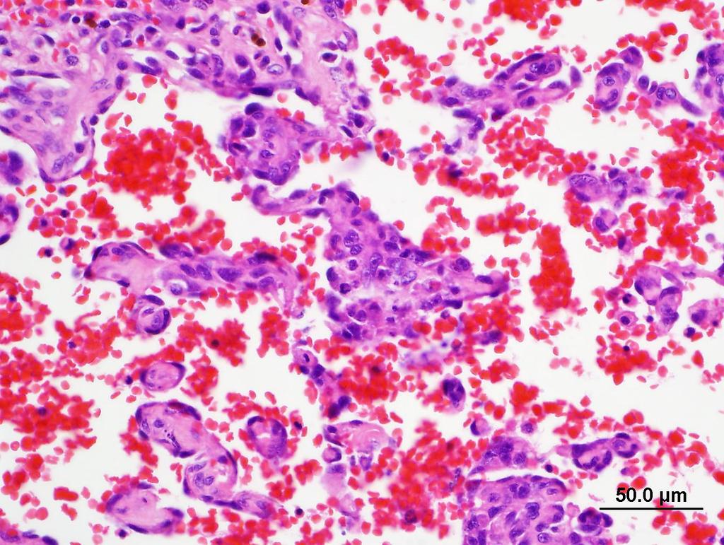
3 Case 1 Description Normal cat heart for comparison The heart is markedly enlarged with thickening of LV free wall, the IVS, and the RV wall. The LV chamber in particular is reduced in size The atria are dilated especially the LA which contains a large brown and tan thrombus that occludes the lumen

4 Case 1 Morph Diagnosis Left ventricular hypertrophy, concentric, marked Right ventricular hypertrophy, mild to moderate Left atrial thrombus, occlusive What are possible causes for the changes in the left ventricle? Usually pressure overload: Subaortic stenosis Systemic hypertension Idiopathic / genetic Hypertrophic cardiomyopathy Hyperthyroidism
5 Case 1 What disease process do you think this represents? Hypertrophic cardiomyopathy Why do cats with this disease develop atrial thrombosis? LA dilation alters laminar blood flow predisposes to thrombosis What is another common site for thrombosis in cats with this disease and what clinical signs might be seen as a result? Caudal abdominal aorta Hind end pain /paresis Cold extremities (hind legs) Lack of femoral pulses Cause of facial paralysis: Possible embolism to the brain (stroke) from the aortic thrombus

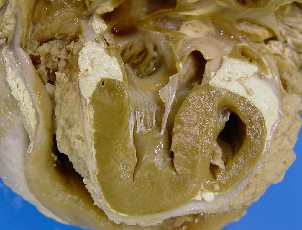
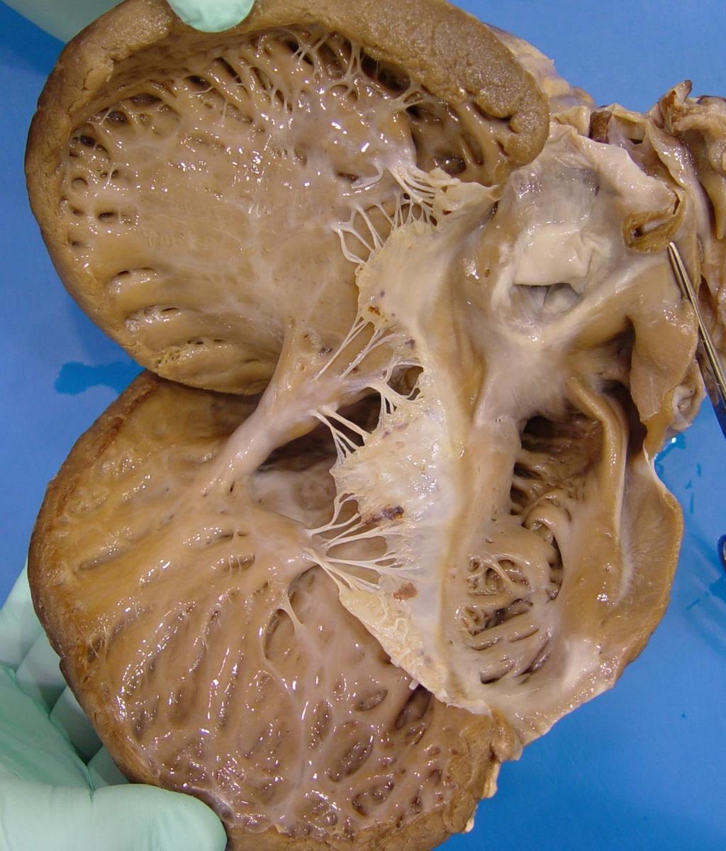
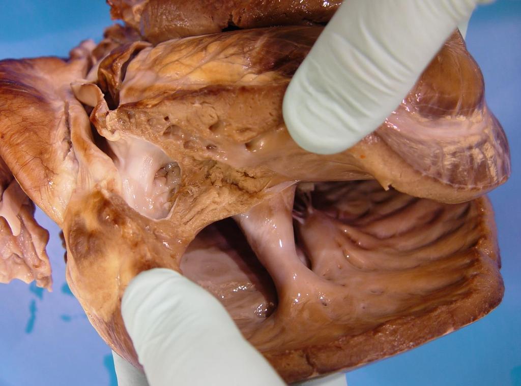
6 Case 2 Signalment: 1.5 year old intact F Newfoundland Dog Clinical History: Murmur detected at a young age Recurrent fever and previous bouts of joint pain Mild elevation in BUN and Creatinine Became anorexic and was euthanized Fluid in the chest and red, heavy lungs seen at necropsy

7 Case 2 Description There is LV hypertrophy and LA dilation A band of fibrous connective tissue encircles and narrows the LV outflow tract beneath the aortic valve The aorta is dilated above the valve The aortic valve is roughened and focally ruptured with a necrotic tract dissecting through to the right atrium The chordae and leaflets of the mitral valve are short and thick and attach from the papillary muscle to the ventricular free wall in the area of stenosis


8 Case 2 Morphologic Diagnosis Subaortic stenosis, severe with poststenotic dilation Left ventricular hypertrophy, moderate to marked Valvular endocarditis, rupture, and tract formation Mitral valve dysplasia

9 Case 2 Describe the hemodynamic alterations resulting from the primary lesion and relate them back to the other findings? Subaortic stenosis narrowing of the orifice causes pressure overload of the LV LV concentric hypertrophy Left heart failure Pulmonary edema and congestion Increased turbulence above the valve can lead to dilation of the aorta The necrotic tract may have occurred as a result of high pressure and weakening of the wall at this site
10 Case 2 Can you relate the lesions back to the clinical findings / history? While subtle, there is endocarditis Malformed valves are predisposed to the development of endocarditis The source of bacteria is often undetermined presumed bacteremia Fever may occur during periods of bacteremia or showering anorexia Small thromboemboli, which may be septic, can be released causing ischemia/infection in the organs Renal infarcts in this case may have caused urea and creatinine Possible ischemic injury in the limbs or septic showering of the joints may cause lameness
 the owners opted for")
11 Case 3 Signalment: Aged male mixed breed dog Clinical History: 10 day history of lethargy Using imaging, fluid was detected in the abdomen and thorax: thoraco- and abdominocentesis transudate A 3 rd degree heart block was detected (ECG) the owners opted for euthanasia


12 Case 3 Description A large (~8 x 4 x 5 cm) irregular solid tan and black mass infiltrates the RA obliterating the lumen and extending into the RV and IVS. The mass encompassed the aorta at the heart base. Both ventricles are dilated Multiple small (<0.5 cm) red nodular masses are present randomly within the lung.


13 Case 3 What time of disease process is this? Malignant Neoplasia What are some differentials for this lesion? Hemangiosarcoma Rhabdomyosarcoma Chemodectoma Aortic body tumour Carotid body tumour Ectopic thyroid carcinoma Lymphoma How would you reach a final diagnosis? Histology!

14 Case 3 Morph Diagnosis Hemangiosarcoma, right atrium and lung

15 Case 3 Morph Diagnosis Hemangiosarcoma, right atrium and lung Can you relate this lesion back to the clinical signs and other postmortem findings? RA mass could have prevented conduction of electrical activity via the AV node to the ventricles 3 rd degree heart block The presence of this mass resulted in congestive RHF (impedance of venous flow from the vena cava) ascites and hydrothorax Arrhythmia and CHF lethargy Metastasis to the lung from the primary mass


16 Case 3 What is a more common outcome for these tumours? RA rupture hemopericardium cardiac tamponade PBVD, Saunders, 2017


17 Case 4 Signalment: 31 day old ram lamb. Clinical History: Lamb was sick for 10 days and seemed to respond briefly to antibiotic treatment Became sick again a week later with no response to antibiotics. The lamb is now in poor body condition the owner opted to euthanize
18 Case 4
19 Case 4 Description The pericardial sac is markedly dilated (~ 15 cm diameter) and thickened by dense fibrous connective tissue with a thick covering layer of slightly friable tan shaggy material Similar changes are present in the epicardium Both the LV and RV are hyertrophied Fibrous adhesions span between the pericardium and pleura and there is mild CV consolidation of the lung Morph Diagnosis Organizing fibrinous pericarditis, diffuse, chronic, severe Biventricular hypertrophy Bronchopneumonia and fibrous adhesions
20 Case 4 Possible etiology? Bacterial infection (sepsis) Trueperella pyogenes Pasteurella multocida Staphylococcus aureus E coli Submit a swab for bacteriology Antibiotic treatment may hamper microbial growth What would the most likely underlying disease process be if this was a cow? Traumatic reticuloperitonitis (rare in lambs) Why is there LV and RV hypertrophy? Fibrosis of the epicardium and adhesion to the pericardial sac can limit diastolic expansion and cardiac output (= constrictive pericarditis)

21 Case 5 Signalment: 21 year old horse Clinical History: Donated to AVC Poor dentition and weight loss Recurrent bouts of colic
22 Case 5 Description The cranial mesenteric artery has a markedly thickened and firm (fibrotic) wall The lumen varies in caliber with areas of dilation and narrowing The intima is roughed and corrugated with brown to orange discolouration Morph Diagnosis Arteritis, proliferative, segmental, chronic, severe, with dilation (aneurysm) and fibrosis Etiology Strongylus vulgaris (L4) migration
 and fibrosis Etiology Strongylus vulgaris (L4)")
23 Case 5 Description The cranial mesenteric artery has a markedly thickened and firm (fibrotic) wall The lumen varies in caliber with areas of dilation and narrowing The intima is roughed and corrugated with brown to orange discolouration Morph Diagnosis Arteritis, proliferative, segmental, chronic, severe, with dilation (aneurysm) and fibrosis Etiology Strongylus vulgaris (L4) migration
 Can you relate")

24 Case 5 How might this result in colic? Thrombosis can result in infarction of the intestine (collateral circulation makes this less likely) Can you relate this lesion to that seen in the small intestine? Arteritis and the resulting endothelial damage promote the formation of a thrombus at the affected site (also altered blood flow in an aneurysm) pieces can break off as thromboemboli which lodge downstream in this case an embolus has lodged in a mesenteric vessel


25 Case 6 Signalment: 7 week old Holstein bull calf Clinical History : Calf was ill-thrift, lethargic and had bluish mucous membranes Found dead one morning

26 Case 6

27 Case 6

28 Case 6 Description The heart is enlarged and somewhat globose in shape A thick muscular band narrows the right ventricular outflow tract The leaflets of the pulmonic valve are white-tan, thick and rugous with partial fusion of the leaflets leaving a central irregular perforation measuring ~ 0.75 cm There is moderate to marked RV hypertrophy and the LV ventricle appears dilated A 3 cm diameter opening is present high within the IVS and the aorta overrides this opening The ductus arteriosis is patent with a 1 cm diameter lumen The foramen ovale is covered by a perforated valve (probe patent)

29 Case 6 Morphologic Diagnoses Pulmonic Stenosis Ventricular septal defect Right and left ventricular hypertrophy Over-riding aorta Patent ductus arteriosis Patent foramen ovale (~ASD)


30 Case 6 Which of the identified changes would be found in tetralogy of Fallot? Pulmonic stenosis Ventricular septal defect Over-riding aorta Right ventricular hypertrophy Pulmonic stenosis Ventricular septal defect Over-riding aorta Right ventricular hypertrophy Which of these are congenital and which are acquired? Congenital Acquired Pentology of Fallot Patent foramen ovale OR Patent Ductus Arteriosis
31 Case 6 What are the hemodynamic alterations in this case? R to L shunt through VSD Cyanosis Pulmonic stenosis RV pressure overload RV hypertrophy Right heart failure Shunt through the ASD and PDA? PDA and ASD are not though to contribute much to clinical disease when present along with tetralogy


32
CARDIOVASCULAR PATHOLOGY LABORATORY CASES AND SLIDES. VPM Lisa Miller CARDIOVASCULAR LAB
 CARDIOVASCULAR PATHOLOGY LABORATORY CASES AND SLIDES VPM 222 2009 Lisa Miller CARDIOVASCULAR LAB Case 1 Heart - Echoplane section from a 7-month-old Labrador retriever Give two morphologic diagnoses 1.
Right-Sided Congestive Heart Failure Basics
 Right-Sided Congestive Heart Failure Basics OVERVIEW Failure of the right side of the heart to pump blood at a sufficient rate to meet the needs of the body or to prevent blood from pooling within the
Inflammation Laboratory 2. Shannon Martinson: VPM 152: March 2012
 Inflammation Laboratory 2 Shannon Martinson: http://people.upei.ca/smartinson VPM 152: March 2012 Reminder - Creating a Morphologic Diagnosis for Inflammatory Lesions Organ and Process Exudate Distribution
Respiratory Pathology Lab 2: Lung. Shannon Martinson,
 Respiratory Pathology Lab 2: Lung Shannon Martinson, 2017 http://people.upei.ca/smartinson/ Case 1 Signalment: 9 month old DSH cat History: Poor doer with stunted growth One month of lethargy one day the
Saluki heart pathology study
 Heart conditions by MaryDee Sist, DVM Originally published in Baraka Book, Autumn-Winter 2001 For the last decade I have been involved in Saluki heart research. Ouroriginalgoalwastoexaminethe incidence
CMS Limitations Guide - Radiology Services
 CMS Limitations Guide - Radiology Services Starting October 1, 2015, CMS will update their existing medical necessity limitations on tests and procedures to correspond to ICD-10 codes. This limitations
Adult Echocardiography Examination Content Outline
 Adult Echocardiography Examination Content Outline (Outline Summary) # Domain Subdomain Percentage 1 2 3 4 5 Anatomy and Physiology Pathology Clinical Care and Safety Measurement Techniques, Maneuvers,
The Cardiovascular System Part I: Heart Outline of class lecture After studying part I of this chapter you should be able to:
 The Cardiovascular System Part I: Heart Outline of class lecture After studying part I of this chapter you should be able to: 1. Describe the functions of the heart 2. Describe the location of the heart,
CONGENITAL HEART DISEASE (CHD)
") CONGENITAL HEART DISEASE (CHD) DEFINITION It is the result of a structural or functional abnormality of the cardiovascular system at birth GENERAL FEATURES OF CHD Structural defects due to specific disturbance
CONGENITAL HEART DISEASE (CHD) DEFINITION It is the result of a structural or functional abnormality of the cardiovascular system at birth GENERAL FEATURES OF CHD Structural defects due to specific disturbance
Heart Disorders. Cardiovascular Disorders (Part B-1) Module 5 -Chapter 8. Overview Heart Disorders Vascular Disorders
 Module 5 -Chapter 8. Overview Heart Disorders Vascular Disorders") Cardiovascular Disorders (Part B-1) Module 5 -Chapter 8 Overview Heart Disorders Vascular Disorders Susie Turner, MD 1/7/13 Heart Disorders Coronary Artery Disease Cardiac Arrhythmias Congestive Heart
Cardiovascular Disorders (Part B-1) Module 5 -Chapter 8 Overview Heart Disorders Vascular Disorders Susie Turner, MD 1/7/13 Heart Disorders Coronary Artery Disease Cardiac Arrhythmias Congestive Heart
Alfonso López. Cardiac Hypertrophy and Dilation
 Cardiac Hypertrophy and Dilation Alfonso López Professor of Anatomic Pathology Dept. Pathology and Microbiology Atlantic Veterinary College University of Prince Edward Island Canada Jan 23, 2012 Compensatory
Anatomy & Physiology
 1 Anatomy & Physiology Heart is divided into four chambers, two atrias & two ventricles. Atrioventricular valves (tricuspid & mitral) separate the atria from ventricles. they open & close to control flow
Adult Congenital Heart Disease: What All Echocardiographers Should Know Sharon L. Roble, MD, FACC Echo Hawaii 2016
 1 Adult Congenital Heart Disease: What All Echocardiographers Should Know Sharon L. Roble, MD, FACC Echo Hawaii 2016 DISCLOSURES I have no disclosures relevant to today s talk 2 Why should all echocardiographers
Cardiovascular Nursing Practice: A Comprehensive Resource Manual and Study Guide for Clinical Nurses 2 nd Edition
 Cardiovascular Nursing Practice: A Comprehensive Resource Manual and Study Guide for Clinical Nurses 2 nd Edition Table of Contents Volume 1 Chapter 1: Cardiovascular Anatomy and Physiology Basic Cardiac
By Dickens ATURWANAHO & ORIBA DAN LANGOYA MAKchs, MBchB CONGENTAL HEART DISEASE
 By Dickens ATURWANAHO & ORIBA DAN LANGOYA MAKchs, MBchB CONGENTAL HEART DISEASE Introduction CHDs are abnormalities of the heart or great vessels that are present at birth. Common type of heart disease
Cardiovascular System
 System examination The initial examination of the cardiovascular system should be done in-situ on opening the thoracic cavity, to evaluate abnormalities of size and position. The four major areas for gross
Chapter 14. Circulatory System Images. VT-122 Anatomy & Physiology II
 Chapter 14 Circulatory System Images VT-122 Anatomy & Physiology II The mediastinum Dog heart Dog heart Cat heart Dog heart ultrasound Can see pericardium as distinct bright line Pericardial effusion Fluid
PATHOLOGY OF THE CARDIOVASCULAR SYSTEM
 PATHOLOGY OF THE CARDIOVASCULAR SYSTEM Lecture 3: Pericardium and Endocardium Shannon Martinson, 2018 VPM 2220 Systemic Pathology II http://people.upei.ca/smartinson/ PERICARDIUM AND EPICARDIUM Normal
Atlas of Practical Cardiac Applications of MRI
 Atlas of Practical Cardiac Applications of MRI Atlas of Practical Cardiac Applications of MRI Guillcm Pons-LIado, MD. Director, Cardiac Imaging Unit, Cardiology Department, Hospital de la Santa Creu i
Liver Pathology Lab 1. Shannon Martinson, 2017
 Liver Pathology Lab 1 Shannon Martinson, 2017 http://people.upei.ca/smartinson/ Case 1 Signalment: 10 year old MC DSH cat History: Inappetence and weight loss Fluid in the abdomen noted on US Esophageal
Pathophysiology: Left To Right Shunts
 Pathophysiology: Left To Right Shunts Daphne T. Hsu, MD dh17@columbia.edu Learning Objectives Learn the relationships between pressure, blood flow, and resistance Review the transition from fetal to mature
Heart and Lungs. LUNG Coronal section demonstrates relationship of pulmonary parenchyma to heart and chest wall.
 Heart and Lungs Normal Sonographic Anatomy THORAX Axial and coronal sections demonstrate integrity of thorax, fetal breathing movements, and overall size and shape. LUNG Coronal section demonstrates relationship
Inflammation Laboratory 1
 Inflammation Laboratory 1 Lab1 Emphasis: The exudates of acute inflammation Descriptions Morphologic Diagnoses Shannon Martinson: http://people.upei.ca/smartinson VPM 152: March 2013 Describing Lesions
Uptofate Study Summary
 CONGENITAL HEART DISEASE Uptofate Study Summary Acyanotic Atrial septal defect Ventricular septal defect Patent foramen ovale Patent ductus arteriosus Aortic coartation Pulmonary stenosis Cyanotic Tetralogy
Pathophysiology: Left To Right Shunts
 Pathophysiology: Left To Right Shunts Daphne T. Hsu, MD dh17@columbia.edu Learning Objectives Learn the relationships between pressure, blood flow, and resistance Review the transition from fetal to mature
HISTORY. Question: What category of heart disease is suggested by this history? CHIEF COMPLAINT: Heart murmur present since early infancy.
 HISTORY 18-year-old man. CHIEF COMPLAINT: Heart murmur present since early infancy. PRESENT ILLNESS: Although normal at birth, a heart murmur was heard at the six week check-up and has persisted since
The Cardiovascular System. Chapter 15. Cardiovascular System FYI. Cardiology Closed systemof the heart & blood vessels. Functions
 Chapter 15 Cardiovascular System FYI The heart pumps 7,000 liters (4000 gallons) of blood through the body each day The heart contracts 2.5 billion times in an avg. lifetime The heart & all blood vessels
Congenital Heart Defects
 Normal Heart Congenital Heart Defects 1. Patent Ductus Arteriosus The ductus arteriosus connects the main pulmonary artery to the aorta. In utero, it allows the blood leaving the right ventricle to bypass
Myocardial Infarction
 Myocardial Infarction MI = heart attack Defined as necrosis of heart muscle resulting from ischemia. A very significant cause of death worldwide. of these deaths, 33% -50% die before they can reach the
Absent Pulmonary Valve Syndrome
 Absent Pulmonary Valve Syndrome Fact sheet on Absent Pulmonary Valve Syndrome In this condition, which has some similarities to Fallot's Tetralogy, there is a VSD with narrowing at the pulmonary valve.
Atrioventricular Valve Dysplasia
 Atrioventricular Valve Dysplasia How does the heart work? The heart is the organ responsible for pumping blood to and from all tissues of the body. The heart is divided into right and left sides. The job
Cardiac Pathology 2: Heart Failure, Ischemic Heart Disease and other assorted stuff. Kris=ne Kra>s, M.D.
 Cardiac Pathology 2: Heart Failure, Ischemic Heart Disease and other assorted stuff Kris=ne Kra>s, M.D. Cardiac Pathology Outline Blood Vessels Heart I Heart II Cardiac Pathology Outline Blood Vessels
Circulatory Disturbances 5: Thrombosis, Embolism, Infarction, Shock
 Circulatory Disturbances 5: Thrombosis, Embolism, Infarction, Shock Shannon Martinson, Feb 2016 http://people.upei.ca/smartinson/ VPM 152 General Pathology Thrombosis, Embolism, Infarction, Shock Learning
Echocardiography as a diagnostic and management tool in medical emergencies
 Echocardiography as a diagnostic and management tool in medical emergencies Frank van der Heusen MD Department of Anesthesia and perioperative Care UCSF Medical Center Objective of this presentation Indications
Cardiac Catheterization Cases Primary Cardiac Diagnoses Facility 12 month period from to PRIMARY DIAGNOSES (one per patient)
") PRIMARY DIAGNOSES (one per patient) Septal Defects ASD (Atrial Septal Defect) PFO (Patent Foramen Ovale) ASD, Secundum ASD, Sinus venosus ASD, Coronary sinus ASD, Common atrium (single atrium) VSD (Ventricular
PRIMARY DIAGNOSES (one per patient) Septal Defects ASD (Atrial Septal Defect) PFO (Patent Foramen Ovale) ASD, Secundum ASD, Sinus venosus ASD, Coronary sinus ASD, Common atrium (single atrium) VSD (Ventricular
Figure 10.1A Transparency Master 79
 Brain Carotid arteries Jugular vein Right front leg Lungs (inflated) Cranial Right atrium To left front leg Left subclavian Bronchus capillaries Brachiocephalic vein Left atrium Dorsal aorta Right ventricle
"Lecture Index. 1) Heart Progenitors. 2) Cardiac Tube Formation. 3) Valvulogenesis and Chamber Formation. 4) Epicardium Development.
 Heart Progenitors. 2) Cardiac Tube Formation. 3) Valvulogenesis and Chamber Formation. 4) Epicardium Development.") "Lecture Index 1) Heart Progenitors. 2) Cardiac Tube Formation. 3) Valvulogenesis and Chamber Formation. 4) Epicardium Development. 5) Septation and Maturation. 6) Changes in Blood Flow during Development.
"Lecture Index 1) Heart Progenitors. 2) Cardiac Tube Formation. 3) Valvulogenesis and Chamber Formation. 4) Epicardium Development. 5) Septation and Maturation. 6) Changes in Blood Flow during Development.
Index. Note: Page numbers of article titles are in boldface type.
 Index Note: Page numbers of article titles are in boldface type. A Acute coronary syndrome(s), anticoagulant therapy in, 706, 707 antiplatelet therapy in, 702 ß-blockers in, 703 cardiac biomarkers in,
Case 47 Clinical Presentation
 93 Case 47 C Clinical Presentation 45-year-old man presents with chest pain and new onset of a murmur. Echocardiography shows severe aortic insufficiency. 94 RadCases Cardiac Imaging Imaging Findings C
Heart Disease in Dogs: An Overview
 Heart Disease in Dogs: An Overview Heart disease in dogs is a commonly diagnosed condition. A dog s heart, lungs, and blood vessels combine to form his circulatory system. The heart is the central player
Cardiovascular Disease
 Cardiovascular Disease Certification Examination Blueprints Blueprint for the Full-Day, Multiple-Choice Questions Component of the Exam: Purpose of the exam The exam is designed to evaluate the knowledge,
An aneurysm is a localized abnormal dilation of a blood vessel or the heart Types: 1-"true" aneurysm it involves all three layers of the arterial
 An aneurysm is a localized abnormal dilation of a blood vessel or the heart Types: 1-"true" aneurysm it involves all three layers of the arterial wall (intima, media, and adventitia) or the attenuated
1) Severe, crushing substernal chest pain 2) radiate to the neck, jaw, epigastrium, or left arm. 3- rapid and weak pulse 4- nausea (posterior MI).
 Severe, crushing substernal chest pain 2) radiate to the neck, jaw, epigastrium, or left arm. 3- rapid and weak pulse 4- nausea (posterior MI).") 1) Severe, crushing substernal chest pain 2) radiate to the neck, jaw, epigastrium, or left arm. 3- rapid and weak pulse 4- nausea (posterior MI). 5- cardiogenic shock (massive MIs >40% of the left ventricle)
1) Severe, crushing substernal chest pain 2) radiate to the neck, jaw, epigastrium, or left arm. 3- rapid and weak pulse 4- nausea (posterior MI). 5- cardiogenic shock (massive MIs >40% of the left ventricle)
Pulmonic Stenosis. How does the heart work?
 Pulmonic Stenosis How does the heart work? The heart is the organ responsible for pumping blood to and from all tissues of the body. The heart is divided into right and left sides. The job of the right
DEVELOPMENT OF THE CIRCULATORY SYSTEM L E C T U R E 5
 DEVELOPMENT OF THE CIRCULATORY SYSTEM L E C T U R E 5 REVIEW OF CARDIAC ANATOMY Heart 4 chambers Base and apex Valves Pericardial sac 3 layers: epi, myo, endo cardium Major blood vessels Aorta and its
Septal Defects. How does the heart work?
 Septal Defects How does the heart work? The heart is the organ responsible for pumping blood to and from all tissues of the body. The heart is divided into right and left sides. The job of the right side
HISTORY. Question: What category of heart disease is suggested by the fact that a murmur was heard at birth?
 HISTORY 23-year-old man. CHIEF COMPLAINT: Decreasing exercise tolerance of several years duration. PRESENT ILLNESS: The patient is the product of an uncomplicated term pregnancy. A heart murmur was discovered
ECHOCARDIOGRAPHIC APPROACH TO CONGENITAL HEART DISEASE: THE UNOPERATED ADULT
 ECHOCARDIOGRAPHIC APPROACH TO CONGENITAL HEART DISEASE: THE UNOPERATED ADULT Karen Stout, MD, FACC Divisions of Cardiology University of Washington Medical Center Seattle Children s Hospital NO DISCLOSURES
10/16/2014. CCRN Review - Cardiovascular. CCRN Review - Cardiovascular. CCRN Review - Cardiovascular
 Hypertrophic (IHSS) Diagnosis Chest x ray cardiomegaly Electrocardiography LV hypertrophy, ST segment T was changes, Q waves in inferior & precordial leads Atrial & ventricular dysrhythmias Hypertrophic
Notes by Sandra Dankwa 2009 HF- Heart Failure DS- Down Syndrome IE- Infective Endocarditis ET- Exercise Tolerance. Small VSD Symptoms -asymptomatic
 Congenital Heart Disease: Notes. Condition Pathology PC Ix Rx Ventricular septal defect (VSD) L R shuntsdefect anywhere in the ventricle, usually perimembranous (next to the tricuspid valve) 30% 1)small
Cardiac MRI: Clinical Application to Disease
 Cardiac MRI: Clinical Application to Disease Jessi Smith, MD Cardiothoracic imaging, Indiana University Slides courtesy of Stacy Rissing, MD Outline Imaging planes Disease findings Pulse sequences used
ATHEROSCLEROSIS. Secondary changes are found in other coats of the vessel wall.
 ATHEROSCLEROSIS Atherosclerosis Atherosclerosis is a disease process affecting the intima of the aorta and large and medium arteries, taking the form of focal thickening or plaques of fibrous tissue and
A DAYS CARDIOVASCULAR UNIT GUIDE DUE WEDNESDAY 4/12
 A DAYS CARDIOVASCULAR UNIT GUIDE DUE WEDNESDAY 4/12 MONDAY TUESDAY WEDNESDAY THURSDAY FRIDAY 3/20 - B 3/21 - A 3/22 - B 3/23 - A 3/24 - B 3/27 - A Dissection Ethics Debate 3/28 - B 3/29 - A Intro to Cardiovascular
Infective Endocarditis عبد المهيمن أحمد
 Infective Endocarditis إعداد : عبد المهيمن أحمد أحمد علي Infective endocarditis Inflammation of the heart valve or endocardium of the heart. The agents are usually bacterial, but other organisms can also
b) List the steps that may occur in hemostasis with a brief explanation of what happens in those steps.
 List the steps that may occur in hemostasis with a brief explanation of what happens in those steps.") UNIT 6: CARDIOVASCULAR SYSTEM 1) List the three general functions of BLOOD. REVIEW QUESTIONS Blood 2) a) What are the three formed elements /cellular elements in blood (use anatomy vocabulary)? b) List
UNIT 6: CARDIOVASCULAR SYSTEM 1) List the three general functions of BLOOD. REVIEW QUESTIONS Blood 2) a) What are the three formed elements /cellular elements in blood (use anatomy vocabulary)? b) List
Cardiovascular Disorders. Heart Disorders. Diagnostic Tests for CV Function. Bio 375. Pathophysiology
 Cardiovascular Disorders Bio 375 Pathophysiology Heart Disorders Heart disease is ranked as a major cause of death in the U.S. Common heart diseases include: Congenital heart defects Hypertensive heart
Paediatrics Revision Session Cardiology. Emma Walker 7 th May 2016
 Paediatrics Revision Session Cardiology Emma Walker 7 th May 2016 Cardiovascular Examination! General:! Make it fun!! Change how you act depending on their age! Introduction! Introduce yourself & check
MI Acute occlusion of the proximal left anterior descending (LAD) artery is the cause of 40% to 50% of all MIs. *
 artery is the cause of 40% to 50% of all MIs. *") MI *33% -50% die before hospital lethal arrhythmia Sudden Cardiac Death. * Arrhythmias are caused by electrical abnormalities of the ischemic myocardium and conduction system. *Acute occlusion of the proximal
MI *33% -50% die before hospital lethal arrhythmia Sudden Cardiac Death. * Arrhythmias are caused by electrical abnormalities of the ischemic myocardium and conduction system. *Acute occlusion of the proximal
Cardiac Radiology In-Training Test Questions for Diagnostic Radiology Residents
 Cardiac Radiology In-Training Test Questions for Diagnostic Radiology Residents March, 2013 Sponsored by: Commission on Education Committee on Residency Training in Diagnostic Radiology 2013 by American
Radiology of the respiratory/cardiac diseases (part 2)
") Cardiology Cycle - Lecture 6 436 Teams Radiology of the respiratory/cardiac diseases (part 2) Objectives Done By Team Leaders: Khalid Alshehri Hanin Bashaikh Team Members: Leena Alwakeel Aroob Alhuthail
Cardiology Cycle - Lecture 6 436 Teams Radiology of the respiratory/cardiac diseases (part 2) Objectives Done By Team Leaders: Khalid Alshehri Hanin Bashaikh Team Members: Leena Alwakeel Aroob Alhuthail
Clinical significance of cardiac murmurs: Get the sound and rhythm!
 Clinical significance of cardiac murmurs: Get the sound and rhythm! Prof. dr. Gunther van Loon, DVM, PhD, Ass Member ECVDI, Dip ECEIM Dept. of Large Animal Internal Medicine Ghent University, Belgium Murmurs
Pericardial Effusion
 Pericardial Effusion How does the heart work? The heart is the organ responsible for pumping blood to and from all tissues of the body. The heart is divided into right and left sides. The job of the right
HISTORY. Question: What type of heart disease is suggested by this history? CHIEF COMPLAINT: Decreasing exercise tolerance.
 HISTORY 15-year-old male. CHIEF COMPLAINT: Decreasing exercise tolerance. PRESENT ILLNESS: A heart murmur was noted in childhood, but subsequent medical care was sporadic. Easy fatigability and slight
Cardiovascular manifestations of HIV
 Cardiovascular manifestations of HIV Prabhakar Rajiah, MBBS, MD, FRCR Associate Professor of Radiology Associate Director, Cardiac CT and MRI University of Texas Southwestern Medical Center, Dallas, USA
Chapter 2 Cardiac Interpretation of Pediatric Chest X-Ray
 Chapter 2 Cardiac Interpretation of Pediatric Chest X-Ray Ra-id Abdulla and Douglas M. Luxenberg Key Facts The cardiac silhouette occupies 50 55% of the chest width on an anterior posterior chest X-ray
The production of murmurs is due to 3 main factors:
 Heart murmurs The production of murmurs is due to 3 main factors: high blood flow rate through normal or abnormal orifices forward flow through a narrowed or irregular orifice into a dilated vessel or
Veterinary Internal Medicine. Cardiology. Dr. Khaled M. Al-Qudah
 Veterinary Internal Medicine Cardiology Dr. Khaled M. Al-Qudah 1 Pericarditis; Endocarditis; Myocarditis 2 Pericarditis Dr. Khaled M. Al-Qudah 3 Inflammation of the pericardial sac, characterized by: Audible
Test Review Circulatory System Chapters
 Test Review Circulatory System Chapters 13-2010 1. The tissue that forms the tight fitting sac around the heart is the a. parietal pericardium c. myocardium b. visceral pericardium d. endocardium 2. Which
Cardiac MRI: Clinical Application to Disease
 Cardiac MRI: Clinical Application to Disease Stacy Rissing, MD! Cardiothoracic imaging, Indiana University! Outline Imaging planes Disease findings Pulse sequences used for each indication Pathophysiology
CARDIOVASCULAR DISEASE Maintenance of Certification (MOC) Examination Blueprint
 Examination Blueprint") CARDIOVASCULAR DISEASE Maintenance of Certification (MOC) Examination Blueprint ABIM invites diplomates to help develop the Cardiovascular Disease MOC exam blueprint Based on feedback from physicians that
CARDIOVASCULAR DISEASE Maintenance of Certification (MOC) Examination Blueprint ABIM invites diplomates to help develop the Cardiovascular Disease MOC exam blueprint Based on feedback from physicians that
Anomalous Systemic Venous Connection Systemic venous anomaly
 World Database for Pediatric and Congenital Heart Surgery Appendix B: Diagnosis (International Paediatric and Congenital Cardiac Codes (IPCCC) and definitions) Anomalous Systemic Venous Connection Systemic
Practical radiography in small animal practice III: cases with a heart murmur
 Practical radiography in small animal practice III: cases with a heart murmur Francisco Llabrés-Díaz, Davies Veterinary Specialists, Manor Farm Business Park, Higham Gobion, Hertfordshire SG5 3HR, England
Atrial Fibrillaton. Key: RA: right atrium RV: right ventricle PA: pulmonic artery LA: left atrium LV: left ventricle AO: aorta
 Atrial Fibrillaton How does the heart work? The heart is the organ responsible for pumping blood to and from all tissues of the body. The heart is divided into right and left sides. The job of the right
pulmonary valve on, 107 pulmonary valve vegetations on, 113
 INDEX Adriamycin-induced cardiomyopathy, 176 Amyloidosis, 160-161 echocardiographic abnormalities in, 160 intra-mural tumors similar to, 294 myocardial involvement in, 160-161 two-dimensional echocardiography
10. Thick deposits of lipids on the walls of blood vessels, called, can lead to serious circulatory issues. A. aneurysm B. atherosclerosis C.
 Heart Student: 1. carry blood away from the heart. A. Arteries B. Veins C. Capillaries 2. What is the leading cause of heart attack and stroke in North America? A. alcohol B. smoking C. arteriosclerosis
Restrictive Cardiomyopathy in Cats (a Type of Heart-Muscle Disease) Basics
 Basics") Restrictive Cardiomyopathy in Cats (a Type of Heart-Muscle Disease) Basics OVERVIEW The heart of the cat is composed of four chambers; the top two chambers are the left and right atria and the bottom two
Restrictive Cardiomyopathy in Cats (a Type of Heart-Muscle Disease) Basics OVERVIEW The heart of the cat is composed of four chambers; the top two chambers are the left and right atria and the bottom two
PATHOLOGY OF THE CARDIOVASCULAR SYSTEM
 PATHOLOGY OF THE CARDIOVASCULAR SYSTEM Lecture 5: Vascular System Shannon Martinson, 2018 VPM 2220 Systemic Pathology II http://people.upei.ca/smartinson/ VASCULAR SYSTEM The vascular system is formed
the Cardiovascular System I
 the Cardiovascular System I By: Dr. Nabil A Khouri MD, MsC, Ph.D MEDIASTINUM 1. Superior Mediastinum 2. inferior Mediastinum Anterior mediastinum. Middle mediastinum. Posterior mediastinum Anatomy of
5 DISTURBANCES IN CIRCULATION. Congestion / Hyperemia Haemorrhage Thrombosis Embolism Ischemia Infarction Oedema Shock Sludged blood Model Questions
 5 DISTURBANCES IN CIRCULATION Congestion / Hyperemia Haemorrhage Thrombosis Embolism Ischemia Infarction Oedema Shock Sludged blood Model Questions CONGESTION/ HYPEREMIA Hyperemia is increased amount of
Echocardiographic Evaluation of the Cardiomyopathies. Stephanie Coulter, MD, FACC, FASE April, 2016
 Echocardiographic Evaluation of the Cardiomyopathies Stephanie Coulter, MD, FACC, FASE April, 2016 Cardiomyopathies (CMP) primary disease intrinsic to cardiac muscle Dilated CMP Hypertrophic CMP Infiltrative
CVS LAB Questions Pulse Batch :
 CVS LAB Questions Pulse Batch : ** NOTE : here I will just put the qus that were with photos, then the qus that were without photos. 1 All of the following are true regarding ATH in this pic, except :
Eponymous cardiovascular abnormalities- Imaging review and historical perspectives
 Eponymous cardiovascular abnormalities- Imaging review and historical perspectives Poster No.: C-2567 Congress: ECR 2015 Type: Educational Exhibit Authors: Y. Ahmed, P. Rajiah; Cleveland, Ohio/US Keywords:
3/10/2009 PRESENTERS : Patience B. Patience B. Adjei Adjei Patience Ogunbode Peace 1
 PRESENTERS : Patience B. Adjei Patience Ogunbode Peace 1 Introduction to Aortic Dissection An aortic dissection also called dissecting aneurysm or dissecting hematoma is a fatal disorder in which the inner
Common Codes for ICD-10
 Common Codes for ICD-10 Specialty: Cardiology *Always utilize more specific codes first. ABNORMALITIES OF HEART RHYTHM ICD-9-CM Codes: 427.81, 427.89, 785.0, 785.1, 785.3 R00.0 Tachycardia, unspecified
5.8 Congenital Heart Disease
 5.8 Congenital Heart Disease Congenital heart diseases (CHD) refer to structural or functional heart diseases, which are present at birth. Some of these lesions may be discovered later. prevalence of Chd
Hemodynamic Disorders, Thrombosis, and Shock. Richard A. McPherson, M.D.
 Hemodynamic Disorders, Thrombosis, and Shock Richard A. McPherson, M.D. Edema The accumulation of abnormal amounts of fluid in intercellular spaces of body cavities. Inflammation and release of mediators
Disturbances of Circulation. Histopathology Lab #2 (Web)
") Disturbances of Circulation Histopathology Lab #2 (Web) Paul Hanna Winter 2015 Slide #96 History: pig was fine in the morning & found dead in the afternoon there was ~100 mls of clear fluid in the pericardial
Disturbances of Circulation Histopathology Lab #2 (Web) Paul Hanna Winter 2015 Slide #96 History: pig was fine in the morning & found dead in the afternoon there was ~100 mls of clear fluid in the pericardial
Heart failure congestive heart failure, or CHF
 Heart failure Heart failure (also called congestive heart failure, or CHF) is a frequent end point of many of the conditions In the United States alone, CHF affects nearly 5 million individuals annually,
Cardiovascular System
 Cardiovascular System angio BELLWORK Day One: Define using technology hemo/hema cardio Medical Therapeutics Standards 11) Outline the gross normal structure and function of all body systems and summarize
Miscellaneous Cardiology Topics pregnancy - congenital - myocarditis - pericardial disease. Pregnancy and Cardiovascular Disease MCQ
 Miscellaneous Cardiology Topics pregnancy - congenital - myocarditis - pericardial disease Maan Jokhadar, MD, FACC Emory Center for Advanced Heart Failure Therapy Emory Adult Congenital Heart Center Pregnancy
Cardiac Pathology & Rehabilitation
 Cardiac Pathology & Rehabilitation Which of the following best describes the physical activity performed in my leisure time? A. I perform vigorous physical activity 3X/week for 20 minutes each time B.
EVALUATION OF PREGNANT PATIENTS WITH HEART DISEASE. Karen Stout, MD University of Washington Seattle Children s Seattle, WA
 EVALUATION OF PREGNANT PATIENTS WITH HEART DISEASE Karen Stout, MD University of Washington Seattle Children s Seattle, WA CASE PRESENTATION 24 year old woman with aortic regurgitation referred for evaluation
Large veins of the thorax Brachiocephalic veins
 Large veins of the thorax Brachiocephalic veins Right brachiocephalic vein: formed at the root of the neck by the union of the right subclavian & the right internal jugular veins. Left brachiocephalic
List of Videos. Video 1.1
 Video 1.1 Video 1.2 Video 1.3 Video 1.4 Video 1.5 Video 1.6 Video 1.7 Video 1.8 The parasternal long-axis view of the left ventricle shows the left ventricular inflow and outflow tract. The left atrium
Congenital Heart Disease
 Congenital Heart Disease Mohammed Alghamdi, MD, FRCPC, FAAP, FACC Associate Professor and Consultant Pediatric Cardiology, Cardiac Science King Fahad Cardiac Centre King Saud University INTRODUCTION CHD
Inflammation Laboratory 1
 Inflammation Laboratory 1 Lab1 Emphasis: The exudates of acute inflammation Descriptions Morphologic Diagnoses Shannon Martinson: http://people.upei.ca/smartinson VPM 152: February 2012 Describing Lesions
III./ Cerebral embolism
 III./11.3 Neurological consequences of cardiac disorders Epidemiology Cardiovascular disorders are the leading cause of death in Western countries, thus the prevention, pathology and treatment of these
Pathology of pulmonary vascular disease. Dr.Ashraf Abdelfatah Deyab. Assistant Professor of Pathology Faculty of Medicine Almajma ah University
 Pathology of pulmonary vascular disease Dr.Ashraf Abdelfatah Deyab Assistant Professor of Pathology Faculty of Medicine Almajma ah University Pulmonary vascular disease Type of pulmonary circulation: Types