Echocardiographic Assessment of the Left Ventricle
|
|
|
- Clement Randolph O’Brien’
- 5 years ago
- Views:
Transcription

1 Echocardiographic Assessment of the Left Ventricle Theodora Zaglavara, MD, PhD, BSCI/BSCCT Department of Cardiovascular Imaging INTERBALKAN EUROPEAN MEDICAL CENTER

2 2015 The quantification of cardiac chamber size and function is the cornerstone of cardiac imaging Echocardiography is the most commonly used noninvasive imaging modality Real-time images of the beating heart, availability, portability


3 Importance of Patient Position Steep-lateral decubitus position Raised left arm Cut-out mattress to permit visualization of the true apex while avoiding LV foreshortening Imaging at quiet respiration or end-expiration
4 Optimize Image Acquisition Before Measurements Identify end-diastole and end-systole based on MV motion and cavity size rather than reliance on ECG Average cardiac cycles in AF, avoid postectopic beats Image at minimum depth necessary Highest possible transducer frequency Frame rate 30/s, Harmonic Imaging Maximize endocardial border delineation LV chamber measurements should be reported indexed for BSA


5 Quantification of the Left Ventricle Linear Measurements


6 2D Guided Measurements Assures orientation perpenticular to LV long axis Lower frame rates Global assessment in a symmetrically contracting LV Evaluates basal regional funcion in LV with RWMA The use of linear measurements to calculate LVEF and LV volumes is not recommended in clinical practice
/EDV Limited in cases of foreshortened apex, endocardial dropout.")
7 LV Volumetric Measurements: Biplane Disc Method A4C A2C Currently the recommended method is biplane method of discs (modified Simpson s rule) Total LV volume is calculated from a stack of elliptical disks Less geometrical assumptions LVEF= (EDV-ESV)/EDV Limited in cases of foreshortened apex, endocardial dropout. LV EDD: Frame after MVC- largest frame LV ESD: Frame preceding MVOsmallest frame

 (length)]/6 Limited published data on")
8 The Area Length Method An alternative method to calculate volumes when apical endocardial definition precludes accurate tracing The LV is assumed to be bullet shaped Volume= [5 (area) (length)]/6 Limited published data on normal population

9 Matrix Array Transducers: Triplane LV Imaging in one Cardiac Cycle


10 Real-time 3D Echo: Semi-automated method for LV volumes
11 3-Dimensional Assessment of LV Volume and Mass 3D echocardiography does not rely on geometric assumptions for volume/mass calculation. In patients with good image quality are accurate and reproducible. Studies comparing 3D echo LV volumes or mass with other gold standards (eg MRI) have confirmed 3D echo to be accurate. Should be used when available and feasible.
12 3 D Echo Limitations Operator experience with rendering necessary 3-D image quality greatly depends on the quality of the 2-D image and the ability to obtain a motion and artifact free 3-D data set Lower temporal resolution Necessity for regular rhythm Stiching artifacts with gated full volume acquisition Manual or automated endocardial tracing to obtain volumes can be subjected to errors Less published data on normal values


13 Endocardial Border Enhancement Helpful in patients with suboptimal acoustic window Provides higher volumes that are closer to MRI However no established normal values available at present Should be used in all patients in whom quantitative assessment of LVEF is important for prognosis or decision making. VLMI and LMI and Harmonic Imaging should be used for LVO.


14 Normal Values for LV Volumes and Dimensions in Men and Women
15 Normal Range and Severity Cut off Values for 2-D derived EF


16 2 D Strain by Speckle Tracking Longitudinal Radial ircumferencial

17 Global Longitutinal Strain (2-D)

18 Global Longitutinal Strain (4-D)

19

20

21

22

23


24 Το κείμενό σας Cardio- Oncology 2014!!!


25 LV Regional Function by Visual Assessment


26 2-D Calculation of LV Mass The area- length formula and the truncated elipsoid formula from SAX and A4C views A circular area is assumed The bi-plane Simpson s method may be used in the presence of RWMA Measurements at end diastole, papillary muscles must be excluded Reference range is up to 88 g/m²for women and up to 102 g/m² for men


27 3- Dimensional Calculation of LV Mass Measurement of LV mass by RT-3DE is obtained by subtracting the endocardial from the epicardial volume and multiplying the obtained myocardial volume by its specific weight. (1.05 g/ml)

28 Relative Wall Thickness/ LV Remodeling Increased arterial stiffness AS, hypertension, isometric exercise RWT= 2x PWT/ LVDD Chronic AR, MR, athletic heart
29 Thank you for your Attention!!
Cardiac Chamber Quantification by Echocardiography
 Cardiac Chamber Quantification by Echocardiography Maryam Bokhamseen, RCS, RCDS, EACVI Echotechnologist ǁ, Non invasive Cardiac Laboratory King Abdulaziz Cardiac Center. Outline: Introduction. Background
10/7/2013. Systolic Function How to Measure, How Accurate is Echo, Role of Contrast. Thanks to our Course Director: Neil J.
 Systolic Function How to Measure, How Accurate is Echo, Role of Contrast Neil J. Weissman, MD MedStar Health Research Institute & Professor of Medicine Georgetown University Washington, D.C. No Disclosures
Quantification of Cardiac Chamber Size
 2017 KSE 2017-11-25 Quantification of Cardiac Chamber Size Division of Cardiology Keimyung University Dongsan Medical Center In-Cheol Kim M.D., Ph.D. LV size and function Internal linear dimensions PLX
LV FUNCTION ASSESSMENT: WHAT IS BEYOND EJECTION FRACTION
 LV FUNCTION ASSESSMENT: WHAT IS BEYOND EJECTION FRACTION Jamilah S AlRahimi Assistant Professor, KSU-HS Consultant Noninvasive Cardiology KFCC, MNGHA-WR Introduction LV function assessment in Heart Failure:
Martin G. Keane, MD, FASE Temple University School of Medicine
 Martin G. Keane, MD, FASE Temple University School of Medicine Measurement of end-diastolic LV internal diameter (LVIDd) made by properly-oriented M-Mode techniques in the Parasternal Long Axis View (PLAX):
MAYON VOLCANO: FAST FACTS
 MAYON VOLCANO: FAST FACTS Type of Volcano: Stratovolcano Elevation: 2.46 km Base Diameter: 20 km Base Circumference: 62.8 km Area: 314.1 km 2 Reference: http://www.phivolcs.dost.gov.ph/html/update_vmepd/volcano/volcanolist/mayon.htm
Chamber Quantitation Guidelines: What is New?
 Chamber Quantitation Guidelines: What is New? Roberto M Lang, MD J AM Soc Echocardiogr 2005; 18:1440-1463 1 Approximately 10,000 citations iase in itune Cardiac Chamber Quantification: What is New? Database
Global left ventricular circumferential strain is a marker for both systolic and diastolic myocardial function
 Global left ventricular circumferential strain is a marker for both systolic and diastolic myocardial function Toshinari Onishi 1, Samir K. Saha 2, Daniel Ludwig 1, Erik B. Schelbert 1, David Schwartzman
Evaluation of Left Ventricular Function and Hypertrophy Gerard P. Aurigemma MD
 Evaluation of Left Ventricular Function and Hypertrophy Gerard P. Aurigemma MD Board Review Course 2017 43 year old health assistant Severe resistant HTN LT BSA 2 Height 64 1 Here is the M mode echocardiogram
좌심실수축기능평가 Cardiac Function
 Basic Echo Review Course 좌심실수축기능평가 Cardiac Function Seonghoon Choi Cardiology Hallym university LV systolic function Systolic function 좌심실수축기능 - 심근의수축으로심실에서혈액을대동맥으로박출하는기능 실제임상에서 LV function 의의미 1Diagnosis
Velocity Vector Imaging as a new approach for cardiac magnetic resonance: Comparison with echocardiography
 Velocity Vector Imaging as a new approach for cardiac magnetic resonance: Comparison with echocardiography Toshinari Onishi 1, Samir K. Saha 2, Daniel Ludwig 1, Erik B. Schelbert 1, David Schwartzman 1,
Recommendations for chamber quantification *
 Eur J Echocardiography (2006) 7, 79e108 GUIDELINES Recommendations for chamber quantification * Roberto M. Lang, Michelle Bierig, Richard B. Devereux, Frank A. Flachskampf *, Elyse Foster, Patricia A.
3D-stress echocardiography Bernard Cosyns, MD, PhD
 3D-stress echocardiography Bernard Cosyns, MD, PhD No Disclosure The Pro-Technology bias Sicari et al. Cardiovascular Ultrasound 2006, 4:11 Overview 2D stress echocardiography: main limitations 3D echocardiography:
Advanced Multi-Layer Speckle Strain Permits Transmural Myocardial Function Analysis in Health and Disease:
 Advanced Multi-Layer Speckle Strain Permits Transmural Myocardial Function Analysis in Health and Disease: Clinical Case Examples Jeffrey C. Hill, BS, RDCS Echocardiography Laboratory, University of Massachusetts
Conflict of Interests
 The Left Ventricle: How Should We Quantify Its Size and Function; Is It Time for 3D in Everyone? Roberto M Lang, MD Conflict of Interests Philips Medical Imaging Research Grants Speakers bureau Advisory
Assessment of cardiac function with 3D echocardiography. Đánh giá chức năng tim bằng siêu âm tim 3D
 Assessment of cardiac function with 3D echocardiography Đánh giá chức năng tim bằng siêu âm tim 3D TS. BS. Nguyễn Thị Thu Hoài Viện Tim Mạch Quốc Gia Việt Nam TỪ SIÊU ÂM M-mode ĐẾN SIÊU ÂM 3D TỪ SIÊU ÂM
Prof. JL Zamorano Hospital Universitario Ramón y Cajal
 Prof. JL Zamorano Hospital Universitario Ramón y Cajal Fully Automated Quantification Software Adaptive analytical algorithm consists in knowledge-based identification of global shape and specific adaptation
Heart Failure in Women: Dr Goh Ping Ping Cardiologist Asian Heart & Vascular Centre
 Heart Failure in Women: More than EF? Dr Goh Ping Ping Cardiologist Asian Heart & Vascular Centre Overview Review pathophysiology as it relates to diagnosis and management Rational approach to workup:
Tissue Doppler and Strain Imaging. Steven J. Lester MD, FRCP(C), FACC, FASE
, FACC, FASE") Tissue Doppler and Strain Imaging Steven J. Lester MD, FRCP(C), FACC, FASE Relevant Financial Relationship(s) None Off Label Usage None a. Turn the wall filters on and turn down the receiver gain. b. Turn
Tissue Doppler and Strain Imaging Steven J. Lester MD, FRCP(C), FACC, FASE Relevant Financial Relationship(s) None Off Label Usage None a. Turn the wall filters on and turn down the receiver gain. b. Turn
Tissue Doppler and Strain Imaging
 Tissue Doppler and Strain Imaging Steven J. Lester MD, FRCP(C), FACC, FASE Relevant Financial Relationship(s) None Off Label Usage None 1 Objective way with which to quantify the minor amplitude and temporal
Tissue Doppler and Strain Imaging
 Tissue Doppler and Strain Imaging Steven J. Lester MD, FRCP(C), FACC, FASE Relevant Financial Relationship(s) None Off Label Usage None 1 Objective way with which to quantify the minor amplitude and temporal
RIGHT VENTRICULAR SIZE AND FUNCTION
 RIGHT VENTRICULAR SIZE AND FUNCTION Edwin S. Tucay, MD, FPCC, FPCC, FPSE Philippine Society of Echocardiography Quezon City, Philippines Echo Mission, BRTTH, Legaspi City, July 1-2, 2016 NO DISCLOSURE
How To Perform Strain Imaging; Step By Step Approach. Maryam Bo Khamseen Echotechnoligist II EACVI, ARDMS, RCS King Abdulaziz Cardiac Center- Riyadh
 How To Perform Strain Imaging; Step By Step Approach Maryam Bo Khamseen Echotechnoligist II EACVI, ARDMS, RCS King Abdulaziz Cardiac Center- Riyadh Outlines: Introduction Describe the basic of myocardium
How NOT to miss Hypertrophic Cardiomyopathy? Adaya Weissler-Snir, MD University Health Network, University of Toronto
 How NOT to miss Hypertrophic Cardiomyopathy? Adaya Weissler-Snir, MD University Health Network, University of Toronto Introduction Hypertrophic cardiomyopathy is the most common genetic cardiomyopathy,
CHAPTER. Quantification in cardiac MRI. This chapter was adapted from:
 CHAPTER Quantification in cardiac MRI This chapter was adapted from: Quantification in cardiac MRI Rob J. van der Geest, Johan H.C. Reiber Journal of Magnetic Resonance Imaging 1999, Volume 10, Pages 602-608.
Alicia Armour, MA, BS, RDCS
 Alicia Armour, MA, BS, RDCS No disclosures Review 2D Speckle Strain (briefly) Discuss some various patient populations & disease pathways where Strain can be helpful Discuss how to acquire images for Strain
Basic Assessment of Left Ventricular Systolic Function
 WINFOCUS BASIC ECHO (WBE) Basic Assessment of Left Ventricular Systolic Function Ritesh Dhar, MD Director, Echocardiography Lab and Staff Cardiologist Intermountain Medical Center Murray, Utah Outline
On the feasibility of speckle reduction in echocardiography using strain compounding
 Title On the feasibility of speckle reduction in echocardiography using strain compounding Author(s) Guo, Y; Lee, W Citation The 2014 IEEE International Ultrasonics Symposium (IUS 2014), Chicago, IL.,
Assessment of LV systolic function
 Tutorial 5 - Assessment of LV systolic function Assessment of LV systolic function A knowledge of the LV systolic function is crucial in the undertanding of and management of unstable hemodynamics or a
Coronary artery disease (CAD) risk factors
 risk factors") Background Coronary artery disease (CAD) risk factors CAD Risk factors Hypertension Insulin resistance /diabetes Dyslipidemia Smoking /Obesity Male gender/ Old age Atherosclerosis Arterial stiffness precedes
Background Coronary artery disease (CAD) risk factors CAD Risk factors Hypertension Insulin resistance /diabetes Dyslipidemia Smoking /Obesity Male gender/ Old age Atherosclerosis Arterial stiffness precedes
Mechanisms of False Positive Exercise Electrocardiography: Is False Positive Test Truly False?
 Mechanisms of False Positive Exercise Electrocardiography: Is False Positive Test Truly False? Masaki Izumo a, Kengo Suzuki b, Hidekazu Kikuchi b, Seisyo Kou b, Keisuke Kida b, Yu Eguchi b, Nobuyuki Azuma
Reproducibility and Accuracy of Echocardiographic Measurements of Left Ventricular Parameters Using Real-Time Three-Dimensional Echocardiography
 Journal of the American College of Cardiology Vol. 44, No. 4, 2004 2004 by the American College of Cardiology Foundation ISSN 0735-1097/04/$30.00 Published by Elsevier Inc. doi:10.1016/j.jacc.2004.05.050
2/2/2011. Strain and Strain Rate Imaging How, Why and When? Movement vs Deformation. Doppler Myocardial Velocities. Movement. Deformation.
 Strain and Strain Rate Imaging How, Why and When? João L. Cavalcante, MD Advanced Cardiac Imaging Fellow Cleveland Clinic Foundation Disclosures: No conflicts of interest Movement vs Deformation Movement
Automated Volumetric Cardiac Ultrasound Analysis
 Whitepaper Automated Volumetric Cardiac Ultrasound Analysis ACUSON SC2000 Volume Imaging Ultrasound System Bogdan Georgescu, Ph.D. Siemens Corporate Research Princeton, New Jersey USA Answers for life.
4D Auto LAQ (Left Atrial Quantification)
") 4D Auto LAQ (Left Atrial Quantification) Introduction There has been an increased interest in quantification of the left atrium (LA) for various types of diseases; e.g. heart failure, heart valve diseases,
4D Auto LAQ (Left Atrial Quantification) Introduction There has been an increased interest in quantification of the left atrium (LA) for various types of diseases; e.g. heart failure, heart valve diseases,
Conflict of interest: none declared
 The value of left ventricular global longitudinal strain assessed by three-dimensional strain imaging in the early detection of anthracycline-mediated cardiotoxicity C. Mornoş, A. Ionac, D. Cozma, S. Pescariu,
Myocardial Strain Imaging in Cardiac Diseases and Cardiomyopathies.
 Myocardial Strain Imaging in Cardiac Diseases and Cardiomyopathies. Session: Cardiomyopathy Tarun Pandey MD, FRCR. Associate Professor University of Arkansas for Medical Sciences Disclosures No relevant
HYPERTROPHY: Behind the curtain. V. Yotova St. Radboud Medical University Center, Nijmegen
 HYPERTROPHY: Behind the curtain V. Yotova St. Radboud Medical University Center, Nijmegen Disclosure of interest: none Relative wall thickness (cm) M 0.22 0.42 0.43 0.47 0.48 0.52 0.53 F 0.24 0.42 0.43
Value of echocardiography in chronic dyspnea
 Value of echocardiography in chronic dyspnea Jahrestagung Schweizerische Gesellschaft für /Schweizerische Gesellschaft für Pneumologie B. Kaufmann 16.06.2016 Chronic dyspnea Shortness of breath lasting
Right Ventricular Strain in Normal Healthy Adult Filipinos: A Retrospective, Cross- Sectional Pilot Study
 Right Ventricular Strain in Normal Healthy Adult Filipinos: A Retrospective, Cross- Sectional Pilot Study By Julius Caesar D. de Vera, MD Jonnah Fatima B. Pelat, MD Introduction Right ventricle contributes
Quantifying LV function how good are we?
 Quantifying LV function how good are we? Professor Alan G Fraser Wales Heart Research Institute Cardiff University, U.K. Support for research from Hitachi Aloka, & GE Ultrasound Visual assessment of synchronicity
Altered left ventricular geometry and torsional mechanics in high altitude-induced pulmonary hypertension:
 Altered left ventricular geometry and torsional mechanics in high altitude-induced pulmonary hypertension: a 3-D echocardiographic study B.W. De Boeck,* S. Kiencke, C. Dehnert, K. Auinger, # M. Maggiorini,
Qualitative and Quantitative Assessment of Perfusion
 APCDE 2011 Qualitative and Quantitative Assessment of Perfusion Hyun Ju Yoon Chonnam National University Hospital Gwangju, Korea ISCHEMIC CASCADE Blood flow mismatch Perfusion defects on nuclear imaging,
Prospect Cardiac Packages. S-Sharp
 Prospect Cardiac Packages S-Sharp B mode: Teichholz: Teichholz formula LV Volume 2D: modified Simpson's rule method ALM: area length method LV Volume (Intg.): integral method M mode: Long axis: Teichholz
Adel Hasanin Ahmed 1 LV MORPHOLOGY
 Adel Hasanin Ahmed 1 LV MORPHOLOGY The left ventricular wall comprises three layers- middle circumferential layer and superficial and deep longitudinal layers: 1. Subepicardial longitudinal layer (25%
Contrast-enhanced echocardiography improves agreement on the assessment of ejection fraction and left ventricular function. A multicentre study
 Eur J Echocardiography 7 Suppl. 2 (2006) S16 S21 Contrast-enhanced echocardiography improves agreement on the assessment of ejection fraction and left ventricular function. A multicentre study Rainer Hoffmann*
Automated Image Analysis Techniques for Cardiovascular Magnetic Resonance Imaging
 Automated Image Analysis Techniques for Cardiovascular Magnetic Resonance Imaging Robertus Jacobus van der Geest 2011 Printed by: Drukkerij Mostert & van Onderen, Leiden. ISBN 978-94-90858-04-9 2011, R.J.
Nancy Goldman Cutler, MD Beaumont Children s Hospital Royal Oak, Mi
 Nancy Goldman Cutler, MD Beaumont Children s Hospital Royal Oak, Mi Identify increased LV wall thickness (WT) Understand increased WT in athletes Understand hypertrophic cardiomyopathy (HCM) Enhance understanding
THE LEFT ATRIUM HOW CAN ECHO HELP US?
 THE LEFT ATRIUM HOW CAN ECHO HELP US? Dr. Dragos COZMA BACKGROUND Left atrium (LA) dilation can occur in a broad spectrum of cardiovascular diseases including hypertension, left ventricular dysfunction,
Velocity, strain and strain rate: Doppler and Non-Doppler methods. Thoraxcentre, Erasmus MC,Rotterdam
 Velocity, strain and strain rate: Doppler and Non-Doppler methods J Roelandt J. Roelandt Thoraxcentre, Erasmus MC,Rotterdam Basics of tissue Doppler imaging Instantaneous annular velocity profiles IVCT
LV geometric and functional changes in VHD: How to assess? Mi-Seung Shin M.D., Ph.D. Gachon University Gil Hospital
 LV geometric and functional changes in VHD: How to assess? Mi-Seung Shin M.D., Ph.D. Gachon University Gil Hospital LV inflow across MV LV LV outflow across AV LV LV geometric changes Pressure overload
Three-dimensional Wall Motion Tracking:
 Three-dimensional Wall Motion Tracking: A Novel Echocardiographic Method for the Assessment of Ventricular Volumes, Strain and Dyssynchrony Jeffrey C. Hill, BS, RDCS, FASE Jennifer L. Kane, RCS Gerard
Novel echocardiographic modalities: 3D echo, speckle tracking and strain rate imaging. Potential roles in sports cardiology. Stefano Caselli, MD, PhD
 Novel echocardiographic modalities: 3D echo, speckle tracking and strain rate imaging. Potential roles in sports cardiology. Stefano Caselli, MD, PhD Ospedale San Pietro Fatebenefratelli Rome, Italy Differential
OPTIMIZING ECHO ACQUISTION FOR STRAIN AND DIASTOLOGY
 OPTIMIZING ECHO ACQUISTION FOR STRAIN AND DIASTOLOGY October 8, 2017 Deborah Agler, ACS, RDCS, FASE Coordinator of Education and Training Cleveland Clinic General Principles Diastology Clinical Data Heart
Strain and Strain Rate Imaging How, Why and When?
 Strain and Strain Rate Imaging How, Why and When? João L. Cavalcante, MD Advanced Cardiac Imaging Fellow Cleveland Clinic Foundation Disclosures: No conflicts of interest Movement vs Deformation Movement
Quantitation of right ventricular dimensions and function
 SCCS Basics of cardiac assessment Quantitation of right ventricular dimensions and function Tomasz Kukulski, MD PhD Dept of Cardiology, Congenital Heart Disease and Electrotherapy Silesian Medical University
Chamber Quantitation Guidelines - Update II
 Chamber Quantitation Guidelines - Update II Right Heart Measurements Steven A. Goldstein MD FACC FASE Professor of Medicine Georgetown University Medical Center MedStar Heart Institute Washington Hospital
Left Ventricular Volumes from Three-Dimensional. Echocardiography by Rapid Freehand Scanning using
 Left Ventricular Volumes from Three-Dimensional Echocardiography by Rapid Freehand Scanning using Digital Scan Line Data Stig A. Slørdahl, MD, PhD, Sevald Berg, MSc, Asbjørn Støylen*, MD, Stein Samstad,
Measuring cardiac tissue motion and strain
 Ultrasound Measuring cardiac tissue motion and strain Automated Cardiac Motion Quantification A.I. (acmq A.I. ) David Prater, MS, Clinical Scientist, Philips Jane Vogel, MD, Senior Product Manager, Philips
Multiple Gated Acquisition (MUGA) Scanning
 Scanning") Multiple Gated Acquisition (MUGA) Scanning Dmitry Beyder MPA, CNMT Nuclear Medicine, Radiology Barnes-Jewish Hospital / Washington University St. Louis, MO Disclaimers/Relationships Standard of care research
Multiple Gated Acquisition (MUGA) Scanning Dmitry Beyder MPA, CNMT Nuclear Medicine, Radiology Barnes-Jewish Hospital / Washington University St. Louis, MO Disclaimers/Relationships Standard of care research
Managing Hypertrophic Cardiomyopathy with Imaging. Gisela C. Mueller University of Michigan Department of Radiology
 Managing Hypertrophic Cardiomyopathy with Imaging Gisela C. Mueller University of Michigan Department of Radiology Disclosures Gadolinium contrast material for cardiac MRI Acronyms Afib CAD Atrial fibrillation
Review of Cardiac Imaging Modalities in the Renal Patient. George Youssef
 Review of Cardiac Imaging Modalities in the Renal Patient George Youssef ECHO Left ventricular hypertrophy (LVH) assessment Diastolic dysfunction Stress ECHO Cardiac CT angiography Echocardiography - positives
Tissue Doppler Imaging in Congenital Heart Disease
 Tissue Doppler Imaging in Congenital Heart Disease L. Youngmin Eun, M.D. Department of Pediatrics, Division of Pediatric Cardiology, Kwandong University College of Medicine The potential advantage of ultrasound
ASCeXAM / ReASCE. Practice Board Exam Questions Monday Morning
 ASCeXAM / ReASCE Practice Board Exam Questions Monday Morning Ultrasound Physics Artifacts Doppler Physics Imaging, Knobology, and Artifacts Echocardiographic Evaluation of the RV Tricuspid and Pulmonary
Conflict of Interests
 New Approaches to Systolic Function: 4D Roberto M Lang, MD Conflict of Interests Philips Medical Imaging Research Grants Speakers bureau Advisory bureau Tomtec Research Grants Epsilon Research Grants 1
Η ηχωκαρδιολογία στην διάγνωση κα πρόγνωση της καρδιακής ανεπάρκειας µε µειωµένο και φυσιολογικό κλάσµα εξώθησης
 Η ηχωκαρδιολογία στην διάγνωση κα πρόγνωση της καρδιακής ανεπάρκειας µε µειωµένο και φυσιολογικό κλάσµα εξώθησης Βασίλειος Σαχπεκίδης Επιµελητής Β Καρδιολογίας Γ.Ν. Παπαγεωργίου Θεσσαλονίκη ESC Guidelines
Impaired Regional Myocardial Function Detection Using the Standard Inter-Segmental Integration SINE Wave Curve On Magnetic Resonance Imaging
 Original Article Impaired Regional Myocardial Function Detection Using the Standard Inter-Segmental Integration Ngam-Maung B, RT email : chaothawee@yahoo.com Busakol Ngam-Maung, RT 1 Lertlak Chaothawee,
Right Heart Evaluation ASE Guidelines Review. Chris Mann RDCS, RCS, FASE Faculty, Echocardiography Pitt Community College Greenville, NC
 Right Heart Evaluation ASE Guidelines Review Chris Mann RDCS, RCS, FASE Faculty, Echocardiography Pitt Community College Greenville, NC Objectives Briefly review right atrial and right ventricular anatomy
IRM cardiaque en cancérologie: le rôle du radiologue
 IRM cardiaque en cancérologie: le rôle du radiologue Laurent MACRON Centre Cardiologique du Nord (CCN) Saint Denis Centre Cardiologique du Nord - Saint Denis - France CMR in oncology Characterisation of
Ιπποκράτειες μέρες καρδιολογίας Θεσσαλονίκη, 9-10 Μαρτίου Φωτεινή Α. Λαζαρίδου Επιμελήτρια Α Γενικό Νοσοκομείο Αγιος Παύλος, Θεσσαλονίκη
 Ιπποκράτειες μέρες καρδιολογίας Θεσσαλονίκη, 9-10 Μαρτίου 2018 Φωτεινή Α. Λαζαρίδου Επιμελήτρια Α Γενικό Νοσοκομείο Αγιος Παύλος, Θεσσαλονίκη RV shape Triangular shape in frontal plane crescent shape in
Questions on Chamber Quantitation
 Questions on Chamber Quantitation @RobertoMLang Which of the following statements is true? 1. The aortic annulus should be measured in midsystole. 2. The aortic annulus should be measured in enddiastole.
PART II ECHOCARDIOGRAPHY LABORATORY OPERATIONS ADULT TRANSTHORACIC ECHOCARDIOGRAPHY TESTING
 PART II ECHOCARDIOGRAPHY LABORATORY OPERATIONS ADULT TRANSTHORACIC ECHOCARDIOGRAPHY TESTING STANDARD - Primary Instrumentation 1.1 Cardiac Ultrasound Systems SECTION 1 Instrumentation Ultrasound instruments
Advanced imaging of the left atrium - strain, CT, 3D, MRI -
 Advanced imaging of the left atrium - strain, CT, 3D, MRI - Monica Rosca, MD Carol Davila University of Medicine and Pharmacy, Bucharest, Romania Declaration of interest: I have nothing to declare Case
DISCLOSURE. Myocardial Mechanics. Relevant Financial Relationship(s) Off Label Usage
 Off Label Usage") 7th Annual Team Echocardiography: The Heart of Cardiovascular Medicine Tissue Doppler, Strain, Speckle: What? How? Christopher J Kramer RDCS Aurora Medical Group Advanced Cardiovascular Services, Aurora
7th Annual Team Echocardiography: The Heart of Cardiovascular Medicine Tissue Doppler, Strain, Speckle: What? How? Christopher J Kramer RDCS Aurora Medical Group Advanced Cardiovascular Services, Aurora
Objectives. CMR Volumetric Analysis 8/25/11. CMR Volumetric Analysis Technique. Cardiac imaging plane acquisition. CMR Volumetric Analysis
 Objectives Cynthia K. Rigsby Children s Memorial Hospital Chicago, IL CMR volumetric analysis Techniques Normalized data Sources of error CMR phase contrast flow analysis Techniques What we can do with
Research Article Changes in Mitral Annular Ascent with Worsening Echocardiographic Parameters of Left Ventricular Diastolic Function
 Scientifica Volume 216, Article ID 633815, 4 pages http://dx.doi.org/1.1155/216/633815 Research Article Changes in Mitral Annular Ascent with Worsening Echocardiographic Parameters of Left Ventricular
Pulmonary arteriovenous fistula
 International Journal of Medical Imaging 2014; 2(2): 34-38 Published online April 10, 2014 (http://www.sciencepublishinggroup.com/j/ijmi) doi: 10.11648/j.ijmi.20140202.16 Pulmonary arteriovenous fistula
Cardiovascular Imaging Stress Echo
 Cardiovascular Imaging Stress Echo Theodora A Zaglavara, MD, PhD Cardiac Imaging Department INTERBALKAN MEDICAL CENTER Thessaloniki GREECE Evolution of Stress Echo: From Innovation to a Widely Established
The new Guidelines: Focus on Chronic Heart Failure
 The new Guidelines: Focus on Chronic Heart Failure Petros Nihoyannopoulos MD, FRCP, FESC Professor of Cardiology Imperial College London and National & Kapodistrian University of Athens 2 3 4 The principal
Echocardiographic Cardiovascular Risk Stratification: Beyond Ejection Fraction
 Echocardiographic Cardiovascular Risk Stratification: Beyond Ejection Fraction October 4, 2014 James S. Lee, M.D., F.A.C.C. Associates in Cardiology, P.A. Silver Spring, M.D. Disclosures Financial none
Three-dimensional echocardiography in the clinical world
 Three-dimensional echocardiography in the clinical world Dr. JL Zamorano Director CV Institute University Clinic SC, Madrid Advantages of 3D. Spatial manipulation. Optimal alineation of structures. Views
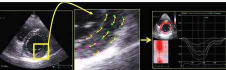
POSITION PAPER. Keywords Adult echocardiography Transthoracic echocardiography Ventricular function Normal values
 European Heart Journal Cardiovascular Imaging (2015) 16, 233 271 doi:10.1093/ehjci/jev014 POSITION PAPER Recommendations for Cardiac Chamber Quantification by Echocardiography in Adults: An Update from
Certificate in Clinician Performed Ultrasound (CCPU) Syllabus. Rapid Cardiac Echo (RCE)
 Syllabus. Rapid Cardiac Echo (RCE)") Certificate in Clinician Performed Ultrasound (CCPU) Syllabus Rapid Cardiac Echo (RCE) Purpose: Rapid Cardiac Echocardiography (RCE) This unit is designed to cover the theoretical and practical curriculum
Certificate in Clinician Performed Ultrasound (CCPU) Syllabus Rapid Cardiac Echo (RCE) Purpose: Rapid Cardiac Echocardiography (RCE) This unit is designed to cover the theoretical and practical curriculum
Cardiac Risk Assessment Using 2D and 3D Transthoracic Echocardiography in Patients Undergoing Haemodialysis
 Cardiac Risk Assessment Using 2D and 3D Transthoracic Echocardiography in Patients Undergoing Haemodialysis A thesis submitted to The University of Manchester for the degree of Doctor of Philosophy (phd)
The importance of left atrium in LV diastolic function
 II Baltic Heart Failure Meeting and Congress of Latvian Society of Cardiology The importance of left atrium in LV diastolic function Dr. Artem Kalinin Eastern Clinical University Hospital Riga 30.09.2010.
ECHOCARDIOGRAPHY CHAPTER INTRODUCTION
 CHAPTER 2 ECHOCARDIOGRAPHY 2.1 INTRODUCTION The use of ultrasound in the diagnosis of cardiac disease has been available for more than four decades with the diagnostic potential of this modality first
Guideline-Driven Care in Cardio- Oncology: Utilizing Recommendations Across Disciplines
 Guideline-Driven Care in Cardio- Oncology: Utilizing Recommendations Across Disciplines Jennifer Liu, MD FACC FASE Director of CV Laboratories Associate Professor of Clinical Medicine Memorial Sloan Kettering
Feasibility and limitations of 2D speckle tracking echocardiography
 ORIGINAL ARTICLE 204 A prospective study in daily clinical practice Feasibility and limitations of 2D speckle tracking echocardiography Lina Melzer, Anja Faeh-Gunz, Barbara Naegeli, Burkhardt Seifert*,
How does the heart pump? From sarcomere to ejection volume
 How does the heart pump? From sarcomere to ejection volume Piet Claus Cardiovascular Imaging and Dynamics Department of Cardiovascular Diseases University Leuven, Leuven, Belgium Course on deformation
Incorporating the New Echo Guidelines Into Everyday Practice
 Incorporating the New Echo Guidelines Into Everyday Practice Clinical Case RIGHT VENTRICULAR FAILURE Gustavo Restrepo MD President Elect Interamerican Society of Cardiology Director Fellowship Training
VECTORS OF CONTRACTION
 1/3/216 Strain, Strain Rate, and Torsion: Myocardial Mechanics Simplified and Applied VECTORS OF CONTRACTION John Gorcsan, MD University of Pittsburgh, Pittsburgh, PA Shortening Thickening Twisting No
Left Ventricular Dyssynchrony in Patients Showing Diastolic Dysfunction without Overt Symptoms of Heart Failure
 ORIGINAL ARTICLE DOI: 10.3904/kjim.2010.25.3.246 Left Ventricular Dyssynchrony in Patients Showing Diastolic Dysfunction without Overt Symptoms of Heart Failure Jae Hoon Kim, Hee Sang Jang, Byung Seok
Assessment of right ventricular contraction by speckle tracking echocardiography in pulmonary hypertension patients.
 Biomedical Research 2017; 28 (1): 173-177 ISSN 0970-938X www.biomedres.info Assessment of right ventricular contraction by speckle tracking echocardiography in pulmonary hypertension patients. Yudong Peng,
Lingyun Kong, Chao Yu, Jihong Guo and Tiangang Zhu. Abstract. Introduction. Department of Cardiology, Peking University People s Hospital.
 24 Original Article Comparison of Left Ventricular Global Longitudinal Strain Measured with Real Time Triplane and 2-Dimensional Echocardiography in Patients with Atrial Fibrillation Lingyun Kong, Chao
Normal values for cardiovascular magnetic resonance in adults and children
 Kawel-Boehm et al. Journal of Cardiovascular Magnetic Resonance (2015) 17:29 DOI 10.1186/s12968-015-0111-7 REVIEW Normal values for cardiovascular magnetic resonance in adults and children Nadine Kawel-Boehm
Right ventricular adaptation in endurance athletes. António Freitas. No conflict of interest
 The role of echocardiography in sports cardiology Right ventricular adaptation in endurance athletes. António Freitas Cardiology Department - Fernando Fonseca Hospital Lisbon Sports Medicine Centre - Lisbon
Revealing new insights. irotate electronic rotation and xplane adjustable biplane imaging. Ultrasound cardiology. irotate and xplane
 Ultrasound cardiology irotate and xplane Revealing new insights irotate electronic rotation and xplane adjustable biplane imaging Annemien van den Bosch and Jackie McGhie Department of Cardiology, Erasmus
British Society of Echocardiography
 British Society of Echocardiography Affiliated to the British Cardiac Society A Minimum Dataset for a Standard Adult Transthoracic Echocardiogram From the British Society of Echocardiography Education
Measurement of Ventricular Volumes and Function: A Comparison of Gated PET and Cardiovascular Magnetic Resonance
 BRIEF COMMUNICATION Measurement of Ventricular Volumes and Function: A Comparison of Gated PET and Cardiovascular Magnetic Resonance Kim Rajappan, MBBS 1,2 ; Lefteris Livieratos, MSc 2 ; Paolo G. Camici,
General Cardiovascular Magnetic Resonance Imaging
 2 General Cardiovascular Magnetic Resonance Imaging 19 Peter G. Danias, Cardiovascular MRI: 150 Multiple-Choice Questions and Answers Humana Press 2008 20 Cardiovascular MRI: 150 Multiple-Choice Questions