Prof. JL Zamorano Hospital Universitario Ramón y Cajal
|
|
|
- Catherine Shaw
- 5 years ago
- Views:
Transcription


1 Prof. JL Zamorano Hospital Universitario Ramón y Cajal


2 Fully Automated Quantification Software Adaptive analytical algorithm consists in knowledge-based identification of global shape and specific adaptation of endocardial border High frame rate single-beat 3DE images, is accurate when compared with conventional volumetric analysis of 4-beat full-volume data sets, thus avoiding is stich artifacts. Medvedofsky D et al. J Am Soc Echocardiogr. 2017;30:
3 Valve and & Cardiac chambers
4

5 Time-consuming Requires training in 3DE analysis Accuracy varies with expertise Reproducibility varies among individuals Accurate Fast Minimal user interaction Reproducible

6 Correlations and time analysis Correlations between HM manual 3D TTE measurements were strong (r = 0.87 to 0.96). Automated 3DE vs CMR p< DE Time Analysis p< Bias of manual 3 DE compared with CMR: EDV -54; ESV: -37; EF: 2; LAV: -33 Tsang W et al. JACC Cardiovasc Imaging. 2016;9:

 Clinical characteristic of the study patients Medvedofsky D et al.")
7 Prospective validation in a multicentre setting Comparison of automated measurements made by six participating sites with and without corrections against reference values generated by Core Lab 180 patients were included divided into four group according to the biplane 2DE LVEF (group 1<_20%, group 2= 21 40%, group 3= 41 55%, group 4 >55%) Clinical characteristic of the study patients Medvedofsky D et al. Eur Heart J Cv Imaging. 2017[]

8 Results of multicenter validation study Experienced readers in differents part of the world can obtain accurate and reproducible automated measurements Automated 3DE Intra and inter observer variability Border correction was deemed necessary in the majority of patients, however, the fully automated analysis was accurate and with endocardial border editing, the accuracy improved only slightly on the average Comparison of the Sites HM to the Core Lab s HM (N=180) Medvedofsky D et al. Eur Heart J Cardiovasc Imaging. 2017]
9 Application of 3DE automated quantification in pre MitraCLip implant LV ESV 2D (ml) LV EDV 2D (ml) LV EF 2D (%) LA vol 2D (ml) LVEF%: LVEF%: (2DEF-HMEF) LVEF%: (2DEF-3DEF) Patients with severe MR usually show relevant remodelling of left chambers causing limitation in the evaluation of geometry and function of both left atrium and ventricle performed with bi- and tridimensional standard analysis Patients with indication of Mitraclip (n=32) were screened All patients underwent a complete standard echo and 3D automated quantification (Heart Model) for the evaluation of geometry and function of LV and LA Correlation between 2D and 3D automated quantification Bland-Altman analysis of 2D, Manual 3D & automated quantification Variables Study Population Sex (M/F)% 80.6(25)/(6)19.4% r 2 = 0.67 r 2 = 0.92 Age(years) 75.4 ± 8.5 NYHA class Class II Class III-IV 23.4% 77.8% LVEF%: (2DEF-3DEF) LVEF%: (2DEF-HMEF) SD Mean SD Afib (%) 52% LV EDV HM (ml) LV EF HM (%) LVEF%: (2DEF+HMEF)/2 r 2 = 0.88 r 2 = SD Mean SD Unpublished data LV ESV HM (ml) LA vol HM (ml) LVEF%: (2DEF+3DEF)/2


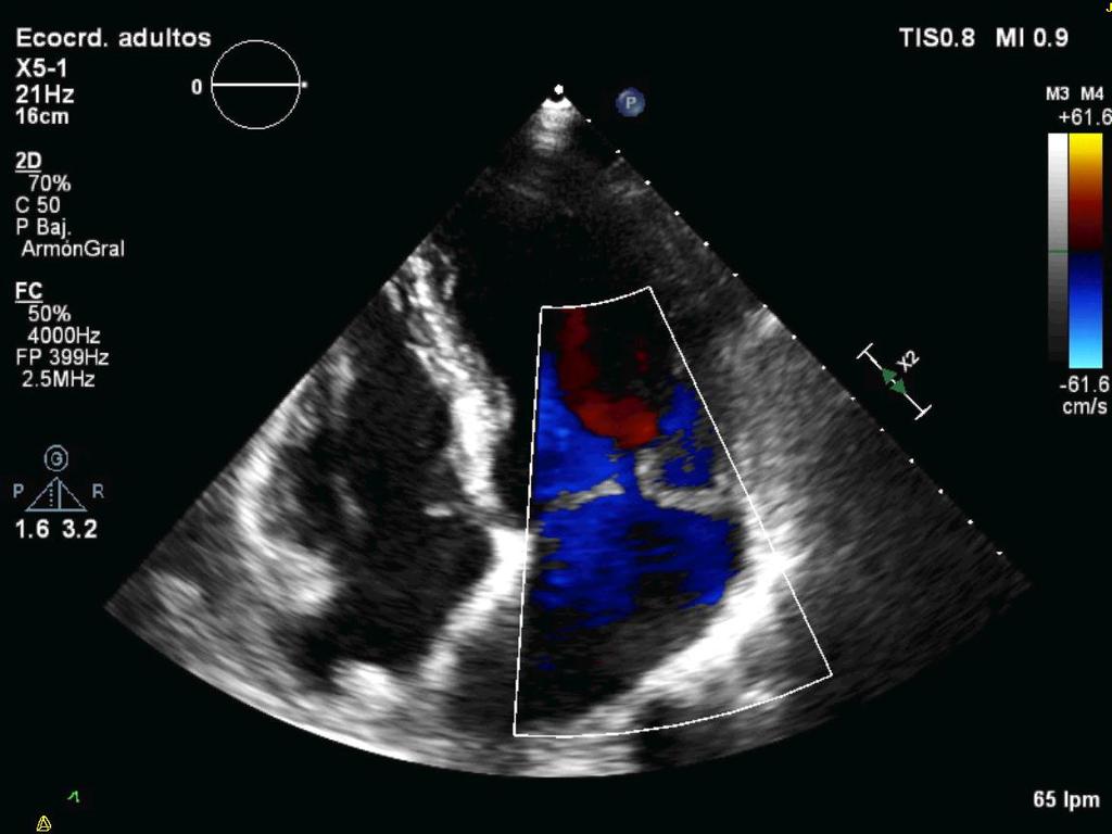
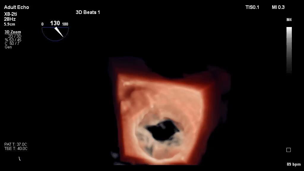
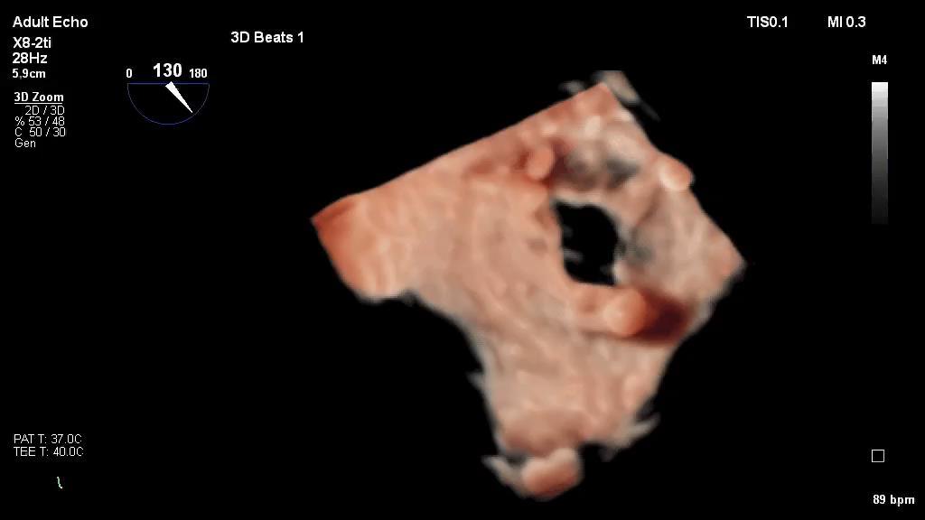
10 AO STENOSIS RT3DE imaging improves the accuracy of the quantification of aortic stenosis. Planimetry of the aortic valve with RT3DE images showed good agreement with the standard 2D TEE technique, flow-derived methods, and cardiac catheterization data with the advantage of improved reproducibility. Analysis of RT3DE revealed that in half of the subjects, the LV outflow tract cross section is not round but rather elliptical. Aortic Valve Heart. 2007;93:

11 Aortic area with RT 3D echo?

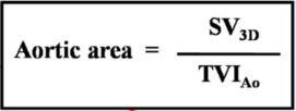
12 Aortic area: RT3D-Doppler hybrid approach

 60 50 40 30 20")
13 AVA(cm2): (2DCE-HM) 2D CE derived AVA(cm2) 3D full automatic software in the evaluation of aortic stenosis severity in TAVI patients Stroke volume and valvular area analysis are strongly recommended for a correct evaluation of AS, especially in patients with reduced flow but severe AS. METHODS: 88 consecutive patients undergoing TAVI (mean age 83.4 ± 6.84; 61.8% female) AVA was calculated by conventional CE (AVACE) and by CE calculated from the stroke volume obtained by 3D analysis (AVAHM). Feasibility of 3D full automatic software was of 73.6% in our study population Determination of LF and NF patients according AVAEC and AVAHM ns Bland-Altman analysis of AVA by 2DCE or 3D automated quantification 70 HM derived AVA(cm2) ns SD + 0,41 Mean Unpublished data 10 0 Normal Flow (%) Low Flow (%) AVA_EC AVA_HM AVA(cm2): (2DCE+HM)/ SD


 in all patients by TEE the day before or the same day")

. A moderate significant correlation was obtained between AVA3DP and AVAHM (r=0,53, p<0,05) but no between")
 AVA 3DP (cm2) Mean difference")
14 3D full automatic software in the evaluation of aortic stenosis severity in TAVI patients. The aim of our study was to evaluate the usefulness of a new 3D automatic quantitative software for aortic valve area (AVA) assessment compared to transesophageal echocardiography (TEE) 3D planimetry in patients undergoing TAVI. METHODS: 18 consecutive patients undergoing TAVI were prospectively included AVA was calculated by conventional CE (AVACE) and by CE calculated from the stroke volume obtained by 3D analysis (AVAHM). 3D planimetry of the aortic valve area was performed (AVA3DP) in all patients by TEE the day before or the same day of the procedure Patients with very poor acoustic window and those studies that needed boundary correction were excluded. Images for AVA calculation by CE and 3D TEE planimetry RESULTS: 18 patients were included (mean age 84±4 years, 20% men). A moderate significant correlation was obtained between AVA3DP and AVAHM (r=0,53, p<0,05) but no between AVA3DP and AVACE. Acquisition and imaging post-processing for 3D images required less than 2 minutes in all cases. 3D- images and automated volumes calculation by Heart Model software AVA CE (cm2) AVA 3DP (cm2) Mean difference (cm2) P value 0,72 0,64 0,08 0,028 AVA HM AVA 3DP Mean difference P value (cm2) (cm2) (cm2) 0,68 0,62 0,06 0,29 C. Fernandez-Golfin et al EuroEcho Imaging Leipzig December 2016
15 Clinical case MITRAL REGURGITATION 56 years old male Barlow s disease. MVP. Asymptomatic severe MR. July 2014 January 2015 July 2015 LVEF Teich 76% 72% LVEF Simpson 70% 68%??? LVEDV 155 ml 150 ml LVESV 45 ml 60 ml LA Volume 110 ml 114 ml

16 MITRAL REGURGITATION January 2015

17 MITRAL REGURGITATION July 2015
18 MITRAL REGURGITATION July 2014 January 2015 July 2015 LVEF Teich 76% 72% 60% LVEF Simpson 70% 68% 58% LVEDV 155 ml 150 ml 170 ml LVESV 45 ml 60 ml 70 ml LA Volume 110 ml 114 ml 112 ml

19 MITRAL REGURGITATION
20 The near future..

21
22 Conclusion Automated software is highly feasible and rapid, allowing the simultaneously measures of LA and LV volumes, LV SV and LVEF This software represent an accurate and robust alternative to conventional manual methodology and is more reproducible. Integration of 3DE quantification with standard measurement may implement pre-interventional screening in patients with valvular diseases (MR or AS) as well as post interventional follow up due to its lower inter and intra operator variability.
23 Quantification is a must. CONCLUSIONS Importance in everyday clinical decision making Heart Model allows fast, easy, automatic and reproducible quantitation of LV and LA volumes and EF Measurements are not only comparable to manual but also CMR values. Promises to facilitate the integration of 3D-TTE based chamber quantification into clinical practice.
Conflict of Interests
 The Left Ventricle: How Should We Quantify Its Size and Function; Is It Time for 3D in Everyone? Roberto M Lang, MD Conflict of Interests Philips Medical Imaging Research Grants Speakers bureau Advisory
When Does 3D Echo Make A Difference?
 When Does 3D Echo Make A Difference? Wendy Tsang, MD, SM Assistant Professor, University of Toronto Toronto General Hospital, University Health Network 1 Practical Applications of 3D Echocardiography Recommended
Three-dimensional echocardiography in the clinical world
 Three-dimensional echocardiography in the clinical world Dr. JL Zamorano Director CV Institute University Clinic SC, Madrid Advantages of 3D. Spatial manipulation. Optimal alineation of structures. Views
Assessment of cardiac function with 3D echocardiography. Đánh giá chức năng tim bằng siêu âm tim 3D
 Assessment of cardiac function with 3D echocardiography Đánh giá chức năng tim bằng siêu âm tim 3D TS. BS. Nguyễn Thị Thu Hoài Viện Tim Mạch Quốc Gia Việt Nam TỪ SIÊU ÂM M-mode ĐẾN SIÊU ÂM 3D TỪ SIÊU ÂM
Conflict of Interests
 New Approaches to Systolic Function: 4D Roberto M Lang, MD Conflict of Interests Philips Medical Imaging Research Grants Speakers bureau Advisory bureau Tomtec Research Grants Epsilon Research Grants 1
EDITOR S PICK CURRENT STATUS OF FULLY AUTOMATED SOFTWARE WITH THREE-DIMENSIONAL ECHOCARDIOGRAPHY FOR THE QUANTIFICATION OF LEFT VENTRICULAR FUNCTION
 ITOR S PICK This paper, courtesy of Yang and Takeuchi, provides a timely and well-considered update on the current status of fully-automated software with three-dimensional echocardiography for quantifying
ECHO HAWAII. Role of Stress Echo in Valvular Heart Disease. Not only ischemia! Cardiomyopathy. Prosthetic Valve. Diastolic Dysfunction
 Role of Stress Echo in Valvular Heart Disease ECHO HAWAII January 15 19, 2018 Kenya Kusunose, MD, PhD, FASE Tokushima University Hospital Japan Not only ischemia! Cardiomyopathy Prosthetic Valve Diastolic
Valvular disease : Ο ρόλος του CMR. Sophie Mavrogeni MD FESC. Onassis Cardiac Surgery Center Athens Greece
 Valvular disease : Ο ρόλος του CMR Sophie Mavrogeni MD FESC Onassis Cardiac Surgery Center Athens Greece Aortic Valve CMR EVALUATION OF AORTIC VALVE Phase-contrast CMR of the aorta to determine aortic
«Paradoxical» low-flow, low-gradient AS with preserved LV function: A Silent Killer
 «Paradoxical» low-flow, low-gradient AS with preserved LV function: A Silent Killer Philippe Pibarot, DVM, PhD, FACC, FAHA, FESC, FASE Canada Research Chair in Valvular Heart Diseases Université LAVAL
Echocardiographic Assessment of the Left Ventricle
 Echocardiographic Assessment of the Left Ventricle Theodora Zaglavara, MD, PhD, BSCI/BSCCT Department of Cardiovascular Imaging INTERBALKAN EUROPEAN MEDICAL CENTER 2015 The quantification of cardiac chamber
Chamber Quantitation Guidelines: What is New?
 Chamber Quantitation Guidelines: What is New? Roberto M Lang, MD J AM Soc Echocardiogr 2005; 18:1440-1463 1 Approximately 10,000 citations iase in itune Cardiac Chamber Quantification: What is New? Database
Prosthesis-Patient Mismatch or Prosthetic Valve Stenosis?
 EuroValves 2015, Nice Prosthesis-Patient Mismatch or Prosthetic Valve Stenosis? Philippe Pibarot, DVM, PhD, FACC, FAHA, FASE FESC Canada Research Chair in Valvular Heart Diseases Université LAVAL Disclosure
Quantifying LV function how good are we?
 Quantifying LV function how good are we? Professor Alan G Fraser Wales Heart Research Institute Cardiff University, U.K. Support for research from Hitachi Aloka, & GE Ultrasound Visual assessment of synchronicity
Multi-imaging modality approach. Covadonga Fernández-Golfín Cardiac Imaging Unit. Cardiology Department. Ramón y Cajal Hospital.
 Multi-imaging modality approach Covadonga Fernández-Golfín Cardiac Imaging Unit. Cardiology Department. Ramón y Cajal Hospital.Madrid Faculty Disclosure Covadonga Fernández-Golfín I have no financial relationships
ORIGINAL ARTICLE. Keywords Real-time 3D echocardiography. Daily practice. Observer experience. CMR. Introduction
 Neth Heart J (2014) 22:383 390 DOI 10.1007/s12471-014-0577-1 ORIGINAL ARTICLE Assessment of LV ejection fraction using real-time 3D echocardiography in daily practice: direct comparison of the volumetric
Reproducibility and Accuracy of Echocardiographic Measurements of Left Ventricular Parameters Using Real-Time Three-Dimensional Echocardiography
 Journal of the American College of Cardiology Vol. 44, No. 4, 2004 2004 by the American College of Cardiology Foundation ISSN 0735-1097/04/$30.00 Published by Elsevier Inc. doi:10.1016/j.jacc.2004.05.050
Valvular Regurgitation: Can We Do Better Than Colour Doppler?
 Valvular Regurgitation: Can We Do Better Than Colour Doppler? A/Prof David Prior St Vincent s Hospital Melbourne Sports Cardiology Valvular Regurgitation Valve regurgitation volume loads the ventricles
Echocardiographic evaluation of mitral stenosis
 Echocardiographic evaluation of mitral stenosis Euroecho 2011 Philippe Unger, MD, FESC Erasme Hospital, ULB, Brussels, Belgium I have nothing to declare EuroHeart Survey Etiology of single native left-sided
HIGHLIGHT SESSION. Imaging. J. L. Zamorano Gomez (Madrid, ES) Disclosures: Speaker Philips
 Disclosures: Speaker Philips") Imaging. J. L. Zamorano Gomez (Madrid, ES) Disclosures: Speaker Philips Agenda ECHO Diagnosis & Prognosis : Functional MR Severity Aortic Stenosis CT How to select pts for TAVI Adding prognostic info to
Imaging. J. L. Zamorano Gomez (Madrid, ES) Disclosures: Speaker Philips Agenda ECHO Diagnosis & Prognosis : Functional MR Severity Aortic Stenosis CT How to select pts for TAVI Adding prognostic info to
The correlation of AVA measured by transthoracic, transesophageal echocardiography and cardiac CT
 The correlation of AVA measured by transthoracic, transesophageal echocardiography and cardiac CT R.Petr, H.Linkova, J.Knot, E.Paskova, J.Daniel, M.Labos Cardiocenter of University Hospital Kralovske Vinohrady
Reverse left atrium and left ventricle remodeling after aortic valve interventions
 Reverse left atrium and left ventricle remodeling after aortic valve interventions Alexandra Gonçalves, Cristina Gavina, Carlos Almeria, Pedro Marcos-Alberca, Gisela Feltes, Rosanna Hernández-Antolín,
Dobutamine Stress testing In Low Flow, Low EF, Low Gradient Aortic Stenosis Case Studies
 Dobutamine Stress testing In Low Flow, Low EF, Low Gradient Aortic Stenosis Case Studies Mitral Regurgitation The New ASE Guidelines: Role of 2D/3D and CMR William A. Zoghbi MD, FASE, MACC Professor and
Organic mitral regurgitation
 The best in heart valve disease Organic mitral regurgitation Ewa Szymczyk Department of Cardiology Medical University of Lodz, Poland I have nothing to declare Organic mitral regurgitation leaflet abnormality
New imaging modalities for assessment of TAVI procedure and results. R Dulgheru, MD Heart Valve Clinic CHU, Liege
 New imaging modalities for assessment of TAVI procedure and results R Dulgheru, MD Heart Valve Clinic CHU, Liege Disclosure of Interest I, Raluca Dulgheru, DO NOT HAVE a financial interest/arrangement
3D-stress echocardiography Bernard Cosyns, MD, PhD
 3D-stress echocardiography Bernard Cosyns, MD, PhD No Disclosure The Pro-Technology bias Sicari et al. Cardiovascular Ultrasound 2006, 4:11 Overview 2D stress echocardiography: main limitations 3D echocardiography:
LV geometric and functional changes in VHD: How to assess? Mi-Seung Shin M.D., Ph.D. Gachon University Gil Hospital
 LV geometric and functional changes in VHD: How to assess? Mi-Seung Shin M.D., Ph.D. Gachon University Gil Hospital LV inflow across MV LV LV outflow across AV LV LV geometric changes Pressure overload
Reshape/Coapt: do we need more? Prof. J Zamorano Head of Cardiology University Hospital Ramon y Cajal, Madrid
 Reshape/Coapt: do we need more? Prof. J Zamorano Head of Cardiology University Hospital Ramon y Cajal, Madrid Patient records 76 y.o. male Hypertension. Dyslipidemia. OPLD. Smoked in the past. Diabetes
Comprehensive Echo Assessment of Aortic Stenosis
 Comprehensive Echo Assessment of Aortic Stenosis Smonporn Boonyaratavej, MD, MSc King Chulalongkorn Memorial Hospital Bangkok, Thailand Management of Valvular AS Medical and interventional approaches to
MAYON VOLCANO: FAST FACTS
 MAYON VOLCANO: FAST FACTS Type of Volcano: Stratovolcano Elevation: 2.46 km Base Diameter: 20 km Base Circumference: 62.8 km Area: 314.1 km 2 Reference: http://www.phivolcs.dost.gov.ph/html/update_vmepd/volcano/volcanolist/mayon.htm
TAVR: Echo Measurements Pre, Post And Intra Procedure
 2017 ASE Florida, Orlando, FL October 10, 2017 8:00 8:25 AM 25 min TAVR: Echo Measurements Pre, Post And Intra Procedure Muhamed Sarić MD, PhD, MPA Director of Noninvasive Cardiology Echo Lab Associate
좌심실수축기능평가 Cardiac Function
 Basic Echo Review Course 좌심실수축기능평가 Cardiac Function Seonghoon Choi Cardiology Hallym university LV systolic function Systolic function 좌심실수축기능 - 심근의수축으로심실에서혈액을대동맥으로박출하는기능 실제임상에서 LV function 의의미 1Diagnosis
Optimal Imaging Technique Prior to TAVI -Echocardiography-
 2014 KSC meeting Optimal Imaging Technique Prior to TAVI -Echocardiography- Geu-Ru Hong, M.D. Ph D Associate Professor of Medicine Division of Cardiology, Severance Cardiovascular Hospital Yonsei University
Severity of AS Degree of AV calcification (? Bicuspid AV), annulus size, & aortic root
, annulus size, & aortic root") The role of Cardiac Imaging modalities in evaluation & selection of patients for Trans-catheter Aortic Valve Implantation Dr.Saeed AL Ahmari Consultant Cardiologist Prince Sultan Cardaic Center, Riyadh
The role of Cardiac Imaging modalities in evaluation & selection of patients for Trans-catheter Aortic Valve Implantation Dr.Saeed AL Ahmari Consultant Cardiologist Prince Sultan Cardaic Center, Riyadh
Aortic stenosis aetiology: morphology of calcific AS,
 How to improve patient selection in aortic stenosis? Fausto J. Pinto, FESC Aortic stenosis aetiology: morphology of calcific AS, bicuspid valve, and rheumatic AS (Adapted from C. Otto, Principles of
Disclosures Rebecca T. Hahn, MD, FASE
 The New ASE Guidelines for Native Valvular Regurgitation Mitral Regurgitation The New ASE Guidelines: Role of 2D/3D and CMR (With caveats and comments from R. Hahn) William A. Zoghbi MD, FASE, MACC Professor
Νεότερα ςτην Υπερηχοκαρδιογραφία. Βαςίλειοσ Καμπερίδησ Clinical research fellow in Cardiology
 Νεότερα ςτην Υπερηχοκαρδιογραφία Βαςίλειοσ Καμπερίδησ Clinical research fellow in Cardiology Disclosures ESC training grant EACVI research grant HCS training grant ELIKAR research grant Evolution of Echocardiography
What echo measurements are key prior to MitraClip?
 APHP CHU Bichat - Claude Bernard What echo measurements are key prior to MitraClip? Eric Brochet,MD Cardiology Department Hopital Bichat Paris France No disclosure Conflict of interest Case 69 y.o man
The best in heart valve disease Aortic valve stenosis
 The best in heart valve disease Aortic valve stenosis Marie Moonen, MD, PhD Heart Valve Clinic, University of Liège, CHU Sart Tilman, BELGIUM My declaration of interest : I have nothing to declare Prevalence
Revealing new insights. irotate electronic rotation and xplane adjustable biplane imaging. Ultrasound cardiology. irotate and xplane
 Ultrasound cardiology irotate and xplane Revealing new insights irotate electronic rotation and xplane adjustable biplane imaging Annemien van den Bosch and Jackie McGhie Department of Cardiology, Erasmus
AS with reduced LV ejection fraction: Contractile reserve should be systematically assessed: PRO
 AS with reduced LV ejection fraction: Contractile reserve should be systematically assessed: PRO Jean-Luc MONIN, MD, PhD Henri Mondor University Hospital Créteil, FRANCE Potential conflicts of interest
Balloon mitral valvuloplasty
 Echo for selecting and monitoring procedures in mitral valve diseases Balloon mitral valvuloplasty Eric Brochet,MD Cardiology Department Hôpital Bichat. Paris France Conflict of interest : none Balloon
10/7/2013. Systolic Function How to Measure, How Accurate is Echo, Role of Contrast. Thanks to our Course Director: Neil J.
 Systolic Function How to Measure, How Accurate is Echo, Role of Contrast Neil J. Weissman, MD MedStar Health Research Institute & Professor of Medicine Georgetown University Washington, D.C. No Disclosures
What the Cardiologist needs to know from Medical Images
 What the Cardiologist needs to know from Medical Images Gerald Maurer Department of Cardiology Medical University of Vienna What kinds of Cardiologists Plumbers Electricians Photographers And then there
Percutaneous Mitral Valve Repair: What Can We Treat and What Should We Treat
 Percutaneous Mitral Valve Repair: What Can We Treat and What Should We Treat Innovative Procedures, Devices & State of the Art Care for Arrhythmias, Heart Failure & Structural Heart Disease October 8-10,
Severe left ventricular dysfunction and valvular heart disease: should we operate?
 Severe left ventricular dysfunction and valvular heart disease: should we operate? Laurie SOULAT DUFOUR Hôpital Saint Antoine Service de cardiologie Pr A. COHEN JESFC 16 janvier 2016 Disclosure : No conflict
HAND held echocardiography a dangerous toy?
 HAND held echocardiography a dangerous toy? Eva Gerdts Professor of Medicine University of Bergen Visiting Professor of Medicine Federico II University, Napoli Diagnostic accuracy Vscan vs high end echocardiograph
Prosthetic valve dysfunction: stenosis or regurgitation
 Prosthetic valve dysfunction: stenosis or regurgitation Jean G. Dumesnil MD, FRCP(C), FACC, FASE(Hon) Quebec Heart and Lung Institute, Québec, Québec No disclosures Possible Causes of High Gradients in
Three-dimensional Wall Motion Tracking:
 Three-dimensional Wall Motion Tracking: A Novel Echocardiographic Method for the Assessment of Ventricular Volumes, Strain and Dyssynchrony Jeffrey C. Hill, BS, RDCS, FASE Jennifer L. Kane, RCS Gerard
A patient with aortic stenosis and LV dysfunction EuroECHO & Other Imaging Modalities 2012 Athens, Greece
 A patient with aortic stenosis and LV dysfunction EuroECHO & Other Imaging Modalities 2012 Athens, Greece Jean-Luc MONIN, MD, PhD. University Hospital, Créteil, FRANCE My disclosures: Lecture and/ or consulting
Introduction. Aims. Keywords
 European Journal of Echocardiography (2010) 11, 359 368 doi:10.1093/ejechocard/jep217 Validation of a novel automated border-detection algorithm for rapid and accurate quantitation of left ventricular
Mild paravalvular regurgitation is not an independent predictor of mortality following TAVI
 Mild paravalvular regurgitation is not an independent predictor of mortality following TAVI Philippe Pibarot, DVM, PhD, FACC, FAHA, FASE Canada Research Chair in Valvular Heart Diseases INSTITUT UNIVERSITAIRE
2019 Qualified Clinical Data Registry (QCDR) Performance Measures
 Performance Measures") 2019 Qualified Clinical Data Registry (QCDR) Performance Measures Description: This document contains the 18 performance measures approved by CMS for inclusion in the 2019 Qualified Clinical Data Registry
2019 Qualified Clinical Data Registry (QCDR) Performance Measures Description: This document contains the 18 performance measures approved by CMS for inclusion in the 2019 Qualified Clinical Data Registry
LV function in ischemic heart failure - decreased correlation between Echo and CMR
 LV function in ischemic heart failure - decreased correlation between Echo and CMR Poster No.: C-0590 Congress: ECR 2011 Type: Scientific Exhibit Authors: K. Gruszczy#ska, L. Krzych, K. Golba, P. Ulbrych,
Evaluation of Ejection Fraction in Patients with Cardiac Resynchronization Therapy by Two and Three Dimensional Echocardiography
 58 Original article Evaluation of Ejection Fraction in Patients with Cardiac Resynchronization Therapy by Two and Three Dimensional Echocardiography Anil OM Department of cardiology, Manmohan Cardiothoracic
Low Gradient AS Normal LVEF
 Low Gradient AS Normal LVEF Shahbudin H. Rahimtoola MB, FRCP, MACP, MACC, FESC, D.Sc.(Hon) Distinguished Professor University of Southern California Griffith Professor of Cardiology Professor of Medicine
Coronary artery disease (CAD) risk factors
 risk factors") Background Coronary artery disease (CAD) risk factors CAD Risk factors Hypertension Insulin resistance /diabetes Dyslipidemia Smoking /Obesity Male gender/ Old age Atherosclerosis Arterial stiffness precedes
Background Coronary artery disease (CAD) risk factors CAD Risk factors Hypertension Insulin resistance /diabetes Dyslipidemia Smoking /Obesity Male gender/ Old age Atherosclerosis Arterial stiffness precedes
LA Function analysis Marcia Barbosa Vice Presidente - Brazilian Soc of Cardiology President-elect - Interamerican Soc of Cardiology
 LA Function analysis Marcia Barbosa Vice Presidente - Brazilian Soc of Cardiology President-elect - Interamerican Soc of Cardiology Belo Horizonte Brazil DECLARATION OF CONFLICT OF INTEREST Nothing to
Direct Planimetry of Mitral Valve Regurgitation Orifice Area by Real-time 3D Transesophageal Echocardiography
 Direct Planimetry of Mitral Valve Regurgitation Orifice Area by Real-time 3D Transesophageal Echocardiography Ertunc Altiok, Sandra Hamada, Silke van Hall, Mehtap Hanenberg, Eva Grabskaya, Michael Becker,
Aortic Valve Replacement Improves Outcome in Patients with Preserved Ejection Fraction: PRO!
 ESC 2011, Paris Controversies in Low-Flow, Low-Gradient Aortic Stenosis Aortic Valve Replacement Improves Outcome in Patients with Preserved Ejection Fraction: PRO! Philippe Pibarot, DVM, PhD, FACC, FAHA,
DON T FORGET TO OPTIMISE DEVICE PROGRAMMING
 CRT:NON-RESPONDERS OR NON-PROGRESSORS? DON T FORGET TO OPTIMISE DEVICE PROGRAMMING Prof. ALİ OTO,MD,FESC,FACC,FHRS Chairman,Department of Cardiology Hacettepe University Faculty of Medicine,Ankara Causes
Planimetric and continuity equation assessment of aortic valve area (AVA): comparison between cardiac magnetic resonance (cmr) and echocardiography
: comparison between cardiac magnetic resonance (cmr) and echocardiography") Planimetric and continuity equation assessment of aortic valve area (AVA): comparison between cardiac magnetic resonance (cmr) and echocardiography Poster No.: C-2058 Congress: ECR 2011 Type: Scientific
Planimetric and continuity equation assessment of aortic valve area (AVA): comparison between cardiac magnetic resonance (cmr) and echocardiography Poster No.: C-2058 Congress: ECR 2011 Type: Scientific
True morphology of mitral regurgitant flow assessed by three- dimensional transesophageal echocardiography
 DOI: 10.1111/echo.13395 ORIGINAL INVESTIGATION True morphology of mitral regurgitant flow assessed by three- dimensional transesophageal echocardiography Martin Lombardero M.D. Ruth Henquin D.L.S.H.T.M.,
Aortic valve Stenosis: Insights in the evaluation of LV function. Erwan DONAL Cardiologie CHU Rennes
 Aortic valve Stenosis: Insights in the evaluation of LV function Erwan DONAL Cardiologie CHU Rennes erwan.donal@chu-rennes.fr Preload Afterload Myocardial Fiber Shortening Circumferential Longitudinal
imagine 2018 Echocardiography Today - Do, Learn, Do More, Learn More November 3-4, 2018 Georgia Technology Hotel and Conference Center
 Piedmont Heart presents imagine 2018 Echocardiography Today - Do, Learn, Do More, Learn More November 3-4, 2018 Georgia Technology Hotel and Conference Center Program Co-Directors Mani A. Vannan MBBS FASE
Imaging heart. UK Biobank Annual Meeting 13 th June 2016
 NIHR Barts and The London Cardiovascular Biomedical Research Unit National Institute for Health Research Cardiovascular Biomedical Research Unit at Barts Imaging heart UK Biobank Annual Meeting 13 th June
Back to Basics: Common Errors In Quantitation In Everyday Practice
 Back to Basics: Common Errors In Quantitation In Everyday Practice Deborah Agler, ACS, RDCS, FASE October 9, 2017 ASE: Echo Florida Rebecca T. Hahn, MD Director of Interventional Echocardiography Professor
Hemodynamic Assessment. Assessment of Systolic Function Doppler Hemodynamics
 Hemodynamic Assessment Matt M. Umland, RDCS, FASE Aurora Medical Group Milwaukee, WI Assessment of Systolic Function Doppler Hemodynamics Stroke Volume Cardiac Output Cardiac Index Tei Index/Index of myocardial
Titel Kardiologie-SG.ch hot topics in heart failure and mitral regurgitation
 Titel Kardiologie-SG.ch hot topics in heart failure and mitral regurgitation where and how to quantify mitral regurgitation: Echolab, Cathlab or MRI? Philipp K. Haager, St. Gallen Measuring mitral regurgitation?
Low Gradient Severe AS: Who Qualifies for TAVR? Andrzej Boguszewski MD, FACC, FSCAI Vice Chairman, Cardiology Mid-Michigan Health Associate Professor
 Low Gradient Severe AS: Who Qualifies for TAVR? Andrzej Boguszewski MD, FACC, FSCAI Vice Chairman, Cardiology Mid-Michigan Health Associate Professor Michigan State University, Central Michigan University
Affecting the elderly Requiring new approaches. Echocardiographic Evaluation of Hemodynamic Severity. Increasing prevalence Mostly degenerative
 Echocardiographic Evaluation of Hemodynamic Severity Steven J. Lester MD, FACC, FRCP(C), FASE Mayo Clinic, Arizona Relevant Financial Relationship(s) None Off Label Usage None A re-emerging public-health
TAVI EN INSUFICIENCIA AORTICA
 TAVI EN INSUFICIENCIA AORTICA Cesar Moris Profesor Cardiología Director Departamento del Corazón Hospital Universitario Central de Asturias Universidad de Oviedo OVIEDO -- ESPAÑA CONFLICTO DE INTERESES
Quantification of Cardiac Chamber Size
 2017 KSE 2017-11-25 Quantification of Cardiac Chamber Size Division of Cardiology Keimyung University Dongsan Medical Center In-Cheol Kim M.D., Ph.D. LV size and function Internal linear dimensions PLX
Alicia Armour, MA, BS, RDCS
 Alicia Armour, MA, BS, RDCS No disclosures Review 2D Speckle Strain (briefly) Discuss some various patient populations & disease pathways where Strain can be helpful Discuss how to acquire images for Strain
Load and Function - Valvular Heart Disease. Tom Marwick, Cardiovascular Imaging Cleveland Clinic
 Load and Function - Valvular Heart Disease Tom Marwick, Cardiovascular Imaging Cleveland Clinic Indications for surgery in common valve lesions Risks Operative mortality Failed repair - to MVR Operative
Catheter-based mitral valve repair MitraClip System
 Percutaneous Mitral Valve Repair: Results of the EVEREST II Trial William A. Gray MD Director of Endovascular Services Associate Professor of Clinical Medicine Columbia University Medical Center The Cardiovascular
Echocardiographie de la Tétralogie de Fallot opérée
 Echocardiographie de la Tétralogie de Fallot opérée Diala Khraiche M3C-Necker Enfants malades, Université Paris Descartes Paris, France. Disclosure Statement of Financial Interest I currently have, or
New Cardiovascular Devices and Interventions: Non-Contrast MRI for TAVR Abhishek Chaturvedi Assistant Professor. Cardiothoracic Radiology
 New Cardiovascular Devices and Interventions: Non-Contrast MRI for TAVR Abhishek Chaturvedi Assistant Professor Cardiothoracic Radiology Disclosure I have no disclosure pertinent to this presentation.
Exercise Testing/Echocardiography in Asymptomatic AS
 Exercise Testing/Echocardiography in Asymptomatic AS Raluca Dulgheru, MD Heart Valve Clinic, University of Liège, CHU Sart Tilman, BELGIUM Disclosure related to this presentation: None VALVULAR HEART DISEASE
ASE Guidelines on Aortic Regurgitation What Do I Measure? Case Studies
 ASE Guidelines on Aortic Regurgitation What Do I Measure? Case Studies Mitral Regurgitation The New ASE Guidelines: Role of 2D/3D and CMR William A. Zoghbi MD, FASE, MACC Professor and Chairman, Department
Part II: Fundamentals of 3D Echocardiography: Acquisition and Application
 Part II: Fundamentals of 3D Echocardiography: Acquisition and Application Dr. Bruce Bollen 3D matrix array TEE probes provide options for both 2D and 3D imaging. Indeed, their utility in obtaining multiple
Ioannis Alexanian, MD, PhD Department of Cardiology General Hospital of Chest Diseases Sotiria Athens
 MITRAL REGURGITATION IN PATIENT WITH SEVERE AORTIC VALVE STENOSIS Ioannis Alexanian, MD, PhD Department of Cardiology General Hospital of Chest Diseases Sotiria Athens I HAVE NOTHING TO DECLARE Management
Advanced Evaluation of Left Ventricular Function in Degenerative MR. Dr Julien Magne, PhD University of Liege, CHU Sart Tilman, Liege, Belgium
 Advanced Evaluation of Left Ventricular Function in Degenerative MR Dr Julien Magne, PhD University of Liege, CHU Sart Tilman, Liege, Belgium Conflict of Interest Disclosure None Case Clinical data Previous
Comparison of Cardiac MDCT with MRI and Echocardiography in the Assessement of Left Ventricular Function
 Comparison of Cardiac MDCT with MRI and Echocardiography in the Assessement of Left Ventricular Function Poster No.: C-0969 Congress: ECR 2012 Type: Scientific Exhibit Authors: B. Kara, Y. Paksoy, C. Erol,
Martin G. Keane, MD, FASE Temple University School of Medicine
 Martin G. Keane, MD, FASE Temple University School of Medicine Measurement of end-diastolic LV internal diameter (LVIDd) made by properly-oriented M-Mode techniques in the Parasternal Long Axis View (PLAX):
The V Wave. January, 2007 Joe M. Moody, Jr, MD UTHSCSA and ALMMVAH. Ref: Kern MJ. Hemodynamic Rounds, 2 nd ed
 The V Wave January, 2007 Joe M. Moody, Jr, MD UTHSCSA and ALMMVAH Ref: Kern MJ. Hemodynamic Rounds, 2 nd ed. 1999. Normal Hemodynamic Values Cardiac index 2.8-4.2 (mean 3.4 L/min/m 2 ) Stroke volume 30-65
Aortic Regurgitation and Aortic Aneurysm - Epidemiology and Guidelines -
 Reconstruction of the Aortic Valve and Root - A Practical Approach - Aortic Regurgitation and Aortic Aneurysm Wednesday 14 th September - 9.45 Practice must always be founded on sound theory. Leonardo
3D Printing & Echocardiography
 Echo Hawaii Jan 18, 2018 3D Printing & Echocardiography Stephen H. Little, MD John S. Dunn Chair in Cardiovascular Research and Education, Associate professor, Weill Cornell Medicine Rapid Prototyping
Importance of CRT team for optimization of the results: a European point of view
 Importance of CRT team for optimization of the results: a European point of view Matteo Bertini, MD, PhD Arcispedale S. Anna Azienda Ospedaliero-Universitaria Cona-Ferrara No conflict of interest to declare
Imaging of the Heart Todd Tessendorf MD FACC
 Imaging of the Heart Todd Tessendorf MD FACC Outline Imaging Modalities for Structural Heart Disease ECHO, MRI Imaging Modalities for Ischemic Heart Disease SPECT, PET, CCTA Show lots of pretty pictures
Low Gradient Severe? AS
 Low Gradient Severe? AS Philippe Pibarot, DVM, PhD, FACC, FAHA, FESC, FASE Canada Research Chair in Valvular Heart Diseases Institut Universitaire de Cardiologie et de Pneumologie de Québec / Québec Heart
New Imaging for Aortic Valve Disease. Anthony DeMaria Judy and Jack White Chair Director, Sulpizio CV Center University of California, San Diego
 New Imaging for Aortic Valve Disease Anthony DeMaria Judy and Jack White Chair Director, Sulpizio CV Center University of California, San Diego Imaging in Aortic Stenosis Valve morphology calcification
Sténose aortique à Bas Débit et Bas Gradient
 3.6 m/s Sténose aortique à Bas Débit et Bas Gradient Philippe Pibarot, DVM, PhD, FACC, FAHA, FESC, FASE Canada Research Chair in Valvular Heart Diseases Doctorate Honoris Causa, Université de Liège Institut
Paravalvular Regurgitation is a Risk Factor Following TAVI
 Paravalvular Regurgitation is a Risk Factor Following TAVI Philippe Pibarot, DVM, PhD, FACC, FESC, FASE Canada Research Chair in Valvular Heart Disease INSTITUT UNIVERSITAIRE DE CARDIOLOGIE ET DE PNEUMOLOGIE
powerful versatile mobile
 M9 Premium Compact Ultrasound System powerful versatile mobile M9 Premium Compact Ultrasound System powerful versatile mobile Powerful Platform With the next generation of technologies, the Mindray M9
Noncoronary Cardiac MDCT
 Noncoronary Cardiac MDCT David A. Bluemke, M.D., Ph.D. Professor, of Radiology and Medicine Johns Hopkins University School of Medicine Baltimore, Maryland Toshiba Disclosures Grant support Noncoronary
Guidelines in perspective?
 EuroValve 2016 Challenges in the management Secondary MR Guidelines in perspective? Luc A. Pierard, MD, PhD Professor of Medicine Head of the Department of Cardiology, Liège, Belgium Heart Valve Clinic,
The difficult patient with mitral regurgitation
 Clinical pathways The difficult patient with mitral regurgitation Stress echo can be the best tool Challenging cases Maria João Andrade, Lisbon PT Management of Severe Chronic Organic MR Echo Exercise
Aortic Valve Stenosis: Flow and Gradient stratification and association with TAVR outcomes
 Aortic Valve Stenosis: Flow and Gradient stratification and association with TAVR outcomes Kostis Raisakis General Hospital of Athens «G. Gennimatas» Severe Aortic Stenosis Peak Velocity 4 m/s Up to 40%
Role of Stress Echo in Valvular Heart Disease. Satoshi Nakatani Osaka University Graduate School of Medicine Osaka, Japan
 Role of Stress Echo in Valvular Heart Disease Satoshi Nakatani Osaka University Graduate School of Medicine Osaka, Japan Exercise echocardiography Dobutamine echocardiography Usefulness of exercise echo
Basic Assessment of Left Ventricular Systolic Function
 WINFOCUS BASIC ECHO (WBE) Basic Assessment of Left Ventricular Systolic Function Ritesh Dhar, MD Director, Echocardiography Lab and Staff Cardiologist Intermountain Medical Center Murray, Utah Outline