Conflict of Interests
|
|
|
- Rosalind Nichols
- 5 years ago
- Views:
Transcription


1 The Left Ventricle: How Should We Quantify Its Size and Function; Is It Time for 3D in Everyone? Roberto M Lang, MD Conflict of Interests Philips Medical Imaging Research Grants Speakers bureau Advisory bureau Tomtec Research Grants Epsilon Research Grants 1


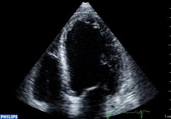

2 Eye ball How do we Assess LV Function? Qualitative Assessment Subjective Experience dependent Lack of standardization Large inter- and intraobserver variability J AM Soc Echocardiogr 2005; 18:


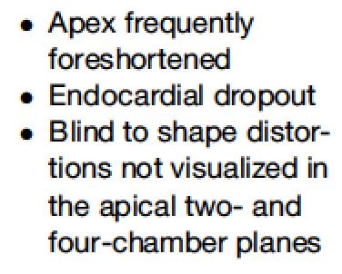
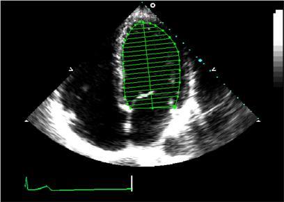
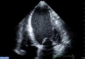
3 Left Ventricular Volumetric Measurement 1 Biplane Disk Summation Area Length Method 2 Quantitation of 2D Echocardiography: 2016 Hand tracing Correct view? Foreshortening? Correct shape? Geometry dependent? Tracing errors? Correct trace? 3


4 Cardiac Chamber Quantification: What is New? Database Deformation Imaging RT3DE Eur Heart J Cardiovasc Imaging Mar;16(3): J Am Soc Echocardiogr 2015;28:1-39 4


5 Chamber Quantification Uses of 3D Echocardiography Recommended Promising Clinical Trials Areas of active Research LV Volumes LV Mass LV Shape MV anatomy RV Volumes LV Dyssynchrony MV Stenosis Ao Anatomy LA Volumes Guidance of Transcatheter Procedures Ao Stenosis MV Regurgitation Prosthetic Valves Lang RM, Badano L et al, JASE


6 Why is 3D More Accurate? long axis (cm) A4C * 7 2D 3D Mor-Avi V, Lang RM et al., Circulation :


7 Validation by MRI Improved accuracy: validation against CMR EDV, ESV Excellent correlation (r²>0.85) but RT3DE underestimates volumes Jacobs LD, et al. Eur Heart J 2005; 27:460-8 Sugeng L, et al. Circulation 2006; EUROECHO 114: Madrid Jenkins C, et al. J Am Soc Echocardiogr 2007; 20:962-8 Soliman OI, et al. Am Soc Echocardiogr 2007; 20: Sources of error: 3D Egg-shaped phantom Small difference between the 2 boundaries resulted in an 11% difference in the measured volume of the 3D shell! Mor-Avi V.,Lang RM et al, JACC Cardiovasc Img 2008: 1:


8 LV Volumes: 3DE Advantages Avoid image foreshortening No geometric assumptions More accurate and reproducible Disadvantages Low temporal resolution Less data on normals Limitations to the Implementation of 3D Echocardiography in the Laboratory Time-consuming Requires training in 3DE analysis Accuracy varies with expertise Reproducibility varies among EUROECHO Madrid individuals and institutions 8


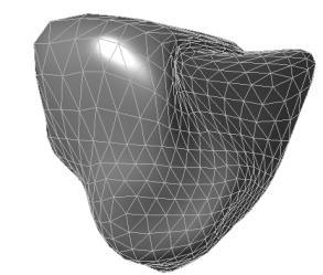
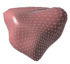
9 Real-Time Automated 3D TTE Left Heart Chamber Quantification using an Adaptive Analytics Algorithm Automated Model-Based Segmentation of the Heart CT MRI 3DE TIME 3 9


 Library of")


10 Real-Time Automated Transthoracic Three- Dimensional Echocardiographic Left Heart Chamber Heart Quantification Model Overview using an Adaptive Analytics Algorithm Generic Model Final Model Tsang w, Lang RM. JACC Imaging (in press) Library of ~1000 Images Local Adaptation Adjustments for: variations in dropout acoustic clutter ventricular shape dataset orientation Real-Time Automated Transthoracic Three- Dimensional Echocardiographic Left Heart Chamber Quantification using an Adaptive Analytics Algorithm Dilated Banana Sigmoid Septum Normal Tsang w, Lang RM. JACC Imaging (in press) 10


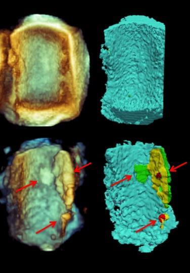
11 Real-Time Automated Transthoracic Three-Dimensional Echocardiographic Left Heart HeartModel A.I. Chamber Quantification using an Adaptive Analytics Algorithm Algorithm Align & Orient Model Adjust Local Borders Automatically Corrects Foreshortening Avoids Geometric Assumptions 15 Initial fully automated model Global/regional corrections LV Focused Views Final model 4-chamber 2-chamber 3-chamber LA Focused Views 11


12 A LV Focused Views 4-chamber 2-chamber 3-chamber B LA Focused Views C 4-chamber 2-chamber 3-chamber Fully Automated Cardiac Chamber Quantification 3D RV End- Diastolic Volume 3D LV End- Diastolic Volume 3D RV End- Systolic Volume 3D RV Ejection Fraction 3D RA Volume at LV End-Systole EUROECHO Madrid 3D LV End- Systolic Volume 3D LV Ejection Fraction 3D LA Volume at LV End- Systole Tsang w, Lang RM. JACC Imaging (in press) 12

13 Time Savings 2h and 56 min 63% 1h and 6 min 82% 33 min EUROECHO Madrid Uses of 3D Echocardiography Recommended Promising Clinical Trials Areas of active Research LV Volumes LV Mass LV Shape MV anatomy RV Volumes LV Dyssynchrony MV Stenosis Ao Anatomy LA Volumes Guidance of Transcatheter Procedures Ao Stenosis MV Regurgitation Prosthetic Valves Lang RM, Badano L et al, JASE


14 LV Remodeling LV SHAPE LV Function 27 Pre-Operative 6 Months 12 Months Pre-operative 6 months 12 months Maffessanti F, Caiani EG, Tamborini G, Muratori M, Sugeng L, Weinert L, Alamanni F, Zanobini M, Mor-Avi V, Lang RM, Pepi M. Am J Cardiol 2010 September 15;106(6):

 MR 0-2 MR 3-4 33±8 41±8* Increased LV sphericity associated with")

15 2D Sphericity Index 2DE Si = Biplane EDV Volume of sphere with the *100 LV long axis as diameter [Marsan et al, Ann Thorac Surg 2011;91:113-22] Application to 2D echocardiographic images, LV EDV was obtained by apical biplane Simpson s rule, to study in men the pathogenesis of MR. [Van Dantzig et al, Am Heart J 1996;131;865] LV sphericity (%) MR 0-2 MR ±8 41±8* Increased LV sphericity associated with severity of MR, independent of LV volumes. 3D Sphericity Index 3D dataset reconstructed from multiple 2D echo apical freehand acquisitions SI V 3D LVV 3 4 D 3 2 [Mannaerts et al. Eur Heart J 2004;25:680-7] Unlike the traditional 2D indices, the 3D sphericity index can predict, accurately and soon in the sub-acute phase after AMI, which patient is likely to undergo LV remodeling. 15

 Shape index:")

![Ultrasound Med Biol 2009;35:1953-62] New shape indexes based on LV](/docs-images/83/87264491/images/16-3.jpg "Contour Evolution through the cardiac cycle in a normal subject ES")
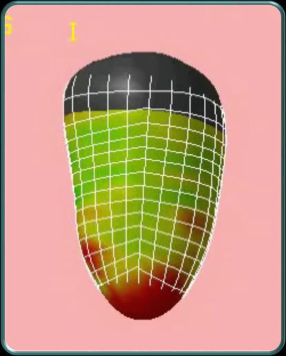
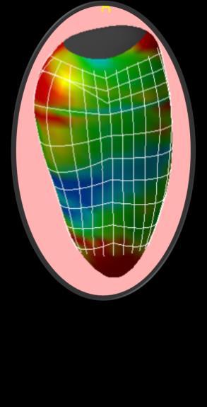
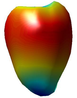
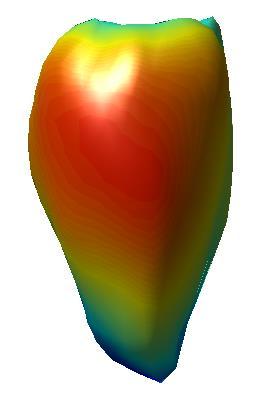
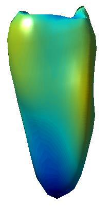
16 LV shape LV volume 2/20/2017 New shape indexes based on LV Contour Application to LV surfaces extracted from RT3DE:.5 s( ) s norm 0 index norm 1 A A max A max Reference shape signal was defined from LV moments of inertia (I X, I Y, I Z ) Shape index: degree of similarity between LV and a reference shape Two shape indices: sphericity S and conicity C A 1 [Maffessanti et al, Ultrasound Med Biol 2009;35: ] New shape indexes based on LV Contour Evolution through the cardiac cycle in a normal subject ES ES time Normal subject, 36 yrs time 16


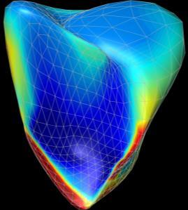
17 LV Shape: Regional Curvature Salgo, Tsang, Lang et al. JASE 2011: 25(1):80-8 Basic IDEA EUROECHO Madrid 34 17


![1980;61:626-33] 1](/docs-images/83/87264491/images/18-3.jpg "k1 R 1 1 k2 R 2 k")

18 Curvature: the amount by which k =1/r a geometric object deviates from being flat. A Circ 1980;61:626-33] 1 k1 R 1 1 k2 R 2 k =1/r R 1 3D Curvature Segmentation 3D RV Shape in Pressure overload 36 18


19 LV Shape Analysis Salgo I, Tsang W, Lang RM et al., J Am Soc Echocardiogr 2011: 25(1):


20 LV SEPTAL & APICAL REMODELING Salgo I, Tsang W, Lang RM et al., J Am Soc Echocardiogr 2011: 25(1):80-8 Salgo, Tsang, Lang et al. JASE 2011: 25(1):


21 3D Regional LV Remodeling: A New Prognosticator of Outcomes in NIDCM M. Cristina Abduch, Ivan Salgo, Wilson Mathias, Roberto Lang RV Shape: Regional Curvature 0.6 R Normalized Curvature k n Curvature < 0 Curvature > 0 Curvature = 0 Convexity Concavity 21


22 Free wall surface Septal wall surface 2/20/2017 Normal RV Shape PAH Normal Indexed Curvature Indexed Curvature


23 LVAD Thoratec HeartMate II Ascending aorta Outflow cannula Left ventricle Inflow cannula 3D CT reconstruction RAMP Study 3D analysis 76mm 8000 ED 69mm 8400 ES Increasing LVAD speed (in rpm) 23
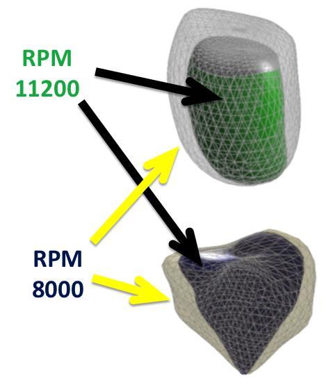
24 LV Endocardial surfaces in LVAD RPM = 8000 RPM = 9600 RPM = Radius (mm) LV Endocardial surfaces in LVAD 24
25 Multiple images from a patient are registered and overlaid or merged Fused images may be created from multiple images from the same imaging modality, or by combining information from multiple modalities, such as MRI, CT, PET and SPECT. U N I V E R S I T Y O F C H I C A G O N O N - I N V A S I V E C A R D I A C I M A G I N G L A B O R A T O R Y Software Image registration Spatial compounding Mosaicing (Stitching) U N I V E R S I T Y O F C H I C A G O N O N - I N V A S I V E C A R D I A C I M A G I N G L A B O R A T O R Y 25


26 HYBRID SYSTEM SPECT-CT fusion imaging integrating anatomy and perfusion O. Gaemperli; et al European Heart Journal Vol 28, N o 2, January 200. U N I V E R S I T Y O F C H I C A G O N O N - I N V A S I V E C A R D I A C I M A G I N G L A B O R A T O R Y 26


27 Anterior view Lateral view 2/20/2017 Longitudinal Circumferential Radial 27


 U N I V E R S")

28 Stress CT perfusion Resting longitudinal strain 2/20/ <50% LAD stenosis >70% RCA stenosis Antero-septal view Antero-lateral view Infero-septal view Software Image registration Spatial compounding Mosaicing (Stitching) U N I V E R S I T Y O F C H I C A G O N O N - I N V A S I V E C A R D I A C I M A G I N G L A B O R A T O R Y 28


29 Grau et al [IEEE Trans Med Imaging 2007;26: ]: extension to the combination of parasternal and apical RT3DE datasets. To combine multiple ultrasound images obtained from different transducer positions to improve the quality of the compounded image. Rajpoot et al [Med Imaging Analysis 2009; ]: combination of multiple apical RT3DE datasets. Yao et al [Phys Med Biol 2011;56:6109] : compounding of up to 10 datasets U N I V E R S I T Y O F C H I C A G O N O N - I N V A S I V E C A R D I A C I M A G I N G L A B O R A T O R Y Software Image registration Spatial compounding Mosaicing (Stitching) U N I V E R S I T Y O F C H I C A G O N O N - I N V A S I V E C A R D I A C I M A G I N G L A B O R A T O R Y 29

30 30


31 Promises and Perspectives 3D Chamber Quantification, LV &RV Shape and Image Fusion Where have we been? Where are we going? Feasible Improved accuracy Improved reproducibility Mechanistic insights Improved temporal and spatial resolution Improved integration into clinical practice Automation Establish outcome measures/guidelines Decreased costs 31

32 32
Conflict of Interests
 New Approaches to Systolic Function: 4D Roberto M Lang, MD Conflict of Interests Philips Medical Imaging Research Grants Speakers bureau Advisory bureau Tomtec Research Grants Epsilon Research Grants 1
Assessment of cardiac function with 3D echocardiography. Đánh giá chức năng tim bằng siêu âm tim 3D
 Assessment of cardiac function with 3D echocardiography Đánh giá chức năng tim bằng siêu âm tim 3D TS. BS. Nguyễn Thị Thu Hoài Viện Tim Mạch Quốc Gia Việt Nam TỪ SIÊU ÂM M-mode ĐẾN SIÊU ÂM 3D TỪ SIÊU ÂM
Chamber Quantitation Guidelines: What is New?
 Chamber Quantitation Guidelines: What is New? Roberto M Lang, MD J AM Soc Echocardiogr 2005; 18:1440-1463 1 Approximately 10,000 citations iase in itune Cardiac Chamber Quantification: What is New? Database
3D-stress echocardiography Bernard Cosyns, MD, PhD
 3D-stress echocardiography Bernard Cosyns, MD, PhD No Disclosure The Pro-Technology bias Sicari et al. Cardiovascular Ultrasound 2006, 4:11 Overview 2D stress echocardiography: main limitations 3D echocardiography:
When Does 3D Echo Make A Difference?
 When Does 3D Echo Make A Difference? Wendy Tsang, MD, SM Assistant Professor, University of Toronto Toronto General Hospital, University Health Network 1 Practical Applications of 3D Echocardiography Recommended
Three-dimensional echocardiography in the clinical world
 Three-dimensional echocardiography in the clinical world Dr. JL Zamorano Director CV Institute University Clinic SC, Madrid Advantages of 3D. Spatial manipulation. Optimal alineation of structures. Views
Prof. JL Zamorano Hospital Universitario Ramón y Cajal
 Prof. JL Zamorano Hospital Universitario Ramón y Cajal Fully Automated Quantification Software Adaptive analytical algorithm consists in knowledge-based identification of global shape and specific adaptation
EDITOR S PICK CURRENT STATUS OF FULLY AUTOMATED SOFTWARE WITH THREE-DIMENSIONAL ECHOCARDIOGRAPHY FOR THE QUANTIFICATION OF LEFT VENTRICULAR FUNCTION
 ITOR S PICK This paper, courtesy of Yang and Takeuchi, provides a timely and well-considered update on the current status of fully-automated software with three-dimensional echocardiography for quantifying
Automated Volumetric Cardiac Ultrasound Analysis
 Whitepaper Automated Volumetric Cardiac Ultrasound Analysis ACUSON SC2000 Volume Imaging Ultrasound System Bogdan Georgescu, Ph.D. Siemens Corporate Research Princeton, New Jersey USA Answers for life.
10/7/2013. Systolic Function How to Measure, How Accurate is Echo, Role of Contrast. Thanks to our Course Director: Neil J.
 Systolic Function How to Measure, How Accurate is Echo, Role of Contrast Neil J. Weissman, MD MedStar Health Research Institute & Professor of Medicine Georgetown University Washington, D.C. No Disclosures
Quantification of Cardiac Chamber Size
 2017 KSE 2017-11-25 Quantification of Cardiac Chamber Size Division of Cardiology Keimyung University Dongsan Medical Center In-Cheol Kim M.D., Ph.D. LV size and function Internal linear dimensions PLX
Cardiac Chamber Quantification by Echocardiography
 Cardiac Chamber Quantification by Echocardiography Maryam Bokhamseen, RCS, RCDS, EACVI Echotechnologist ǁ, Non invasive Cardiac Laboratory King Abdulaziz Cardiac Center. Outline: Introduction. Background
RIGHT VENTRICULAR SIZE AND FUNCTION
 RIGHT VENTRICULAR SIZE AND FUNCTION Edwin S. Tucay, MD, FPCC, FPCC, FPSE Philippine Society of Echocardiography Quezon City, Philippines Echo Mission, BRTTH, Legaspi City, July 1-2, 2016 NO DISCLOSURE
LV FUNCTION ASSESSMENT: WHAT IS BEYOND EJECTION FRACTION
 LV FUNCTION ASSESSMENT: WHAT IS BEYOND EJECTION FRACTION Jamilah S AlRahimi Assistant Professor, KSU-HS Consultant Noninvasive Cardiology KFCC, MNGHA-WR Introduction LV function assessment in Heart Failure:
Advanced imaging of the left atrium - strain, CT, 3D, MRI -
 Advanced imaging of the left atrium - strain, CT, 3D, MRI - Monica Rosca, MD Carol Davila University of Medicine and Pharmacy, Bucharest, Romania Declaration of interest: I have nothing to declare Case
좌심실수축기능평가 Cardiac Function
 Basic Echo Review Course 좌심실수축기능평가 Cardiac Function Seonghoon Choi Cardiology Hallym university LV systolic function Systolic function 좌심실수축기능 - 심근의수축으로심실에서혈액을대동맥으로박출하는기능 실제임상에서 LV function 의의미 1Diagnosis
Left atrial function. Aliakbar Arvandi MD
 In the clinic Left atrial function Abstract The left atrium (LA) is a left posterior cardiac chamber which is located adjacent to the esophagus. It is separated from the right atrium by the inter-atrial
Echocardiographic Assessment of the Left Ventricle
 Echocardiographic Assessment of the Left Ventricle Theodora Zaglavara, MD, PhD, BSCI/BSCCT Department of Cardiovascular Imaging INTERBALKAN EUROPEAN MEDICAL CENTER 2015 The quantification of cardiac chamber
Novel echocardiographic modalities: 3D echo, speckle tracking and strain rate imaging. Potential roles in sports cardiology. Stefano Caselli, MD, PhD
 Novel echocardiographic modalities: 3D echo, speckle tracking and strain rate imaging. Potential roles in sports cardiology. Stefano Caselli, MD, PhD Ospedale San Pietro Fatebenefratelli Rome, Italy Differential
Revealing new insights. irotate electronic rotation and xplane adjustable biplane imaging. Ultrasound cardiology. irotate and xplane
 Ultrasound cardiology irotate and xplane Revealing new insights irotate electronic rotation and xplane adjustable biplane imaging Annemien van den Bosch and Jackie McGhie Department of Cardiology, Erasmus
Reproducibility and Accuracy of Echocardiographic Measurements of Left Ventricular Parameters Using Real-Time Three-Dimensional Echocardiography
 Journal of the American College of Cardiology Vol. 44, No. 4, 2004 2004 by the American College of Cardiology Foundation ISSN 0735-1097/04/$30.00 Published by Elsevier Inc. doi:10.1016/j.jacc.2004.05.050
Global left ventricular circumferential strain is a marker for both systolic and diastolic myocardial function
 Global left ventricular circumferential strain is a marker for both systolic and diastolic myocardial function Toshinari Onishi 1, Samir K. Saha 2, Daniel Ludwig 1, Erik B. Schelbert 1, David Schwartzman
MAYON VOLCANO: FAST FACTS
 MAYON VOLCANO: FAST FACTS Type of Volcano: Stratovolcano Elevation: 2.46 km Base Diameter: 20 km Base Circumference: 62.8 km Area: 314.1 km 2 Reference: http://www.phivolcs.dost.gov.ph/html/update_vmepd/volcano/volcanolist/mayon.htm
Martin G. Keane, MD, FASE Temple University School of Medicine
 Martin G. Keane, MD, FASE Temple University School of Medicine Measurement of end-diastolic LV internal diameter (LVIDd) made by properly-oriented M-Mode techniques in the Parasternal Long Axis View (PLAX):
Echocardiographic Evaluation of Primary Mitral Regurgitation
 Echocardiographic Evaluation of Primary Mitral Regurgitation Roberto M Lang, MD 0-10 o ME 4CH Med A2 P2 50-70 o Commissural P3 P1 A2 80-100 o ME 2CH P3 A2 A1 A1 125-135 o - ME Long axis P2 A2 P3 A3 P2
Ιπποκράτειες μέρες καρδιολογίας Θεσσαλονίκη, 9-10 Μαρτίου Φωτεινή Α. Λαζαρίδου Επιμελήτρια Α Γενικό Νοσοκομείο Αγιος Παύλος, Θεσσαλονίκη
 Ιπποκράτειες μέρες καρδιολογίας Θεσσαλονίκη, 9-10 Μαρτίου 2018 Φωτεινή Α. Λαζαρίδου Επιμελήτρια Α Γενικό Νοσοκομείο Αγιος Παύλος, Θεσσαλονίκη RV shape Triangular shape in frontal plane crescent shape in
Challenge on Endocardial Three-dimensional Ultrasound Segmentation (CETUS)
") Challenge on Endocardial Three-dimensional Ultrasound Segmentation (CETUS) Olivier Bernard 1,BrechtHeyde 2, Martino Alessandrini 2, Daniel Barbosa 3, Sorina Camarasu-Pop 1, Frederic Cervenansky 1, Sebastien
Challenge on Endocardial Three-dimensional Ultrasound Segmentation (CETUS) Olivier Bernard 1,BrechtHeyde 2, Martino Alessandrini 2, Daniel Barbosa 3, Sorina Camarasu-Pop 1, Frederic Cervenansky 1, Sebastien
Cardiology for the Practitioner Advanced Cardiac Imaging: Worth the pretty pictures?
 Keenan Research Centre Li Ka Shing Knowledge Institute Cardiology for the Practitioner Advanced Cardiac Imaging: Worth the pretty pictures? Howard Leong-Poi, MD, FRCPC Associate Professor of Medicine St.
Certificate in Clinician Performed Ultrasound (CCPU) Syllabus. Rapid Cardiac Echo (RCE)
 Syllabus. Rapid Cardiac Echo (RCE)") Certificate in Clinician Performed Ultrasound (CCPU) Syllabus Rapid Cardiac Echo (RCE) Purpose: Rapid Cardiac Echocardiography (RCE) This unit is designed to cover the theoretical and practical curriculum
Certificate in Clinician Performed Ultrasound (CCPU) Syllabus Rapid Cardiac Echo (RCE) Purpose: Rapid Cardiac Echocardiography (RCE) This unit is designed to cover the theoretical and practical curriculum
Cardiac MRI in ACHD What We. ACHD Patients
 Cardiac MRI in ACHD What We Have Learned to Apply to ACHD Patients Faris Al Mousily, MBChB, FAAC, FACC Consultant, Pediatric Cardiology, KFSH&RC/Jeddah Adjunct Faculty, Division of Pediatric Cardiology
Quantifying LV function how good are we?
 Quantifying LV function how good are we? Professor Alan G Fraser Wales Heart Research Institute Cardiff University, U.K. Support for research from Hitachi Aloka, & GE Ultrasound Visual assessment of synchronicity
CHAPTER. Quantification in cardiac MRI. This chapter was adapted from:
 CHAPTER Quantification in cardiac MRI This chapter was adapted from: Quantification in cardiac MRI Rob J. van der Geest, Johan H.C. Reiber Journal of Magnetic Resonance Imaging 1999, Volume 10, Pages 602-608.
Tissue Doppler and Strain Imaging
 Tissue Doppler and Strain Imaging Steven J. Lester MD, FRCP(C), FACC, FASE Relevant Financial Relationship(s) None Off Label Usage None 1 Objective way with which to quantify the minor amplitude and temporal
Tissue Doppler and Strain Imaging. Steven J. Lester MD, FRCP(C), FACC, FASE
, FACC, FASE") Tissue Doppler and Strain Imaging Steven J. Lester MD, FRCP(C), FACC, FASE Relevant Financial Relationship(s) None Off Label Usage None a. Turn the wall filters on and turn down the receiver gain. b. Turn
Tissue Doppler and Strain Imaging Steven J. Lester MD, FRCP(C), FACC, FASE Relevant Financial Relationship(s) None Off Label Usage None a. Turn the wall filters on and turn down the receiver gain. b. Turn
Value of echocardiography in chronic dyspnea
 Value of echocardiography in chronic dyspnea Jahrestagung Schweizerische Gesellschaft für /Schweizerische Gesellschaft für Pneumologie B. Kaufmann 16.06.2016 Chronic dyspnea Shortness of breath lasting
Multimodality Comparison of Quantitative Volumetric Analysis of the Right Ventricle
 JACC: CARDIOVASCULAR IMAGING VOL. 3, NO. 1, 21 21 BY THE AMERICAN COLLEGE OF CARDIOLOGY FOUNDATION ISSN 1936-878X/1/$36. PUBLISHED BY ELSEVIER INC. DOI:1.116/j.jcmg.29.9.17 Multimodality Comparison of
Echo assessment of the failing heart
 Echo assessment of the failing heart Mark K. Friedberg, MD The Labatt Family Heart Center The Hospital for Sick Children Toronto, Ontario, Canada Cardiac function- definitions Cardiovascular function:
Quantitation of right ventricular dimensions and function
 SCCS Basics of cardiac assessment Quantitation of right ventricular dimensions and function Tomasz Kukulski, MD PhD Dept of Cardiology, Congenital Heart Disease and Electrotherapy Silesian Medical University
The importance of left atrium in LV diastolic function
 II Baltic Heart Failure Meeting and Congress of Latvian Society of Cardiology The importance of left atrium in LV diastolic function Dr. Artem Kalinin Eastern Clinical University Hospital Riga 30.09.2010.
Echocardiographie de la Tétralogie de Fallot opérée
 Echocardiographie de la Tétralogie de Fallot opérée Diala Khraiche M3C-Necker Enfants malades, Université Paris Descartes Paris, France. Disclosure Statement of Financial Interest I currently have, or
Part II: Fundamentals of 3D Echocardiography: Acquisition and Application
 Part II: Fundamentals of 3D Echocardiography: Acquisition and Application Dr. Bruce Bollen 3D matrix array TEE probes provide options for both 2D and 3D imaging. Indeed, their utility in obtaining multiple
TAVR: Echo Measurements Pre, Post And Intra Procedure
 2017 ASE Florida, Orlando, FL October 10, 2017 8:00 8:25 AM 25 min TAVR: Echo Measurements Pre, Post And Intra Procedure Muhamed Sarić MD, PhD, MPA Director of Noninvasive Cardiology Echo Lab Associate
Cardiovascular Imaging Endpoints in Oncology Clinical Trials
 Cardiovascular Imaging Endpoints in Oncology Clinical Trials Bonnie Ky, MD, MSCE Assistant Professor of Medicine and Epidemiology Director, Penn Cardio-Oncology Center of Excellence Director, Penn Center
Alicia Armour, MA, BS, RDCS
 Alicia Armour, MA, BS, RDCS No disclosures Review 2D Speckle Strain (briefly) Discuss some various patient populations & disease pathways where Strain can be helpful Discuss how to acquire images for Strain
Disclosures Rebecca T. Hahn, MD, FASE
 The New ASE Guidelines for Native Valvular Regurgitation Mitral Regurgitation The New ASE Guidelines: Role of 2D/3D and CMR (With caveats and comments from R. Hahn) William A. Zoghbi MD, FASE, MACC Professor
Tissue Doppler and Strain Imaging
 Tissue Doppler and Strain Imaging Steven J. Lester MD, FRCP(C), FACC, FASE Relevant Financial Relationship(s) None Off Label Usage None 1 Objective way with which to quantify the minor amplitude and temporal
An intensive interactive course for 3D echocardiography: is crop till you drop an effective learning strategy?
 European Journal of Echocardiography (2008) 9, 373 380 doi:10.1016/j.euje.2007.06.011 An intensive interactive course for 3D echocardiography: is crop till you drop an effective learning strategy? Carly
Quantitative Assessment of Pulmonary Regurgitation by Echocardiography in Patients After Repaired TOF
 Quantitative Assessment of Pulmonary Regurgitation by Echocardiography in Patients After Repaired TOF 2013. 4. 20. 서울대학교어린이병원소아청소년과 권보상 W. B. TOF (large VSD, infundibular stenosis) 19 mo, 8.5 kg Indication
ORIGINAL ARTICLE. Keywords Real-time 3D echocardiography. Daily practice. Observer experience. CMR. Introduction
 Neth Heart J (2014) 22:383 390 DOI 10.1007/s12471-014-0577-1 ORIGINAL ARTICLE Assessment of LV ejection fraction using real-time 3D echocardiography in daily practice: direct comparison of the volumetric
Comparison of Cardiac MDCT with MRI and Echocardiography in the Assessement of Left Ventricular Function
 Comparison of Cardiac MDCT with MRI and Echocardiography in the Assessement of Left Ventricular Function Poster No.: C-0969 Congress: ECR 2012 Type: Scientific Exhibit Authors: B. Kara, Y. Paksoy, C. Erol,
Impaired Regional Myocardial Function Detection Using the Standard Inter-Segmental Integration SINE Wave Curve On Magnetic Resonance Imaging
 Original Article Impaired Regional Myocardial Function Detection Using the Standard Inter-Segmental Integration Ngam-Maung B, RT email : chaothawee@yahoo.com Busakol Ngam-Maung, RT 1 Lertlak Chaothawee,
LV function in ischemic heart failure - decreased correlation between Echo and CMR
 LV function in ischemic heart failure - decreased correlation between Echo and CMR Poster No.: C-0590 Congress: ECR 2011 Type: Scientific Exhibit Authors: K. Gruszczy#ska, L. Krzych, K. Golba, P. Ulbrych,
Right Ventricular Strain in Normal Healthy Adult Filipinos: A Retrospective, Cross- Sectional Pilot Study
 Right Ventricular Strain in Normal Healthy Adult Filipinos: A Retrospective, Cross- Sectional Pilot Study By Julius Caesar D. de Vera, MD Jonnah Fatima B. Pelat, MD Introduction Right ventricle contributes
Echo in Heart Failure
 Echo in Heart Failure Karima Addetia, MD Heart Failure: Definition A clinical syndrome that results from impairment of ventricular filling or ejection of blood. Manifestations include dyspnea and fatigue,
THE LEFT ATRIUM HOW CAN ECHO HELP US?
 THE LEFT ATRIUM HOW CAN ECHO HELP US? Dr. Dragos COZMA BACKGROUND Left atrium (LA) dilation can occur in a broad spectrum of cardiovascular diseases including hypertension, left ventricular dysfunction,
Left Ventricular Assist Device: What Should I Report?
 2017 SOTA, Tucson, AZ February 21, 2017 11:15 11:40 AM 25 min Left Ventricular Assist Device: What Should I Report? Muhamed Sarić MD, PhD, MPA Director of Noninvasive Cardiology Echo Lab Associate Professor
Introduction. Aims. Keywords
 European Journal of Echocardiography (2010) 11, 359 368 doi:10.1093/ejechocard/jep217 Validation of a novel automated border-detection algorithm for rapid and accurate quantitation of left ventricular
Altered left ventricular geometry and torsional mechanics in high altitude-induced pulmonary hypertension:
 Altered left ventricular geometry and torsional mechanics in high altitude-induced pulmonary hypertension: a 3-D echocardiographic study B.W. De Boeck,* S. Kiencke, C. Dehnert, K. Auinger, # M. Maggiorini,
Η ηχωκαρδιολογία στην διάγνωση κα πρόγνωση της καρδιακής ανεπάρκειας µε µειωµένο και φυσιολογικό κλάσµα εξώθησης
 Η ηχωκαρδιολογία στην διάγνωση κα πρόγνωση της καρδιακής ανεπάρκειας µε µειωµένο και φυσιολογικό κλάσµα εξώθησης Βασίλειος Σαχπεκίδης Επιµελητής Β Καρδιολογίας Γ.Ν. Παπαγεωργίου Θεσσαλονίκη ESC Guidelines
Conflict of Interests
 Introduction to Interventional Echocardiography Roberto M Lang, MD Tomtec Conflict of Interests Research Grants Philips Medical Imaging Research Grants Speakers bureau Advisory bureau 1 Structural Heart
True morphology of mitral regurgitant flow assessed by three- dimensional transesophageal echocardiography
 DOI: 10.1111/echo.13395 ORIGINAL INVESTIGATION True morphology of mitral regurgitant flow assessed by three- dimensional transesophageal echocardiography Martin Lombardero M.D. Ruth Henquin D.L.S.H.T.M.,
Three-dimensional Wall Motion Tracking:
 Three-dimensional Wall Motion Tracking: A Novel Echocardiographic Method for the Assessment of Ventricular Volumes, Strain and Dyssynchrony Jeffrey C. Hill, BS, RDCS, FASE Jennifer L. Kane, RCS Gerard
Echocardiographic assessment of the right ventricle in paediatric pulmonary hypertension.
 Echocardiographic assessment of the right ventricle in paediatric pulmonary hypertension. Mark K. Friedberg, MD No disclosures Outline RV response to increased afterload Echo assessment of RV function
Basic Assessment of Left Ventricular Systolic Function
 WINFOCUS BASIC ECHO (WBE) Basic Assessment of Left Ventricular Systolic Function Ritesh Dhar, MD Director, Echocardiography Lab and Staff Cardiologist Intermountain Medical Center Murray, Utah Outline
HYPERTROPHY: Behind the curtain. V. Yotova St. Radboud Medical University Center, Nijmegen
 HYPERTROPHY: Behind the curtain V. Yotova St. Radboud Medical University Center, Nijmegen Disclosure of interest: none Relative wall thickness (cm) M 0.22 0.42 0.43 0.47 0.48 0.52 0.53 F 0.24 0.42 0.43
Beginner s Guide to Strain: What should be in your lab in Disclosures
 Beginner s Guide to Strain: What should be in your lab in 2018 Bonita Anderson DMU (Cardiac), MApplSc (Med Ultrasound), ACS, AMS, FASE None Disclosures Calculation of Strain Strain can be Positive Strain
What echo measurements are key prior to MitraClip?
 APHP CHU Bichat - Claude Bernard What echo measurements are key prior to MitraClip? Eric Brochet,MD Cardiology Department Hopital Bichat Paris France No disclosure Conflict of interest Case 69 y.o man
Strain imaging in children: from Tissue Doppler to 3 D
 Strain imaging in children: from Tissue Doppler to 3 D Mark kk. Friedberg Fi Outline Deformation in the fetus and neonate Deformation in pediatric cardiomyopathy y (briefly!) Deformation in Congenital
Three-dimensional speckle tracking echocardiography for the evaluation of segmental myocardial deformation
 ORIGINAL RESEARCH Three-dimensional speckle tracking echocardiography for the evaluation of segmental myocardial deformation Janine Baum, Florian Beeres, Silke Van Hall, Yang Chul Boering, Eva Susanne
Velocity Vector Imaging as a new approach for cardiac magnetic resonance: Comparison with echocardiography
 Velocity Vector Imaging as a new approach for cardiac magnetic resonance: Comparison with echocardiography Toshinari Onishi 1, Samir K. Saha 2, Daniel Ludwig 1, Erik B. Schelbert 1, David Schwartzman 1,
Effect of loading and geometry on functional parameters
 Effect of loading and geometry on functional parameters Piet Claus Cardiovascular Imaging and Dynamics Department of Cardiovascular Diseases Leuven University, Leuven, Belgium 5 th European Echocardiography
Tissue Doppler Imaging in Congenital Heart Disease
 Tissue Doppler Imaging in Congenital Heart Disease L. Youngmin Eun, M.D. Department of Pediatrics, Division of Pediatric Cardiology, Kwandong University College of Medicine The potential advantage of ultrasound
Chamber Quantitation Guidelines - Update II
 Chamber Quantitation Guidelines - Update II Right Heart Measurements Steven A. Goldstein MD FACC FASE Professor of Medicine Georgetown University Medical Center MedStar Heart Institute Washington Hospital
Myocardial Strain Imaging in Cardiac Diseases and Cardiomyopathies.
 Myocardial Strain Imaging in Cardiac Diseases and Cardiomyopathies. Session: Cardiomyopathy Tarun Pandey MD, FRCR. Associate Professor University of Arkansas for Medical Sciences Disclosures No relevant
JOINT MEETING 2 Tricuspid club Chairpersons: G. Athanassopoulos, A. Avgeropoulou, M. Khoury, G. Stavridis
 JOINT MEETING 2 Tricuspid club Chairpersons: G. Athanassopoulos, A. Avgeropoulou, M. Khoury, G. Stavridis Similarities and differences in Tricuspid vs. Mitral Valve Anatomy and Imaging. Echo evaluation
3D Echo for Evaluation of Tricuspid Regurgitation Jong-Min Song, MD, PhD
 3D Echo for Evaluation of Tricuspid Regurgitation Jong-Min Song, MD, PhD Asan Medical Center University of Ulsan College of Medicine Seoul, Korea Causes of TR Primary causes (25%) Rheumatic Myxomatous
2D/3D in Evaluation of Atrial Septum
 2D/3D in Evaluation of Atrial Septum Roberto M Lang, MD OSTIUM SECUNDUM ASD: 2D AND 3D TNSESOPHAGEAL ECHO 1 Biplane views 90 0 3D Acquisi on Acquire 3D volume Lang RM et al. JASE 2012;25:3 46. Right atrial
Left Ventricular Volumes from Three-Dimensional. Echocardiography by Rapid Freehand Scanning using
 Left Ventricular Volumes from Three-Dimensional Echocardiography by Rapid Freehand Scanning using Digital Scan Line Data Stig A. Slørdahl, MD, PhD, Sevald Berg, MSc, Asbjørn Støylen*, MD, Stein Samstad,
DECLARATION OF CONFLICT OF INTEREST. None
 DECLARATION OF CONFLICT OF INTEREST None Hot Topics in Echocardiography: The position of the EAE EAE / ASE recommendation about Echo Assessment of Cardiac Mechanics Jens-Uwe Voigt Dpt. of Cardiovascular
Elections to EACVI Board
 Elections to EACVI Board 2018-2020 Application for the position: EACVI Councillor (Echocardiography) 1. Your Identity Title Family Name(s) First Name(s) M.D., Ph.D. Muraru Denisa Birth Date 03 April 1979
Right ventricular adaptation in endurance athletes. António Freitas. No conflict of interest
 The role of echocardiography in sports cardiology Right ventricular adaptation in endurance athletes. António Freitas Cardiology Department - Fernando Fonseca Hospital Lisbon Sports Medicine Centre - Lisbon
Advanced Multi-Layer Speckle Strain Permits Transmural Myocardial Function Analysis in Health and Disease:
 Advanced Multi-Layer Speckle Strain Permits Transmural Myocardial Function Analysis in Health and Disease: Clinical Case Examples Jeffrey C. Hill, BS, RDCS Echocardiography Laboratory, University of Massachusetts
Assessment of LV systolic function
 Tutorial 5 - Assessment of LV systolic function Assessment of LV systolic function A knowledge of the LV systolic function is crucial in the undertanding of and management of unstable hemodynamics or a
MITRAL STENOSIS. Joanne Cusack
 MITRAL STENOSIS Joanne Cusack BSE Breakdown Recognition of rheumatic mitral stenosis Qualitative description of valve and sub-valve calcification and fibrosis Measurement of orifice area by planimetry
Assessment of left ventricular systolic function by deformation imaging derived from speckle tracking: a comparison between 2D and 3D echo modalities
 European Heart Journal Cardiovascular Imaging (2014) 15, 316 323 doi:10.1093/ehjci/jet103 Assessment of left ventricular systolic function by deformation imaging derived from speckle tracking: a comparison
How to Assess Dyssynchrony
 How to Assess Dyssynchrony Otto A. Smiseth, Professor, MD, PhD Oslo University Hospital None Conflicts of interest Cardiac resynchronization therapy effect on mortality Cleland JG et al, N Engl J Med
Nancy Goldman Cutler, MD Beaumont Children s Hospital Royal Oak, Mi
 Nancy Goldman Cutler, MD Beaumont Children s Hospital Royal Oak, Mi Identify increased LV wall thickness (WT) Understand increased WT in athletes Understand hypertrophic cardiomyopathy (HCM) Enhance understanding
Normal TTE Examination, Doppler Echocardiography and Normal Antegrade Flow Patterns
 Normal TTE Examination, Doppler Echocardiography and Normal Antegrade Flow Patterns Pravin Patil, MD FACC FASE Associate Professor of Medicine Director, Cardiovascular Disease Training Program Lewis Katz
Adult Echocardiography Examination Content Outline
 Adult Echocardiography Examination Content Outline (Outline Summary) # Domain Subdomain Percentage 1 2 3 4 5 Anatomy and Physiology Pathology Clinical Care and Safety Measurement Techniques, Maneuvers,
Imaging and heart failure
 Imaging and heart failure Jeroen J Bax Dept of Cardiology Leiden Univ Medical Center The Netherlands Davos, feb 2013 Research grants: Medtronic, Biotronik, Boston, St Jude, BMS imaging, GE Healthcare,
THE RIGHT VENTRICLE IN PULMONARY HYPERTENSION R. DRAGU
 THE RIGHT VENTRICLE IN PULMONARY HYPERTENSION R. DRAGU Cardiology Dept. Rambam Health Care Campus Rappaport Faculty of Medicine Technion, Israel Why the Right Ventricle? Pulmonary hypertension (PH) Right
MITRAL VALVE PATHOLOGY WITH TRICUSPID REGURGITATION (AND PHT)
") UNIVERSITY OF PADUA, SCHOOL OF MEDICINE Department of Cardiac,Thoracic and Vascular Sciences Padua, Italy MITRAL VALVE PATHOLOGY WITH TRICUSPID REGURGITATION (AND PHT) Luigi P. Badano**, MD, PhD, FESC,
UNIVERSITY OF PADUA, SCHOOL OF MEDICINE Department of Cardiac,Thoracic and Vascular Sciences Padua, Italy MITRAL VALVE PATHOLOGY WITH TRICUSPID REGURGITATION (AND PHT) Luigi P. Badano**, MD, PhD, FESC,
Strain/Untwisting/Diastolic Suction
 What Is Diastole and How to Assess It? Strain/Untwisting/Diastolic Suction James D. Thomas, M.D., F.A.C.C. Cardiovascular Imaging Center Department of Cardiology Cleveland Clinic Foundation Cleveland,
Reverse left atrium and left ventricle remodeling after aortic valve interventions
 Reverse left atrium and left ventricle remodeling after aortic valve interventions Alexandra Gonçalves, Cristina Gavina, Carlos Almeria, Pedro Marcos-Alberca, Gisela Feltes, Rosanna Hernández-Antolín,
Incorporating the New Echo Guidelines Into Everyday Practice
 Incorporating the New Echo Guidelines Into Everyday Practice Clinical Case RIGHT VENTRICULAR FAILURE Gustavo Restrepo MD President Elect Interamerican Society of Cardiology Director Fellowship Training
PROSTHETIC VALVE BOARD REVIEW
 PROSTHETIC VALVE BOARD REVIEW The correct answer D This two chamber view shows a porcine mitral prosthesis with the typical appearance of the struts although the leaflets are not well seen. The valve
Little is known about the degree and time course of
 Differential Changes in Regional Right Ventricular Function Before and After a Bilateral Lung Transplantation: An Ultrasonic Strain and Strain Rate Study Virginija Dambrauskaite, MD, Lieven Herbots, MD,
4D Auto LAQ (Left Atrial Quantification)
") 4D Auto LAQ (Left Atrial Quantification) Introduction There has been an increased interest in quantification of the left atrium (LA) for various types of diseases; e.g. heart failure, heart valve diseases,
4D Auto LAQ (Left Atrial Quantification) Introduction There has been an increased interest in quantification of the left atrium (LA) for various types of diseases; e.g. heart failure, heart valve diseases,
Strain Imaging: Myocardial Mechanics Simplified and Applied
 9/28/217 Strain Imaging: Myocardial Mechanics Simplified and Applied John Gorcsan III, MD Professor of Medicine Director of Clinical Research Division of Cardiology VECTORS OF CONTRACTION Shortening Thickening
HIGHLIGHT SESSION. Imaging. J. L. Zamorano Gomez (Madrid, ES) Disclosures: Speaker Philips
 Disclosures: Speaker Philips") Imaging. J. L. Zamorano Gomez (Madrid, ES) Disclosures: Speaker Philips Agenda ECHO Diagnosis & Prognosis : Functional MR Severity Aortic Stenosis CT How to select pts for TAVI Adding prognostic info to
Imaging. J. L. Zamorano Gomez (Madrid, ES) Disclosures: Speaker Philips Agenda ECHO Diagnosis & Prognosis : Functional MR Severity Aortic Stenosis CT How to select pts for TAVI Adding prognostic info to