Echo in Heart Failure
|
|
|
- Bryan Fields
- 5 years ago
- Views:
Transcription


1 Echo in Heart Failure Karima Addetia, MD Heart Failure: Definition A clinical syndrome that results from impairment of ventricular filling or ejection of blood. Manifestations include dyspnea and fatigue, exercise intolerance, fluid retention, which may lead to pulmonary +/- splanchnic congestion +/- peripheral edema. Some patients have exercise intolerance but little fluid retention, others complain primarily of edema, dyspnea, or fatigue There is no single diagnostic test for HF. It is a clinical diagnosis based on a the history and physical examination Yancy et al 2013 ACCF/AHA Heart Failure Guideline 1

2 Heart Failure: Prevalence Estimated 23 million people with HF worldwide In the US: cases diagnosed/year 1 million hospitalizations and 3.4 million outpatient visits Rate of rise in HF cases has outpaced the rate of transplantation Prevalence of Common Cardiovascular and Lung Diseases, U.S., 2004, NHLBI report Death from specific cardiovascular, Lung and Blood Diseases, U.S., 2004 NHLBI report Mechanical Assist Devices A valuable option for patients with end stage systolic heart failure (Stage D) ESC Definition of Advanced HF 2

 oduration of Support: 6 ~ 12 months omaximize survival until transplant 2.")
3 Why offer an LVAD? Wait times for cardiac transplantation are long Large numbers of patients with endstage heart failure 1-y survival on LVAD support awaiting transplant ~55-85% > LVADs are implanted world-wide HeartMate III HeartMate II HeartWare LVADs: Clinical Indications 1. Bridge-to-Transplantation (BTT) oduration of Support: 6 ~ 12 months omaximize survival until transplant 2. Destination Therapy (DT) oduration of support is indefinite opurpose: maximize functional capacity and quality of life o1-y survival approaching 80% 3. Myocardial Recovery + Potential Explant omaximize LV reverse remodeling 3


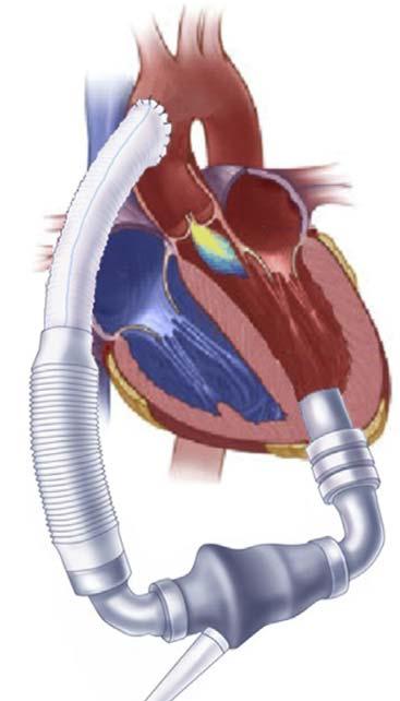
4 LVAD Circuit Removal of blood from LV apex return blood to aorta via graft Three main internal components to LVAD: 1. Inflow cannula at LV apex 2. Mechanical impeller 3. Outflow graft connected to the ascending aorta Rasalingam R. J Am Soc Echocardiogr 2011;24: HeartMate II HVAD (HeartWare) Rasalingam R. J Am Soc Echocardiogr 2011;24:
5 HeartMate II HVAD (HeartWare) Rasalingam R. J Am Soc Echocardiogr 2011;24: HeartMate II HVAD (HeartWare) Rasalingam R. J Am Soc Echocardiogr 2011;24:
6 HeartMate II HVAD (HeartWare) Rasalingam R. J Am Soc Echocardiogr 2011;24: HeartMate II HVAD (HeartWare) Ascending Ao Ascending Ao Rasalingam R. J Am Soc Echocardiogr 2011;24:
7 ECHO: Clinical Indications 1. Surveillance odrift from baseline echo 2. Determine optimal device settings owith or without speed changes oto select the optimal LVAD speed setting 3. Assess complications othrombosis oinflow/outflow cannula obstruction 4. Assessment of LV recovery The Surveillance Echo 7
8 LVAD Parameters LVAD Evaluation 1. LV size 2. Aortic valve opening 3. Aortic insufficiency 4. Inter-ventricular septum position 5. Right ventricle size and function 6. Mitral regurgitation 7. Inflow cannula 8. Outflow cannula 9. Thrombosis 10. Pericardium 8

9 LVAD Surveillance Echo LV size/function AV opening MR Inflow Septum and RV Outflow 76mm 69mm AI ED ES Pericardium Inter-ventricular septum 9


10 Position of Inter-ventricular Septum Inefficient LV unloading Efficient LV compression Markedly unloaded LV Aortic Valve Opening 10


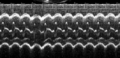
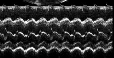
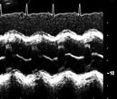
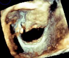
11 Aortic valve opening: 2D Echo Closed Intermittent opening Opening normally Aortic valve opening: M-mode Echo Closed Intermittent opening Regular opening Opening every beat 11


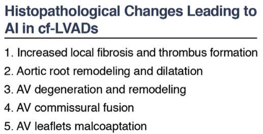
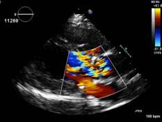
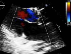
12 Impact of Aortic valve opening Aggarwal A. Ann Thorac Surg 2013;95:493 9 Aortic Insufficiency 12

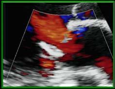
13 Aortic Insufficiency 1) Often occurs during both systole and diastole (pan-cyclical) and not only during diastole 2) Overall volume load that the ventricle sees is greater Continuous AI 3) Eccentric and often poorly measured by traditional echocardiographic measures such as vena contracta and PISA Time Course of Aortic Insufficiency N = 237 patients with HeartMate II CF-LVADs 32 patients had mod or severe AI 13
14 Thrombosis 14


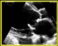
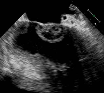
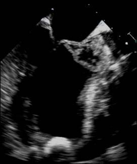
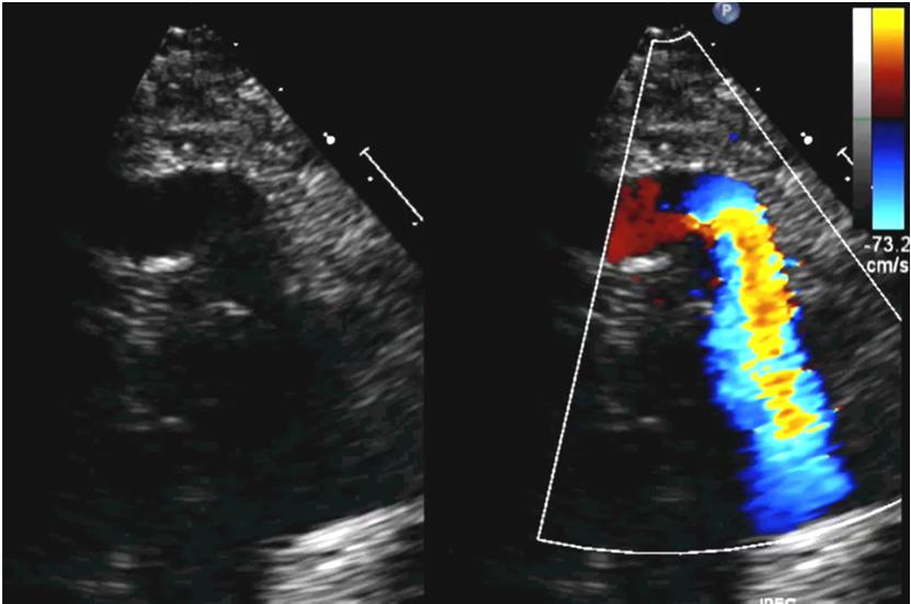
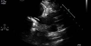
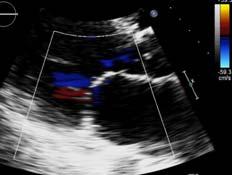
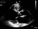
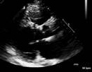
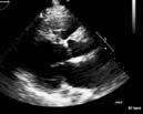
15 Aortic Root Thrombosis Other Thrombosis 50 year-old man 1 month PSLAXstatus post HeartMate II LVAD. He was ready to go home after a lengthy admission. This echo was done prior to discharge. A4C A5C PSLAX 15

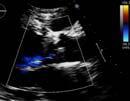
16 Other Thrombosis LV perspective Mid-esophageal 4CH PSLAX LAX Malposition Occlusion Thrombosis Inflow cannula 16


17 The Normal Inflow Cannula Inflow cannula is usually positioned in the LV apex and oriented within the LV toward the mitral valve The Abnormal Inflow Cannula Mechanisms of inflow cannula obstruction othrombus oinlet occlusion by trabeculations ocannula angulation into the myocardium omalposition due to LV under-filling CANNULA CANNULA LV LV 17


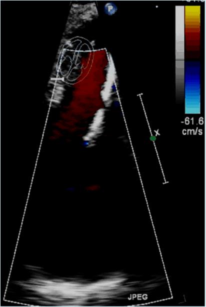
18 The Abnormal Inflow Cannula 74 year-old F with an LVAD complains of shortness of breath, fatigue and exercise intolerance RV 34 mm RA The Abnormal Inflow Cannula 74 year-old female with an LVAD complains of shortness of breath, fatigue and exercise intolerance 3.6 m/s 18


19 The Abnormal Inflow Cannula RV CANNULA LV LV CANNULA LV Kink Obstruction Thrombosis Outflow cannula 19

 (N= 44)")
 from the")


20 The Normal Outflow Cannula AA AA Transthoracic echocardiogram Trans-esophageal echo Normal reference for outflow cannula peak velocities depend on LVAD type 57 patients with LVADs: Thoratec HeartMate II (HMII) (N= 44) HeartWare (HW) (N= 13) LVAD outflow peak velocities were measured with Doppler echocardiography (TTE) from the right parasternal window to establish the average velocity as well as the upper and lower normal reference limit (defined as ± 2SD around the mean). The upper reference limit was then used as a screening threshold for outflow cannula malfunction. 20
 3 2 1 0 0 10 20 30 40 Pa ent 0 0 5 10 Pa ent Proposed lower reference")


21 Thoratec HeartMate II HeartWare HVAD 4 4 Peak Velocity (m/sec) Peak Velocity (m/sec) Pa ent Pa ent Proposed lower reference limit Average peak velocity Cited reference limit Proposed upper reference limit m Confired outlet cannula malfunc on No evidence of outlet cannula malfunc on 1.94 ± 0.42 m/sec, with an upper and lower reference limits of 2.78 and 1.10 m/sec, 2.36 ± 0.53 m/sec, with an upper and lower reference limits of 3.42 and 1.3 m/sec Abnormalities in the outflow cannula: Case 1 61 year-old man s/p HMII for ICM. Peak outflow cannula velocity of 3.9 m/s on 13d follow-up echo Outflow cannula velocity 3.9 m/s 21


22 Kink/bend in the outflow graft Abnormalities in the outflow cannula: Case 2 22


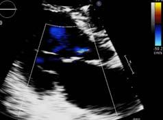
23 Stenosis in the outflow graft Abnormalities in the outflow cannula: Case 3 48 yo woman status post HeartWare LVAD for ICM as BTT. Admitted with shortness of breath/chest pain RHC: RAP = 23 mmhg PAP = 52/34 mpap = 40 mmhg PCWP = 29 mmhg SVR = 1644 dynes/sec/cm-5 Outflow velocity 3.4 m/s 23


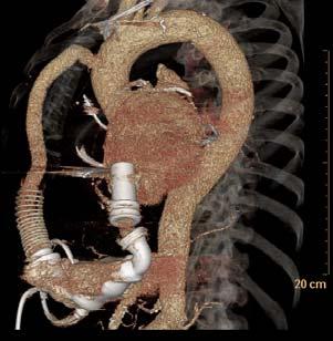
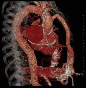
24 Abnormalities in the outflow cannula: Case 3 Turbulence Rt. supra sternal Parasternal long-axis Abnormalities in the outflow cannula: Case 3 Aortogram Balloon Stent 24
25 Abnormalities in the outflow cannula: Case 3 Post stent echo LVAD speed optimization 25


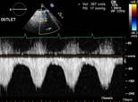
26 Cardiac Output Lie Run Walk The Ramp Study The RAMP study is stopped if suction events occur and/or if the LVEDD <3.0 cm 76mm ED mm ES HMII HVAD RVSP Increasing LVAD speed (in RPM) 26

27 The RAMP Study 1. Optimize device speed without compromising cardiac function: i. Mean arterial BP >65 mmhg ii. Maintain inter-ventricular septum position in midline iii. Intermittent aortic valve opening iv. No more than mild mitral regurgitation 2. Evaluate for LVAD malfunction such as device-related thrombosis Pre-RAMP Considerations Ensure: INR >1.8 PTT >60 No LV thrombus No aortic root thrombus Aortic root clot 27


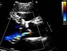
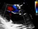
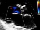
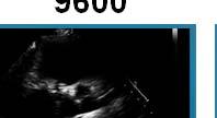
28 RAMP test protocol LVAD parameters Patient parameters Echo parameters Optimization of LV unloading Increasing LVAD pump speed 8000 RPM 9600 RPM RPM 74 mm 69 mm 63 mm 8000 RPM 9600 RPM RPM Rightward 28


29 Optimization of Ao valve opening Increasing LVAD pump speed 8000 RPM 9200 RPM RPM Optimization of Aortic Insufficiency Increasing LVAD pump speed 8000 RPM 9600 RPM RPM 29


30 Optimization of Mitral Regurgitation Increasing LVAD pump speed 8000 RPM 9600 RPM RPM Example RAMP Study LVEDD=77 mm LVEDD=75 mm LVEDD=69 mm LVEDD=69 mm LVEDD=62 mm AI=2 AI=2.5 AI=2.5 AI=3 AI=3 MR=3 MR=3 MR=3 MR=2 MR=1 AVO = 10/10 AVO = 5/10 AVO = 3/10 AV = Closed AV = Closed 30
31 Example RAMP Study Speed PI Power CVP PAsys PAd mpap PCWP PASAT CO Speed LVEDD Septum AVO AI MR Midline 10/ Midline 5/ Midline 3/ Midline Closed Leftward Closed 3 1 LVAD Malfunction: Case 31


32 LVAD Malfunction: A Case 50 year-old female o Idiopathic CM o HMII 6 months ago o 2-d history of shortness of breath and fatigue o Labs consistent with hemolysis o A RAMP study was performed LVAD Recall: Normal LVAD Function RPM AV opening AV closed Increasing aortic regurgitation Decreasing mitral regurgitation Shift in interventricular septum to the left 32


33 RAMP Study in our patient RPM Device inspection in this patient Thrombus Consider LVAD thrombosis: 1.Worsening HF 2.Signs of hemolysis Elevated LDH Low haptoglobin 3.Device malfunction (power spikes/flow alarms) 4.Echo-Ramp test demonstrates lack of change in LVEDD with increasing LVAD speeds Visible thrombus in the inlet stator 33


34 Septum: Malposition LV dimension Pericardium: Atypical Effusion Tamponade Hemorrhage Right ventricle: Failure Tricuspid regurgitation Thrombosis Aortic valve: Opening Regurgitation Thrombosis Mitral valve: Regurgitation Thrombosis Outflow cannula: Kink Obstruction Thrombosis Inflow cannula: Malposition Occlusion Malfunction Thrombosis Prevalence of Common Cardiovascular and Lung Diseases, U.S., 2004, NHLBI report Death from specific cardiovascular, Lung and Blood Diseases, U.S., 2004 NHLBI report 34
Diagnosis of Device Thrombosis
 Diagnosis of Device Thrombosis Andrew Civitello MD, FACC Medical Director, Heart Transplant Program Director, Fellowship Co-Director, Baylor St. Luke's Medical Center / Texas Heart Institute Trends in
UNIVERSITY OF UTAH HEALTH CARE HOSPITALS AND CLINICS
 UNIVERSITY OF UTAH HEALTH CARE HOSPITALS AND CLINICS CARDIAC MECHANICAL SUPPORT PROGRAM GUIDELINES CARDIAC MECHANICAL SUPPORT: LVAD BASICS FREQUENT SCENARIOS AND TROUBLESHOOTING Review Date: July 2011
Left Ventricular Assist Device: What Should I Report?
 2017 SOTA, Tucson, AZ February 21, 2017 11:15 11:40 AM 25 min Left Ventricular Assist Device: What Should I Report? Muhamed Sarić MD, PhD, MPA Director of Noninvasive Cardiology Echo Lab Associate Professor
Mechanical Cardiac Support and Cardiac Transplant: The Role for Echocardiography
 Mechanical Cardiac Support and Cardiac Transplant: The Role for Echocardiography David Langholz, M.D., F.A.C.C. Co-Director Cardiovascular Imaging Fredrick Meijer Heart and Vascular Institute Spectrum
Echo Emergencies. Outline. Michael H. Picard, MD Massachusetts General Hospital Harvard Medical School No disclosures
 Echo Emergencies Michael H. Picard, MD Massachusetts General Hospital Harvard Medical School No disclosures Outline Common emergency / on call scenarios Tamponade Pulmonary embolism/rv strain Cardiogenic
Modern Left Ventricular Assist Devices (LVAD) : An Intro, Complications, and Emergencies
 : An Intro, Complications, and Emergencies") Modern Left Ventricular Assist Devices (LVAD) : An Intro, Complications, and Emergencies ERIC T. ROME D.O. HEART FAILURE, MECHANICAL ASSISTANCE AND TRANSPLANTATION CVI Left Ventricular Assist Device An
Modern Left Ventricular Assist Devices (LVAD) : An Intro, Complications, and Emergencies ERIC T. ROME D.O. HEART FAILURE, MECHANICAL ASSISTANCE AND TRANSPLANTATION CVI Left Ventricular Assist Device An
LEFT VENTRICULAR ASSIST DEVICE COMPLICATIONS. Daniel Vargas, MD Section of Cardiothoracic Imaging University of Colorado Anschutz Medical Campus
 LEFT VENTRICULAR ASSIST DEVICE COMPLICATIONS Daniel Vargas, MD Section of Cardiothoracic Imaging University of Colorado Anschutz Medical Campus OBJECTIVES Review the most common LVAD-related complications.
Ventricular Assisting Devices in the Cathlab. Unrestricted
 Ventricular Assisting Devices in the Cathlab Unrestricted What is a VAD? A single system device that is surgically attached to the left ventricle of the heart and to the aorta for left ventricular support
The Balancing Act Bleeding and Thrombosis in MCS. Muhammad Adil Soofi
 The Balancing Act Bleeding and Thrombosis in MCS Muhammad Adil Soofi Road Map Survival and complications with LVAD What is the Burden of thrombosis and bleeding Why Bleeding and Thrombosis happen When
Risk Factors for Adverse Outcome after HeartMate II Jennifer Cowger, MD, MS St. Vincent Heart Center of Indiana
 Risk Factors for Adverse Outcome after HeartMate II Jennifer Cowger, MD, MS St. Vincent Heart Center of Indiana Advanced Heart Failure, Transplant, & Mechanical Circulatory Support Relevant Financial Relationship
Evaluation of the Right Ventricle in Candidates for Right Ventricular Assist Device Implantation.
 Evaluation of the Right Ventricle in Candidates for Right Ventricular Assist Device Implantation. Evaluation of RVAD Function. Ioannis A Paraskevaidis Attikon University Hospital Historical Perspective
Echocardiographic Structural Assessment Pre- LVAD
 None Disclosures Echocardiographic Structural Assessment Pre- LVAD LVEF ( 25% for DT LVAD) Right ventricle Valvular disease Intra- cardiac shunts Intra- cardiac thrombi Ascending aorta Case 1 50 yo M
เอกราช อร ยะช ยพาณ ชย
 30 July 2016 เอกราช อร ยะช ยพาณ ชย Heart Failure and Transplant Cardiology aekarach.a@chula.ac.th Disclosure Speaker, CME service: Merck, Otsuka, Servier Consultant, non-cme service: Novartis, Menarini
Role of Echocardiography in Patients With Intravascular Hemolysis Due to Suspected Continuous-Flow LVAD Thrombosis
 JACC: CARDIOVASCULAR IMAGING VOL. 6, NO. 11, 2013 ª 2013 BY THE AMERICAN COLLEGE OF CARDIOLOGY FOUNDATION ISSN 1936-878X/$36.00 PUBLISHED BY ELSEVIER INC. http://dx.doi.org/10.1016/j.jcmg.2013.06.006 ORIGINAL
Appendix II: ECHOCARDIOGRAPHY ANALYSIS
 Appendix II: ECHOCARDIOGRAPHY ANALYSIS Two-Dimensional (2D) imaging was performed using the Vivid 7 Advantage cardiovascular ultrasound system (GE Medical Systems, Milwaukee) with a frame rate of 400 frames
Ventricular Assist Devices for Permanent Therapy: Current Status and Future
 Ventricular Assist Devices for Permanent Therapy: Current Status and Future Prospects Francis D. Pagani MD PhD Professor of Cardiac Surgery University of Michigan April 28 th, 2012 Disclosures NHLBI and
Curtin University is a trademark of Curtin University of Technology CRICOS Provider Code 00301J
 Exercise-based cardiac rehabilitation for people with ventricular assist devices: Associate Professor Andrew Maiorana School of Physiotherapy and Exercise Science, Curtin University; Advanced Heart Failure
MCSD Pump Thrombosis : Industry Perspective
 MCSD Pump Thrombosis : Industry Perspective John B. O Connell MD Vice President, Medical Affairs Thoratec Corporation 1 1 Thoratec Asia Pacific Mechanical Circulatory Support (MCS) Conference Agenda 15-17
TGA atrial vs arterial switch what do we need to look for and how to react
 TGA atrial vs arterial switch what do we need to look for and how to react Folkert Meijboom, MD, PhD, FES Dept ardiology University Medical entre Utrecht The Netherlands TGA + atrial switch: Follow-up
Aortic Insufficiency: How Often Does It Occur and When To Treat
 Aortic Insufficiency: How Often Does It Occur and When To Treat Simon Maltais, MD PhD Vice-Chair of Clinical Practice Director of MCS Program Department of Cardiovascular Surgery Mayo Clinic, Rochester,
HeartMate 3 Left Ventricular Assist System Catalog #106524US LVAS KIT, HM3
 URGENT MEDICAL DEVICE RECALL HeartMate 3 Left Ventricular Assist System Catalog #106524US LVAS KIT, HM3 May 21, 2018 Dear Physician, We are providing additional information to the letter we recently issued
HEARTWARE HVAD WAVEFORM APP INSTRUCTIONS
 HEARTWARE HVAD WAVEFORM APP INSTRUCTIONS TABLE OF CONTENTS Welcome... 3 HVAD Waveforms 1. Characteristics... 4 2. Theory of Operation... 5 3. Ao & LV Pressure... 6 4. HQ Curve... 7 5. PV Loops... 8 Home
Evaluation of Left Ventricular Diastolic Dysfunction by Doppler and 2D Speckle-tracking Imaging in Patients with Primary Pulmonary Hypertension
 ESC Congress 2011.No 85975 Evaluation of Left Ventricular Diastolic Dysfunction by Doppler and 2D Speckle-tracking Imaging in Patients with Primary Pulmonary Hypertension Second Department of Internal
LVAD Complications, Recovery
 LVAD Complications, Recovery Abbas Ardehali, M.D., F.A.C.S. Professor of Surgery and Medicine, Division of Cardiac Surgery William E. Connor Chair in Cardiothoracic Transplantation Director, UCLA Heart,
Mechanical Support in the Failing Fontan-Kreutzer
 Mechanical Support in the Failing Fontan-Kreutzer Stephanie Fuller MD, MS Thomas L. Spray Endowed Chair in Congenital Heart Surgery Associate Professor, The Perelman School of Medicine at the University
Certificate in Clinician Performed Ultrasound (CCPU) Syllabus. Rapid Cardiac Echo (RCE)
 Syllabus. Rapid Cardiac Echo (RCE)") Certificate in Clinician Performed Ultrasound (CCPU) Syllabus Rapid Cardiac Echo (RCE) Purpose: Rapid Cardiac Echocardiography (RCE) This unit is designed to cover the theoretical and practical curriculum
Certificate in Clinician Performed Ultrasound (CCPU) Syllabus Rapid Cardiac Echo (RCE) Purpose: Rapid Cardiac Echocardiography (RCE) This unit is designed to cover the theoretical and practical curriculum
PROSTHETIC VALVE BOARD REVIEW
 PROSTHETIC VALVE BOARD REVIEW The correct answer D This two chamber view shows a porcine mitral prosthesis with the typical appearance of the struts although the leaflets are not well seen. The valve
Destination Therapy For Advanced Heart Failure
 Destination Therapy For Advanced Heart Failure Kevin Guffey, RN Vad Coordinator Tacoma General Hospital April 28, 2012 Current HF Estimates 300 Million Population HF=2.5% of Population 6.5-7 Million Patients
New murmur: acute valvular regurgitations. A.Pasquet, MD,PhD. UCL -Cliniques Saint Luc
 New murmur: acute valvular regurgitations. A.Pasquet, MD,PhD UCL -Cliniques Saint Luc Acute valvular regurgitation Clinical case Mr Dupont, a 53 y old men, without any particular medical history On Thursday
ASCeXAM / ReASCE. Practice Board Exam Questions Monday Morning
 ASCeXAM / ReASCE Practice Board Exam Questions Monday Morning Ultrasound Physics Artifacts Doppler Physics Imaging, Knobology, and Artifacts Echocardiographic Evaluation of the RV Tricuspid and Pulmonary
HEMODYNAMIC ASSESSMENT
 HEMODYNAMIC ASSESSMENT INTRODUCTION Conventionally hemodynamics were obtained by cardiac catheterization. It is possible to determine the same by echocardiography. Methods M-mode & 2D echo alone can provide
Left Ventricular Assist Device Malfunction: A Systematic Approach to Diagnosis
 Journal of the American College of Cardiology Vol. 43, No. 9, 2004 2004 by the American College of Cardiology Foundation ISSN 0735-1097/04/$30.00 Published by Elsevier Inc. doi:10.1016/j.jacc.2003.11.055
Complications of Left Ventricular Assist Device Chronic Support. Dr. Tal Hasin RMC, Beilinson, Petach-Tiqva, Israel
 Complications of Left Ventricular Assist Device Chronic Support. Dr. Tal Hasin RMC, Beilinson, Petach-Tiqva, Israel No disclosures Probability of survival Survival (%) Survival with LVAD Destination Bridge
Review Article Focused Review on Transthoracic Echocardiographic Assessment of Patients with Continuous Axial Left Ventricular Assist Devices
 SAGE-Hindawi Access to Research Cardiology Research and Practice Volume 2011, Article ID 187434, 11 pages doi:10.4061/2011/187434 Review Article Focused Review on Transthoracic Echocardiographic Assessment
Doppler Basic & Hemodynamic Calculations
 Doppler Basic & Hemodynamic Calculations August 19, 2017 Smonporn Boonyaratavej MD Division of Cardiology, Department of Medicine Chulalongkorn University Cardiac Center, King Chulalongkorn Memorial Hospital
Echocardiographic Cardiovascular Risk Stratification: Beyond Ejection Fraction
 Echocardiographic Cardiovascular Risk Stratification: Beyond Ejection Fraction October 4, 2014 James S. Lee, M.D., F.A.C.C. Associates in Cardiology, P.A. Silver Spring, M.D. Disclosures Financial none
What are the indications for Tricuspid valve repair during LVAD Implant RANJIT JOHN, MD UNIVERSITY OF MINNESOTA
 What are the indications for Tricuspid valve repair during LVAD Implant RANJIT JOHN, MD UNIVERSITY OF MINNESOTA Contraindications for LVAD Lack of social support system Nonreversible end organ failure
Choose the grading of diastolic function in 82 yo woman
 Question #1 Choose the grading of diastolic function in 82 yo woman E= 80 cm/s A= 70 cm/s LAVI < 34 ml/m 2 1= Grade 1 2= Grade 2 3= Grade 3 4= Normal 5= Indeterminate 2018 MFMER 3712003-1 Choose the grading
Echocardiography as a diagnostic and management tool in medical emergencies
 Echocardiography as a diagnostic and management tool in medical emergencies Frank van der Heusen MD Department of Anesthesia and perioperative Care UCSF Medical Center Objective of this presentation Indications
Ventricular Assist Device. Lauren Bartlett 10/5/16 BME 281, section 1
 Ventricular Assist Device Lauren Bartlett 10/5/16 BME 281, section 1 What is a Ventricular Assist Device (VAD)? Electromechanical device for assisting cardiac circulation Used to partially or completely
Mechanical Cardiac Support in Acute Heart Failure. Michael Felker, MD, MHS Associate Professor of Medicine Director of Heart Failure Research
 Mechanical Cardiac Support in Acute Heart Failure Michael Felker, MD, MHS Associate Professor of Medicine Director of Heart Failure Research Disclosures Research Support and/or Consulting NHLBI Amgen Cytokinetics
Cardiac ultrasound protocols
 Cardiac ultrasound protocols IDEXX Telemedicine Consultants Two-dimensional and M-mode imaging planes Right parasternal long axis four chamber Obtained from the right side Displays the relative proportions
What are the best diagnostic tools to quantify aortic regurgitation?
 What are the best diagnostic tools to quantify aortic regurgitation? Agnès Pasquet, MD, PhD Pôle de Recherche Cardiovasculaire Institut de Recherche Expérimentale et Clinique Université catholique de Louvain
New Trends and Indications for LVADs
 New Trends and Indications for LVADs Mark S. Slaughter, MD Professor and Chief Division of Thoracic and Cardiovascular Surgery University of Louisville Natural History of Heart Failure 100 10 Class III
1/21/2016. HeartMate II Indications for Use. Ventricular Assist Device Overview. Jon G. Echterling MSN, CCRN, FNP-BC. Learning Objectives
 Ventricular Assist Device Overview Jon G. Echterling MSN, CCRN, FNP-BC February 5, 2016 Learning Objectives Identify the components and operation of the HeartMate II LVAD Describe the path blood follows
DOPPLER HEMODYNAMICS (1) QUANTIFICATION OF PRESSURE GRADIENTS and INTRACARDIAC PRESSURES
 QUANTIFICATION OF PRESSURE GRADIENTS and INTRACARDIAC PRESSURES") THORAXCENTRE DOPPLER HEMODYNAMICS (1) QUANTIFICATION OF PRESSURE GRADIENTS and INTRACARDIAC PRESSURES J. Roelandt DOPPLER HEMODYNAMICS Intracardiac pressures and pressure gradients Volumetric measurement
THORAXCENTRE DOPPLER HEMODYNAMICS (1) QUANTIFICATION OF PRESSURE GRADIENTS and INTRACARDIAC PRESSURES J. Roelandt DOPPLER HEMODYNAMICS Intracardiac pressures and pressure gradients Volumetric measurement
EMS: Care of the VAD Patient. Brittany Butzler BSN RN VAD Coordinator Froedtert and the Medical College of WI
 EMS: Care of the VAD Patient Brittany Butzler BSN RN VAD Coordinator Froedtert and the Medical College of WI Disclosure No relevant financial relationships by planners or presenters Left Ventricular Assist
Prosthetic valve dysfunction: stenosis or regurgitation
 Prosthetic valve dysfunction: stenosis or regurgitation Jean G. Dumesnil MD, FRCP(C), FACC, FASE(Hon) Quebec Heart and Lung Institute, Québec, Québec No disclosures Possible Causes of High Gradients in
TRANSTHORACIC ECHOCARDIOGRAPHY (TTE) An overview for Perioperative Care Dr Andrew Cluer, Sydney, Australia 2015
 An overview for Perioperative Care Dr Andrew Cluer, Sydney, Australia 2015") TRANSTHORACIC ECHOCARDIOGRAPHY (TTE) An overview for Perioperative Care Dr Andrew Cluer, Sydney, Australia 2015 This piece of work is not meant to teach students echo interpretation, but instead offers
TRANSTHORACIC ECHOCARDIOGRAPHY (TTE) An overview for Perioperative Care Dr Andrew Cluer, Sydney, Australia 2015 This piece of work is not meant to teach students echo interpretation, but instead offers
Little is known about the degree and time course of
 Differential Changes in Regional Right Ventricular Function Before and After a Bilateral Lung Transplantation: An Ultrasonic Strain and Strain Rate Study Virginija Dambrauskaite, MD, Lieven Herbots, MD,
Left Ventricular Assist Devices (LVADs): Overview and Future Directions
: Overview and Future Directions") Left Ventricular Assist Devices (LVADs): Overview and Future Directions FATIMA KARAKI, M.D. PGY-3, DEPARTMENT OF MEDICINE WASHINGTON UNIVERSITY IN ST. LOUIS ST. LOUIS, MISSOURI, USA St. Louis, Missouri,
Left Ventricular Assist Devices (LVADs): Overview and Future Directions FATIMA KARAKI, M.D. PGY-3, DEPARTMENT OF MEDICINE WASHINGTON UNIVERSITY IN ST. LOUIS ST. LOUIS, MISSOURI, USA St. Louis, Missouri,
cardiac imaging planes planning basic cardiac & aortic views for MR
 cardiac imaging planes planning basic cardiac & aortic views for MR Dianna M. E. Bardo, M. D. Assistant Professor of Radiology & Cardiovascular Medicine Director of Cardiac Imaging cardiac imaging planes
Repeated Ramp Tests on Stable LVAD Patients Reveal Patient-Specific Hemodynamic Fingerprint
 ASAIO Journal 2017 Adult Circulatory Support Repeated Ramp Tests on Stable LVAD Patients Reveal Patient-Specific Hemodynamic Fingerprint TERUHIKO IMAMURA,* DANIEL BURKHOFF, DANIEL RODGERS,* SIRTAZ ADATYA,*
Ventricular Assist Device: Are Early Interventions Superior? Hamang Patel, MD Section of Cardiomyopathy & Heart Transplantation
 Ventricular Assist Device: Are Early Interventions Superior? Hamang Patel, MD Section of Cardiomyopathy & Heart Transplantation Objectives Current rationale behind use of MCS Patient Selection Earlier?
British Society of Echocardiography
 British Society of Echocardiography Affiliated to the British Cardiac Society A Minimum Dataset for a Standard Adult Transthoracic Echocardiogram From the British Society of Echocardiography Education
Congenital. Unicuspid Bicuspid Quadricuspid
 David Letterman s Top 10 Aortic Stenosis The victim can be anyone: Echo is the question and the answer!!!! Hilton Head Island Echocardiography Conference 2012 Timothy E. Paterick, MD, JD, MBA Christopher
Right Ventricular Failure: Prediction, Prevention and Treatment
 Right Ventricular Failure: Prediction, Prevention and Treatment 3 rd European Training Symposium for Heart Failure Cardiologists and Cardiac Surgeons University Hospital Bern June 24-25, 2016 Disclosures:
Evaluation and treatment of pump thrombosis and hemolysis
 Featured Article Evaluation and treatment of pump thrombosis and hemolysis Vakhtang Tchantchaleishvili 1, Fabio Sagebin 1, Ronald E. Ross 1, William Hallinan 2, Karl Q. Schwarz 2, H. Todd Massey 1 1 Division
Conflict of Interests
 The Left Ventricle: How Should We Quantify Its Size and Function; Is It Time for 3D in Everyone? Roberto M Lang, MD Conflict of Interests Philips Medical Imaging Research Grants Speakers bureau Advisory
Index. K Knobology, TTE artifact, image resolution, ultrasound, 14
 A Acute aortic regurgitation (AR), 124 128 Acute aortic syndrome (AAS) classic aortic dissection diagnosis, 251 263 evolutive patterns, 253 255 pathology, 250 251 classifications, 247 248 incomplete aortic
Hemodynamic Assessment. Assessment of Systolic Function Doppler Hemodynamics
 Hemodynamic Assessment Matt M. Umland, RDCS, FASE Aurora Medical Group Milwaukee, WI Assessment of Systolic Function Doppler Hemodynamics Stroke Volume Cardiac Output Cardiac Index Tei Index/Index of myocardial
Disclosures. Cardiac Ultrasound. Introductory Case. 80 y/o male Syncope at home Emesis x 3 in ambulance Looks sick. No pain.
 Disclosures Cardiac Ultrasound Justin A Davis, MD MPH RDMS Subchief for Emergency Ultrasound Kaiser Permanente East Bay Medical Center I have nothing to disclose. Introductory Case HR 118 BP 65/43 RR 27
Dobutamine Stress testing In Low Flow, Low EF, Low Gradient Aortic Stenosis Case Studies
 Dobutamine Stress testing In Low Flow, Low EF, Low Gradient Aortic Stenosis Case Studies Mitral Regurgitation The New ASE Guidelines: Role of 2D/3D and CMR William A. Zoghbi MD, FASE, MACC Professor and
MITRAL STENOSIS. Joanne Cusack
 MITRAL STENOSIS Joanne Cusack BSE Breakdown Recognition of rheumatic mitral stenosis Qualitative description of valve and sub-valve calcification and fibrosis Measurement of orifice area by planimetry
LV geometric and functional changes in VHD: How to assess? Mi-Seung Shin M.D., Ph.D. Gachon University Gil Hospital
 LV geometric and functional changes in VHD: How to assess? Mi-Seung Shin M.D., Ph.D. Gachon University Gil Hospital LV inflow across MV LV LV outflow across AV LV LV geometric changes Pressure overload
Complications of VAD therapy - RV failure
 Complications of VAD therapy - RV failure Nana Afari-Armah, MD Advanced heart failure and transplant cardiology Temple University Hospital 3/24/18 Goals Understand the role of the right ventricle in LVAD
PART II ECHOCARDIOGRAPHY LABORATORY OPERATIONS ADULT TRANSTHORACIC ECHOCARDIOGRAPHY TESTING
 PART II ECHOCARDIOGRAPHY LABORATORY OPERATIONS ADULT TRANSTHORACIC ECHOCARDIOGRAPHY TESTING STANDARD - Primary Instrumentation 1.1 Cardiac Ultrasound Systems SECTION 1 Instrumentation Ultrasound instruments
Echo Assessment Pre-TAVI
 Disclosure Statement of Financial Interest Within the past 12 months, I or my spouse/partner have had a financial Interest /arrangement or affiliation with the organization(s) listed below Echocardiographic
Left Ventricular Assist Devices LVAD. North Country EMS Program Agency 3/21/12
 Left Ventricular Assist Devices LVAD North Country EMS Program Agency 3/21/12 Objectives Describe indications for and functions of ventricular assist devices (LVAD) Differentiate assessment findings of
ECHOCARDIOGRAPHY DATA REPORT FORM
 Patient ID Patient Study ID AVM - - Date of form completion / / 20 Initials of person completing the form mm dd yyyy Study period Preoperative Postoperative Operative 6-month f/u 1-year f/u 2-year f/u
HISTORY. Question: What category of heart disease is suggested by this history? CHIEF COMPLAINT: Heart murmur present since early infancy.
 HISTORY 18-year-old man. CHIEF COMPLAINT: Heart murmur present since early infancy. PRESENT ILLNESS: Although normal at birth, a heart murmur was heard at the six week check-up and has persisted since
Quantitation of right ventricular dimensions and function
 SCCS Basics of cardiac assessment Quantitation of right ventricular dimensions and function Tomasz Kukulski, MD PhD Dept of Cardiology, Congenital Heart Disease and Electrotherapy Silesian Medical University
Right Ventricle Steven J. Lester MD, FACC, FRCP(C), FASE Mayo Clinic, Arizona
, FASE Mayo Clinic, Arizona") Right Ventricle Steven J. Lester MD, FACC, FRCP(C), FASE Mayo Clinic, Arizona 1. In which scenario will applying the simplified Bernoulli equation to the peak tricuspid regurgitation velocity and adding
Right Ventricle Steven J. Lester MD, FACC, FRCP(C), FASE Mayo Clinic, Arizona 1. In which scenario will applying the simplified Bernoulli equation to the peak tricuspid regurgitation velocity and adding
The background of the Cardiac Sonographer Network News masthead is a diagnostic image:
 Number 5 Welcome Number 5 Welcome to the newsletter created just for you: sonographers who perform pediatric echocardiograms in primarily adult echo labs. Each issue features tips on echocardiography of
CARDIOVASCULAR SYSTEM
 CARDIOVASCULAR SYSTEM Overview Heart and Vessels 2 Major Divisions Pulmonary Circuit Systemic Circuit Closed and Continuous Loop Location Aorta Superior vena cava Right lung Pulmonary trunk Base of heart
Advanced Applica,on of Point- of- Care Echocardiography in Cri,cal Care. Dr. Mark Tutschka Dr. Rob ArnAield
 Advanced Applica,on of Point- of- Care Echocardiography in Cri,cal Care Dr. Mark Tutschka Dr. Rob ArnAield OBJECTIVES Provide an overview of common advanced echocardiographic techniques suitable for use
Atrial Septal Defects
 Supplementary ACHD Echo Acquisition Protocol for Atrial Septal Defects The following protocol for echo in adult patients with atrial septal defects (ASDs) is a guide for performing a comprehensive assessment
Evaluation of the Right Ventricle and Risk Stratification for Sudden Cardiac Death
 Evaluation of the Right Ventricle and Risk Stratification for Sudden Cardiac Death Presenters: Sabrina Phillips, MD FACC FASE Director, Adult Congenital Heart Disease Services The University of Oklahoma
Heart Failure Dr ahmed almutairi Assistant professor internal medicin dept
 Heart Failure Dr ahmed almutairi Assistant professor internal medicin dept (MBBS)(SBMD) Introduction Epidemiology Pathophysiology diastolic/systolic Risk factors Signs and symptoms Classification of HF
Giovanni Di Salvo MD, PhD, FESC Second University of Naples Monaldi Hospital
 Giovanni Di Salvo MD, PhD, FESC Second University of Naples Monaldi Hospital VSD is one of the most common congenital cardiac abnormalities in the newborn. It can occur as an isolated finding or in combination
Adel Hasanin Ahmed 1
 Adel Hasanin Ahmed 1 PERICARDIAL DISEASE The pericardial effusion ends anteriorly to the descending aorta and is best visualised in the PLAX. PSAX is actually very useful sometimes for looking at posterior
ORIGINAL ARTICLE. Alexander M. Bernhardt a, *, Theo M.M.H. De By b, Hermann Reichenspurner a and Tobias Deuse a. Abstract INTRODUCTION
 European Journal of Cardio-Thoracic Surgery 48 (2015) 158 162 doi:10.1093/ejcts/ezu406 Advance Access publication 29 October 2014 ORIGINAL ARTICLE Cite this article as: Bernhardt AM, De By TMMH, Reichenspurner
You Won t Believe What I Saw on. Disclosures. Goals. Dimensions 2013 October 18 th Michael Pfeiffer, MD. No Financial Disclosures
 You Won t Believe What I Saw on that ECHO! Dimensions 2013 October 18 th Michael Pfeiffer, MD Disclosures No Financial Disclosures Goals Review unusual and unique echocardiographic images. Briefly present
ECHO HAWAII. My home. Pulmonary Hypertension and Pulmonary Embolism: Role of Echo U.S.A. Japan. Hawaii Island 1/9/2018
 Pulmonary Hypertension and Pulmonary Embolism: Role of Echo ECHO HAWAII January 15 19, 2018 Kenya Kusunose, MD, PhD, FASE Tokushima University Hospital Japan My home Japan U.S.A Hawaii Island 1 Economy
The Doppler Examination. Katie Twomley, MD Wake Forest Baptist Health - Lexington
 The Doppler Examination Katie Twomley, MD Wake Forest Baptist Health - Lexington OUTLINE Principles/Physics Use in valvular assessment Aortic stenosis (continuity equation) Aortic regurgitation (pressure
Comprehensive Hemodynamics By Doppler Echocardiography. The Echocardiographic Swan-Ganz Catheter.
 Comprehensive Hemodynamics By Doppler Echocardiography. The Echocardiographic Swan-Ganz Catheter. Itzhak Kronzon, MD, FASE, FACC, FESC, FAHA, FACP, FCCP North Shore HS, LIJ/Lenox Hill Hospital, New York
P = 4V 2. IVC Dimensions 10/20/2014. Comprehensive Hemodynamic Evaluation by Doppler Echocardiography. The Simplified Bernoulli Equation
 Comprehensive Hemodynamic Evaluation by Doppler Echocardiography Itzhak Kronzon, MD North Shore LIJ/ Lenox Hill Hospital New York, NY Disclosure: Philips Healthcare St. Jude Medical The Simplified Bernoulli
Pediatric Echocardiography Examination Content Outline
 Pediatric Echocardiography Examination Content Outline (Outline Summary) # Domain Subdomain Percentage 1 Anatomy and Physiology Normal Anatomy and Physiology 10% 2 Abnormal Pathology and Pathophysiology
The Development of Aortic Insufficiency in Continuous-Flow Left Ventricular Assist Device Supported Patients
 The Development of Aortic Insufficiency in Continuous-Flow Left Ventricular Assist Device Supported Patients Ashim Aggarwal, MD, MRCP, Rashmi Raghuvir, MD, Paula Eryazici, MD, Gregory Macaluso, MD, Priya
Complications of Contemporary Continuous Flow LVAD Therapy
 Complications of Contemporary Continuous Flow LVAD Therapy Daniel J. Goldstein MD FACS FACC Associate Professor Vice-Chair, Dept Cardiothoracic Surgery Disclosures Thoratec Inc Medical Advisory Board Chair,
Heart Failure. Cardiac Anatomy. Functions of the Heart. Cardiac Cycle/Hemodynamics. Determinants of Cardiac Output. Cardiac Output
 Cardiac Anatomy Heart Failure Professor Qing ZHANG Department of Cardiology, West China Hospital www.blaufuss.org Cardiac Cycle/Hemodynamics Functions of the Heart Essential functions of the heart to cover
SONOGRAPHER & NURSE LED VALVE CLINICS
 SONOGRAPHER & NURSE LED VALVE CLINICS Frequency of visits and alerts AORTIC STENOSIS V max > 4.0 m/s or EOA < 1.0 cm 2 V max 3.5 4.0 m/s + Ca+ V max 3.0 4.0 m/s or EOA 1.0-1.5 cm 2 V max 2.5 3.0 m/s every
Analysis of Pump Thrombosis in the Intermacs Database
 Analysis of Pump Thrombosis in the Intermacs Database Michael Acker William Measey Professor of Surgery Chief of Division of Cardiovascular Surgery Director of Heart and Vascular Center University of Pennsylvania
Stress Testing in Valvular Disease
 2017 ASE Florida Orlando, FL October 10, 2017 2:40 2:50 PM 10 min Grand Harbor Ballroom South Stress Testing in Valvular Disease Muhamed Sarić MD, PhD, MPA Director of Noninvasive Cardiology Echo Lab Associate
Ray Matthews MD Professor of Clinical Medicine Chief of Cardiology University of Southern California
 High Risk PCI Making Possible the Impossible Ray Matthews MD Professor of Clinical Medicine Chief of Cardiology University of Southern California Disclosures Abiomed Research Support Consulting Agreement
RIGHT VENTRICULAR SIZE AND FUNCTION
 RIGHT VENTRICULAR SIZE AND FUNCTION Edwin S. Tucay, MD, FPCC, FPCC, FPSE Philippine Society of Echocardiography Quezon City, Philippines Echo Mission, BRTTH, Legaspi City, July 1-2, 2016 NO DISCLOSURE
Echo Doppler Assessment of Right and Left Ventricular Hemodynamics.
 Echo Doppler Assessment of Right and Left Ventricular Hemodynamics. Itzhak Kronzon, MD, FASE, FACC, FESC, FAHA, FACP, FCCP Northwell, Lenox Hill Hospital, New York Professor of Cardiology Hofstra University
Valve Disease Board Review Questions
 Valve Disease Board Review Questions Dennis A. Tighe, MD, FASE University of Massachusetts Medical School Worcester, MA Case 1 History A 61 year-old man Presents to hospital with worsening shortness of
None. Declaration of conflict of interest
 None Declaration of conflict of interest New Long Term Circulatory Support Technology and Treatment Strategies Stephen Westaby Oxford, UK Cardiac Transplantation: Facts from the UNOS Database Median survival
EMS and Nursing Considerations in VAD Patient Care
 EMS and Nursing Considerations in VAD Patient Care B R I T T A N Y B U T Z L E R B S N R N V A D C O O R D I N A T O R F R O E D T E R T A N D T H E M E D I C A L C O L L E G E O F W I 1 0 / 2 5 / 1 8