Right Ventricle Steven J. Lester MD, FACC, FRCP(C), FASE Mayo Clinic, Arizona
|
|
|
- Edwin Singleton
- 5 years ago
- Views:
Transcription

1 Right Ventricle Steven J. Lester MD, FACC, FRCP(C), FASE Mayo Clinic, Arizona 1. In which scenario will applying the simplified Bernoulli equation to the peak tricuspid regurgitation velocity and adding an estimate of right atrial pressure likely not result in an under estimation the right ventricular systolic pressure. a. When there is pulmonary valve stenosis. b. When there is severe tricuspid regurgitation c. When there is marked dilation of the right ventricle d. When you are unable to align the continuous wave Doppler signal parallel to the intercept angle of the axis of flow. 1


2 Assumptions 1. Velocity is only dependent on pressure. (fr-fo) = 2fo v (cosθ) / c Doppler Frequency Shift Equation If V1 is significant ( >1.5 m/sec) Presence of anemia (viscous friction) Assumptions 1.Velocity is only dependent on pressure. Flow = Area x Velocity Heart Rate Contractility 2


3 Velocity = Flow Area Assumptions 1.Velocity is only dependent on pressure. 2.You can accurately estimate right atrial pressure. - Non simultaneous - Peak systole 3

4 Systolic RV RA pressure equalization R V RA 4


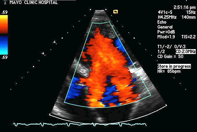
5 Assumptions 1.Velocity is only dependent on pressure. 2.You can accurately estimate right atrial pressure. 3.Right ventricular systolic pressure = Pulmonary artery systolic pressure TRV = 4.8 m/sec 1 = 2.64 cm 2 = 1.96 cm RVSP = 4 X TR vmax2 + estimated RA pressure = 4 (4.8) = 105 mmhg 1 2 5

 of older")
 The RVEF would be the same in both the older women and younger men.")
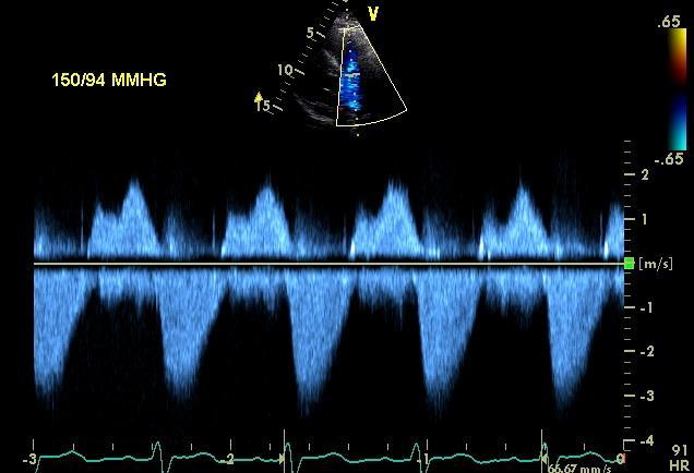
6 RV-PA Gradient = 36 mmhg 3 SPAP = RVSP PV gradient = = 69 mmhg 2. Women have smaller right ventricular end-diastolic volumes when compared to men. Also older age is associated with smaller volumes. Therefore, you would expect the right ventricular ejection fraction (RVEF) of older women compared to younger men to be? a) The RVEF would be the same in both the older women and younger men. b) The RVEF would be higher in older women compared to younger men c) The RVEF would be lower in older women compared to younger men. 6


7 3. A 47 year old women has a dilated right ventricle with reduced right ventricular function along with signs and symptoms of right heart failure. The calculated right ventricular index of myocardial performance (RIMP) determined by pulsed wave Doppler was She was treated with diuretics and clinically improved as her right atrial pressure decreased. You now recalculate the RIMP and you would expect the value to? a. Higher than prior to treatment with diuretics b. Lower than prior to treatment with diuretics c. Unchanged arterial ventricular atrial 7


8 Effect of decrease in LA pressure Lasix 80 mg Ac Mo Ao 215 Tei index = = Tei index = = Courtesy Dr. Hatle 4. With respect to the S velocity, an example shown below, the following statement is true. S = 0.14 m/s a. The S velocity obtained by pulsed wave tissue Doppler will be lower than the S velocity if obtained by color tissue Doppler. b. The S velocity represents global right ventricular function. c. S velocity will be reduced following open heart surgery. d. The value of S is not angle dependent. 8

9 5. 3D methods with which to calculate right ventricular (RV) volumes and ejection fraction may be superior to 2D methods for the following reason. a. 3D methods of calculating RV volumes and ejection fraction are less load dependent than 2D methods. b. 3D methods of calculating RV volumes and ejection fraction are less dependent on image quality then 2D methods. c. 3D but not 2D methods with which to calculate RV volumes and ejection fraction correlate with values obtained by MRI. d. 3D methods include the RV outflow tract contribution to overall function while 2D methods do not. 9
Hemodynamic Assessment. Assessment of Systolic Function Doppler Hemodynamics
 Hemodynamic Assessment Matt M. Umland, RDCS, FASE Aurora Medical Group Milwaukee, WI Assessment of Systolic Function Doppler Hemodynamics Stroke Volume Cardiac Output Cardiac Index Tei Index/Index of myocardial
Affecting the elderly Requiring new approaches. Echocardiographic Evaluation of Hemodynamic Severity. Increasing prevalence Mostly degenerative
 Echocardiographic Evaluation of Hemodynamic Severity Steven J. Lester MD, FACC, FRCP(C), FASE Mayo Clinic, Arizona Relevant Financial Relationship(s) None Off Label Usage None A re-emerging public-health
Doppler Basic & Hemodynamic Calculations
 Doppler Basic & Hemodynamic Calculations August 19, 2017 Smonporn Boonyaratavej MD Division of Cardiology, Department of Medicine Chulalongkorn University Cardiac Center, King Chulalongkorn Memorial Hospital
RVOTO adult and post-op
 Right ventricular outflow tract obstruction in the adult: native and post-op Helmut Baumgartner Westfälische Wilhelms-Universität Münster Adult Congenital and Valvular Heart Disease Center University of
Echo Doppler Assessment of Right and Left Ventricular Hemodynamics.
 Echo Doppler Assessment of Right and Left Ventricular Hemodynamics. Itzhak Kronzon, MD, FASE, FACC, FESC, FAHA, FACP, FCCP Northwell, Lenox Hill Hospital, New York Professor of Cardiology Hofstra University
Appendix II: ECHOCARDIOGRAPHY ANALYSIS
 Appendix II: ECHOCARDIOGRAPHY ANALYSIS Two-Dimensional (2D) imaging was performed using the Vivid 7 Advantage cardiovascular ultrasound system (GE Medical Systems, Milwaukee) with a frame rate of 400 frames
HEMODYNAMIC ASSESSMENT
 HEMODYNAMIC ASSESSMENT INTRODUCTION Conventionally hemodynamics were obtained by cardiac catheterization. It is possible to determine the same by echocardiography. Methods M-mode & 2D echo alone can provide
Tricuspid and Pulmonary Valve Disease
 Tricuspid and Pulmonary Valve Disease Lawrence Rudski MD FRCPC FACC FASE Professor of Medicine Director, Division of Cardiology Jewish General Hospital McGill University Question 1 All of the following
The Doppler Examination. Katie Twomley, MD Wake Forest Baptist Health - Lexington
 The Doppler Examination Katie Twomley, MD Wake Forest Baptist Health - Lexington OUTLINE Principles/Physics Use in valvular assessment Aortic stenosis (continuity equation) Aortic regurgitation (pressure
Right Heart Hemodynamics: Echo-Cath Discrepancies
 Department of cardiac, thoracic and vascular sciences University of Padua, School of Medicine Padua, Italy Right Heart Hemodynamics: Echo-Cath Discrepancies Luigi P. Badano, MD, PhD, FESC, FACC **Dr. Badano
DOPPLER HEMODYNAMICS (1) QUANTIFICATION OF PRESSURE GRADIENTS and INTRACARDIAC PRESSURES
 QUANTIFICATION OF PRESSURE GRADIENTS and INTRACARDIAC PRESSURES") THORAXCENTRE DOPPLER HEMODYNAMICS (1) QUANTIFICATION OF PRESSURE GRADIENTS and INTRACARDIAC PRESSURES J. Roelandt DOPPLER HEMODYNAMICS Intracardiac pressures and pressure gradients Volumetric measurement
THORAXCENTRE DOPPLER HEMODYNAMICS (1) QUANTIFICATION OF PRESSURE GRADIENTS and INTRACARDIAC PRESSURES J. Roelandt DOPPLER HEMODYNAMICS Intracardiac pressures and pressure gradients Volumetric measurement
RIGHT VENTRICULAR SIZE AND FUNCTION
 RIGHT VENTRICULAR SIZE AND FUNCTION Edwin S. Tucay, MD, FPCC, FPCC, FPSE Philippine Society of Echocardiography Quezon City, Philippines Echo Mission, BRTTH, Legaspi City, July 1-2, 2016 NO DISCLOSURE
ASCeXAM / ReASCE. Practice Board Exam Questions Monday Morning
 ASCeXAM / ReASCE Practice Board Exam Questions Monday Morning Ultrasound Physics Artifacts Doppler Physics Imaging, Knobology, and Artifacts Echocardiographic Evaluation of the RV Tricuspid and Pulmonary
Evaluation of the Right Ventricle and Risk Stratification for Sudden Cardiac Death
 Evaluation of the Right Ventricle and Risk Stratification for Sudden Cardiac Death Presenters: Sabrina Phillips, MD FACC FASE Director, Adult Congenital Heart Disease Services The University of Oklahoma
P = 4V 2. IVC Dimensions 10/20/2014. Comprehensive Hemodynamic Evaluation by Doppler Echocardiography. The Simplified Bernoulli Equation
 Comprehensive Hemodynamic Evaluation by Doppler Echocardiography Itzhak Kronzon, MD North Shore LIJ/ Lenox Hill Hospital New York, NY Disclosure: Philips Healthcare St. Jude Medical The Simplified Bernoulli
Pulmonary Hypertension. Echocardiography: Pearls & Pitfalls
 Pulmonary Hypertension Echocardiography: Pearls & Pitfalls Αθανάσιος Γ. Κουτσάκης Ειδικευόμενος Καρδιολογίας Α Καρδιολογική Κλινική ΑΠΘ Σεμινάρια Ομάδων Εργασίας Ελληνικής Καρδιολογικής Εταιρείας Ιωάννινα,
Incorporating the New Echo Guidelines Into Everyday Practice
 Incorporating the New Echo Guidelines Into Everyday Practice Clinical Case RIGHT VENTRICULAR FAILURE Gustavo Restrepo MD President Elect Interamerican Society of Cardiology Director Fellowship Training
Echocardiography in Adult Congenital Heart Disease
 Echocardiography in Adult Congenital Heart Disease Michael Vogel Kinderherz-Praxis München CHD missed in childhood Subsequent lesions after repaired CHD Follow-up of cyanotic heart disease CHD missed in
Candice Silversides, MD Toronto Congenital Cardiac Centre for Adults University of Toronto Toronto, Canada
 PVR Following Repair of TOF Now? When? Candice Silversides, MD Toronto Congenital Cardiac Centre for Adults University of Toronto Toronto, Canada Late Complications after TOF repair Repair will be necessary
Comprehensive Hemodynamics By Doppler Echocardiography. The Echocardiographic Swan-Ganz Catheter.
 Comprehensive Hemodynamics By Doppler Echocardiography. The Echocardiographic Swan-Ganz Catheter. Itzhak Kronzon, MD, FASE, FACC, FESC, FAHA, FACP, FCCP North Shore HS, LIJ/Lenox Hill Hospital, New York
Chamber Quantitation Guidelines - Update II
 Chamber Quantitation Guidelines - Update II Right Heart Measurements Steven A. Goldstein MD FACC FASE Professor of Medicine Georgetown University Medical Center MedStar Heart Institute Washington Hospital
Tricuspid and Pulmonary Valve Disease
 Tricuspid and Pulmonary Valve Disease Lawrence Rudski MD FRCPC FACC FASE Professor of Medicine Director, Division of Cardiology Jewish General Hospital McGill University Right Sided Failure Edema Gut congestion
Non-Invasive Bed-Side Assessment of Pulmonary Vascular Resistance in Critically Ill Pediatric Patients with Acute Respiratory Distress Syndrome
 Aim of the Work This study aimed to evaluate the degree of pulmonary hypertension as well as alterations in the pulmonary vascular resistance in critically ill children with ARDS using bed- side echocardiography.
Brief View of Calculation and Measurement of Cardiac Hemodynamics
 Cronicon OPEN ACCESS EC CARDIOLOGY Review Article Brief View of Calculation and Measurement of Cardiac Hemodynamics Samah Alasrawi* Pediatric Cardiologist, Al Jalila Children Heart Center, Dubai, UAE *
Aortic Stenosis: Spectrum of Disease, Low Flow/Low Gradient and Variants
 Aortic Stenosis: Spectrum of Disease, Low Flow/Low Gradient and Variants Martin G. Keane, MD, FASE Professor of Medicine Lewis Katz School of Medicine at Temple University Basic root structure Parasternal
Diastolic Function: What the Sonographer Needs to Know. Echocardiographic Assessment of Diastolic Function: Basic Concepts 2/8/2012
 Diastolic Function: What the Sonographer Needs to Know Pat Bailey, RDCS, FASE Technical Director Beaumont Health System Echocardiographic Assessment of Diastolic Function: Basic Concepts Practical Hints
TGA atrial vs arterial switch what do we need to look for and how to react
 TGA atrial vs arterial switch what do we need to look for and how to react Folkert Meijboom, MD, PhD, FES Dept ardiology University Medical entre Utrecht The Netherlands TGA + atrial switch: Follow-up
SONOGRAPHER & NURSE LED VALVE CLINICS
 SONOGRAPHER & NURSE LED VALVE CLINICS Frequency of visits and alerts AORTIC STENOSIS V max > 4.0 m/s or EOA < 1.0 cm 2 V max 3.5 4.0 m/s + Ca+ V max 3.0 4.0 m/s or EOA 1.0-1.5 cm 2 V max 2.5 3.0 m/s every
Echo in Pulmonary HTN
 Echo in Pulmonary HTN Steven A. Goldstein MD FACC FASE Professor of Medicine Georgetown University Medical Center MedStar Heart Institute Washington Hospital Center Monday, October 10, 2017 Pulmonary Artery
PROSTHETIC VALVE BOARD REVIEW
 PROSTHETIC VALVE BOARD REVIEW The correct answer D This two chamber view shows a porcine mitral prosthesis with the typical appearance of the struts although the leaflets are not well seen. The valve
Uncommon Doppler Echocardiographic Findings of Severe Pulmonic Insufficiency
 Uncommon Doppler Echocardiographic Findings of Severe Pulmonic Insufficiency Rahul R. Jhaveri, MD, Muhamed Saric, MD, PhD, FASE, and Itzhak Kronzon, MD, FASE, New York, New York Background: Two-dimensional
ECHO HAWAII. My home. Pulmonary Hypertension and Pulmonary Embolism: Role of Echo U.S.A. Japan. Hawaii Island 1/9/2018
 Pulmonary Hypertension and Pulmonary Embolism: Role of Echo ECHO HAWAII January 15 19, 2018 Kenya Kusunose, MD, PhD, FASE Tokushima University Hospital Japan My home Japan U.S.A Hawaii Island 1 Economy
Quantitation of right ventricular dimensions and function
 SCCS Basics of cardiac assessment Quantitation of right ventricular dimensions and function Tomasz Kukulski, MD PhD Dept of Cardiology, Congenital Heart Disease and Electrotherapy Silesian Medical University
Diagnostic approach to heart disease
 Diagnostic approach to heart disease Initial work up History Physical exam Chest radiographs ECG Special studies Echocardiography Cardiac catheterization Echocardiography principles Technique of producing
How to Assess and Treat Obstructive Lesions
 How to Assess and Treat Obstructive Lesions Erwin Oechslin, MD, FESC, FRCPC, Director, Congenital Cardiac Centre for Adults Peter Munk Cardiac Centre University Health Network/Toronto General Hospital
Doppler-echocardiographic findings in a patient with persisting right ventricular sinusoids
 Zurich Open Repository and Archive University of Zurich Main Library Strickhofstrasse 39 CH-8057 Zurich www.zora.uzh.ch Year: 1990 Doppler-echocardiographic findings in a patient with persisting right
Cardiac ultrasound protocols
 Cardiac ultrasound protocols IDEXX Telemedicine Consultants Two-dimensional and M-mode imaging planes Right parasternal long axis four chamber Obtained from the right side Displays the relative proportions
Fig.1 Normal appearance of RV in SAX:
 Tutorial 7 - Assessment of the right heart Assessment of the Right heart The right heart assessment clinically and echocardiographically is not a very important part of mainstream cardiology. In the ICU,
Cases in Adult Congenital Heart Disease
 Cases in Adult Congenital Heart Disease Sabrina Phillips, MD FACC FASE Associate Professor of Medicine The University of Oklahoma Health Sciences Center No Disclosures I Have Palpitations 18 Year old Man
Imaging Assessment of the Pulmonary Valve in Stenosis/Atresia and Regurgitation
 Imaging Assessment of the Pulmonary Valve in Stenosis/Atresia and Regurgitation Craig E Fleishman, MD FACC FASE The Heart Center at Arnold Palmer Hospital for Children SCAI Fall Fellows Course 2014 Las
The Fontan circulation. Folkert Meijboom
 The Fontan circulation Folkert Meijboom What to expect? Why a Fontan-circulation Indications How does it work Types of Fontan circulation Historical overview Role of echocardiography What to expect? Why
cardiac imaging planes planning basic cardiac & aortic views for MR
 cardiac imaging planes planning basic cardiac & aortic views for MR Dianna M. E. Bardo, M. D. Assistant Professor of Radiology & Cardiovascular Medicine Director of Cardiac Imaging cardiac imaging planes
Left Ventricular Outflow Tract Obstruction
 Left Ventricular Outflow Tract Obstruction Department of Paediatrics Left Ventricular Outflow Tract Obstruction Subvalvular aortic stenosis Aortic Stenosis Supravalvular aortic stenosis Aortic Coarctation
Institute of Cardiology Warsaw, Poland
 Mateusz Śpiewak, Elżbieta K. Biernacka, Łukasz A. Małek, Jolanta Miśko, Joanna Petryka, Mirosław Kowalski, Barbara Miłosz, Magdalena Żabicka, Piotr Hoffman, Witold Rużyłło Institute of Cardiology Warsaw,
ECHOCARDIOGRAPHY DATA REPORT FORM
 Patient ID Patient Study ID AVM - - Date of form completion / / 20 Initials of person completing the form mm dd yyyy Study period Preoperative Postoperative Operative 6-month f/u 1-year f/u 2-year f/u
Difficult echocardiography in an adult patient with repaired congenital heart disease
 Difficult echocardiography in an adult patient with repaired congenital heart disease Markus Schwerzmann, MD Center for Congenital Heart Disease University of Bern Clinical history 19 year old male, 168
PART II ECHOCARDIOGRAPHY LABORATORY OPERATIONS ADULT TRANSTHORACIC ECHOCARDIOGRAPHY TESTING
 PART II ECHOCARDIOGRAPHY LABORATORY OPERATIONS ADULT TRANSTHORACIC ECHOCARDIOGRAPHY TESTING STANDARD - Primary Instrumentation 1.1 Cardiac Ultrasound Systems SECTION 1 Instrumentation Ultrasound instruments
Imaging Assessment of Aortic Stenosis/Aortic Regurgitation
 Imaging Assessment of Aortic Stenosis/Aortic Regurgitation Craig E Fleishman, MD FACC FASE The Heart Center at Arnold Palmer Hospital for Children, Orlando SCAI Fall Fellows Course 2014 Las Vegas Disclosure
imagine 2018 Echocardiography Today - Do, Learn, Do More, Learn More November 3-4, 2018 Georgia Technology Hotel and Conference Center
 Piedmont Heart presents imagine 2018 Echocardiography Today - Do, Learn, Do More, Learn More November 3-4, 2018 Georgia Technology Hotel and Conference Center Program Co-Directors Mani A. Vannan MBBS FASE
HISTORY. Question: What category of heart disease is suggested by the fact that a murmur was heard at birth?
 HISTORY 23-year-old man. CHIEF COMPLAINT: Decreasing exercise tolerance of several years duration. PRESENT ILLNESS: The patient is the product of an uncomplicated term pregnancy. A heart murmur was discovered
Department of Pediatrics and Child Health, Kurume University School of Medicine, Kurume, 830 Japan. Received for publication October 26, 1992
 THE KURUME MEDICAL JOURNAL Vol.39, p.291-296, 1992 Jon-Invasive Evaluation of Pulmonary Arterial and Right Ventricular Pressures with Contrast Enhanced Doppler Signals of Tricuspid Regurgitation Flow Using
Pulmonary hypertension in clinical practice: are we focusing on the problem?
 Pulmonary hypertension in clinical practice: are we focusing on the problem? Odd Bech-Hanssen, MD, PhD Cardiology/Clinical Physiology Sahlgrenska University Hospital Gothenburg, Sweden Definition Mean
ECHO HAWAII. Role of Stress Echo in Valvular Heart Disease. Not only ischemia! Cardiomyopathy. Prosthetic Valve. Diastolic Dysfunction
 Role of Stress Echo in Valvular Heart Disease ECHO HAWAII January 15 19, 2018 Kenya Kusunose, MD, PhD, FASE Tokushima University Hospital Japan Not only ischemia! Cardiomyopathy Prosthetic Valve Diastolic
Transthoracic echocardiography in the evaluation of pediatric pulmonary hypertension and ventricular dysfunction
 REVIEW ARTICLE Transthoracic echocardiography in the evaluation of pediatric pulmonary hypertension and ventricular dysfunction Martin Koestenberger, 1 Mark K. Friedberg, 2 Eirik Nestaas, 3 Ina Michel-Behnke,
Disclosure. RV is not the innocent bystander 10/1/16. Assessment and Management of Pulmonary Heart Disease in the Female Patient
 Assessment and Management of Pulmonary Heart Disease in the Female Patient Oct 1, 2016 Deborah Women s Heart Center Susan E Wiegers, MD, FASE, FACC Professor of Medicine Senior Associate Dean of Faculty
Cardiac Cycle MCQ. Professor of Cardiovascular Physiology. Cairo University 2007
 Cardiac Cycle MCQ Abdel Moniem Ibrahim Ahmed, MD Professor of Cardiovascular Physiology Cairo University 2007 1- Regarding the length of systole and diastole: a- At heart rate 75 b/min, the duration of
Tissue Doppler and Strain Imaging. Steven J. Lester MD, FRCP(C), FACC, FASE
, FACC, FASE") Tissue Doppler and Strain Imaging Steven J. Lester MD, FRCP(C), FACC, FASE Relevant Financial Relationship(s) None Off Label Usage None a. Turn the wall filters on and turn down the receiver gain. b. Turn
Tissue Doppler and Strain Imaging Steven J. Lester MD, FRCP(C), FACC, FASE Relevant Financial Relationship(s) None Off Label Usage None a. Turn the wall filters on and turn down the receiver gain. b. Turn
Methods Forty patients representing all consecutive patients referred to. our laboratory on randomly selected days were chosen for
 DIAGNOSTIC METHODS DOPPLER ECHOCARDIOGRAPHY Comparison of multiple views for the evaluation of pulmonary arterial blood flow by Doppler echocardiography GEORGE W. LIGHTY, JR., M.D., PH.D., ANTHONY GARGIULO,
Tissue Doppler and Strain Imaging
 Tissue Doppler and Strain Imaging Steven J. Lester MD, FRCP(C), FACC, FASE Relevant Financial Relationship(s) None Off Label Usage None 1 Objective way with which to quantify the minor amplitude and temporal
Interventions in Adult Congenital Heart Disease: Role of CV Imaging. Associate Professor. ACHD mortality. Pillutla. Am Heart J 2009;158:874-9
 Interventions in Adult Congenital Heart Disease: Role of CV Imaging Sangeeta Shah MD, FACC, FASE Associate Professor ACHD mortality Pillutla. Am Heart J 2009;158:874-9 Adult Congenital Heart Disease Heterogenity
Echocardiography in BPD. Hythem Nawaytou MBBCH Assistant Professor Pediatric Cardiology UCSF - Benioff Children s Hospital
 Echocardiography in BPD Hythem Nawaytou MBBCH Assistant Professor Pediatric Cardiology UCSF - Benioff Children s Hospital Role of echo in BPD Measuring the PA pressure» rule in PH?» rule out PH?» accurately
5 Working With Measurements
 5 Working With Measurements Measurement Overview Measurements accompanying ultrasound images supplement other clinical procedures available to the attending physician. Accuracy of the measurements is determined
THE RIGHT VENTRICLE IN PULMONARY HYPERTENSION R. DRAGU
 THE RIGHT VENTRICLE IN PULMONARY HYPERTENSION R. DRAGU Cardiology Dept. Rambam Health Care Campus Rappaport Faculty of Medicine Technion, Israel Why the Right Ventricle? Pulmonary hypertension (PH) Right
found that some patients without stenotic lesions had blood velocity or pressure measurement across the
 Br Heart J 1985; 53: 640-4 Increased blood velocities in the heart and great vessels of patients with congenital heart disease An assessment of their significance in the absence of valvar stenosis STANLEY
Adult Congenital Heart Disease: What All Echocardiographers Should Know Sharon L. Roble, MD, FACC Echo Hawaii 2016
 1 Adult Congenital Heart Disease: What All Echocardiographers Should Know Sharon L. Roble, MD, FACC Echo Hawaii 2016 DISCLOSURES I have no disclosures relevant to today s talk 2 Why should all echocardiographers
Echocardiography as a diagnostic and management tool in medical emergencies
 Echocardiography as a diagnostic and management tool in medical emergencies Frank van der Heusen MD Department of Anesthesia and perioperative Care UCSF Medical Center Objective of this presentation Indications
Stage of Valvular AS. Outline 10/14/16. Low-flow and Other Challenges to the Assessment of Aortic Stenosis. Severe AS
 Low-flow and Other Challenges to the Assessment of Aortic Stenosis Nithima Ratanasit, MD, FACC, FASE Siriraj Hospital, Mahidol University Bangkok, Thailand Outline Types of low-flow aortic stenosis Assessment
Prosthetic valve dysfunction: stenosis or regurgitation
 Prosthetic valve dysfunction: stenosis or regurgitation Jean G. Dumesnil MD, FRCP(C), FACC, FASE(Hon) Quebec Heart and Lung Institute, Québec, Québec No disclosures Possible Causes of High Gradients in
Prosthesis-Patient Mismatch or Prosthetic Valve Stenosis?
 EuroValves 2015, Nice Prosthesis-Patient Mismatch or Prosthetic Valve Stenosis? Philippe Pibarot, DVM, PhD, FACC, FAHA, FASE FESC Canada Research Chair in Valvular Heart Diseases Université LAVAL Disclosure
Valvular Regurgitation: Can We Do Better Than Colour Doppler?
 Valvular Regurgitation: Can We Do Better Than Colour Doppler? A/Prof David Prior St Vincent s Hospital Melbourne Sports Cardiology Valvular Regurgitation Valve regurgitation volume loads the ventricles
Quantification of Mitral Stenosis: Planimetry, pressure Half time, Continuity Common Errors
 Quantification of Mitral Stenosis: Planimetry, pressure Half time, Continuity Common Errors Christopher J Kramer RDCS Advanced Cardiovascular Services Aurora Health Care Milwaukee, WI No Disclosures Baumgartner,
2019 Qualified Clinical Data Registry (QCDR) Performance Measures
 Performance Measures") 2019 Qualified Clinical Data Registry (QCDR) Performance Measures Description: This document contains the 18 performance measures approved by CMS for inclusion in the 2019 Qualified Clinical Data Registry
2019 Qualified Clinical Data Registry (QCDR) Performance Measures Description: This document contains the 18 performance measures approved by CMS for inclusion in the 2019 Qualified Clinical Data Registry
MITRAL STENOSIS. Joanne Cusack
 MITRAL STENOSIS Joanne Cusack BSE Breakdown Recognition of rheumatic mitral stenosis Qualitative description of valve and sub-valve calcification and fibrosis Measurement of orifice area by planimetry
Pulmonary Hypertension: Echocardiographic Evaluation of Pulmonary Hypertension and Right Ventricular Function. Irmina Gradus-Pizlo, MD
 Pulmonary Hypertension: Echocardiographic Evaluation of Pulmonary Hypertension and Right Ventricular Function Irmina Gradus-Pizlo, MD Disclosures: Nothing to disclose Overview Is pulmonary hypertension
Echocardiographie de la Tétralogie de Fallot opérée
 Echocardiographie de la Tétralogie de Fallot opérée Diala Khraiche M3C-Necker Enfants malades, Université Paris Descartes Paris, France. Disclosure Statement of Financial Interest I currently have, or
Assessment of fetal heart function and rhythm
 Assessment of fetal heart function and rhythm The fetal myocardium Early Gestation Myofibrils 30% of myocytes Less sarcoplasmic reticula Late Gestation Myofibrils 60% of myocytes Increased force per unit
Basic Approach to the Echocardiographic Evaluation of Ventricular Diastolic Function
 Basic Approach to the Echocardiographic Evaluation of Ventricular Diastolic Function J A F E R A L I, M D U N I V E R S I T Y H O S P I T A L S C A S E M E D I C A L C E N T E R S T A F F C A R D I O T
Stephen G. Worthley. Cardiovascular Research Centre, Royal Adelaide Hospital and University of Adelaide, Adelaide, South Australia 5000, Australia
 RIGHT VENTRICULAR SPECKLE TRACKING STRAIN HAS A CLOSER CORRELATION WITH RIGHT VENTRICULAR EJECTION FRACTION THAN OTHER ECHOCARDIOGRAPHIC INDICES OF RIGHT VENTRICULAR FUNCTION: A COMPARISON WITH CARDIAC
The Doppler echocardiographic assessment of valvular regurgitation in normal children

Right Heart Evaluation ASE Guidelines Review. Chris Mann RDCS, RCS, FASE Faculty, Echocardiography Pitt Community College Greenville, NC
 Right Heart Evaluation ASE Guidelines Review Chris Mann RDCS, RCS, FASE Faculty, Echocardiography Pitt Community College Greenville, NC Objectives Briefly review right atrial and right ventricular anatomy
Right Heart Catheterization. Franz R. Eberli MD Chief of Cardiology Stadtspital Triemli, Zurich
 Right Heart Catheterization Franz R. Eberli MD Chief of Cardiology Stadtspital Triemli, Zurich Right Heart Catheterization Pressure measurements Oxygen saturation measurements Cardiac output, Vascular
RV dysfunction and failure PATHOPHYSIOLOGY. Adam Torbicki MD, Dept Chest Medicine Institute of Tuberculosis and Lung Diseases Warszawa, Poland
 RV dysfunction and failure PATHOPHYSIOLOGY Adam Torbicki MD, Dept Chest Medicine Institute of Tuberculosis and Lung Diseases Warszawa, Poland Normal Right Ventricle (RV) Thinner wall Weaker myocytes Differences
Chamber Quantitation Guidelines II Right Heart Measurements
 Chamber Quantitation Guidelines II Right Heart Measurements Steven A. Goldstein MD FACC FASE Director, Noninvasive Cardiology MedStar Heart Institute Washington Hospital Center Sunday, October 9, 2016
Assessing the Impact on the Right Ventricle
 Advances in Tricuspid Regurgitation Congress of the European Society of Cardiology (ESC) Munich, August 25-29, 2012 Assessing the Impact on the Right Ventricle Stephan Rosenkranz, MD Clinic III for Internal
LV geometric and functional changes in VHD: How to assess? Mi-Seung Shin M.D., Ph.D. Gachon University Gil Hospital
 LV geometric and functional changes in VHD: How to assess? Mi-Seung Shin M.D., Ph.D. Gachon University Gil Hospital LV inflow across MV LV LV outflow across AV LV LV geometric changes Pressure overload
COMPLEX CONGENITAL HEART DISEASE: WHEN IS IT TOO LATE TO INTERVENE?
 COMPLEX CONGENITAL HEART DISEASE: WHEN IS IT TOO LATE TO INTERVENE? Aurora S. Gamponia, MD, FPPS, FPCC, FPSE OBJECTIVES Identify complex congenital heart disease at high risk or too late for intervention
The background of the Cardiac Sonographer Network News masthead is a diagnostic image:
 Number 5 Welcome Number 5 Welcome to the newsletter created just for you: sonographers who perform pediatric echocardiograms in primarily adult echo labs. Each issue features tips on echocardiography of
Advanced Applica,on of Point- of- Care Echocardiography in Cri,cal Care. Dr. Mark Tutschka Dr. Rob ArnAield
 Advanced Applica,on of Point- of- Care Echocardiography in Cri,cal Care Dr. Mark Tutschka Dr. Rob ArnAield OBJECTIVES Provide an overview of common advanced echocardiographic techniques suitable for use
MASSACHUSETTS INSTITUTE OF TECHNOLOGY
 Harvard-MIT Division of Health Sciences and Technology HST.542J: Quantitative Physiology: Organ Transport Systems Instructors: Roger Mark and Jose Venegas MASSACHUSETTS INSTITUTE OF TECHNOLOGY Departments
Assessment of LV systolic function
 Tutorial 5 - Assessment of LV systolic function Assessment of LV systolic function A knowledge of the LV systolic function is crucial in the undertanding of and management of unstable hemodynamics or a
Tissue Doppler and Strain Imaging
 Tissue Doppler and Strain Imaging Steven J. Lester MD, FRCP(C), FACC, FASE Relevant Financial Relationship(s) None Off Label Usage None 1 Objective way with which to quantify the minor amplitude and temporal
Evaluation of Left Ventricular Diastolic Dysfunction by Doppler and 2D Speckle-tracking Imaging in Patients with Primary Pulmonary Hypertension
 ESC Congress 2011.No 85975 Evaluation of Left Ventricular Diastolic Dysfunction by Doppler and 2D Speckle-tracking Imaging in Patients with Primary Pulmonary Hypertension Second Department of Internal
Usually we DON T need to go beyond the gradient
 Aortic Stenosis Going Beyond the Gradient James D. Thomas, MD, FACC, FASE Director, Center for Heart Valve Disease Bluhm Cardiovascular Institute Professor of Medicine, Feinberg School of Medicine, Northwestern
Presenter: Steven Brust, HCS-D, HCS-H Product Manager, Home Health Coding Center
 Presenter: Steven Brust, HCS-D, HCS-H Product Manager, Home Health Coding Center Pinpoint & properly assign the appropriate heart failure codes Left- vs. Right-sided Left ventricular failure (LVF) may
Quantification of MR
 Valvular Regurgitation: Putting the New Guidelines into Practice James D. Thomas, MD, FACC, FASE, FESC Director, Center for Heart Valve Disease Bluhm Cardiovascular Institute Professor of Medicine, Feinberg
Little is known about the degree and time course of
 Differential Changes in Regional Right Ventricular Function Before and After a Bilateral Lung Transplantation: An Ultrasonic Strain and Strain Rate Study Virginija Dambrauskaite, MD, Lieven Herbots, MD,
Congenital. Unicuspid Bicuspid Quadricuspid
 David Letterman s Top 10 Aortic Stenosis The victim can be anyone: Echo is the question and the answer!!!! Hilton Head Island Echocardiography Conference 2012 Timothy E. Paterick, MD, JD, MBA Christopher
Swan Song: Echocardiography as a Pulmonary Artery Catheter? Interdepartmental Division of Critical Care Medicine
 Swan Song: Echocardiography as a Pulmonary Artery Catheter? The swan is without spot, and it sings sweetly as it dies, that song ending its life Leonardo Da Vinci Curr Opin Anesthesiol 2016, 29:36 45 Circulation.
Adult Echocardiography Examination Content Outline
 Adult Echocardiography Examination Content Outline (Outline Summary) # Domain Subdomain Percentage 1 2 3 4 5 Anatomy and Physiology Pathology Clinical Care and Safety Measurement Techniques, Maneuvers,
Cardiac Ausculation in the Elderly
 Cardiac Ausculation in the Elderly 박성하 신촌세브란스병원심장혈관병원심장내과 Anatomy Surface projection of the Heart and Great Vessels Evaluating pulsation Superior vena cava Rt. pulmonary artery Right atrium Right ventricle