Lung ultrasound in the critically ill patient Pleural Effusions
|
|
|
- Blake Howard
- 5 years ago
- Views:
Transcription


1 Lung ultrasound in the critically ill patient Pleural Effusions Rohit Patel, MD University of Florida Health Director, Critical Care Ultrasound Surgical ICU Center for Intensive Care Gainesville, Florida
2 Critical Care Ultrasound A lines Bat sign B lines B3 lines B7 lines BLUE protocol BLUE points Comet tails Sinusoid sign Jellyfish sign Lung point Merlin's space PLAPS Point Quad sign Seashore sign Shred sign Stratosphere sign Tissue like sign Z lines
3 What can I find? Pneumothorax Hemothorax Other pleural effusions Alveolar consolidation Pulmonary edema and/or extravascular lung water Pulmonary embolism?
4 What are the questions? Focused abdominal sonography in trauma Focused assessment Shock, hypoxia, oligoanuria, fever, etc.? with sonography in trauma
5 Principles of lung ultrasound Nearly all acute disorders of the thorax come in contact with the surface Pleural effusions and pneumothorax always reaches pleural line Alveolar consolidations touch chest wall in nearly all acute conditions Acute interstitial changes involve deep areas as well as superficial

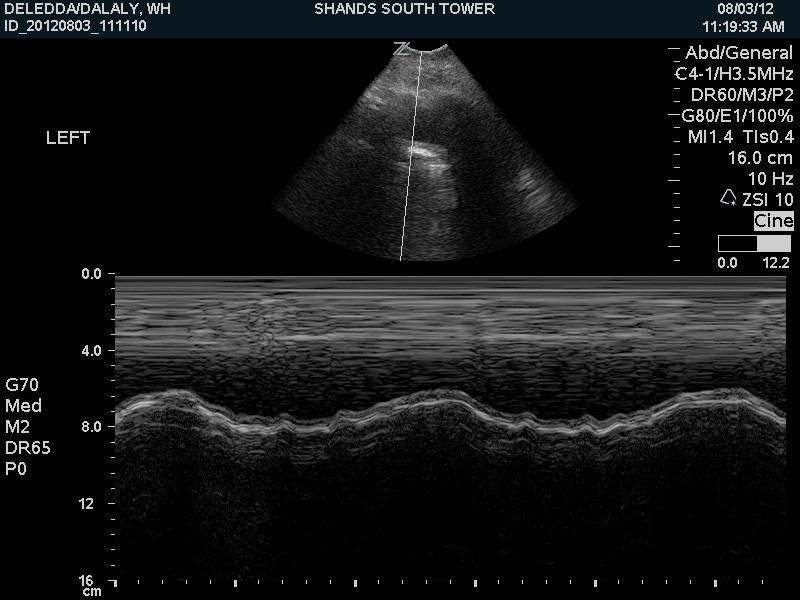
6 Pleural effusions Focused Questions: How much fluid is present? Where is best location to do procedure? Probe: Abdominal probe
7 Pleural effusion - introduction Common in critically ill patients - 62% in MICU, 41% present on admission This application in itself might be reason enough to use US Evaluates volume, nature of effusion, and best location Collects in dependent areas Traditionally located from abdominal exam, but to be more consistent should be done through PLAPS point - "one shot" should be this location Mattison. Chest. 1997

8 Principles of lung ultrasound Anterior zone = "BLUE hands" Lateral zone Posterior zone Lichtenstein. Textbook. Whole body ultrasonography in the critically ill.
9 Upper BLUE point: between third and fourth finger of BLUE hand, at palmar insertion Lower BLUE point: middle of palm of lower BLUE hand; allows for avoidance of heart in most cases Phrenic line: continuation of this line locates the lateral place PLAPS point: "posterior and/or lateral alveolar and/or pleural syndrome" -- intersection between the posterior axillary line and the transversal line continuing posteriorly to the lower BLUE point; also can have extended PLAPS points
10 Principles of lung ultrasound Stage 1: anterior wall Stage 2: adds lateral wall from anterior to posterior axillary line Stage 3: external part of the posterior wall; aim from back towards sky; no visual control of probe so need to hold with whole hand; can depress bed if need to Stage 4: patients must be positioned laterally or sitting; can also study the apex


11 Principles of lung ultrasound Upper and Lower BLUE points Phrenic point PLAPS point Lichtenstein. Textbook. Whole body ultrasonography in the critically ill.
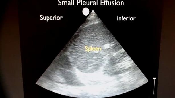
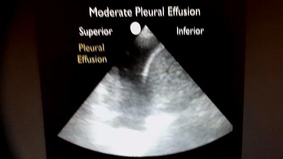
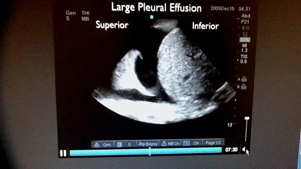
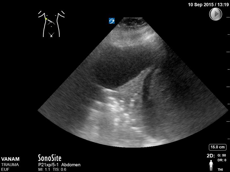
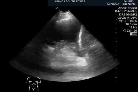
12 Pleural effusion - the signs Quad sign: static sign, borders are pleural line, upper and lower shadows of rib, and the deep border which is always roughly parallel to the pleural line and regular (represents lung surface) Jellyfish sign: aerated lung floats over effusion; as lung becomes more injured it becomes more towards same density as fluid surrounding it (looks like algae...) Sinusoid sign: dynamic sign, respiratory variation of the interpleural distance
13 Pleural effusion - Quad sign More on deep border: This line is called the lung line and is the visceral pleura; and visible when both pleura are separates by a structure that allows ultrasound transmission; the lung itself can be normal, show alveolar consolidation, or B lines Pleural line Rib shadows Lung line Lichtenstein. Textbook. Whole body ultrasonography in the critically ill.
14 Pleural effusion - sinusoid sign Sinusoid sign: dynamic sign, respiratory variation of the interpleural distance Also indicates low viscosity; very viscous or septate will not show Sinusoid sign Pleural line Rib shadows Lung line Lichtenstein. Textbook. Whole body ultrasonography in the critically ill.
15
16 Pleural effusion - jellyfish sign Jellyfish sign: aerated lung floats over effusion; as lung becomes more injured it becomes more towards same density as fluid surrounding it (looks like algae...) Pleural line Rib shadows Lung line Lichtenstein. Textbook. Whole body ultrasonography in the critically ill.
17 Perera P. " Video
18 Perera P. " Video
19
20
21 Pleural effusion - are we done? Transudate, exudate, purulent pleurisy MICU: cardiac failure (35%), atelectasis (23%), parapneumonic (11%), empyema (1%) Transudates are anechoic, anechoic effusions can be transudates or exudates, and all echoic effusions are exudates Ultrasound not reliable for indicating puncture is not necessary Mattison. Chest Yang. AJR AM J Rad. 1992
22 Pleural effusion - are we done? No movement, no sinusoid sign Plankton sign: visualization within tissue like image of a slow whirling movement of numerous particles; hyperechoic structures should correspond to infectious gas; can be seen in hemothorax (should use PLAPS point) Lichtenstein. Textbook. Whole body ultrasonography in the critically ill.
23 Pleural effusion - are we done? Lichtenstein. Textbook. Whole body ultrasonography in the critically ill.
24 Pleural effusion - are we done? Shred Tissue like echogenicity, Separated by lung line (quad sign) Plankton sign Shred sign indicating alveolar origin Hyperechoic points generating dynamic air bronchograms (indicating non atelectatic origin) Abolished lung sliding, no sinusoid sign, indicate infective process Lichtenstein. Textbook. Whole body ultrasonography in the critically ill.
25
26 Pleural effusion - the numbers Quad and sinusoid signs confirm presence with 97% with gold standard is withdrawal of fluid; 93% with CT as gold standard Ultrasound detects effusions occult on radiography; up to 525 ml can be missed on bedside radiograph Note: effusions allow you to see deep structures like aorta better, consider exploring prior to evacuation Lichtenstein. Intensive care medicine Lichtenstein. Anesthesiology Muller. Radiology Collins. Radiology. 1972
27 Pleural effusion - quantity Minor, moderate, large?...more important might be full clinical state; a diseased lung will not tolerate restrictive syndrome as well Guidelines? Several protocols for indicating volume of effusion PLAPS index: probe at PLAPS point and measuring from pleural line to lung line; probe must be as tangential as possible or will overestimate
28 Pleural effusion - quantity Measure on expiration since small effusions will have lung line touch pleural line on inspiration Usually between 1mm to 4 cm, rarely 5, exceptionally 6, and never 7; a 10 cm value questions technique PLAPS index 0.3 cm = ml 1 cm = ml 2 cm = ml 3.5 cm = ml
29 Pleural effusion - quantity Limitations: lung floating in early fast effusions, massive consolidation will make lung dive towards earth driving back the pleural effusion Looking at anterior or phrenic points (usually indicates abundant effusion) Many other methods Talmor. Surgery Roch. Chest Vignon. Crit Care Med Balik. Intensive Care Med. 2006
30 Pleural effusion - technique 1. Presence of quad, sinusoid sign 2. Safety distance of 15 mm seen over three adjacent interspaces 3. Check for absence of interposition of critical structures: lung, aorta, heart, liver, or spleen; move more posterior if lung comes close with inspiration 4. Should be done immediately at time of ultrasound evaluation 5. Technical notes: remember skin folds and not too posterior where dislodgements, infection, decubitus can occur
Lung ultrasound in the critically ill patient BASICS
 Lung ultrasound in the critically ill patient BASICS Rohit Patel, MD University of Florida Health Director, Critical Care Ultrasound Surgical ICU Center for Intensive Care Gainesville, Florida Introduction
Chest Ultrasound: Pneumothorax
 WINFOCUS BASIC ECHO (WBE) Chest Ultrasound: Pneumothorax Mark Hamlin, MD, MS Associate Professor of Anesthesiology and Surgery University of Vermont College of Medicine Co-Director of Surgical Critical
A Practical Approach to Ultrasound Assessment of Respiratory Distress
 A Practical Approach to Ultrasound Assessment of Respiratory Distress Yanick Beaulieu, MD, FRCPC Director, Bedside Ultrasound Curriculum Division of Cardiology and Critical Care Hôpital du Sacré-Coeur
Ultrasound. FAST Focused Assessment with Sonography in Trauma
 Ultrasound FAST Focused Assessment with Sonography in Trauma Rohit Patel, MD University of Florida Health Director, Critical Care Ultrasound Surgical ICU Center for Intensive Care Gainesville, Florida
This appendix was part of the submitted manuscript and has been peer reviewed. It is posted as supplied by the authors.
 This appendix was part of the submitted manuscript and has been peer reviewed. It is posted as supplied by the authors. - Figure S1: The four quadrant approach lung ultrasound at the bedside. * The anterolateral
Lung sonography in the diagnosis of pneumothorax.
 Lung sonography in the diagnosis of pneumothorax. Poster No.: C-0526 Congress: ECR 2011 Type: Educational Exhibit Authors: K. Stefanidis, K. Vintzilaios, D. D. Cokkinos, E. Antypa, S. Dimopoulos, S. Nanas,
Bedside ultrasound - Lung ultrasound in the Intensive Care Unit
 Bedside ultrasound - Lung ultrasound in the Intensive Care Unit Kishore K. Pichamuthu, Professor, Department of Critical Care, Christian Medical College, Vellore. Summary In an ICU setting, ultrasonographic
OVERVIEW. Need for USG. Weaning assessment. Mechanics of USG. Pneumonia / VAP. Principles of lung USG. Prone position ventilation assessment
 OVERVIEW Need for USG Mechanics of USG Principles of lung USG BLUE protocol Alveolar syndrome Interstitial syndrome Weaning assessment Pneumonia / VAP Prone position ventilation assessment ETT positioning
Pediatric Lung Ultrasound (PLUS) In Diagnosis of Community Acquired Pneumonia (CAP)
 In Diagnosis of Community Acquired Pneumonia (CAP)") Pediatric Lung Ultrasound (PLUS) In Diagnosis of Community Acquired Pneumonia (CAP) Dr Neetu Talwar Senior Consultant, Pediatric Pulmonology Fortis Memorial Research Institute, Gurugram Study To compare
Pediatric Lung Ultrasound (PLUS) In Diagnosis of Community Acquired Pneumonia (CAP) Dr Neetu Talwar Senior Consultant, Pediatric Pulmonology Fortis Memorial Research Institute, Gurugram Study To compare
NON INVASIVE LIFE SAVERS. Ultrasound in PICU
 VOL 1 NO.1 Jan March 2014 54 Table 1. Selected Applications of Point-of-Care Ultrasonography, According to Medical Specialty. Specialty Ultrasound Applications Anesthesia Cardiology Guidance for vascular
We are IntechOpen, the world s leading publisher of Open Access books Built by scientists, for scientists. International authors and editors
 We are IntechOpen, the world s leading publisher of Open Access books Built by scientists, for scientists 3,800 116,000 120M Open access books available International authors and editors Downloads Our
MINERVA MEDICA COPYRIGHT REVIEW ARTICLE D. LICHTENSTEIN. Resuscitation Service, Ambroise-Paré Hospital, Boulogne, France ABSTRACT
 MINERVA ANESTESIOL 2009;75:313-7 REVIEW ARTICLE Lung ultrasound in acute respiratory failure an introduction to the BLUE-protocol D. Resuscitation Service, Ambroise-Paré Hospital, Boulogne, France ABSTRACT
Initially for cardiac echo Subsequent studies non-cardiac applications
 No disclosures But Heavy accent Initially for cardiac echo Subsequent studies non-cardiac applications 1973: Goldberg et al in JCUS 30 mediastinal masses in pts. age 1-84 yrs. 1977: Kangarloo et al in
Objectives. The Extended FAST Exam. Focused Assessment e With Sonography In. Trauma (FAST)
") Northern California Emergency Ultrasound Course Objectives The Extended FAST Exam Rimon Bengiamin, MD, RDMS UC SF Discuss the components of the EFAST exam Evaluate the utility of the EFAST Review how to
Northern California Emergency Ultrasound Course Objectives The Extended FAST Exam Rimon Bengiamin, MD, RDMS UC SF Discuss the components of the EFAST exam Evaluate the utility of the EFAST Review how to
CHEST Recent Advances in Chest Medicine
 CHEST Recent Advances in Chest Medicine Thoracic Ultrasonography for the Pulmonary Specialist Seth J. Koenig, MD; Mangala Narasimhan, DO, FCCP; and Paul H. Mayo, MD, FCCP Thoracic ultrasonography is a
The Intensive Care Unit
 Imaging of the ADRS patient: Risk of transportation and alternative to repetitive radiation exposure Jean-Jacques Rouby Pitié-Salpêtrière Hospital The Intensive Care Unit The Intensive Care Unit Multidisciplinary
Ultrasound basics Part 1
 Ultrasound basics Part 1 'Ultrasound enhanced critical care medicine' Rohit Patel, MD University of Florida Health Director, Critical Care Ultrasound Surgical ICU Center for Intensive Care Gainesville,
Contents& & & 1.! Ultrasound&basics& 1! 2.! Image&generation& 15!
 A l i n e press é % % % Contents& & & 1. Ultrasound&basics& 1 1.1. What,is,ultrasound?, 1 1.2. Ultrasound,probes,send,and,receive,ultrasound, 3 1.3. How,does,ultrasound,behave,travelling,through,tissue?,
Bedside Sonographic Diagnosis of Pneumothorax in Pediatric Patients: A Preliminary Report Chia-Wang Tang 1, Kai-Sheng Hsieh 1 1
 ORIGINAL ARTICLE Bedside Sonographic Diagnosis of in Pediatric Patients: A Preliminary Report Chia-Wang Tang 1, Kai-Sheng Hsieh 1 1 Division of Pediatric Pulmonology, Department of Pediatrics, Kaohsiung
UERMMMC Department of Radiology. Basic Chest Radiology
 UERMMMC Department of Radiology Basic Chest Radiology PHYSICS DENSITIES BONE SOFT TISSUES WATER FAT AIR TELEROENTGENOGRAM Criteria for an Ideal Chest Radiograph 1. Upright 2. Posteroanterior View 3. Full
Definitions and diagnostic implications of terms used in the chest radiograph and lung ultrasound diagnoses of pneumonia.
 Supplementary 1 Definitions and diagnostic implications of terms used in the chest radiograph and lung ultrasound diagnoses of pneumonia. Imaging finding Definition Implication CR Consolidation Interstitial
Interpreting thoracic x-ray of the supine immobile patient: Syllabus
 Interpreting thoracic x-ray of the supine immobile patient: Syllabus Johannes Godt Dep. of Radiology and Nuclear Medicine Oslo University Hospital Ullevål NORDTER 2017, Helsinki Content - Why bedside chest
Ultrasound in the ICU
 Ultrasound in the ICU Kristine E. W. Breyer, MD Assistant Professor Anesthesia & Critical Care Medicine UCSF DISCLOSURES: NONE Definition The Ultrasound Exam Types & Uses Training Clinical Examples Objectives
Ultrasonido Pulmonar. Dra. Adriana Denise Zepeda Mendoza. Medicina de Urgencias Medicina del Enfermo en Estado Crí=co Grupo GMEMI.
 Ultrasonido Pulmonar. Dra. Adriana Denise Zepeda Mendoza. Medicina de Urgencias Medicina del Enfermo en Estado Crí=co Grupo GMEMI. Obje=vos Conocer los diferentes patrones ultrasonográficos pulmonares.
Imaging of small amounts of pleural fluid. Part two - physiologic pleural fluid
 review Imaging of small amounts of pleural fluid. Part two - physiologic pleural fluid Igor Kocijančič Department of Radiology, Institute of Oncology, Ljubljana, Slovenia Background. There are only a few
Ultrasound-guided Aspiration of the Iatrogenic Pneumothorax Caused by Paravertebral Block
 Case Report Korean J Pain 2012 January; Vol. 25, No. 1: 33-37 pissn 2005-9159 eissn 2093-0569 http://dx.doi.org/10.3344/kjp.2012.25.1.33 Ultrasound-guided Aspiration of the Iatrogenic Pneumothorax Caused
Chapter 17 Lung Ultrasound in Anaesthesia and Critical Care Medicine
 Chapter 17 Lung Ultrasound in Anaesthesia and Critical Care Medicine David Canty, Kavi Haji, André Denault, and Alistair Royse Abstract Lung ultrasound (respiratory or thoracic ultrasound) has traditionally
Point-of-care lung ultrasound
 Ultrasound Point-of-care lung ultrasound Philips tutorial Michael B. Stone, MD, RDMS Director, Division of Emergency Ultrasound Department of Emergency Medicine Brigham and Women s Hospital, Boston, MA
Impact of lung ultrasound on clinical decision making in critically ill patients
 Intensive Care Med DOI 10.1007/s00134-013-3133-3 ORIGINAL Nektaria Xirouchaki Eumorfia Kondili George Prinianakis Polychronis Malliotakis Dimitrios Georgopoulos Impact of lung ultrasound on clinical decision
Case 1. A 35-year-old male presented with fever, cough, and purulent sputum for one week. This was his CXR (Fig. 1.1). What is the diagnosis?
. What is the diagnosis?") 1 Interpreting Chest X-Rays CASE 1 Fig. 1.1 Case 1. A 35-year-old male presented with fever, cough, and purulent sputum for one week. This was his CXR (Fig. 1.1). What is the diagnosis? CASE 1 Interpreting
1 Interpreting Chest X-Rays CASE 1 Fig. 1.1 Case 1. A 35-year-old male presented with fever, cough, and purulent sputum for one week. This was his CXR (Fig. 1.1). What is the diagnosis? CASE 1 Interpreting
Lung Ultrasound in Diagnosis of Acute Respiratory Failure: BLUE Protocol Based Evaluation
 Lung Ultrasound in Diagnosis of Acute Respiratory Failure: BLUE Protocol Based Evaluation Niyas. K. Naseer 1, Muhammad Shafeek 2*, Rajani. M 3, Manoj. D. K 3 1Junior Resident, 2* Assistant Professor, 3
Imaging of Pleural Effusion: Comparing Ultrasound, X-Ray and CT findings
 Imaging of Pleural Effusion: Comparing Ultrasound, X-Ray and CT findings Poster No.: C-2067 Congress: ECR 2017 Type: Educational Exhibit Authors: J. M. Almeida, N. Antunes, C. Leal, L. Figueiredo ; Lisboa/PT,
Introduction to transthoracic ultrasound for the pulmonologist
 Review Introduction to transthoracic ultrasound for the pulmonologist Clementine Bostantzoglou 1, Charalampos Moschos 2 1 7 th Pulmonary Department, Sotiria Hospital for Chest Diseases, Athens, Greece
Lung ultrasound: Present and future
 Review Article Lung ultrasound: Present and future Ashish Saraogi Department of Anaesthesiology, National Institute of Medical Sciences and Research, Jaipur, Rajasthan, India ABSTRACT The scope of ultrasound
10/17/2016. Nuts and Bolts of Thoracic Radiology. Objectives. Techniques
 Nuts and Bolts of Thoracic Radiology October 20, 2016 Carleen Risaliti Objectives Understand the basics of chest radiograph Develop a system for interpreting chest radiographs Correctly identify thoracic
POCUS for the Internist: Lungs & Pericardial Effusions
 POCUS for the Internist: Lungs & Pericardial Effusions Jeremy S. Boyd, MD, FACEP Asst. Professor of Emergency Medicine Vanderbilt University Medical Illustrations courtesy of Robinson Ferre, MD, FACEP
Extended FAST Exam. Goal of Trauma Care. Golden Hour of Trauma
 Extended FAST Exam Goal of Trauma Care Golden Hour of Trauma Best INITIAL screening modality in trauma efast 2014 LLSA Article (ACEP Policy Statement) Level B Recommendation: In hemodynamically unstable
The efficacy of bedside chest ultrasound: from accuracy to outcomes
 EUROPEAN RESPIRATORY UPDATE EFFICACY OF BEDSIDE CHEST ULTRASOUND The efficacy of bedside chest ultrasound: from accuracy to outcomes Mark Hew 1,2 and Tunn Ren Tay 1,3 Affiliations: 1 Allergy, Immunology
Perioperative Ultrasonography Ehab Farag, MD, FRCA Hesham Elsharkawy David G. Anthony, M.D.
 Perioperative Ultrasonography Ehab Farag, MD, FRCA Hesham Elsharkawy David G. Anthony, M.D. Cleveland Clinic, Cleveland OH 1 Complications during central venous catheterization (CVC) occur 2% -15% of the
Pulmonary Ultrasound in Emergency Medicine and Critical Care
 Pulmonary Ultrasound in Emergency Medicine and Critical Care www.rmgultrasound.com Author: Virginia M Stewart, MD RDMS RDCS RDMSK Dr Stewart is a practicing Emergency Physician in Eastern Virginia, USA.
Role of Transthoracic Ultrasound in Detection of Pneumonia in ICU Patients
 Med. J. Cairo Univ., Vol. 83, No. 1, June: 307-314, 2015 www.medicaljournalofcairouniversity.net Role of Transthoracic Ultrasound in Detection of Pneumonia in ICU Patients FARES AUF, M.D.; AHMED ABO-NAGLH,
Chest X rays and Case Studies. No disclosures. Outline 5/31/2018. Carlo Manalo, M.D. Department of Radiology Loma Linda University Children s Hospital
 Chest X rays and Case Studies Carlo Manalo, M.D. Department of Radiology Loma Linda University Children s Hospital No disclosures. Outline Importance of history Densities delineated on radiography An approach
4/16/2017. Learning Objectives. Interpretation of the Chest Radiograph. Components. Production of the Radiograph. Density & Appearance
 Interpretation of the Arthur Jones, EdD, RRT Learning Objectives Identify technical defects in chest radiographs Identify common radiographic abnormalities This Presentation is Approved for 1 CRCE Credit
Bacterial pneumonia with associated pleural empyema pleural effusion
 EMPYEMA Synonyms : - Parapneumonic effusion - Empyema thoracis - Bacterial pneumonia - Pleural empyema, pleural effusion - Lung abscess - Complicated parapneumonic effusions (CPE) 1 Bacterial pneumonia
Thoracic Ultrasound: Pictorial review of pneumothorax, the fastest and easiest method to diagnose.
 Thoracic Ultrasound: Pictorial review of pneumothorax, the fastest and easiest method to diagnose. Poster No.: C-1588 Congress: ECR 2014 Type: Educational Exhibit Authors: J. A. Guirola, V. Mayoral Campos,
Lung ultrasound: routine practice for the next generation of internists
 REVIEW Lung ultrasound: routine practice for the next generation of internists H.R.W. Touw 1,2, P.R. Tuinman* 1,3,4, H.P.M.M. Gelissen 1, E. Lust 1, P.W.G. Elbers 1,3,4 Departments of 1 Intensive Care
Unusual new signs of pneumothorax at lung ultrasound
 Unusual new signs of pneumothorax at lung ultrasound Volpicelli et al. Volpicelli et al. Critical Ultrasound Journal 2013, 5:10 Volpicelli et al. Critical Ultrasound Journal 2013, 5:10 SHORT COMMUNICATION
Chest X-ray Interpretation
 Chest X-ray Interpretation Introduction Routinely obtained Pulmonary specialist consultation Inherent physical exam limitations Chest x-ray limitations Physical exam and chest x-ray provide compliment
Top Tips for Pleural Disease in 2012
 Top Tips for Pleural Disease in 2012 The unilateral pleural effusion on the Post Take Ward Round Pleural Effusion on CXR Bedside ultrasound + Pleural aspirate Empyema Nil evidence infection Admit IV antibiotics
Critical Care Monitoring. Indications. Pleural Space. Chest Drainage. Chest Drainage. Potential space. Contains fluid lubricant
 Critical Care Monitoring Indications 1-2- 2 Pleural Space Potential space Contains fluid lubricant Can fill with air, blood, plasma, serum, lymph, pus 3 1 Pleural Space Problems when contain abnormal substances:
Background & Indications Probe Selection
 Teresa S. Wu, MD, FACEP Director, EM Ultrasound Program & Fellowship Co-Director, Simulation Based Training Program & Fellowship Associate Program Director, EM Residency Program Maricopa Medical Center
Identification of lung sliding: a basic ultrasound technique with a steep learning curve
 SIGNA VITAE 2013; 8(1): 31-35 ORIGINAL Identification of lung sliding: a basic ultrasound technique with a steep learning curve MATEJ STRNAD SABINA ZADEL ZALIKA KLEMENC-KETIS MATEJ STRNAD ( ) SABINA ZADEL
Background & Indications
 Teresa S. Wu, MD, FACEP Director, EM Ultrasound Program & Fellowship Co-Director, Simulation Based Training Program & Fellowship Maricopa Medical Center Simulation Curriculum Director Associate Professor,
Radiological Anatomy of Thorax. Dr. Jamila Elmedany & Prof. Saeed Abuel Makarem
 Radiological Anatomy of Thorax Dr. Jamila Elmedany & Prof. Saeed Abuel Makarem Indications for Chest x - A chest x-ray may be used to diagnose and plan treatment for various conditions, including: Diseases/Fractures
Lung ultrasound in follow-up of low birth weight with respiratory distress syndrome: clinical application and reduction of x-rays examinations
 Lung ultrasound in follow-up of low birth weight with respiratory distress syndrome: clinical application and reduction of x-rays examinations Poster No.: C-1724 Congress: ECR 2011 Type: Scientific Paper
Certificate in Clinician Performed Ultrasound (CCPU) Syllabus. Lung
 Syllabus. Lung") Certificate in Clinician Performed Ultrasound (CCPU) Syllabus Lung Page 1 of 8 01/17 Lung Syllabus Purpose: This unit is designed to cover the theoretical and practical curriculum for lung ultrasound in
Certificate in Clinician Performed Ultrasound (CCPU) Syllabus Lung Page 1 of 8 01/17 Lung Syllabus Purpose: This unit is designed to cover the theoretical and practical curriculum for lung ultrasound in
ASSESSMENT OF LUNG PARENCHYMAL ABNORMALITIES
 2016 by the author Thank you for viewing this presentation. We would like to remind you that this material is the property of the author. It is provided to you by the ERS for your personal use only, as
Interactive Lecture. Lecture 7 - Interactive. Radiology of cardiorespiratory disease. Editing File. Done By. Color Coding Important Notes Extra
 Lecture 7 - Interactive 436 Teams Interactive Lecture Radiology of cardiorespiratory disease Done By Team Leaders: Khalid Alshehri Hanin Bashaikh Team Members: Ghaida Alsaeed Maha Alissa Nawwaf AlHarbi
What is cpt code for chest tube placement
 What is cpt code for chest tube placement Search 11-4-2016 Chest Tube Placement (Thoracostomy) and Pleurodesis Thoracostomy inserts a thin plastic tube into the pleural space between the lungs and the
Lung Ultrasound in Small Animals: The Vet BLUE SM
 Lung Ultrasound in Small Animals: The Vet BLUE SM Gregory R. Lisciandro, DVM, Dipl. ABVP, Dipl. ACVECC Hill Country Veterinary Specialists & FASTVet.com, San Antonio, Texas USA FASTVet TM and FAST Saves
Management of Pleural Effusion
 Management of Pleural Effusion Development of Pleural Effusion pulmonary capillary pressure (CHF) capillary permeability (Pneumonia) intrapleural pressure (atelectasis) plasma oncotic pressure (hypoalbuminemia)
Focused Assessment with Sonography in Trauma (FAST) UC Irvine School of Medicine
 UC Irvine School of Medicine") Focused Assessment with Sonography in Trauma (FAST) UC Irvine School of Medicine Purpose of FAST exam Quickly evaluate patient s status in emergency situations Blunt or penetrating trauma Visualize fluid
Focused Assessment with Sonography in Trauma (FAST) UC Irvine School of Medicine Purpose of FAST exam Quickly evaluate patient s status in emergency situations Blunt or penetrating trauma Visualize fluid
Children are not small adults Children are Not Small Adults Anatomic considerations Pliable bony & cartilagenous structures - Significant thoracic inj
 PEDIATRIC CHEST TRAUMA Children are not small adults Role of imaging Spectrum of injury Children are not small adults Children are Not Small Adults Anatomic considerations Pliable bony & cartilagenous
EUROPEAN ASSOCIATION OF VETERINARY DIAGNOSTIC IMAGING EUROPEAN COLLEGE OF VETERINARY DIAGNOSTIC IMAGING
 EISAGOGIKO EUROPEAN ASSOCIATION OF VETERINARY DIAGNOSTIC IMAGING EUROPEAN COLLEGE OF VETERINARY DIAGNOSTIC IMAGING ARISTOTLE UNIVERSITY OF THESSALONIKI SCHOOL OF VETERINARY MEDICINE SECTION OF RADIOLOGY
Pleural Effusion. Exudative pleural effusion - Involve an increase in capillary permeability and impaired pleural fluid resorption
 Pleural Effusion Definition of pleural effusion Accumulation of fluid between the pleural layers Epidemiology of pleural effusion Estimated prevalence of pleural effusion is 320 cases per 100,000 people
Pleural ultrasonography. Pictorial essay.
 Pictorial essay Med Ultrason 2014, Vol. 16, no. 4, 364-371 DOI: 10.11152/mu.201.3.2066.164.racc Pleural ultrasonography. Pictorial essay. Romeo Chira 1, Alexandra Chira 2, Roberta Mânzat Săplăcan 1, Georgiana
Abdominal Ultrasonography
 Abdominal Ultrasonography David A. Masneri, DO, FACEP, FAAEM Assistant Professor of Emergency Medicine Assistant Director, Emergency Medicine Residency Medical Director, Operational Medicine Division Center
Imaging of Thoracic Trauma: Tips and Traps. Arun C. Nachiappan, MD Associate Professor of Clinical Radiology University of Pennsylvania
 Imaging of Thoracic Trauma: Tips and Traps Arun C. Nachiappan, MD Associate Professor of Clinical Radiology University of Pennsylvania None Disclosures Objectives Describe blunt and penetrating traumatic
PLEURAE and PLEURAL RECESSES
 PLEURAE and PLEURAL RECESSES By Dr Farooq Aman Ullah Khan PMC 26 th April 2018 Introduction When sectioned transversely, it is apparent that the thoracic cavity is kidney shaped: a transversely ovoid space
Imaging of Respiratory Disorders: M2 Pathology correlated with Radiology
 Imaging of Respiratory Disorders: M2 Pathology correlated with Radiology by (c) Dr Goh Poh Sun MBBS(Melb), FRCR(UK), FAMS(Singapore), MHPE(Maastricht) Senior Consultant Radiologist and Associate Professor
ECBSE Chest 29/01/2014 / 1:52 1. EFSUMB European Course Book Student Edition Editors: Jan Tuma, Radu Badea, Christoph F. Dietrich.
 ECBSE Chest 29/01/2014 / 1:52 1 EFSUMB European Course Book Student Edition Editors: Jan Tuma, Radu Badea, Christoph F. Dietrich Chest Gebhard Mathis Internistische Praxis, Rankweil, Austria Corresponding
TB Radiology for Nurses Garold O. Minns, MD
 TB Nurse Case Management Salina, Kansas March 31-April 1, 2010 TB Radiology for Nurses Garold O. Minns, MD April 1, 2010 TB Radiology for Nurses Highway Patrol Training Center Salina, KS April 1, 2010
Efficacy of ultrasonography and computed tomography in differentiating transudate from exudate in patients with pleural effusion
 Efficacy of ultrasonography and computed tomography in differentiating transudate from exudate in patients with pleural effusion Purpose: To evaluate USG and CT imaging findings in differentiating transudative
Dana Alrafaiah. - Moayyad Al-Shafei. -Mohammad H. Al-Mohtaseb. 1 P a g e
 - 6 - Dana Alrafaiah - Moayyad Al-Shafei -Mohammad H. Al-Mohtaseb 1 P a g e Quick recap: Both lungs have an apex, base, mediastinal and costal surfaces, anterior and posterior borders. The right lung,
FAST Focused Assessment with Sonography in Trauma
 FAST Focused Assessment with Sonography in Trauma Wilma Rodriguez Mojica,MD,FACR Professor of Radiology UPR School of Medicine Ultrasound Section - Radiological Sciences Department OBJECTIVES Understand
Small animal point of care ultrasound techniques
 Small animal point of care ultrasound techniques The role of veterinary point of care ultrasound in determining the presence or absence of specific pathologies is examined by Jantina McMurray DVM; Søren
Right lung. -fissures:
 -Right lung is shorter and wider because it is compressed by the right copula of the diaphragm by the live.. 2 fissure, 3 lobes.. hilum : 2 bronchi ( ep-arterial, hyp-arterial ), one artery mediastinal
Focused Assessment Sonography of Trauma (FAST) Scanning Protocol
 Scanning Protocol") Focused Assessment Sonography of Trauma (FAST) Scanning Protocol Romolo Gaspari CHAPTER 3 GOAL OF THE FAST EXAM Demonstrate free fluid in abdomen, pleural space, or pericardial space. EMERGENCY ULTRASOUND
Focused Assessment Sonography of Trauma (FAST) Scanning Protocol Romolo Gaspari CHAPTER 3 GOAL OF THE FAST EXAM Demonstrate free fluid in abdomen, pleural space, or pericardial space. EMERGENCY ULTRASOUND
Techniques of examination of the thorax and lungs. Dr. Szathmári Miklós Semmelweis University First Department of Medicine 24. Sept
 Techniques of examination of the thorax and lungs Dr. Szathmári Miklós Semmelweis University First Department of Medicine 24. Sept. 2013. Inspection of the thorax Observe: the shape of chest Deformities
Lung ultrasound in critically ill patients: comparison with bedside chest radiography
 Intensive Care Med (2011) 37:1488 1493 DOI 10.1007/s00134-011-2317-y ORIGINAL Nektaria Xirouchaki Eleftherios Magkanas Katerina Vaporidi Eumorfia Kondili Maria Plataki Alexandros Patrianakos Evaggelia
Sonographic Approach to Diagnosing Pulmonary Consolidation
 Sonographic Approach to Diagnosing Pulmonary Consolidation Remi Targhetta, MD, Roseline Chavagneux, MD, Jean-Marie Bourgeois, MD, Michel Dauzat, MD, Pierre Balmes, MD, Leandre Pourcelot, MD Thirty-nine
The prevalence of pleural effusions is high in ICU
 Usefulness of Ultrasonography in Predicting Pleural Effusions > 500 ml in Patients Receiving Mechanical Ventilation* Antoine Roch, MD PHD; Mirela Bojan, MD; Pierre Michelet, MD; Fanny Romain, MD; Fabienne
ARDS - a must know. Page 1 of 14
 ARDS - a must know Poster No.: C-1683 Congress: ECR 2016 Type: Authors: Keywords: DOI: Educational Exhibit M. Cristian; Turda/RO Education and training, Edema, Acute, Localisation, Education, Digital radiography,
Bony Thorax. Anatomy and Procedures of the Bony Thorax Edited by M. Rhodes
 Bony Thorax Anatomy and Procedures of the Bony Thorax 10-526-191 Edited by M. Rhodes Anatomy Review Bony Thorax Formed by Sternum 12 pairs of ribs 12 thoracic vertebrae Conical in shape Narrow at top Posterior
B-I-2 CARDIAC AND VASCULAR RADIOLOGY
 (YEARS 1 3) CURRICULUM FOR RADIOLOGY 13 B-I-2 CARDIAC AND VASCULAR RADIOLOGY KNOWLEDGE To describe the normal anatomy of the heart and vessels including the lymphatic system as demonstrated by radiographs,
Certificate in Clinician Performed Ultrasound (CCPU) Syllabus. Lung
 Syllabus. Lung") Certificate in Clinician Performed Ultrasound (CCPU) Syllabus Lung ASUM Quality CCPU Syllabi Released: 21 March 2013 Approved by: CEO Lung Purpose: This unit is designed to cover the theoretical and practical
Certificate in Clinician Performed Ultrasound (CCPU) Syllabus Lung ASUM Quality CCPU Syllabi Released: 21 March 2013 Approved by: CEO Lung Purpose: This unit is designed to cover the theoretical and practical
Lecture 3. Inflammatory Processes
 Lecture 3 Inflammatory Processes Process: Increased vascular permeability Water and cellular infiltrations Results: Abscess, ulceration, cavitation Penetration, perforation and fistula formation Scarring,
"Pediatric chest: From x-ray to ultrasound. A pictorial review of 173 patients"
 "Pediatric chest: From x-ray to ultrasound. A pictorial review of 173 patients" Poster No.: C-0451 Congress: ECR 2014 Type: Educational Exhibit Authors: S. FYGETAKI, I. Tritou, S. Stefanaki, S. Antonopoulos,
Introduction & Physics of ED Ultrasound. Objectives. What? - Limited Studies. Who? - ED Docs
 Introduction & Physics of ED Ultrasound Martine Sargent, MD Ultrasound Director, Assistant Professor UCSF Department of Emergency Medicine San Francisco General Hospital & Trauma Center Objectives Who?
Chest Ultrasonography - A Quick and Accurate Diagnostic Tool in Pediatric Emergency Department and Intensive Care Unit
 Review Article Chest Ultrasonography - A Quick and Accurate Diagnostic Tool in Pediatric Emergency Department and Intensive Care Unit Dinakara Prithviraj 1, Suresh A 2 1 Associate Professor, Chief Neonatologist,
Chest Tube Thoracostomy
 Chest Tube Thoracostomy INTRODUCTION A chest tube thoracostomy is commonly done in the ED to evacuate an abnormal accumulation of fluid (blood, empyema) or air from the pleural space under an elective,
Certificate in Clinician Performed Ultrasound (CCPU) Syllabus. Lung
 Syllabus. Lung") Certificate in Clinician Performed Ultrasound (CCPU) Syllabus Lung Page 1 of 8 12/15 Lung Syllabus Purpose: This unit is designed to cover the theoretical and practical curriculum for lung ultrasound in
Certificate in Clinician Performed Ultrasound (CCPU) Syllabus Lung Page 1 of 8 12/15 Lung Syllabus Purpose: This unit is designed to cover the theoretical and practical curriculum for lung ultrasound in
An Introduction to Radiology for TB Nurses
 An Introduction to Radiology for TB Nurses Garold O. Minns, MD September 14, 2017 TB Nurse Case Management September 12 14, 2017 EXCELLENCE EXPERTISE INNOVATION Garold O. Minns, MD has the following disclosures
Shedding Light on Neonatal X-rays. Objectives. Indications for X-Rays 5/14/2018
 Shedding Light on Neonatal X-rays Barbara C. Mordue, MSN, NNP-BC Neonatal Nurse Practitioner LLUH Children s Hospital, NICU Objectives Utilize a systematic approach to neonatal x-ray interpretation Identify
Approach to CXR. Terminology. 1.Identification. Greg Blecher SCH Respir Fellow. Correct patient Correct date and time Correct examination
 Approach to CXR Greg Blecher SCH Respir Fellow From Rob Posteraro http://home.earthlink.net/~rhpos/cxr_interpret.txt.html ; http://home.earthlink.net/~rhpos/cxr_main.txt.html) Approach to viewing Chest
BIOE221. Session 5. Examination of Thorax- Respiratory system. Bioscience Department. Endeavour College of Natural Health endeavour.edu.
 BIOE221 Session 5 Examination of Thorax- Respiratory system Bioscience Department Session Objectives Understand the structure of the thorax and the organs contained in this cavity Understand the importance
Resident Case Review CHEST. Daria Manos CAR 2016
 Resident Case Review CHEST CAR 2016 Daria Manos Disclosure Speakers bureau, Roche CAR 2016 Daria Manos 1. Recognize common and critical chest radiograph and computed tomography signs and use these clues
Procedure: Chest Tube Placement (Tube Thoracostomy)
") Procedure: Chest Tube Placement (Tube Thoracostomy) Basic Information: The insertion and placement of a chest tube into the pleural cavity for the purpose of removing air, blood, purulent drainage, or
Procedure: Chest Tube Placement (Tube Thoracostomy) Basic Information: The insertion and placement of a chest tube into the pleural cavity for the purpose of removing air, blood, purulent drainage, or
Radiological conference. Left upper lobe collapse. Citation Hong Kong Practitioner, 1998, v. 20 n. 9, p
 Title Radiological conference. Left upper lobe collapse Author(s) Wong, LLS; Peh, WCG Citation Hong Kong Practitioner, 1998, v. 20 n. 9, p. 513-517 Issued Date 1998 URL http://hdl.handle.net/10722/44672