Perioperative Ultrasonography Ehab Farag, MD, FRCA Hesham Elsharkawy David G. Anthony, M.D.
|
|
|
- George James
- 5 years ago
- Views:
Transcription

1 Perioperative Ultrasonography Ehab Farag, MD, FRCA Hesham Elsharkawy David G. Anthony, M.D. Cleveland Clinic, Cleveland OH 1
2 Complications during central venous catheterization (CVC) occur 2% -15% of the time in adults and can be severe 1. Shummer et al cases, critical-care physicians landmark techniques 3.3% complication rate (10 cases pneumothorax/hemothorax and one stroke leading to death) 1 Domino KB, et al. Anesthesiology 2004;100: Shummer W, et al. Intensive Care Medicine 2004;33:
3 ASA closed claims 13 deaths from CVC insertion. Hemothorax from arterial injury & pneumothorax: major contributors to mortality 1. 5 million CVCs are performed every year 2 even low incidence (0.5%) of significant complications would result in 25,000 problems. 1 Bowdle TA. ASA Newsletter 2002;66. 2 Feller-Kopman. Crit Care Med 2005;33:
4 The Agency for Healthcare Research and Quality US 1 of 11 practices to improve the patient safety and outcome*. 2003, a meta-analysis by Hind, 18 RCTs favored US guidance vs landmark techniques reduced failure rates increased first-attempt success, reduced complication rates and faster procedure time (P<0.0001) *Rothschild JM. Agency for Healthcare Research and Quality 2001:
5 Subclavian Vein Approach SV offers ideal size for central access. Close proximity to the lung, subclavian artery, and brachial plexus can lead to significant morbidity. Other challenges for accessing the SV with US are deeper location and the presence of the clavicle. 5
6 Femoral Vein Approach Baum et al reviewed 100 CT scans FV and the femoral artery overlap in the anteroposterior plane 65% of the time 1. Same finding confirmed in US study in 50 ICU patients 2. 1 Baum PA, et al. Radiology 1989;173: Hughes P, et al. Anaesthesia 2000;55:
7 TRANSTHORACIC ECHOCARDIOGRAPHY 7
8 Possibilites with Bedside Ultrasound Etiologies of Hemodynamic Decompensation Vasodilatory/Distributive Shock Cardiogenic Shock Heart Failure Regional wall motion abnormalities Monitor Resuscitation Efforts
9 Limited TTE Exam Types FATE (Focus Assessed Transthoracic Echocardiography), FEEL (Focused Echocardiographic Evaluation in Life Support) FEER (Focused Echocardiographic Evaluation in Resuscitation Management), RUSH (Rapid Ultrasound for Shock and Hypotension), CAUSE (Cardiac Arrest Ultrasound Exam) BLEEP (Bedside Limited Echocardiography by Emergency Physician)
10 Our Exam (FATE) Three Anatomic windows Four clock positions: 10:00, 2:00, 3:00 12:00 Five scanning views In the ideal patient!

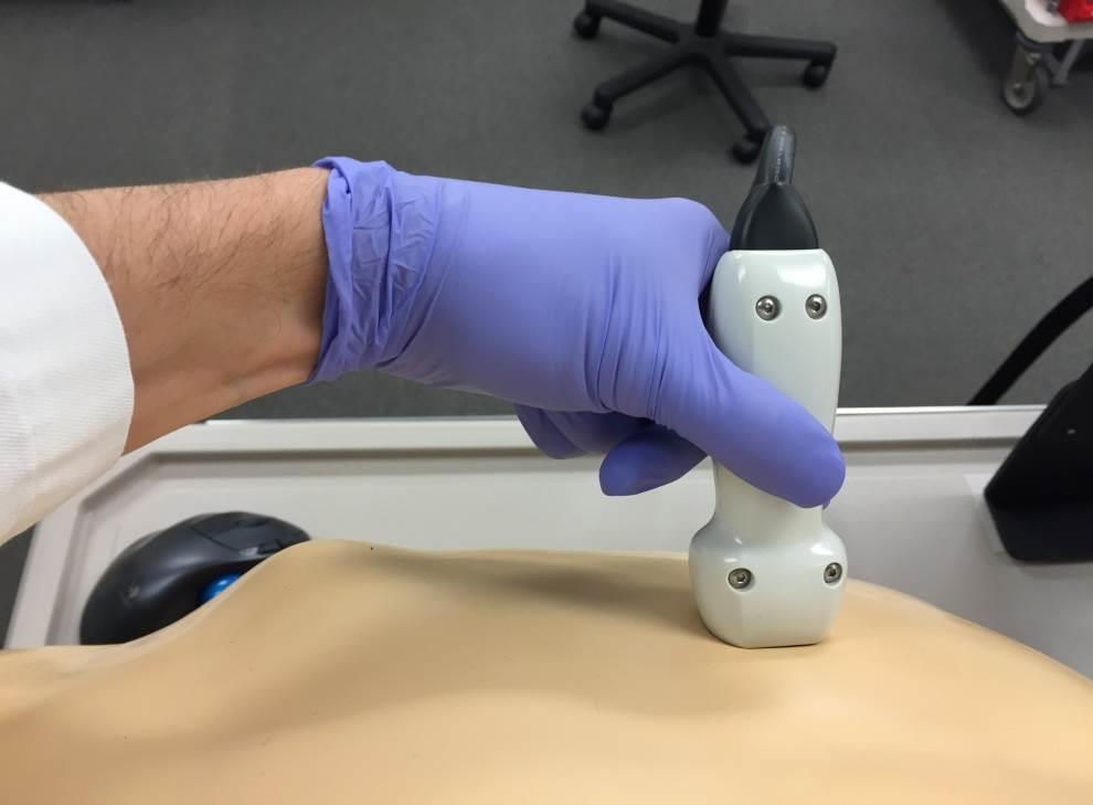
11 Proper probe grasp X
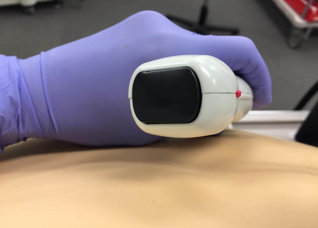
12 Probe Orientation
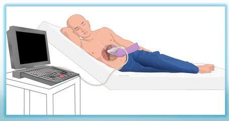
13 Parasternal Long Axis View Parasternal Long Axis Left Upper Sternal Border (3 rd -4 th interspace) Probe Indicator pointed towards right shoulder (10:00) 1

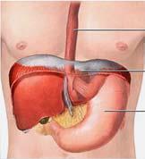
14 Anatomic Correlate Courtesy of Yale University Echocardiography Atlas
15 Parasternal Short Axis View Parasternal Short Axis Left Upper Sternal Border (3 rd -4 th interspace) 2 Probe Indicator pointed towards left shoulder (2:00)

16 Anatomic Correlate Courtesy of Yale University Echocardiography Atlas

17 Apical View Point of Maximal Impulse (PMI) Anterior Axillary Line underneath breast 3 Probe Orientation: 3:00 (patient s left flank)

18 Anatomic Correlate Courtesy of Yale University Echocardiography Atlas
19 Subcostal View Probe in subxiphoid region Probe orientation: 3:00 4 Tilt parallel to back

20 Anatomic Correlate Courtesy of Yale University Echocardiography Atlas
21 IVC View Probe in subxiphoid region Obtain the subcostal view, then rotate the probe Probe orientation: 12:00 5 Tilt towards the back
22 Volume Responsiveness Collapsibility Index = [IVC max (end exhalation) IVC min (end inhalation)] x 100% IVC max (end exhalation) > 50-75% or exhaled diameter < 1 cm suggestive of hypovolemia Distensibility Index = [IVC max (end exhalation) IVC min (end inhalation)] x 100% IVC Mean > 12 % variation: volume responsiveness: 93% PPV; 92% NPV
23 GASTRIC, OPHTHALMIC, AND PULMONARY ULTRASOUND 23
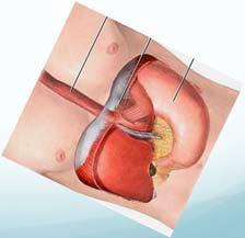
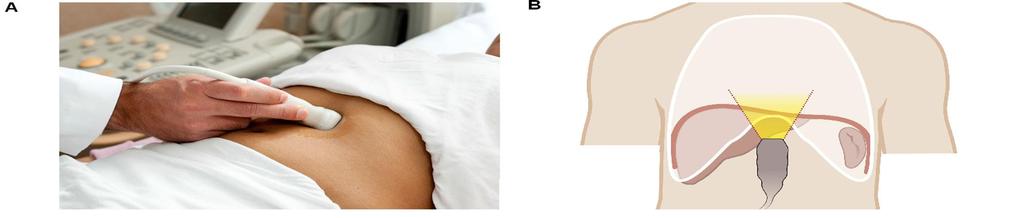
24 Ultrasound for Gastric Content


25 Three outcomes should be assessed: 1-Identify if the stomach is empty or full 2-Identify if the contents is fluid or solid 3-Identify the volume of the content
) Predict volume up to 500 ml. Perlas A, Mitsakakis N, Liu L, et al.")
26 3-Volume of the content (Quantitative) Mathematical models predict GV based on antral CSA. (GV (ml)= right-lateral CSA (cm 2 ) 1.28 age (yr)) Predict volume up to 500 ml. Perlas A, Mitsakakis N, Liu L, et al. Validation of a mathematical model for ultrasound assessment of gastric volume by gastroscopic examination. Anesth Analg 2013;116:

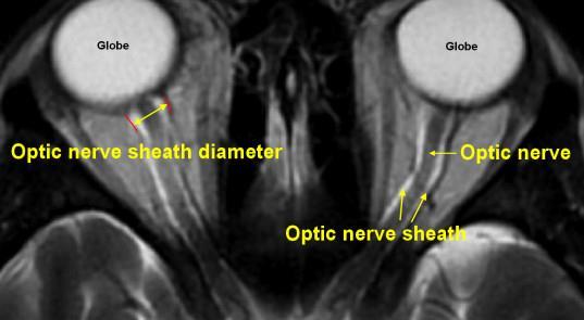
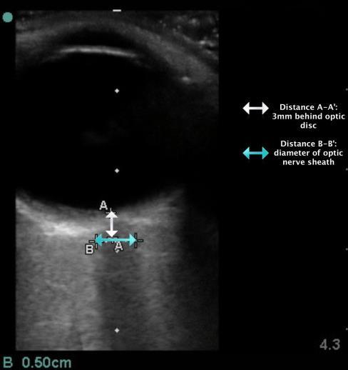
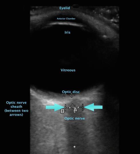
27 Optic nerve sheath Ultrasonography for monitoring intra cranial pressure Optic nerve sheath is a continuation of duramater ICP CSF pressure ONSD Advantages: ICP changes transmitted within minutes Non-invasive No reported complications Disadvantages: Not continuous Cannot quantitate CSF pressure Operator dependent Not widely accepted.
28
29


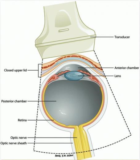
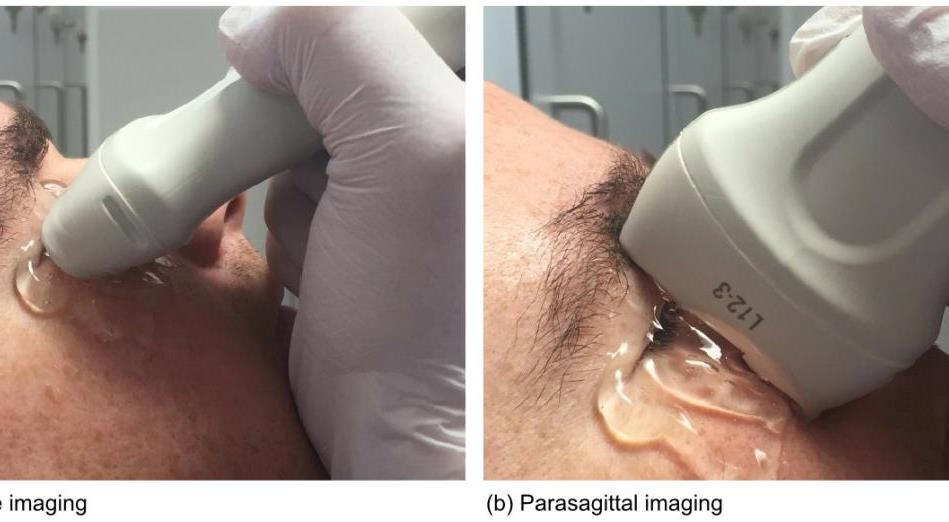
30 FDA approved ultrasound probe and machine for ocular ultrasonography Requirements: Mechanical index < 0.23 Thermal index < 1.0 Scan 1 : longitudinal B- Scan


31 Scan 2 : transverse B- Scan Scan 3 : transverse and confirmatory A- Scan ONSD greater than 5.0 mm reliably predicts an ICP > 20 mm Hg

32 Normal Lung Sliding Sign


33 B-lines or comet-tail artifacts Absence of lung-sliding and Comet-tail is 96.5% specific to pneumothorax.


34 Power slide A-lines
35


36 Pneumothorax stratosphere sign or barcode sign Absent lung sliding and Comet-tail
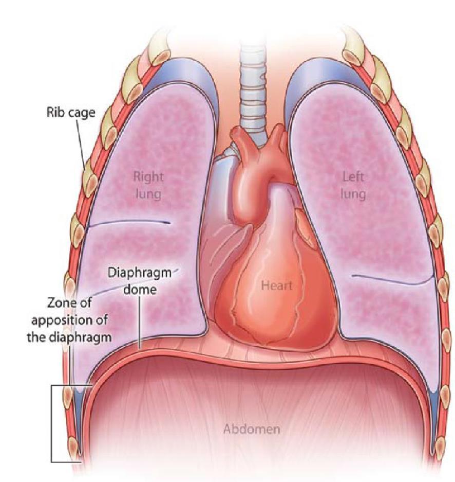
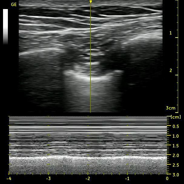
37 Diaphragmatic Motion

38 Intercostal view Neuromuscular Ultrasound for Evaluation of the Diaphragm, Aarti Sarwal et al

39 Anterior Subcostal View


40 Figure 2 M mode ultrasound trace of normal diaphragm movement. In a normal diaphragm a sharp upstroke is demonstrated when the patient sni ffs Figure 1 M mode ultrasound trace of normal diaphragm movement. N ormal trace of diaphragmatic movement. In inspiration, movement is caudal, with the corresponding M mode trace being upwards as the diaphragm moves toward the probe. In expiration, the M mode trace is downwards as the diaphragm moves away from the probe

41

42 Posterior subcostal view Subxiphoid view
Point of Care Ultrasound (PoCUS)
") Point of Care Ultrasound (PoCUS) Competency Assessment Forms AORTA Competency A Focussed Assessment of the Aorta (AAA) Guidance Please follow this guidance as closely as possible to ensure consistency
Point of Care Ultrasound (PoCUS) Competency Assessment Forms AORTA Competency A Focussed Assessment of the Aorta (AAA) Guidance Please follow this guidance as closely as possible to ensure consistency
The faculty will include physicians with international reputations as outstanding ultrasound educators.
 Ultrasound Courses Course Description Whether you re a beginner or a seasoned sonographer, this year s AAEM pre-conference ultrasound course will be worth your time. We will be offering a half day course
Background & Indications Probe Selection
 Teresa S. Wu, MD, FACEP Director, EM Ultrasound Program & Fellowship Co-Director, Simulation Based Training Program & Fellowship Associate Program Director, EM Residency Program Maricopa Medical Center
This appendix was part of the submitted manuscript and has been peer reviewed. It is posted as supplied by the authors.
 This appendix was part of the submitted manuscript and has been peer reviewed. It is posted as supplied by the authors. - Figure S1: The four quadrant approach lung ultrasound at the bedside. * The anterolateral
Background: Bedside ultrasound is emerging as a useful tool in the assessment of
 Abstract: Background: Bedside ultrasound is emerging as a useful tool in the assessment of intravascular volume status by examining measurements of the inferior vena cava (IVC). Many previous studies do
Objectives. The Extended FAST Exam. Focused Assessment e With Sonography In. Trauma (FAST)
") Northern California Emergency Ultrasound Course Objectives The Extended FAST Exam Rimon Bengiamin, MD, RDMS UC SF Discuss the components of the EFAST exam Evaluate the utility of the EFAST Review how to
Northern California Emergency Ultrasound Course Objectives The Extended FAST Exam Rimon Bengiamin, MD, RDMS UC SF Discuss the components of the EFAST exam Evaluate the utility of the EFAST Review how to
Extended FAST Exam. Goal of Trauma Care. Golden Hour of Trauma
 Extended FAST Exam Goal of Trauma Care Golden Hour of Trauma Best INITIAL screening modality in trauma efast 2014 LLSA Article (ACEP Policy Statement) Level B Recommendation: In hemodynamically unstable
Abdominal Ultrasonography
 Abdominal Ultrasonography David A. Masneri, DO, FACEP, FAAEM Assistant Professor of Emergency Medicine Assistant Director, Emergency Medicine Residency Medical Director, Operational Medicine Division Center
Background & Indications Probe Selection
 Teresa S. Wu, MD, FACEP Director, EM Ultrasound Program & Fellowship Co-Director, Simulation Based Training Program & Fellowship Associate Program Director, EM Residency Program Maricopa Medical Center
Intro Case. Outline What We ll Cover. What we won t cover. Cardiac Ultrasound and The RUSH Exam: Bedside Ultrasound in Resuscitation and Shock
 Cardiac Ultrasound and The RUSH Exam: Bedside Ultrasound in Resuscitation and Shock Justin Davis, MD, MPH, RDMS Associate Physician Subchief for Emergency Ultrasound Services Kaiser Oakland Medical Center
Chest Ultrasound: Pneumothorax
 WINFOCUS BASIC ECHO (WBE) Chest Ultrasound: Pneumothorax Mark Hamlin, MD, MS Associate Professor of Anesthesiology and Surgery University of Vermont College of Medicine Co-Director of Surgical Critical
Transthoracic Echocardiography:
 Transthoracic Echocardiography: An essential tool for the obstetric anaesthetist? Brendan Carvalho MBBCh, FRCA Department of Anesthesiology Stanford University, California Focused TTE Stethoscope of the
NON INVASIVE LIFE SAVERS. Ultrasound in PICU
 VOL 1 NO.1 Jan March 2014 54 Table 1. Selected Applications of Point-of-Care Ultrasonography, According to Medical Specialty. Specialty Ultrasound Applications Anesthesia Cardiology Guidance for vascular
Ultrasound in the ICU
 Ultrasound in the ICU Kristine E. W. Breyer, MD Assistant Professor Anesthesia & Critical Care Medicine UCSF DISCLOSURES: NONE Definition The Ultrasound Exam Types & Uses Training Clinical Examples Objectives
Point-of-Care Ultrasound Closer look at the Inferior Vena Cavae &
 Point-of-Care Ultrasound Closer look at the Inferior Vena Cavae & Brief Introduction to Gross Systolic Function Omar S. Darwish, MS, DO Certified in Point-of-Care Ultrasound Hospitalist University of California,
Pediatric Lung Ultrasound (PLUS) In Diagnosis of Community Acquired Pneumonia (CAP)
 In Diagnosis of Community Acquired Pneumonia (CAP)") Pediatric Lung Ultrasound (PLUS) In Diagnosis of Community Acquired Pneumonia (CAP) Dr Neetu Talwar Senior Consultant, Pediatric Pulmonology Fortis Memorial Research Institute, Gurugram Study To compare
Pediatric Lung Ultrasound (PLUS) In Diagnosis of Community Acquired Pneumonia (CAP) Dr Neetu Talwar Senior Consultant, Pediatric Pulmonology Fortis Memorial Research Institute, Gurugram Study To compare
Lung ultrasound in the critically ill patient Pleural Effusions
 Lung ultrasound in the critically ill patient Pleural Effusions Rohit Patel, MD University of Florida Health Director, Critical Care Ultrasound Surgical ICU Center for Intensive Care Gainesville, Florida
Normal TTE Examination, Doppler Echocardiography and Normal Antegrade Flow Patterns
 Normal TTE Examination, Doppler Echocardiography and Normal Antegrade Flow Patterns Pravin Patil, MD FACC FASE Associate Professor of Medicine Director, Cardiovascular Disease Training Program Lewis Katz
A Practical Approach to Ultrasound Assessment of Respiratory Distress
 A Practical Approach to Ultrasound Assessment of Respiratory Distress Yanick Beaulieu, MD, FRCPC Director, Bedside Ultrasound Curriculum Division of Cardiology and Critical Care Hôpital du Sacré-Coeur
Dr. prakruthi Dept. of anaesthesiology, Rrmch, bangalore
 CENTRAL VENOUS CATHETERIZATION Dr. prakruthi Dept. of anaesthesiology, Rrmch, bangalore OBJECTIVES Introduction Indications and Contraindications Complications Technique Basic principles Specifics by Site
Research Article Can we predict the Position of Central Venous Catheter Tip Following Cannulation of Internal Jugular Vein?
 Cronicon OPEN ACCESS ANAESTHESIA Research Article Can we predict the Position of Central Venous Catheter Tip Following Cannulation of Internal Jugular Vein? Pradeep Marur Venkategowda 1, Surath Manimala
Certificate in Clinician Performed Ultrasound (CCPU) Syllabus. Basic Echocardiography in Life Support
 Syllabus. Basic Echocardiography in Life Support") Certificate in Clinician Performed Ultrasound (CCPU) Syllabus Basic Echocardiography in Life Support Page 1 of 7 05/18 ACN 001 679 161 ABN 64 001 679 Basic Echocardiography in Life Support (BELS) Syllabus
Certificate in Clinician Performed Ultrasound (CCPU) Syllabus Basic Echocardiography in Life Support Page 1 of 7 05/18 ACN 001 679 161 ABN 64 001 679 Basic Echocardiography in Life Support (BELS) Syllabus
L o o k L i s t e n F e e l S c a n. Your Pocus Cards For Your Every Day Scanning.
 L o o k L i s t e n F e e l S c a n Your Pocus Cards For Your Every Day Scanning E-FAST Extended Focused Assessment by Sonography in Trauma Subcostal Heart View Pleural Sliding on M-mode (Sea-shore sign)
Patrick C. Cullinan, DO, NBPNS, FCCM, FACOEP, FACOI Associate Clinical Professor, UIWSOM, San Antonio, Texas Adjunct Assistant Professor, University
 Patrick C. Cullinan, DO, NBPNS, FCCM, FACOEP, FACOI Associate Clinical Professor, UIWSOM, San Antonio, Texas Adjunct Assistant Professor, University of Texas Health Science Center, Department of Emergency
Lung sonography in the diagnosis of pneumothorax.
 Lung sonography in the diagnosis of pneumothorax. Poster No.: C-0526 Congress: ECR 2011 Type: Educational Exhibit Authors: K. Stefanidis, K. Vintzilaios, D. D. Cokkinos, E. Antypa, S. Dimopoulos, S. Nanas,
Certificate in Clinician Performed Ultrasound (CCPU) Syllabus. Vascular Access (venous (peripheral and central) and arterial)
 Syllabus. Vascular Access (venous (peripheral and central) and arterial)") Certificate in Clinician Performed Ultrasound (CCPU) Syllabus Vascular Access (venous (peripheral and central) and arterial) Page 1 of 8 04/16 Vascular Access (venous (peripheral and central) and arterial)
Certificate in Clinician Performed Ultrasound (CCPU) Syllabus Vascular Access (venous (peripheral and central) and arterial) Page 1 of 8 04/16 Vascular Access (venous (peripheral and central) and arterial)
Lung ultrasound in the critically ill patient BASICS
 Lung ultrasound in the critically ill patient BASICS Rohit Patel, MD University of Florida Health Director, Critical Care Ultrasound Surgical ICU Center for Intensive Care Gainesville, Florida Introduction
Focused Assessment Sonography of Trauma (FAST) Scanning Protocol
 Scanning Protocol") Focused Assessment Sonography of Trauma (FAST) Scanning Protocol Romolo Gaspari CHAPTER 3 GOAL OF THE FAST EXAM Demonstrate free fluid in abdomen, pleural space, or pericardial space. EMERGENCY ULTRASOUND
Focused Assessment Sonography of Trauma (FAST) Scanning Protocol Romolo Gaspari CHAPTER 3 GOAL OF THE FAST EXAM Demonstrate free fluid in abdomen, pleural space, or pericardial space. EMERGENCY ULTRASOUND
Breakout Session: Transesophageal Echocardiography
 Breakout Session: Transesophageal Echocardiography Doris Ockert, MD Andrew Schroeder, MD University of Wisconsin School of Medicine and Public Health Jutta Novalija, MD, PhD Medical College of Wisconsin
Copyright 2017 American College of Emergency Physicians. All rights reserved.
 POLICY Approved April 2017 Guidelines for the Use of Transesophageal Echocardiography (TEE) in the ED for Cardiac Arrest Approved by the ACEP Board of Directors April 2017 1. Introduction The American
Abdominal Ultrasound
 Abdominal Ultrasound Imaging Control Buttons Depth The organ imaged should take up 3/4 of the screen Frequency = Penetration Use high frequencies (harmonics) for fluid filled and superficial structures
Neck Ultrasound. Faculty Info: Amy Kule, MD
 Neck Ultrasound Date: Friday, October 19, 2018 Time: 11:00 AM Location: SMALL GROUP LABORATORY SSOM L71 Watch: Ø Neck Ultrasound Scanning Protocol (4:00): https://www.youtube.com/watch?v=zozd2x2ll4q Faculty
Certificate in Clinician Performed Ultrasound (CCPU) Syllabus. Rapid Cardiac Echo (RCE)
 Syllabus. Rapid Cardiac Echo (RCE)") Certificate in Clinician Performed Ultrasound (CCPU) Syllabus Rapid Cardiac Echo (RCE) Purpose: Rapid Cardiac Echocardiography (RCE) This unit is designed to cover the theoretical and practical curriculum
Certificate in Clinician Performed Ultrasound (CCPU) Syllabus Rapid Cardiac Echo (RCE) Purpose: Rapid Cardiac Echocardiography (RCE) This unit is designed to cover the theoretical and practical curriculum
CAEP Emergency Ultrasound Committee- Curriculum Working Group Members. Vancouver General Hospital. Lions Gate Hospital. Royal Columbian Hospital
 Appendix A CAEP Emergency Ultrasound Committee- Curriculum Working Group Members Daniel Kim Donna Lee Maja Stachura Justin Ahn Oron Frenkel Vancouver General Hospital Vancouver General Hospital Lions Gate
Introduction & Physics of ED Ultrasound. Objectives. What? - Limited Studies. Who? - ED Docs
 Introduction & Physics of ED Ultrasound Martine Sargent, MD Ultrasound Director, Assistant Professor UCSF Department of Emergency Medicine San Francisco General Hospital & Trauma Center Objectives Who?
Point of Care Ultrasound in the ICU
 Point of Care Ultrasound in the ICU JENNIFER P. KANAAN, M.D. ASSISTANT PROFESSOR OF MEDICINE UNIVERSITY OF CONNECTICUT I have no disclosures 1 Ultrasound Ultrasound imaging is among the fastest, safest
Anatomy notes-thorax.
 Anatomy notes-thorax. Thorax: the part extending from the root of the neck to the abdomen. Parts of the thorax: - Thoracic cage (bones). - Thoracic wall. - Thoracic cavity. ** The thoracic cavity is covered
The Thoracic wall including the diaphragm. Prof Oluwadiya KS
 The Thoracic wall including the diaphragm Prof Oluwadiya KS www.oluwadiya.com Components of the thoracic wall Skin Superficial fascia Chest wall muscles (see upper limb slides) Skeletal framework Intercostal
BEDSIDE ULTRASOUND BEDSIDE ULTRASOUND. Deep Vein Thrombosis. Probe used
 BEDSIDE ULTRASOUND Part 2 Diagnosis of deep vein thrombosis Kishore Kumar Pichamuthu, Professor, Department of Critical Care, CMC, Vellore Summary: Deep vein thrombosis (DVT) is a problem encountered in
Prof. Dr. Iman Riad Mohamed Abdel Aal
 The Use of New Ultrasound Indices to Evaluate Volume Status and Fluid Responsiveness in Septic Shock Patients Thesis Submitted for partial fulfillment of MD degree in Anesthesiology, Surgical Intensive
Initially for cardiac echo Subsequent studies non-cardiac applications
 No disclosures But Heavy accent Initially for cardiac echo Subsequent studies non-cardiac applications 1973: Goldberg et al in JCUS 30 mediastinal masses in pts. age 1-84 yrs. 1977: Kangarloo et al in
Sheet lab 5 Anatomy: CT Scans
 Sheet lab 5 Anatomy: CT Scans In the orientation we see the picture from downward to upward. The first picture is a CT scan at the level of the heart. Left border of the heart is the left ventricle and
AAENP US WORKSHOP 2/25/17
 Know the components of the Rapid Ultrasound for Shock & Hypotension & Extended Focused Assessment Sonography in Trauma & how they can help quickly determine diagnosis. Be comfortable obtaining and interpreting
Guide to Small Animal Vascular Imaging using the Vevo 770 Micro-Ultrasound System
 Guide to Small Animal Vascular Imaging using the Vevo 770 Micro-Ultrasound System January 2007 Objectives: After completion of this module, the participant will be able to accomplish the following: Understand
Small animal point of care ultrasound techniques
 Small animal point of care ultrasound techniques The role of veterinary point of care ultrasound in determining the presence or absence of specific pathologies is examined by Jantina McMurray DVM; Søren
4/16/2017. Learning Objectives. Interpretation of the Chest Radiograph. Components. Production of the Radiograph. Density & Appearance
 Interpretation of the Arthur Jones, EdD, RRT Learning Objectives Identify technical defects in chest radiographs Identify common radiographic abnormalities This Presentation is Approved for 1 CRCE Credit
Sterile Technique & IJ/Femoral Return Demonstration
 Sterile Technique & IJ/Femoral Return Demonstration Sterile Technique Description: This is a return demonstration checklist used to evaluate participants in the simulated hands on skills portions for certification
Point-of-Care Ultrasound Guide for Landmarks, Recording, and Report Content. TJUH/MHD EM Ultrasound Division 2012
 Point-of-Care Ultrasound Guide for Landmarks, Recording, and Report Content TJUH/MHD EM Ultrasound Division 2012 Table of Contents 1 - Objectives 2 - Procedural 3 - AAA 4 - Abdominal OB 5 - Transvaginal
Echocardiography as a diagnostic and management tool in medical emergencies
 Echocardiography as a diagnostic and management tool in medical emergencies Frank van der Heusen MD Department of Anesthesia and perioperative Care UCSF Medical Center Objective of this presentation Indications
EFAST. Extended Focussed Assessment with Sonography for Trauma. Ultrasound Logbook. Name
 EFAST Extended Focussed Assessment with Sonography for Trauma Ultrasound Logbook ame Contents EFAST Accreditation Requirements 25 Abdominal Aorta Report Forms 3 Formative Assessments 1 Summative Assessment
Ultrasonography for Novices
 module 03 Ultrasonography for Novices Stephanie J. Doniger, MD, RDMS, FAAP, FACEP Lei Chen, MD, FAAP Objectives 1Understand the basic principles of ultrasound physics. 2Be familiar with basic controls
Infraclavicular brachial plexus blocks have been designed
 The Supraclavicular Lateral Paravascular Approach for Brachial Plexus Regional Anesthesia: A Simulation Study Using Magnetic Resonance Imaging Øivind Klaastad, MD* and Örjan Smedby, Dr Med Sci *Department
A novel suture-traction method for right internal jugular vein catheterization in left-lateral position in anesthetized patients.
 Biomedical Research 2017; 28 (12): 5628-5632 ISSN 0970-938X www.biomedres.info A novel suture-traction method for right internal jugular vein catheterization in left-lateral position in anesthetized patients.
Intro to Bedside Ultrasound. Cardiac Ultrasound
 Intro to Bedside Ultrasound Cardiac Ultrasound TEACHERS University of California-Irvine School of Medicine Nathan Molina nathan.d.molina@gmail.com Trevor Plescia taplescia90@gmail.com Jack Silva jpsilva42@gmail.com
Basic of Ultrasound Physics E FAST & Renal Examination. Dr Muhammad Umer Ihsan MBBS,MD, DCH CCPU,DDU1,FACEM
 Basic of Ultrasound Physics E FAST & Renal Examination Dr Muhammad Umer Ihsan MBBS,MD, DCH CCPU,DDU1,FACEM What is Sound? Sound is Mechanical pressure waves What is Ultrasound? Ultrasounds are sound waves
Research Article Comparison of Ultrasound Guided Radial Artery Cannulation with Conventional Palpation Technique
 Cronicon OPEN ACCESS ANAESTHESIA Research Article Comparison of Ultrasound Guided Radial Artery Cannulation with Conventional Palpation Technique Amna 1 *, Saira Mehboob 2, Waqas Alam 3, Amna Gulraze 4
ORIGINAL ARTICLE. Role of Ultrasound in Evaluation of Undifferentiated Shock in ICU Settings
 Journal of The Association of Physicians of India Vol. 66 August 2018 13 Role of Ultrasound in Evaluation of Undifferentiated Shock in ICU Settings Tanvi Vaidya 1*, Pradeep D costa 2, Satish Pande 3 ORIGINAL
Diaphragm and intercostal muscles. Dr. Heba Kalbouneh Associate Professor of Anatomy and Histology
 Diaphragm and intercostal muscles Dr. Heba Kalbouneh Associate Professor of Anatomy and Histology Skeletal System Adult Human contains 206 Bones 2 parts: Axial skeleton (axis): Skull, Vertebral column,
Bedside Ultrasound. US Guided Fluid Resuscitation. Michiel J. van Veelen, Emergency Physician, DTM&H
 Bedside Ultrasound US Guided Fluid Resuscitation Michiel J. van Veelen, Emergency Physician, DTM&H Outline Shock and Fluid Resuscitation in ICU Ultrasound in Shock Ultrasound Guided Fluid Resuscitation
Evaluation & Management of Penetrating Wounds to the NECK
 Evaluation & Management of Penetrating Wounds to the NECK Goal Effectively identify patients with a high probability of injury requiring surgical intervention Define the role of diagnostic tests in assessing
Background Focused Assessment with Sonography in Trauma. Johann Baptist Dormagen, MD, PhD
 Focused Assessment with Sonography in Trauma Johann Baptist Dormagen, MD, PhD Unit of Abdominal and Oncologic Radiology Department of Radiology and Nuclear Medicine Oslo University Hospital, Norway 8 th
Contraindications to time critical surgery; when not to proceed from the perspective of: The Physician A/Prof Peter Morley
 Contraindications to time critical surgery; when not to proceed from the perspective of: The Physician A/Prof Peter Morley British Journal of Surgery 2013; 100: 1045 1049 The risk of 30 day mortality
Shock, Monitoring Invasive Vs. Non Invasive
 Shock, Monitoring Invasive Vs. Non Invasive Paula Ferrada MD Assistant Professor Trauma, Critical Care and Emergency Surgery Virginia Commonwealth University Shock Fluid Pressors Ionotrope Intervention
Min Hur, Eun-Hee Kim, In-Kyung Song, Ji-Hyun Lee, Hee-Soo Kim, and Jin Tae Kim INTRODUCTION. Clinical Research
 Anesth Pain Med 2016; 11: 375-379 https://doi.org/10.17085/apm.2016.11.4.375 Clinical Research http://crossmark.crossref.org/dialog/?doi=10.17085/apm.2016.11.4.375&domain=pdf&date_stamp=2016-10-25 pissn
Chest X-ray Interpretation
 Chest X-ray Interpretation Introduction Routinely obtained Pulmonary specialist consultation Inherent physical exam limitations Chest x-ray limitations Physical exam and chest x-ray provide compliment
POLICY ON CREDENTIALING FOR FOCUSSED ECHOCARDIOGRAPHY IN LIFE SUPPORT
 POLICY Document No: P61 Approved: Jul 2000 Last Revised: Feb 2016 Version No: 03 POLICY ON CREDENTIALING FOR FOCUSSED ECHOCARDIOGRAPHY IN LIFE SUPPORT 1. PURPOSE AND SCOPE This document is a policy of
Mitral Regurgitation
 UW MEDICINE PATIENT EDUCATION Mitral Regurgitation Causes, symptoms, diagnosis, and treatment This handout describes mitral regurgitation, a disease of the mitral valve. It explains how this disease is
The role of bedside ultrasound in the diagnosis of pericardial effusion and cardiac tamponade
 Symposium The role of bedside ultrasound in the diagnosis of pericardial effusion and cardiac tamponade Adam Goodman, Phillips Perera, Thomas Mailhot, Diku Mandavia Department of Emergency Medicine, Los
Emergency U/S: Minimum Imaging Guidelines
 Emergency U/S: Minimum Imaging Guidelines Table of contents: Diagnostic imaging guidelines: orta iliary ladder retention Cardiac DVT FST O transabdominal O transvaginal Ocular Renal Soft Tissue Thoracic
ACUSON P500. Ultrasound Anytime, Anywhere. ACUSON P500. siemens.com/acusonp500. siemens.com/acusonp500 1
 Ultrasound Anytime, Anywhere. ACUSON P500 1 Enabling ultrasound imaging anytime, anywhere. 2 Ultrasound Anytime, Anywhere Siemens Healthineers engineered the compact and powerful ACUSON P500, a portable
Ultrasound in critical care
 Stephen Wilson Bsc MBChB MRCP FRCA Andrew Mackay MBChB, FRCA, EDIC, FFICM Matrix Reference 2C01 Key points Focused ultrasound (US) studies can supplement physical examination of critically ill patients.
10/14/2018 Dr. Shatarat
 2018 Objectives To discuss mediastina and its boundaries To discuss and explain the contents of the superior mediastinum To describe the great veins of the superior mediastinum To describe the Arch of
GASTRIC ULTRASOUND. A Point-of-care tool for aspiration risk assessment.
 GASTRIC ULTRASOUND A Point-of-care tool for aspiration risk assessment edu@gastricultrasound.org Indications Any clinical situation where aspiration risk is uncertain. For example: Lack of adherence to
Certificate in Clinician Performed Ultrasound (CCPU) Syllabus. Lung
 Syllabus. Lung") Certificate in Clinician Performed Ultrasound (CCPU) Syllabus Lung Page 1 of 8 01/17 Lung Syllabus Purpose: This unit is designed to cover the theoretical and practical curriculum for lung ultrasound in
Certificate in Clinician Performed Ultrasound (CCPU) Syllabus Lung Page 1 of 8 01/17 Lung Syllabus Purpose: This unit is designed to cover the theoretical and practical curriculum for lung ultrasound in
Ultrasound Guided Vascular Access. 7/25/2016
 Ultrasound Guided Vascular Access 7/25/2016 www.ezono.com 1 Objectives Indications for insertion of central and peripheral lines Complications associated with procedures Role of ultrasound in vascular
Infraclavicular brachial plexus blocks aim at the
 REGIONAL ANESTHESIA AND PAIN MEDICINE SECTION EDITOR DENISE J. WEDEL A Magnetic Resonance Imaging Study of Modifications to the Infraclavicular Brachial Plexus Block Øivind Klaastad, MD*, Finn G. Lilleås,
Interscalene brachial plexus blockade - indications, anatomy, practical performance
 08RC2 Interscalene brachial plexus blockade - indications, anatomy, practical performance Urs Eichenberger Department of Anaesthesiology and Pain Therapy, University Hospital of Bern, Switzerland Saturday,
Identify the lines used in anatomical surface descriptions of the thorax. median line mid-axillary line mid-clavicular line
 L 14 A B O R A T O R Y Thorax THORACIC WALL Identify the lines used in anatomical surface descriptions of the thorax. median line mid-axillary line mid-clavicular line Identify the surface landmarks of
Department of General Medicine, Kilpauk Medical College and Hospital, Chennai, Tamil Nadu, India * Corresponding author
 Original Research Article Study on clinical assessment of volume status and correlation to the respiratory variation in inferior vena cava diameter by echocardiography, a non-invasive method of measuring
Ultrasound-guided supraclavicular block
 THE JOURNAL OF NEW YORK SCHOOL J u l y 2009 V o l u m e OF REGIONAL ANESTHESIA 1 3 Ultrasound-guided supraclavicular block Arthur Atchabahian, MD Department of Anesthesiology, St. Vincent Medical Center,
inerve Guide to Nerves 2009
 inerve Guide to Nerves 2009 A guide to self learning and self assessment Context: The following guide is intended to help interpret the sono-anatomy and follow a systematic stepwise approach to the practice
Hemodynamic Assessment. Assessment of Systolic Function Doppler Hemodynamics
 Hemodynamic Assessment Matt M. Umland, RDCS, FASE Aurora Medical Group Milwaukee, WI Assessment of Systolic Function Doppler Hemodynamics Stroke Volume Cardiac Output Cardiac Index Tei Index/Index of myocardial
Bedside RUQ Ultrasound. Replace Formal ULS? Why Bedside ULS RUQ? RUQ Ultrasound. Bedside ULS is Limited, Goal-Directed
 Bedside RUQ Ultrasound RUQ Ultrasound Why do it How to do it Elizabeth Kwan UCSF Emergency Ultrasound Fellow Why Bedside ULS RUQ? Dx or Rule Out Acute Cholecystitis Cholelithiasis, Choledocolithiasis Earlier
Caroline Polley, BSN, RN, VA-BC Clinical Specialist BD
 Caroline Polley, BSN, RN, VA-BC Clinical Specialist BD Disclosures The speaker is a employee of BD. (Please consult BD product for any indications, contraindications, hazards, warnings, cautions and instructions
Imaging Guide Echocardiography
 Imaging Guide Guide to Small Animal Echocardiography using the Vevo Imaging Systems System Compatibility: This guide contains instructions and suggestions for work on the Vevo2100, VevoLAZR, Vevo 3100
Right lung. -fissures:
 -Right lung is shorter and wider because it is compressed by the right copula of the diaphragm by the live.. 2 fissure, 3 lobes.. hilum : 2 bronchi ( ep-arterial, hyp-arterial ), one artery mediastinal
We are now going to review the diagnosis and management of pericardial collections and tamponade
 We are now going to review the diagnosis and management of pericardial collections and tamponade FEEL COURSE PAGE 1 Paying particular attention to the difference between a collection and cardiac tamponade
Sonoanatomy Of The Brachial Plexus With Single Broad Band-High Frequency (L17-5 Mhz) Linear Transducer
 Linear Transducer") ISPUB.COM The Internet Journal of Anesthesiology Volume 11 Number 2 Sonoanatomy Of The Brachial Plexus With Single Broad Band-High Frequency (L17-5 Mhz) Linear A Thallaj Citation A Thallaj.. The Internet
ISPUB.COM The Internet Journal of Anesthesiology Volume 11 Number 2 Sonoanatomy Of The Brachial Plexus With Single Broad Band-High Frequency (L17-5 Mhz) Linear A Thallaj Citation A Thallaj.. The Internet
Yara saddam & Dana Qatawneh. Razi kittaneh. Maher hadidi
 1 Yara saddam & Dana Qatawneh Razi kittaneh Maher hadidi LECTURE 10 THORAX The thorax extends from the root of the neck to the abdomen. The thorax has a Thoracic wall Thoracic cavity and it is divided
Basics of US Regional Anaesthesia. November 2008
 Basics of US Regional Anaesthesia November 2008 Essential Physics HIGH frequency = great resolution but poor penetration LOW frequency = poor resolution but great penetration Potential Advantages of US
Dana Alrafaiah. - Moayyad Al-Shafei. -Mohammad H. Al-Mohtaseb. 1 P a g e
 - 6 - Dana Alrafaiah - Moayyad Al-Shafei -Mohammad H. Al-Mohtaseb 1 P a g e Quick recap: Both lungs have an apex, base, mediastinal and costal surfaces, anterior and posterior borders. The right lung,
Surface Anatomy and Sonoanatomy for the Occasional Regional Anesthesiologist
 Surface Anatomy and Sonoanatomy for the Occasional Regional Anesthesiologist Edward R. Mariano, M.D., M.A.S. Professor of Anesthesiology, Perioperative & Pain Medicine Stanford University School of Medicine
Large veins of the thorax Brachiocephalic veins
 Large veins of the thorax Brachiocephalic veins Right brachiocephalic vein: formed at the root of the neck by the union of the right subclavian & the right internal jugular veins. Left brachiocephalic
Ultrasound Guided Regional Nerve Blocks
 Ultrasound Guided Regional Nerve Blocks In the country of the blind the one eyed man is King -Deciderius Erasmus (1466-1536) Objectives Benefits of Regional Anesthesia Benefits of US guidance Role of ultrasound
Brachial plexus blockade within the interscalene groove involves local anesthetic
 Interscalene Brachial Plexus Block- How I do it. Part 1 of a 2 part discussion on technique. Stuart Grant Professor of Anesthesiology Duke University Medical Center Durham NC Brachial plexus blockade within
The Role of the FAST exam in the EDRU
 The Role of the FAST exam in the EDRU A. Robb McLean, MD, MHCM Vice Chair of Clinical Operations, Department of Emergency Medicine Joint Trauma Conference June 20, 2017 Disclosures Goals Describe the performance,
Needle visualization with ZONARE ultrasound systems
 Needle visualization with ZONARE ultrasound systems This material provides a general overview of ultrasound guided needle imaging and techniques and is not intended to replace formal training or education
The Normal Echocardiogram
 The Normal Echocardiogram Pravin V. Patil, MD FACC Lewis Katz School of Medicine at Temple University Acknowledgments Dr. Susan Wiegers Dr. Martin Keane Temple Cardiac Sonographers Disclosures No relevant
Advanced Bedside Ultrasound Course for Primary Care Clinicians MUSE 2.0
 M U S E McGill UltraSound Evaluation Program Advanced Bedside Ultrasound Course for Primary Care Clinicians MUSE 2.0 Table of Contents Course description... 2 Introduction... 2 Accreditation... 2 Course
INTRODUCTION. Getting the best scan. Choosing a probe. Choosing the frequency
 Getting the best scan Choosing a probe Select the most appropriate probe for the particular scan required. s vary in their: operating frequency range higher ultrasound frequencies provide better discrimination