FAST Focused Assessment with Sonography in Trauma
|
|
|
- Rosa Hopkins
- 5 years ago
- Views:
Transcription
1 FAST Focused Assessment with Sonography in Trauma Wilma Rodriguez Mojica,MD,FACR Professor of Radiology UPR School of Medicine Ultrasound Section - Radiological Sciences Department
2 OBJECTIVES Understand standard sonographic views of a FAST exam ; and E- FAST evaluation Understand limitations of the FAST exam Review Basic Concepts of Ultrasound Physics Recognize sonographic appearance of intra-abdominal echogenicities
3 BLUNT ABDOMINAL TRAUMA ( BAT ) or Penetrating Injuries : Common Reasons for Presentation at ER ALTERNATIVES FOR EVALUATION DPL : Diagnostic Peritoneal Lavage = historically used to detect bleeding or injury to hollow viscus = invasive = not used for serial assessments = difficult in pregnant patients = replaced by FAST and CT = retains usefulness in the hemodynamically unstable trauma patient, with negative or equivocal FAST exam
4 BLUNT ABDOMINAL TRAUMA ( BAT ) or Penetrating Injuries : Common Reasons for Presentation at ER ALTERNATIVES FOR EVALUATION Abdominal CT exam = better than DPL for intraabdominal injury ( solid organ, bowel wall, mesentery, bladder ) = expensive ; radiation = in the hemodynamically stable patient, CT follows a positive or equivocal FAST scan
5 BLUNT ABDOMINAL TRAUMA ( BAT ) or Penetrating Injuries : Common Reasons for Presentation at ER ALTERNATIVES FOR EVALUATION FAST: focused sonography ** ( widely used as initial exam ) = bedside sonography to DX hemoperitoneum and hemopericardium in abdominal trauma = portable, low cost, high quality machines since 1990 s = non invasive ; no radiation ; rapidly performed = serial exams can be done = safe in pregnant patients and children
6 Comparison parameters for DPL, FAST, and CT DPL FAST CT TIME min 2 4 min Variable REPEATABILITY Possible, rarely done Easy and frequently done Yes RELIABILITY Not organ specific Operator dependent Obesity ; movement SENSITIVITY High Medium High SPECIFICITY Low High High ADVANTAGES Inexpensive; detects bowel injury Noninvasive, rapid, portable; no radiation Noninvasive ; highly accurate DISADVANTAGES Invasive ; misses retroperitoneal, diaphragm injuries Limited by subcutaneous or intra-abdominal air, obesity. Operator dependent Radiation ; expensive ; may miss diaphragm, small bowel, pancreatic injuries

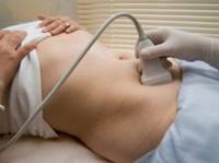
7 FAST : Sonography Screening in Major Trauma Patients Quick evaluation of intraperitoneal cavity and pericardium Detects free fluid : indirect sign of acute hemorrhage and organ injury Supine patient ; convex Mhz probe
8 FAST : Standard Projections ( 1 ) Subxiphoid /Subcostal region : transverse view for pericardial effusion and left liver lobe ( 2 ) RUQ : longitudinal view to assess Morison s pouch: liver - right kidney space ; rt. pleural space * ( 3 ) LUQ : longitudinal view to assess spleen left kidney space; lt.pleural space * ( 4 ) Suprapubic space : long and transverse views; to assess fluid in pouch of Douglas
9 FAST : FOCUSED ASSESSMENT with SONOGRAPHY in TRAUMA When performed correctly, evaluation is done in 2-4 minutes If difficulty arises performing the complete exam, the operator should not waste too much time with the FAST evaluation, if there is any suspicion of hemorrhage If there is intraabdominal bleeding, probability of death increases about 1% for every 3 minutes that elapses before treatment
10 FAST : FOCUSED ASSESSMENT with SONOGRAPHY in TRAUMA Detectability of free fluid is dependent on the volume of fluid present Trendelenburg positions have been used to assess fluid pockets FAST may detect minimum ml of free fluid ( Variable reported sensitivity : ; specificity : ) Rule of thumb : - 5 mm rim of fluid at Morison s pouch : 500 ml free fluid ** - 10 mm rim of fluid at same level : 1,000 ml free fluid **
11 Extended FAST : E- FAST BASIC FAST includes : detection of fluid in upper and lower peritoneal cavity ; pericardial space, pleural spaces ( subxiphoid, RUQ, LUQ, pelvic views ) Other sites incorporated : = Sonographic evaluation at anterior 2 nd or 3 rd intercostal spaces : to assess for pneumothorax = Right and Left Pericolic gutter views : free fluid adjacent to bowel along flanks = Inferior Vena Cava views : intravascular volume status
12 KNOWLEDGE of BASIC ULTRASOUND CONCEPTS will aid in the performance and in the interpretation of the FAST exam
13 - GOOD CONTACT IS IMPORTANT, BETWEEN PATIENT S SKIN AND PROBE, with ACOUSTIC GEL ( to facilitate sound transmission ) - SELECT THE APPROPRIATE PROBE with proper frequency Curved probes for abdomen ; with penetration of sound up to 20 cm = adults : 3. 5 Mhz - 5 Mhz = children : 5 MHZ or higher frequency Curved or linear probes for pneumothorax evaluation - KNOW THE NORMAL ANATOMY OF THE AREA BEING EXAMINED NOTE : You will interpret exams best, when you can supervise images done ; or if you obtain images yourself ** ** ULTRASOUND IS 100% OPERATOR DEPENDENT **

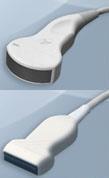
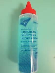
14 Ultrasound equipment Ultrasound Transmission Gel US transducers/probes
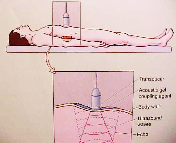
15 HOW DOES ULTRASOUND WORKS 1- Ultrasound transducer receives a short electrical impulse, and generates a pressure wave pulse 2- Pulsed wave propagates down through the tissue 3- Tissue absorbs, scatters, reflects and refracts the wave 4- Reflected waves ( at 90 degrees, perpendicular to probe ) return to the transducer ** 5- Transducer switches to receive mode, and converts the received pressure waves into electrical pulses ( seen in monitor as echoes )** 6- After a fixed period of time, the transducer stops receiving, and transmits the next pressure wave
16
17 ACOUSTIC FREQUENCY Frequency represents cycles per second The unit of acoustic frequency is the hertz (Hz): 1 cycle / second = 1 Hz 1000 cycles / second = 1 KHz 1,000,000 cycles / second = 1 MHz ** Sound frequencies used for diagnostic applications typically range from 2 15 MHz
18 To produce an echo, a reflecting interface must be present At the junction of tissues with different physical properties, acoustic interfaces are present The amount of reflection is determined by difference in acoustic impedances of materials at interface Acoustic impedance is determined by properties of the tissue, and is independent of the frequency
19 RESOLUTION Higher frequency : best resolution / lower penetration of sound waves Lower frequency : better penetration of waves / lower resolution Best image resolution is obtained by using highest frequency possible, although higher frequencies have limited ability to penetrate tissue ** In order to assess deeper anatomic regions in the body, lower frequencies are used, although with some loss of resolution
20 Velocity of propagation Velocity of sound = frequency x wavelength The more closely packed the molecules of the tissues, the faster the speed of sound : = lowest in gases ** = faster in fluids = faster yet in soft tissues = fastest in bones **

21 The average propagation velocity of sound in soft tissues is 1,540 m/sec BONE and AIR create the largest artifacts in sonography ***
22 ATTENUATION Attenuation occurs with the transfer of energy to the tissue ( heating, absorption ) ; as well as with the removal of energy by reflection and scattering *** As sound passes through tissue it looses energy, and the pressure waves decrease in amplitude as they travel further from the source Attenuation depends on the insonating frequency; higher frequencies are attenuated more rapidly than lower frequencies ***
 EXAMPLE : ( A ) Image through liver shows central")

23 A technique used to compensate for attenuation is time gain compensation curve adjustment ( TGC ) EXAMPLE : ( A ) Image through liver shows central band of dark echoes caused by faulty adjustment of TGC curve ( B ) Proper adjustment of TGC curve produces a uniform appearance : operator adjusts the curve *** A B
24 Ultrasound Terminology Echo - free fluid Particulate fluid Echogenic /solid tissue with acoustic interfaces : echoes = hypoechoic = hyperechoic = isoechoic Complex texture = fluid = plus solid material Air / Gas artifacts (dirty shadow) Bone / calcium / calculi ( sharp acoustic shadow )

25 FLUID ECHO FREE FLUID bladder PARTICULATE FLUID ( blood or infection ) cyst


26 ECHOGENIC solid tissue : reflective echoes / acoustic interfaces ( hypoechoic ; hyperechoic ; isoechoic )

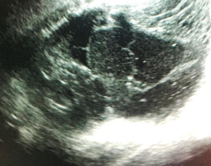
27 Complex texture : mixture of fluid plus echogenic tissue Example : large ovarian dermoid cyst


28 AIR / DIRTY ring down SHADOW BONE / CALCULUS : SHARP ACOUSTIC SHADOW

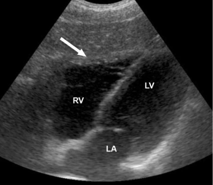
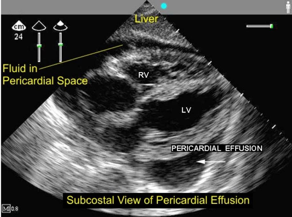
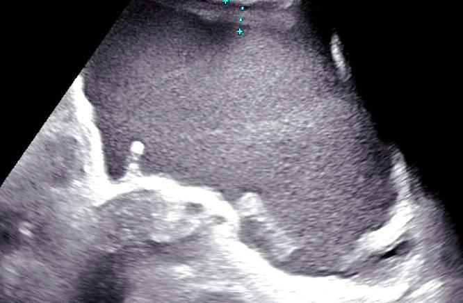
29 Normal FAST : Subxiphoid Subcostal view Echo free

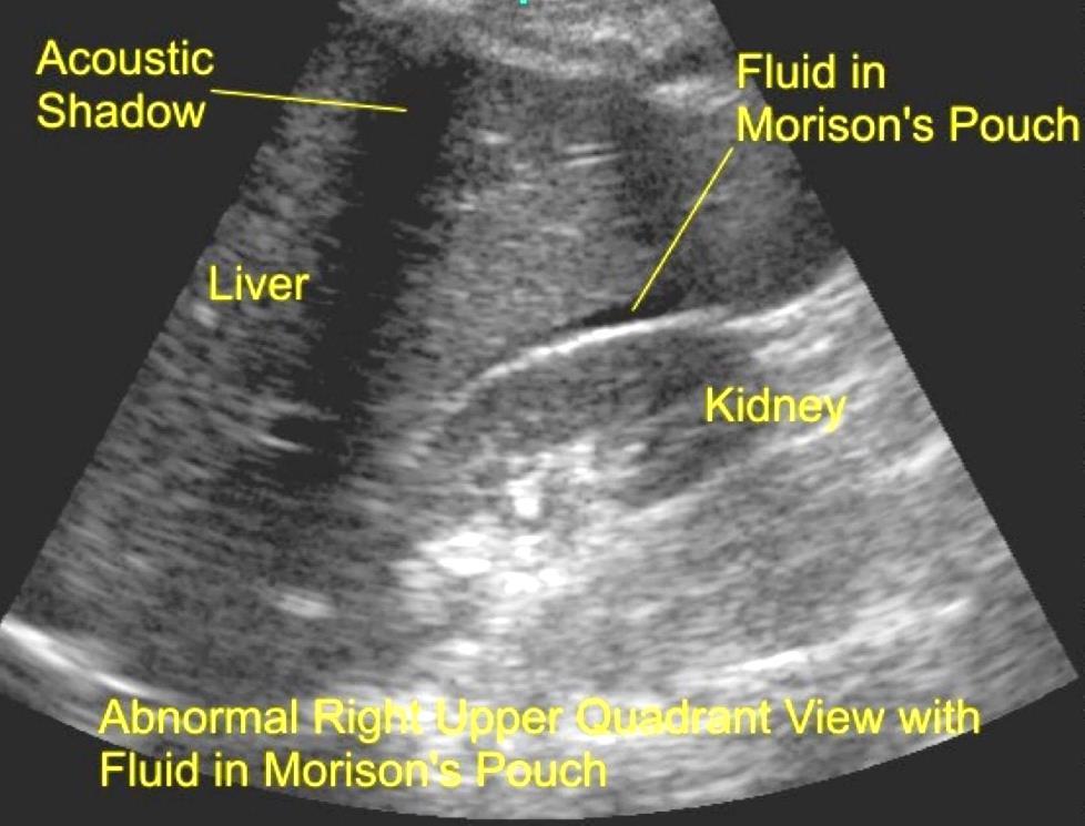
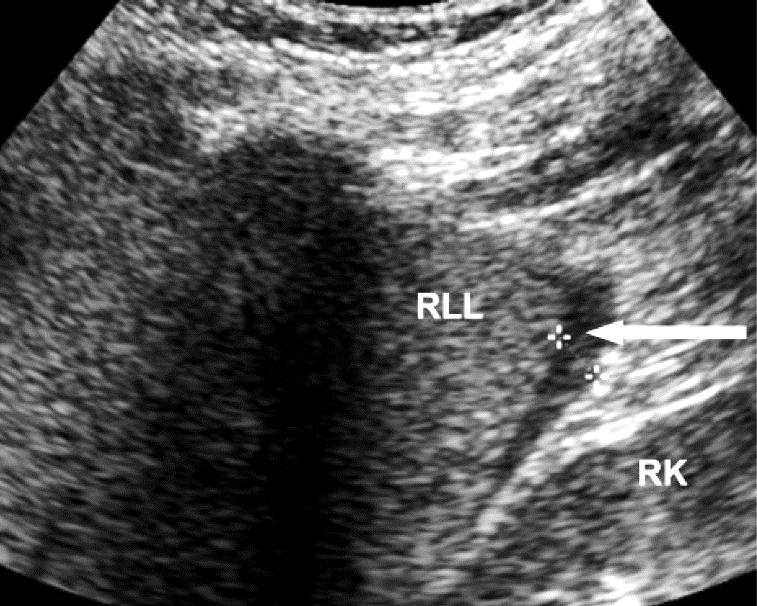
30 RUQ longitudinal view Transverse


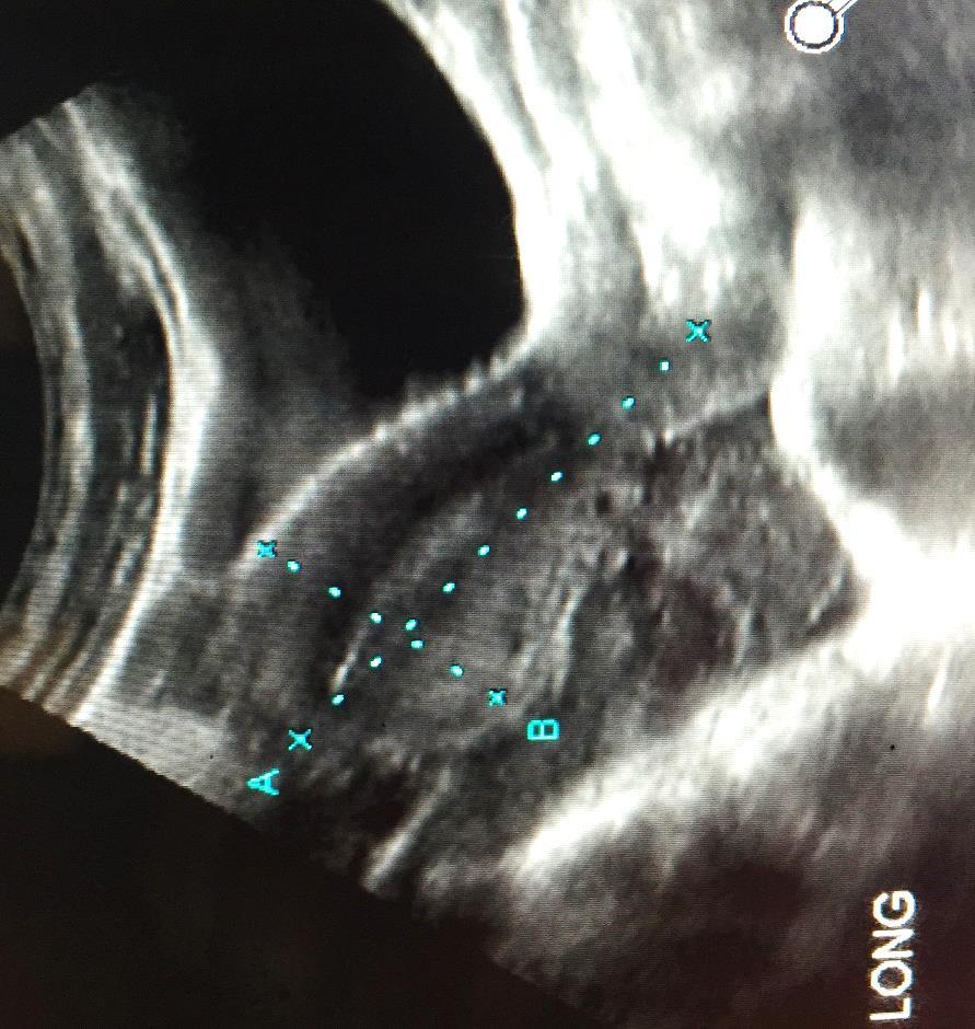
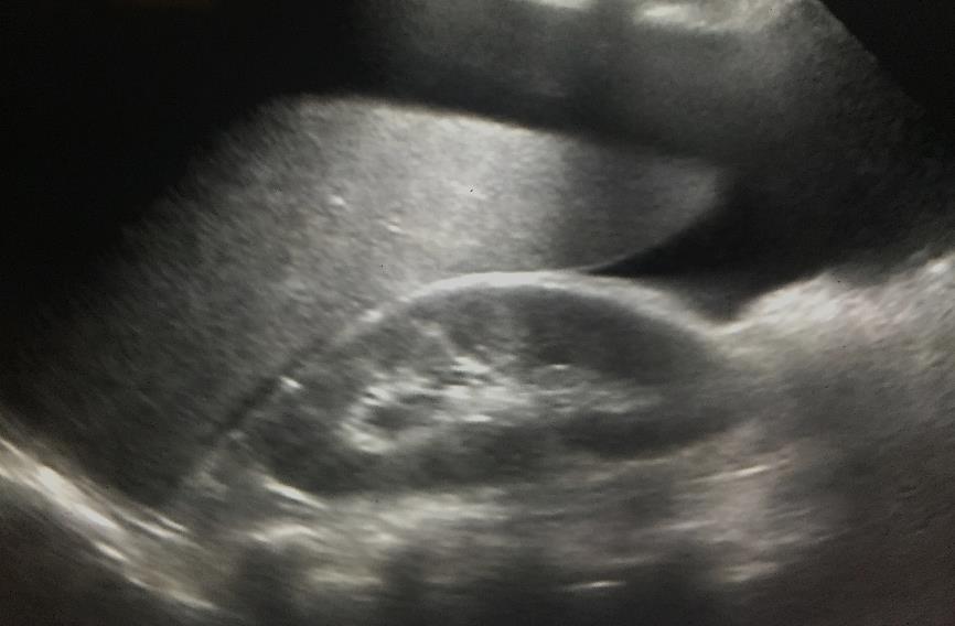
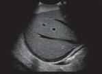
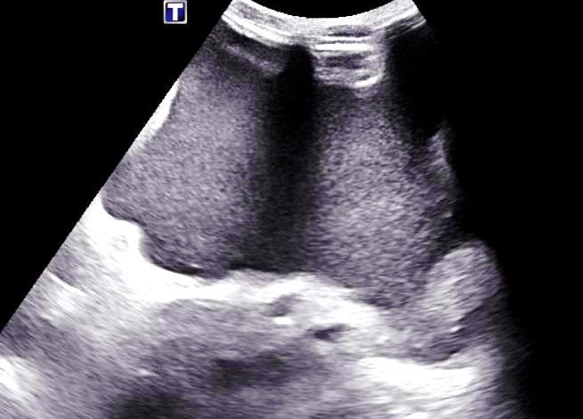
31 PERIHEPATIC PARTICULATE FLUID ( CLOTTED BLOOD ) Complex fluid collection with low level echoes and septations Most likely dx is hematoma in setting of trauma ( D/D biloma ; abscess ) Subcapsular location Infected hematoma or abscess if air present

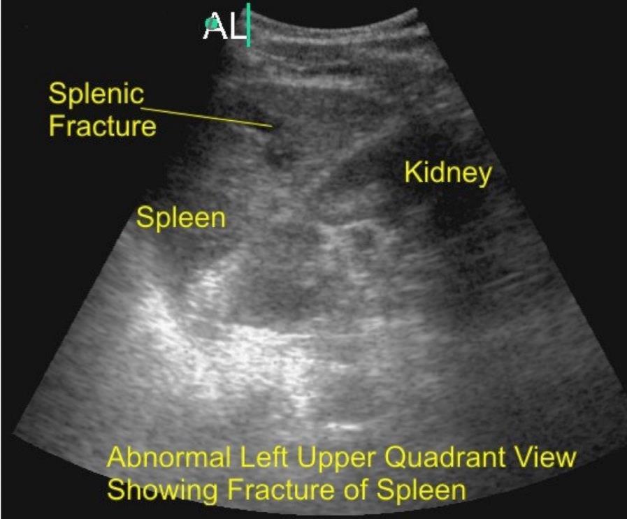

32 LUQ longitudinal view Pleural Fluid


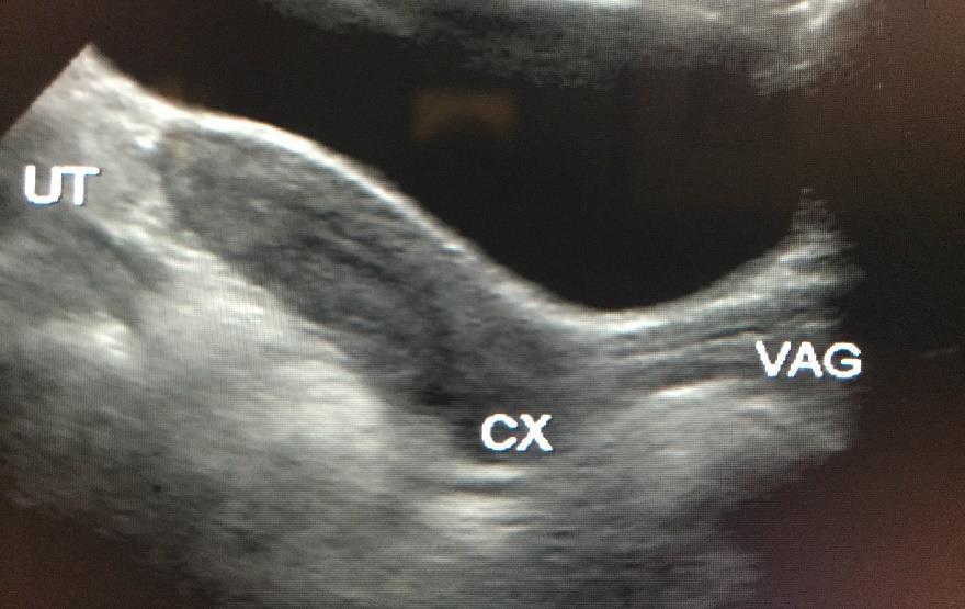
33 PELVIS VIEWS CUL DE SAC Bladder


34 FF bladder Do not confuse posterior sacral promontory and bone absorption, with free fluid


35 PELVIC PSEUDOMASS : REVERBERATION ARTIFACT MAY BE CONFUSED WITH FREE FLUID or CYST


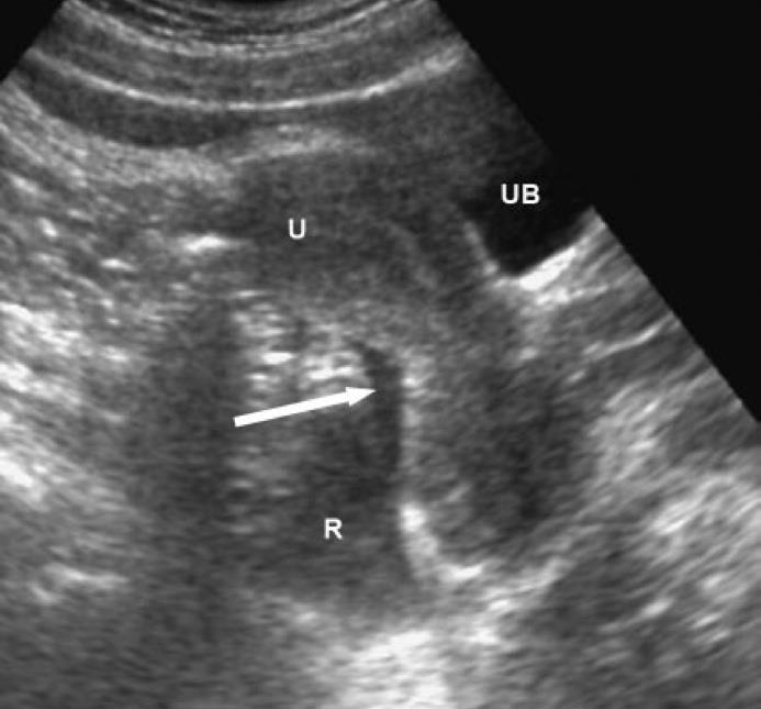
36 A ) BOWEL GAS ARTIFACT : REVERBERATION **** The strong reflection adjacent to the urinary bladder, and the squared appearance of the cul de sac hypoechoic region should make one suspicious of pseudolesion ; not a cyst, and not free fluid B ) DO NOT CONFUSE PELVIC CYSTIC LESIONS WITH FREE FLUID A true cyst has walls on every side

37 TRUE PARTICULATE BLOODY FREE FREE FLUID
38 SOLID ORGAN INJURIES Role of FAST in the diagnosis of injuries to solid organs is limited LIVER : Lacerations range from hypo to hyperechoic Extensive scanning to assess subtle changes would take too much time Sensitivity reported : SPLEEN : Lacerations have variable US appearance ; sensitivity KIDNEYS : Injuries not as common as in spleen and liver Cross sectional imaging needed to assess extent of injury, for treatment
39 SOLID ORGAN INJURIES PANCREAS : Injuries in less than 2% of abdominal trauma cases Subtle changes, best evaluated with CT BOWEL, MESENTERY, BLADDER : Difficult to detect with US


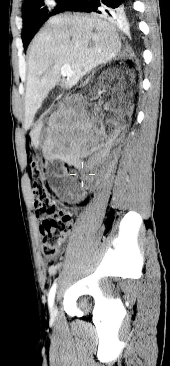
40 SOLID ORGAN INJURIES : LIVER seen best in CT exams

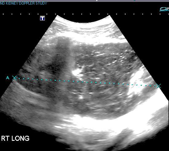
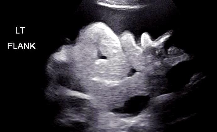
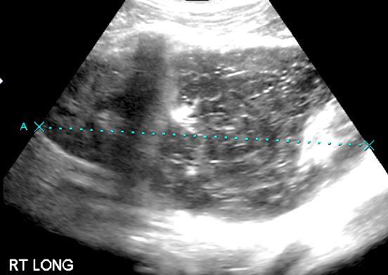
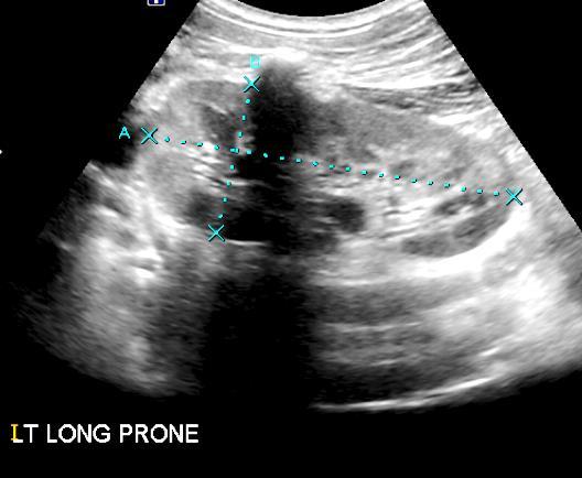
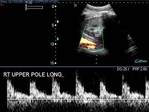
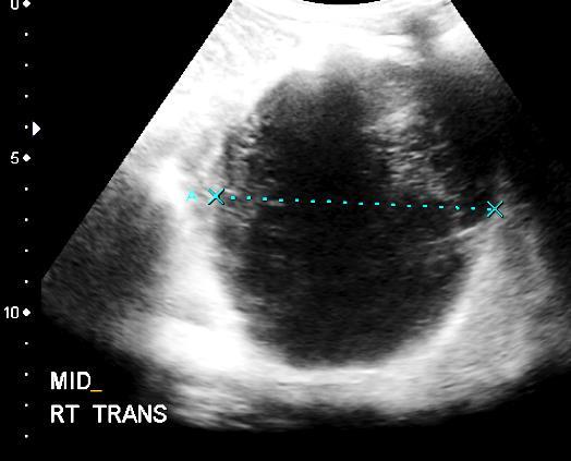
41 RENAL TRAUMA
42
 Probe placed at 2 nd 3 rd intercostal space MC line, between two")
43 EXTENDED FAST : E- FAST PNEUMOTHORAX CT exam remains the gold standard to detect anterior pneumothorax in trauma patients ** Ultrasound has higher sensitivity than supine chest X Rays ** ( sensitivity 95 % ; specificity 91 % : has been reported in ICU cases ) Probe placed at 2 nd 3 rd intercostal space MC line, between two ribs

44 EXTENDED FAST : E- FAST PNEUMOTHORAX
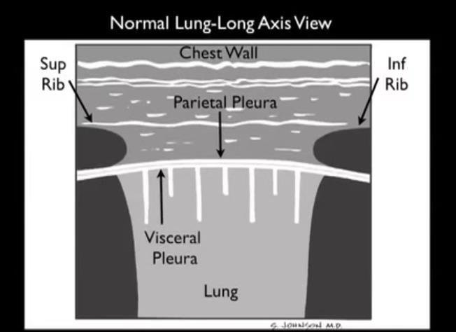
45 EXTENDED FAST : E- FAST PNEUMOTHORAX Normal pleura ( visceral and parietal ) slide on each other in normal lung LUNG SLIDING sign : No Pneumothorax M- mode at same site : anterior - motionless wall : horizontal waves posterior sliding : granular pattern : sand SEASHORE sign : No Pneumothorax


46 EXTENDED FAST : E- FAST PNEUMOTHORAX Normal Pneumothorax Normal Pneumothorax M - mode
47 LIMITATIONS OF FAST EXAM IN MAJOR TRAUMA Detection of free fluid in some injured children Detection of mesenteric, diaphragmatic, or hollow viscus injury Detection of retroperitoneal hemorrhage Technically limited due to patient s obesity ; bowel gas ; degree of injury ; rate of bleeding
48 LIMITATIONS OF FAST EXAM IN MAJOR TRAUMA Bright ambient light in Trauma suite, limits visibility of US monitor Patient movement ; either due to manual chest compressions also being done or combative patient Subcutaneous emphysema : air causes great US artifact Other diagnostic evaluations being done at same time ; small space
49 LIMITATIONS OF FAST EXAM IN MAJOR TRAUMA FALSE POSITIVE diagnosis of free - trauma fluid : = ascites ( chronic liver disease ; renal failure patients ) = ovarian cyst rupture = inflammatory process of abdomen = ventriculoperitoneal shunts = peritoneal dialysis = pre-existing pericardial effusion = pre-existing pleural fluid
50 TRAINING Physicians / Sonologists from a variety of medical specialties may perform the FAST examination ( Trauma Surgeons ; EMERG MED physicians ; ER Radiologists ) Supervised, properly trained sonographers can also obtain the ultrasound images Image interpretation should be performed by a supervising physician
51 Recommended FAST Educational Curriculum and Credentialing Educational Phase ( 4 8 hours ) = didactic course : 1-2 hours ; principles of sonography ; indications, and how to perform and interpret FAST exams = hands on practical session : 3-4 hrs ; should include performance of FAST on models, either simulated or living; with or without intraperitoneal free fluid ( peritoneal dialysis models ) ; video sessions of positive and negative FAST exams Proctored exams = EM and Surgical series, usual proposal FAST exams ( 10 exams should not be enough ) = Competency based certification : non numerical model ** Technical skill is crucial to obtain adequate images **
52 DOCUMENTATION As with all sonograms, focused sonograms require appropriate documentation Images should be stored as part of the medical record Description and interpretation of findings is required
53 SUMMARY : FAST Radiographics : Emergency US in Major Trauma Korner et al Widely available, quick exam for first look Acceptable sensitivity for detection of free fluid ( standard sites ) Poor sensitivity for diagnosis of injury to solid organs Strongly dependent on the operator s skill and experience If initially negative exam, can be repeated
54 RECOMMENDATIONS Radiographics : Emergency US in Major Trauma Korner et al Don t waste time ** ( 2-4 minute evaluation ) Scan for free fluid and pericardial effusion first ( Basic FAST ) Look for pneumothorax, in patients at risk ( E - FAST ) If there is time, look for injuries to solid organs ( although role of FAST for solid organ evaluation is limited **) Use FAST for overview, not for a definite diagnosis of site of injury Stable patient : CT exam Positive or equivocal FAST : Unstable patient : OR
55 REFERENCES 1. AIUM Practice Parameters for the Performance of Focused Assessment with Sonography for Trauma ( FAST ) Examination ; in collaboration with American College of Emergency Physicians ; Korner M., Krotz M., et al,current Role of Emergency Ultrasound in Patients with Major Trauma; Radiographics 2008; 28: Tsui C.,Chung K, et al, Focused Abdominal Sonography for Trauma in the ER for Blunt Abdominal Trauma J. Emergency Medicine 2008; 1: Husain L.,Wayman D, et al, Sonographic Diagnosis of Pneumothorax, J.Emergency Trauma and Shock 2012 :5 : Logan P., Lewis D., FAST: Emergency Ultrasound UK 2004

56
Basic of Ultrasound Physics E FAST & Renal Examination. Dr Muhammad Umer Ihsan MBBS,MD, DCH CCPU,DDU1,FACEM
 Basic of Ultrasound Physics E FAST & Renal Examination Dr Muhammad Umer Ihsan MBBS,MD, DCH CCPU,DDU1,FACEM What is Sound? Sound is Mechanical pressure waves What is Ultrasound? Ultrasounds are sound waves
Focused Assessment Sonography of Trauma (FAST) Scanning Protocol
 Scanning Protocol") Focused Assessment Sonography of Trauma (FAST) Scanning Protocol Romolo Gaspari CHAPTER 3 GOAL OF THE FAST EXAM Demonstrate free fluid in abdomen, pleural space, or pericardial space. EMERGENCY ULTRASOUND
Focused Assessment Sonography of Trauma (FAST) Scanning Protocol Romolo Gaspari CHAPTER 3 GOAL OF THE FAST EXAM Demonstrate free fluid in abdomen, pleural space, or pericardial space. EMERGENCY ULTRASOUND
Abdominal Ultrasonography
 Abdominal Ultrasonography David A. Masneri, DO, FACEP, FAAEM Assistant Professor of Emergency Medicine Assistant Director, Emergency Medicine Residency Medical Director, Operational Medicine Division Center
Certificate in Clinician Performed Ultrasound (CCPU) Syllabus. Extended Focussed Abdominal Scan for Trauma (E-FAST)
 Syllabus. Extended Focussed Abdominal Scan for Trauma (E-FAST)") Certificate in Clinician Performed Ultrasound (CCPU) Syllabus Extended Focussed Abdominal Scan for Trauma (E-FAST) Page 1 of 6 01/17 ACN 001 679 161 ABN 64 001 679 Extended Focussed Abdominal Scan for
Certificate in Clinician Performed Ultrasound (CCPU) Syllabus Extended Focussed Abdominal Scan for Trauma (E-FAST) Page 1 of 6 01/17 ACN 001 679 161 ABN 64 001 679 Extended Focussed Abdominal Scan for
Focused Assessment with Sonography in Trauma (FAST) UC Irvine School of Medicine
 UC Irvine School of Medicine") Focused Assessment with Sonography in Trauma (FAST) UC Irvine School of Medicine Purpose of FAST exam Quickly evaluate patient s status in emergency situations Blunt or penetrating trauma Visualize fluid
Focused Assessment with Sonography in Trauma (FAST) UC Irvine School of Medicine Purpose of FAST exam Quickly evaluate patient s status in emergency situations Blunt or penetrating trauma Visualize fluid
Principles of Ultrasound. Cara C. Prideaux, M.D. University of Utah PM&R Sports Medicine Fellow March 14, 2012
 Principles of Ultrasound Cara C. Prideaux, M.D. University of Utah PM&R Sports Medicine Fellow March 14, 2012 None Disclosures Outline Introduction Benefits and Limitations of US Ultrasound (US) Physics
Objectives. The Extended FAST Exam. Focused Assessment e With Sonography In. Trauma (FAST)
") Northern California Emergency Ultrasound Course Objectives The Extended FAST Exam Rimon Bengiamin, MD, RDMS UC SF Discuss the components of the EFAST exam Evaluate the utility of the EFAST Review how to
Northern California Emergency Ultrasound Course Objectives The Extended FAST Exam Rimon Bengiamin, MD, RDMS UC SF Discuss the components of the EFAST exam Evaluate the utility of the EFAST Review how to
Ultrasound. FAST Focused Assessment with Sonography in Trauma
 Ultrasound FAST Focused Assessment with Sonography in Trauma Rohit Patel, MD University of Florida Health Director, Critical Care Ultrasound Surgical ICU Center for Intensive Care Gainesville, Florida
Background Focused Assessment with Sonography in Trauma. Johann Baptist Dormagen, MD, PhD
 Focused Assessment with Sonography in Trauma Johann Baptist Dormagen, MD, PhD Unit of Abdominal and Oncologic Radiology Department of Radiology and Nuclear Medicine Oslo University Hospital, Norway 8 th
What is Ultrasound? What is Ultrasound? B A. Basic Principles of Ultrasound. Basic Principles of Ultrasound. Basic Principles of Ultrasound
 Introduction to Ultrasound Principles Mani Montazemi, RDMS Baylor College of Medicine Division of Maternal-Fetal Medicine Department of Obstetrics and Gynecology Manager, Maternal Fetal Center Imaging
Extended FAST Exam. Goal of Trauma Care. Golden Hour of Trauma
 Extended FAST Exam Goal of Trauma Care Golden Hour of Trauma Best INITIAL screening modality in trauma efast 2014 LLSA Article (ACEP Policy Statement) Level B Recommendation: In hemodynamically unstable
Lung sonography in the diagnosis of pneumothorax.
 Lung sonography in the diagnosis of pneumothorax. Poster No.: C-0526 Congress: ECR 2011 Type: Educational Exhibit Authors: K. Stefanidis, K. Vintzilaios, D. D. Cokkinos, E. Antypa, S. Dimopoulos, S. Nanas,
Preamble (disclaimer)
") Preamble (disclaimer) PHYSICS AND PRINCIPLES OF HEAD/NECK ULTRASOUND Joseph C. Sniezek, MD FACS LTC, MC, USA Otolaryngology/H&N Surgery Tripler Army Medical Center 1. I am not a physicist 2. ACS has recommended
Preamble (disclaimer) PHYSICS AND PRINCIPLES OF HEAD/NECK ULTRASOUND Joseph C. Sniezek, MD FACS LTC, MC, USA Otolaryngology/H&N Surgery Tripler Army Medical Center 1. I am not a physicist 2. ACS has recommended
This appendix was part of the submitted manuscript and has been peer reviewed. It is posted as supplied by the authors.
 This appendix was part of the submitted manuscript and has been peer reviewed. It is posted as supplied by the authors. - Figure S1: The four quadrant approach lung ultrasound at the bedside. * The anterolateral
Advanced Imaging Practice CSB068
 Advanced Imaging Practice CSB068 Week 1 Peer Review - Evaluation of work by one or more people of similar competence to the producer - A form of self-regulation about improving quality and upholding standards
Background & Indications
 Teresa S. Wu, MD, FACEP Director, EM Ultrasound Program & Fellowship Co-Director, Simulation Based Training Program & Fellowship Maricopa Medical Center Simulation Curriculum Director Associate Professor,
The Focused Assessment with Sonography for Trauma, (FAST) procedure.
 procedure.") The Focused Assessment with Sonography for Trauma, (FAST) procedure. ROBERT H. WRIGLEY Professor Veterinary Diagnostic Imaging University of Sydney Veterinary Teaching Hospital Professor Emeritus Colorado
The Focused Assessment with Sonography for Trauma, (FAST) procedure. ROBERT H. WRIGLEY Professor Veterinary Diagnostic Imaging University of Sydney Veterinary Teaching Hospital Professor Emeritus Colorado
Diagnostic Ultrasound. Sutiporn Khampunnip, M.D.
 Diagnostic Ultrasound Sutiporn Khampunnip, M.D. Definition of Ultrasound Ultrasound is simply sound waves, like audible sound. High-frequency sound and refers to mechanical vibrations above 20 khz. Human
The Role of the FAST exam in the EDRU
 The Role of the FAST exam in the EDRU A. Robb McLean, MD, MHCM Vice Chair of Clinical Operations, Department of Emergency Medicine Joint Trauma Conference June 20, 2017 Disclosures Goals Describe the performance,
Terminology Tissue Appearance
 By Marc Nielsen, MD Advantages/Disadvantages Generation of Image Ultrasound Machine/Transducer selection Modes of Ultrasound Terminology Tissue Appearance Scanning Technique Real-time Portable No ionizing
The Physics of Ultrasound. The Physics of Ultrasound. Claus G. Roehrborn. Professor and Chairman. Ultrasound Physics
 The Physics of Ultrasound Pipe Organ 10-8000 Emission Dog 452-1080 Man 85-1100 Spectrum Bat 10,000-120,000 Porpoise 7000-120,000 Claus G. Roehrborn Professor and Chairman 10 20 Cycles per second Reception
Radiological Investigations of Abdominal Trauma
 76 77 Investigations of Abdominal Trauma Introduction: Trauma to abdominal organs is a common cause of patient morbidity and mortality among trauma patients. Causes of abdominal trauma include blunt injuries,
Ultrasound Physics & Terminology
 Ultrasound Physics & Terminology This module includes the following: Basic physics terms Basic principles of ultrasound Ultrasound terminology and terms Common artifacts seen Doppler principles Terms for
(FAST) Peter Logan FRCS(Ed) FFAEM FACEM David Lewis FRCS FFAEM. Focused Assessment with Sonography for Trauma
 Peter Logan FRCS(Ed) FFAEM FACEM David Lewis FRCS FFAEM. Focused Assessment with Sonography for Trauma") Focused Assessment with Sonography for Trauma (FAST) Peter Logan FRCS(Ed) FFAEM FACEM David Lewis FRCS FFAEM 1 (FAST) Introduction Physical examination of the abdomen in blunt trauma is subjective and
Focused Assessment with Sonography for Trauma (FAST) Peter Logan FRCS(Ed) FFAEM FACEM David Lewis FRCS FFAEM 1 (FAST) Introduction Physical examination of the abdomen in blunt trauma is subjective and
The 2 nd Cambridge Advanced Emergency Ultrasound Course
 The 2 nd Cambridge Advanced Emergency Ultrasound Course Addenbrooke s Hospital Cambridge Sept 2008 1 2 Faculty! UK! USA! Australia! Toshiba! Emergency Medicine! Radiology 3 Programme! Day 1 Introduction
Physical Principles of Ultrasound
 Physical Principles of Ultrasound Grateful appreciation to Richard A. Lopchinsky, MD, FACS and Nancy H. Van Name, RDMS, RTR, and MarleneKattaron, RDMS 2000 UIC All Rights Reserved. Course Objectives Identify
SPECIAL DIAGNOSTIC STUDIES IN BLUNT TRAUMA OLEH : Prof.DR.Dr Abdul Rasyid SpRad (K),Ph.D Dr.Evo Elidar Sp.Rad
,Ph.D Dr.Evo Elidar Sp.Rad") SPECIAL DIAGNOSTIC STUDIES IN BLUNT TRAUMA OLEH : Prof.DR.Dr Abdul Rasyid SpRad (K),Ph.D Dr.Evo Elidar Sp.Rad Trauma Emergency Room layout Ideally the trauma emergency room is centrally located to provide
SPECIAL DIAGNOSTIC STUDIES IN BLUNT TRAUMA OLEH : Prof.DR.Dr Abdul Rasyid SpRad (K),Ph.D Dr.Evo Elidar Sp.Rad Trauma Emergency Room layout Ideally the trauma emergency room is centrally located to provide
Abdominal ultrasound:
 Abdominal ultrasound: Non-traumatic acute abdomen Wittanee Na-ChiangMai, MD Department of Radiology ChiangMai University 26/04/2017 Contents Technique of examination Normal anatomy Emergency conditions
The FAST Exam! Dr. David Easton MD FRCPC Critical Care and Emergency Medicine University of Manitoba Canada
 The FAST Exam! Dr. David Easton MD FRCPC Critical Care and Emergency Medicine University of Manitoba Canada Dr. David Easton MD FRCPC Assistant Professor Section of Critical Care and Emergency Medicine
Introduction & Physics of ED Ultrasound. Objectives. What? - Limited Studies. Who? - ED Docs
 Introduction & Physics of ED Ultrasound Martine Sargent, MD Ultrasound Director, Assistant Professor UCSF Department of Emergency Medicine San Francisco General Hospital & Trauma Center Objectives Who?
ULTRASOUND NOMENCLATURE
 Chapter 1: Ultrasound Nomenclature, Image Orientation, and Basic Instrumentation CYNTHIA SIKOWSKI Ultrasound waves are sound waves that have a frequency exceeding 20,000 Hz. When sound waves are transmitted
Ultrasound Principles cycle Frequency Wavelength Period Velocity
 ! Teresa S. Wu, MD, FACEP Director, EM Ultrasound Program & Fellowship Co-Director, Simulation Based Training Program & Fellowship Associate Program Director, EM Residency Program Maricopa Medical Center
Pediatric Lung Ultrasound (PLUS) In Diagnosis of Community Acquired Pneumonia (CAP)
 In Diagnosis of Community Acquired Pneumonia (CAP)") Pediatric Lung Ultrasound (PLUS) In Diagnosis of Community Acquired Pneumonia (CAP) Dr Neetu Talwar Senior Consultant, Pediatric Pulmonology Fortis Memorial Research Institute, Gurugram Study To compare
Pediatric Lung Ultrasound (PLUS) In Diagnosis of Community Acquired Pneumonia (CAP) Dr Neetu Talwar Senior Consultant, Pediatric Pulmonology Fortis Memorial Research Institute, Gurugram Study To compare
WELCOME! Introduction to Bedside Ultrasound
 WELCOME! Introduction to Bedside Ultrasound TEACHERS University of California-Irvine School of Medicine Nathan Molina nathan.d.molina@gmail.com Trevor Plescia taplescia90@gmail.com Jack Silva jpsilva42@gmail.com
Ultrasound basics Part 1
 Ultrasound basics Part 1 'Ultrasound enhanced critical care medicine' Rohit Patel, MD University of Florida Health Director, Critical Care Ultrasound Surgical ICU Center for Intensive Care Gainesville,
Abdominal Trauma. Nat Krairojananan M.D., FRCST Department of Trauma and Emergency Medicine Phramongkutklao Hospital
 Abdominal Trauma Nat Krairojananan M.D., FRCST Department of Trauma and Emergency Medicine Phramongkutklao Hospital overview Quick review abdominal anatomy Review of mechanism of injury Review of investigation
1 Fundamentals. Basic Definitions and Physics Principles. Fundamentals
 1 To become versed in the language of ultrasonography, it is necessary to review some of the basic principles of physics. The wave physics principles of ordinary (i.e., audible) sound apply to ultrasound
EFAST. Extended Focussed Assessment with Sonography for Trauma. Ultrasound Logbook. Name
 EFAST Extended Focussed Assessment with Sonography for Trauma Ultrasound Logbook ame Contents EFAST Accreditation Requirements 25 Abdominal Aorta Report Forms 3 Formative Assessments 1 Summative Assessment
CONTENTS. Test Number cpd Tanya Reynolds (Nat. Dip. Diag. Rad., B. Tech. Diag. Rad., B. Tech. Ultrasound)
") CONTENTS page 1-15 page 16 BASIC 2-DIMENSIONAL ULTRASOUND PRINCIPLES Multiple Choice Test Test Number cpd 41640 Tanya Reynolds (Nat. Dip. Diag. Rad., B. Tech. Diag. Rad., B. Tech. Ultrasound) Tanya is
CONTENTS page 1-15 page 16 BASIC 2-DIMENSIONAL ULTRASOUND PRINCIPLES Multiple Choice Test Test Number cpd 41640 Tanya Reynolds (Nat. Dip. Diag. Rad., B. Tech. Diag. Rad., B. Tech. Ultrasound) Tanya is
Ultrasound Physics & Doppler
 Ultrasound Physics & Doppler Endocrine University 2018 Mark Lupo, MD, FACE, ECNU Objectives Review the essential components of ultrasound physics in neck sonography Demonstrate the importance of ultrasound
Abdominal Ultrasound
 Abdominal Ultrasound What is Ultrasound Imaging of the Abdomen? What are some common uses of the procedure? How should I prepare? What does the equipment look like? How does the procedure work? How is
US in non-traumatic acute abdomen. Lalita, M.D. Radiologist Department of radiology Faculty of Medicine ChiangMai university
 US in non-traumatic acute abdomen Lalita, M.D. Radiologist Department of radiology Faculty of Medicine ChiangMai university Sagittal Orientation Transverse (Axial) Orientation Coronal Orientation Intercostal
Basic Training Programme. 16 Februrary 2018, ROTTERDAM. Pre and Post-Course Test Answers
 Basic Training Programme 16 Februrary 2018, ROTTERDAM Pre and Post-Course Test Answers Your details: Name: Conference registration number/ BT delegate number: Email address: Are you already performing
Guidelines, Policies and Statements D5 Statement on Abdominal Scanning
 Guidelines, Policies and Statements D5 Statement on Abdominal Scanning Disclaimer and Copyright The ASUM Standards of Practice Board have made every effort to ensure that this Guideline/Policy/Statement
Ultrasound Knobology
 Ultrasound Knobology Raj Dasgupta MD, FACP, FCCP, FASSM Assistant Professor of Clinical Medicine Pulmonary / Critical Care / Sleep Medicine University of Southern California (USC) Objectives Physics of
2. Blunt abdominal Trauma
 Abdominal Trauma 1. Evaluation and management depends on: a. Mechanism (Blunt versus Penetrating) b. Injury complex in addition to abdomen c. Haemodynamic stability assessment: i. Classically patient s
Basic Physics of Ultrasound and Knobology
 WELCOME TO UTMB Basic Physics of Ultrasound and Knobology By Daneshvari Solanki, FRCA Laura B. McDaniel Distinguished Professor Anesthesiology and Pain Medicine University of Texas Medical Branch Galveston,
Chest Ultrasound: Pneumothorax
 WINFOCUS BASIC ECHO (WBE) Chest Ultrasound: Pneumothorax Mark Hamlin, MD, MS Associate Professor of Anesthesiology and Surgery University of Vermont College of Medicine Co-Director of Surgical Critical
Lung ultrasound in the critically ill patient BASICS
 Lung ultrasound in the critically ill patient BASICS Rohit Patel, MD University of Florida Health Director, Critical Care Ultrasound Surgical ICU Center for Intensive Care Gainesville, Florida Introduction
GENERAL ABDOMINAL IMAGING PERITONEAL SPACE, PANCREAS, & SPLEEN. VMB 960 March 25, 2013
 GENERAL ABDOMINAL IMAGING PERITONEAL SPACE, PANCREAS, & SPLEEN VMB 960 March 25, 2013 REFERENCE Chapters 35-36 Pages 650-678 Chapter 37 Pages 694-701 Chapter 3 Pages 38-49 OBJECTIVES Radiography and Ultrasound
Ultrasonography of the Neck as an Adjunct to FNA. Nicole Massoll M.D.
 Ultrasonography of the Neck as an Adjunct to FNA Nicole Massoll M.D. Basic Features of Head and Neck Ultrasound and Anatomy Nicole Massoll M.D. University of Arkansas for Medical Sciences, Little Rock
Emergency Medicine Interest Group (EMIG) 2016
 2016") Emergency Medicine Interest Group (EMIG) 2016 Welcome to the flipped classroom (learning objectives summary) for the 2016 Emergency Medicine Interest Group (EMIG) Procedures Workshop. Overview - Tuesday
Emergency Medicine Interest Group (EMIG) 2016 Welcome to the flipped classroom (learning objectives summary) for the 2016 Emergency Medicine Interest Group (EMIG) Procedures Workshop. Overview - Tuesday
Caudal Edge of the Liver in the Right Upper Quadrant (RUQ) View Is the Most Sensitive Area for Free Fluid on the FAST Exam
 View Is the Most Sensitive Area for Free Fluid on the FAST Exam") Original Research Caudal Edge of the Liver in the Right Upper Quadrant (RUQ) View Is the Most Sensitive Area for Free Fluid on the FAST Exam Viveta Lobo, MD* Michelle Hunter-Behrend, MD* Erin Cullnan,
Original Research Caudal Edge of the Liver in the Right Upper Quadrant (RUQ) View Is the Most Sensitive Area for Free Fluid on the FAST Exam Viveta Lobo, MD* Michelle Hunter-Behrend, MD* Erin Cullnan,
An abdominal ultrasound produces a picture of the organs and other structures in the upper abdomen.
 Scan for mobile link. Ultrasound - Abdomen Ultrasound imaging of the abdomen uses sound waves to produce pictures of the structures within the upper abdomen. It is used to help diagnose pain or distention
Point of Care Ultrasound (PoCUS)
") Point of Care Ultrasound (PoCUS) Competency Assessment Forms AORTA Competency A Focussed Assessment of the Aorta (AAA) Guidance Please follow this guidance as closely as possible to ensure consistency
Point of Care Ultrasound (PoCUS) Competency Assessment Forms AORTA Competency A Focussed Assessment of the Aorta (AAA) Guidance Please follow this guidance as closely as possible to ensure consistency
The faculty will include physicians with international reputations as outstanding ultrasound educators.
 Ultrasound Courses Course Description Whether you re a beginner or a seasoned sonographer, this year s AAEM pre-conference ultrasound course will be worth your time. We will be offering a half day course
Chapter 3. Sonographic Image Interpretation
 Chapter 3 Sonographic Image Interpretation Sonograms are two-dimensional gray-scale images that allow assessment and diagnosis of many anatomic and pathologic changes that can occur in the human body.
LIVER INJURIES PROFF. S.FLORET
 LIVER INJURIES PROFF. S.FLORET Abdominal injuries For anatomical consideration: Abdomen can be divided in four areas Intra thoracic abdomen True abdomen Pelvic abdomen Retroperitoneal abdomen ETIOLOGY
Children's (Pediatric) Ultrasound - Abdomen
 Ultrasound - Abdomen") Scan for mobile link. Children's (Pediatric) Ultrasound - Abdomen Children s (pediatric) ultrasound imaging of the abdomen is a safe, noninvasive test that uses sound waves to produce a clear picture of
Scan for mobile link. Children's (Pediatric) Ultrasound - Abdomen Children s (pediatric) ultrasound imaging of the abdomen is a safe, noninvasive test that uses sound waves to produce a clear picture of
Bedside ultrasound - Lung ultrasound in the Intensive Care Unit
 Bedside ultrasound - Lung ultrasound in the Intensive Care Unit Kishore K. Pichamuthu, Professor, Department of Critical Care, Christian Medical College, Vellore. Summary In an ICU setting, ultrasonographic
Case Conference. Discussion. Indications of Trauma Blue. Trauma Protocol In SKH. Trauma Blue VS. Trauma Red. Supervisor:VS 楊毓錚 Presenter:R1 周光緯
 Case Conference Supervisor:VS 楊毓錚 Presenter:R1 周光緯 Discussion 2010.7.14 2/81 Trauma Protocol In SKH Indications of Trauma Blue Trauma Blue VS. Trauma Red 3/81 Severe trauma mechanism : 1. Trauma to multiple
2 Blunt Abdominal Trauma
 2 Blunt Abdominal Trauma Ricardo Ferrada, Diego Rivera, and Paula Ferrada Pearls and Pitfalls Patients suffering a high-energy trauma have solid viscera rupture in the abdomen and/or aortic rupture in
Small animal point of care ultrasound techniques
 Small animal point of care ultrasound techniques The role of veterinary point of care ultrasound in determining the presence or absence of specific pathologies is examined by Jantina McMurray DVM; Søren
Chapter 14. Imaging Artifacts
 Chapter 14 Image Artifacts The complex physical interactions that occur between an ultrasound beam and human anatomy and the intricate and sophisticated technological components of a sonographic imaging
Category Term Definition Comments 1 Major Categories 1a
 Working Lexicon Categories, Terms & Definitions Category Term Definition Comments 1 Major Categories 1a Physiologic Category (consistent with normal ovarian physiology) Follicle Simple 3 cm in premenopausal
Ultrasound Physics and Knobology Alan Macfarlane. Consultant Anaesthetist Glasgow Royal Infirmary
 Ultrasound Physics and Knobology Alan Macfarlane Consultant Anaesthetist Glasgow Royal Infirmary RAPM 2009; 34: 40-46 Ultrasound Proficiency Understanding US image generation and device operation Image
DIGITAL IMAGE PROCESSING IN ULTRASOUND IMAGES
 DIGITAL IMAGE PROCESSING IN ULTRASOUND IMAGES Kamaljeet Kaur Computer Science & Engineering Department Guru Nanak Dev Engg. College, Ludhiana. Punjab-India meetk.89@gmail.com ABSTRACT-- Image processing
Background & Indications Probe Selection
 Teresa S. Wu, MD, FACEP Director, EM Ultrasound Program & Fellowship Co-Director, Simulation Based Training Program & Fellowship Associate Program Director, EM Residency Program Maricopa Medical Center
Session 2: Ultrasonography for Primary Care Clinicians Learning Objectives
 Session 2: Ultrasonography for Primary Care Clinicians Learning Objectives 1. Assess the main components and functions of a portable ultrasound unit. 2. Identify three clinical applications of portable
Breast Imaging Essentials
 Breast Imaging Essentials Module 9 Transcript 2016 ASRT. All rights reserved. Breast Imaging Essentials Module 9 Breast Ultrasound 1. ASRT Animation 2. Welcome Welcome to Module 9 of Breast Imaging Essentials
Focused Assessment With Sonography for Trauma Examination Reexamining the Importance of the Left Upper Quadrant View
 ORIGINAL RESEARCH Focused Assessment With Sonography for Trauma Examination Reexamining the Importance of the Left Upper Quadrant View Kathleen M. O Brien, MD, Lori A. Stolz, MD, Richard Amini, MD, Austin
Which Blunt Trauma Patients Should Be Studied by Abdominal CT?
 MDCT of Bowel and Mesenteric Injury: How Findings Influence Management 4 th Nordic Trauma Radiology Course 2006 4 th Nordic Trauma Radiology Course 2006 Stuart E. Mirvis, M.D., FACR Department of Radiology
ULTRASOUND. OB/Gyn (Core) Ultrasound PIEZOELECTRIC EFFECT. Principles of Ultrasound Physics and Instrumentation. Nathan Pinkney, BS, CDOS
 Ultrasound PIEZOELECTRIC EFFECT. Principles of Ultrasound Physics and Instrumentation. Nathan Pinkney, BS, CDOS") 1 OB/Gyn (Core) Ultrasound Principles of Ultrasound Physics and Instrumentation Nathan Pinkney, BS, CDOS Philadelphia College of Osteopathic Medicine 2016 ULTRASOUND CATEGORIES OF SOUND INFRASOUND = below
1 OB/Gyn (Core) Ultrasound Principles of Ultrasound Physics and Instrumentation Nathan Pinkney, BS, CDOS Philadelphia College of Osteopathic Medicine 2016 ULTRASOUND CATEGORIES OF SOUND INFRASOUND = below
ASSOC. PROF. DR. SADIK GİRİŞGİN Necmettin Erbakan Uni. Meram Medicine School
 ASSOC. PROF. DR. SADIK GİRİŞGİN Necmettin Erbakan Uni. Meram Medicine School Portable Probe variety Real time imaging Cheap No radiation Repeatable Cost effective 1988 1971 1990 2001 2008 FAST First case
Normal Sonographic Anatomy
 hapter 2:The Liver DUNSTAN ABRAHAM Normal Sonographic Anatomy Homogeneous, echogenic texture (Figure 2-1) Measures approximately 15 cm in length and 10 12.5 cm anterior to posterior; measurement taken
Diploma of Medical Ultrasonography (DMU) Physical Principles of Ultrasound and Instrumentation Syllabus
 Physical Principles of Ultrasound and Instrumentation Syllabus") Diploma of Medical Ultrasonography (DMU) Physical Principles of Ultrasound and Instrumentation Syllabus Page 1 of 7 11/18 Candidates are expected to cover all of the content of this syllabus when preparing
Diploma of Medical Ultrasonography (DMU) Physical Principles of Ultrasound and Instrumentation Syllabus Page 1 of 7 11/18 Candidates are expected to cover all of the content of this syllabus when preparing
Elastography in the. technically difficult patient. EPIQ ultrasound system. Ultrasound
 Ultrasound Elastography in the technically difficult patient EPIQ ultrasound system Chairman Department of Diagnostic Radiology Allegheny General Hospital Pittsburgh, PA, USA You can offer more information
Human Systems. Technology - Ultrasounds
 Human Systems Technology - Ultrasounds What is General Ultrasound Imaging? Ultrasound imaging, also called ultrasound scanning or sonography, involves exposing part of the body to high-frequency sound
My Patient Has Abdominal Pain PoCUS of the Biliary Tract and the Urinary Tract
 My Patient Has Abdominal Pain PoCUS of the Biliary Tract and the Urinary Tract Objectives PoCUS for Biliary Disease PoCUS for Renal Colic PoCUS for Urinary Retention Biliary Disease A patient presents
What is Ultrasound? Resolution Image production Attenuation Imaging modes Ultrasound artifacts... 7
 What is Ultrasound?... 1 Resolution... 3 Image production... 3 Attenuation... 4 Imaging modes... 5 Ultrasound artifacts... 7 0 What is Ultrasound? High frequency sound of frequencies 2-50 MHz is used in
Background & Indications Probe Selection
 Teresa S. Wu, MD, FACEP Director, EM Ultrasound Program & Fellowship Co-Director, Simulation Based Training Program & Fellowship Associate Program Director, EM Residency Program Maricopa Medical Center
NON INVASIVE LIFE SAVERS. Ultrasound in PICU
 VOL 1 NO.1 Jan March 2014 54 Table 1. Selected Applications of Point-of-Care Ultrasonography, According to Medical Specialty. Specialty Ultrasound Applications Anesthesia Cardiology Guidance for vascular
Critical Care Ultrasound Seeing is Believing
 Critical Care Ultrasound Seeing is Believing Philip D Lumb, M.B., B.S., MCCM Los Angeles, California USA Objectives Integrate ultrasound into routine critical care practice Introduce HOLA (Holistic Approach)
A Practical Approach to Ultrasound Assessment of Respiratory Distress
 A Practical Approach to Ultrasound Assessment of Respiratory Distress Yanick Beaulieu, MD, FRCPC Director, Bedside Ultrasound Curriculum Division of Cardiology and Critical Care Hôpital du Sacré-Coeur
An Overview of Ultrasound Testing For Lesion Detection in Human Kidney
 Journal of Tomography System & Sensors Application Vol.1, Issue 1, June 2018 An Overview of Ultrasound Testing For Lesion Detection in Human Kidney Aina Fadhilah Abd Rahim 1, Zawin Najah Abd Halim 1, Jaysuman
Dr Emma Chung. Safety first - Physical principles for excellent imaging
 Safety first - Physical principles for excellent imaging Dr Emma Chung Lecturer in Medical Physics, University of Leicester Clinical Scientist, University Hospitals of Leicester NHS Trust Thanks to Caroline
ASSESSING THE PLAIN ABDOMINAL RADIOGRAPH M A A M E F O S U A A M P O F O
 ASSESSING THE PLAIN ABDOMINAL RADIOGRAPH M A A M E F O S U A A M P O F O Introduction The abdomen (less formally called the belly, stomach, is that part of the body between the thorax (chest) and pelvis,
CSB 046 Complementary Imaging Techniques
 CSB 046 Complementary Imaging Techniques - Quizzes are only ultrasound, final includes nuc med and ultrasound Week 1 Intro to Ultrasound Physics - Uses 1 to 20 MHz frequencies, which is way above the sound
Policies, Standards, and Guidelines. Guidelines for Abdominal Ultrasound Examination
 Policies, Standards, and Guidelines Guidelines for Abdominal Ultrasound Examination Approved by Council Feb 2018 Disclaimer and Copyright The ASUM Standards of Practice Board have made every effort to
Lesson 03: Sound Wave Propagation and Reflection. This lesson contains 15 slides plus 14 multiple-choice questions.
 Lesson 03: Sound Wave Propagation and Reflection This lesson contains 15 slides plus 14 multiple-choice questions. Accompanying text for the slides in this lesson can be found on pages 8 through 14 in
Point-of-Care Ultrasound: An Introduction
 Point-of-Care Ultrasound: An Introduction Delegation Teaching Package for Registered Respiratory Therapists and Anesthesia Assistants Developed by: Rob Bryan RRT, AA Edited by: Kelly Hassall RRT, FCSRT,
THE BASICS OF POINT-OF-CARE ULTRASOUND FOR THE GENERAL PEDIATRICIAN
 THE BASICS OF POINT-OF-CARE ULTRASOUND FOR THE GENERAL PEDIATRICIAN Table of Contents Section 1. Introduction and Knobology 2 Section 2. Pulmonary POC Ultrasound 8 Section 3. efast 14 Section 4. Skin/Soft
Certificate in Clinician Performed Ultrasound (CCPU) Syllabus. Lung
 Syllabus. Lung") Certificate in Clinician Performed Ultrasound (CCPU) Syllabus Lung Page 1 of 8 01/17 Lung Syllabus Purpose: This unit is designed to cover the theoretical and practical curriculum for lung ultrasound in
Certificate in Clinician Performed Ultrasound (CCPU) Syllabus Lung Page 1 of 8 01/17 Lung Syllabus Purpose: This unit is designed to cover the theoretical and practical curriculum for lung ultrasound in
Abdominal Ultrasound
 Abdominal Ultrasound Imaging Control Buttons Depth The organ imaged should take up 3/4 of the screen Frequency = Penetration Use high frequencies (harmonics) for fluid filled and superficial structures
Anatomical Terminology
 Anatomical Terminology Dr. A. Ebneshahidi Anatomy Anatomy : is the study of structures or body parts and their relationships to on another. Anatomy : Gross anatomy - macroscopic. Histology - microscopic.
Archiving in Qpath Defining Adequate
 General Archiving Information for QPath Users As you become familiar with Qpath and how to archive your clips you will want to be sure you are capturing good quality clips for review. The properly captured,
Abdomen Sonography Examination Content Outline
 Abdomen Sonography Examination Content Outline (Outline Summary) # Domain Subdomain Percentage 1 2 3 Anatomy, Perfusion, and Function Pathology, Vascular Abnormalities, Trauma, and Postoperative Anatomy
Learning Objectives. Ultrasound for the Primary Care Provider. Portable Ultrasound: Laptops, Tablets, Plug-in Probes, and Pocket devices
 Learning Objectives Ultrasound for the Primary Care Provider Richard Hoppmann, MD, FACP University of South Carolina School of Medicine Assess the main components and functions of a portable ultrasound
3/20/2017. Disclosures. Ultrasound Fundamentals. Ultrasound Fundamentals. Bone Anatomy. Tissue Characteristics
 Disclosures Images of ultrasound equipment in this presentation are not an endorsement Fundamentals of Musculoskeletal Ultrasound Physics and Knobology Shane A. Shapiro, M.D. Assistant Professor Orthopedic