Motile and non-motile cilia in human pathology: from function to phenotypes
|
|
|
- Buddy Randall
- 5 years ago
- Views:
Transcription
1 Motile and non-motile cilia in human pathology: from function to phenotypes Hannah M. Mitchison, 1* Enza Maria Valente 2,3* 1 Genetics and Genomic Medicine Programme, University College London, UCL Great Ormond Street Institute of Child Health, 30 Guilford Street, London WC1N 1EH, UK; 2 Dept. of Medicine and Surgery, University of Salerno, Salerno, Italy; 3 Neurogenetics Unit, IRCCS Santa Lucia Foundation, via del Fosso di Fiorano, Rome, Italy. * Equal first/co-senior/co-corresponding authors. Running title: Motile and non-motile ciliopathies Conflicts of interest: The authors have no conflicts of interest to declare Word count main text: 6,776 Correspondence to: Hannah M. Mitchison, BSc, PhD UCL Great Ormond Street Institute of Child Health 30 Guilford Street London WC1N 1EH UK Phone: h.mitchison@ucl.ac.uk Enza Maria Valente, MD, PhD Neurogenetics Unit IRCCS Santa Lucia Foundation via del Fosso di Fiorano Rome Italy Phone: em.valente@hsantalucia.it 1
2 Abstract Ciliopathies are inherited human disorders caused by both motile and non-motile cilia dysfunction that form an important and rapidly expanding disease category. Ciliopathies are complex conditions to diagnose, being multisystem disorders characterised by extensive genetic heterogeneity and clinical variability with high levels of lethality. There is marked phenotypic overlap among distinct ciliopathy syndromes that presents a major challenge for their recognition, diagnosis, clinical management, in addition to posing an ongoing task to develop the most appropriate family counselling. The impact of next generation sequencing and high throughput technologies in the last decade has significantly improved our understanding of the biological basis of ciliopathy disorders, enhancing our ability to determine the possible reasons for the extensive overlap in their symptoms and genetic aetiologies. Here we review the diverse functions of cilia in human health and disease and discuss a growing shift away from the classical clinical definitions of ciliopathy syndromes to a more functional categorization. This approach arises from our improved understanding of this unique organelle, revealed through new genetic and cell biological insights into the discrete functioning of subcompartments of the cilium (basal body, transition zone, intraflagellar transport, motility). Mutations affecting these distinct ciliary protein modules can confer different genetic diseases and new clinical classifications are possible to define, according to the nature and extent of organ involvement. Key words: cilia, ciliopathies, signaling, ciliogenesis, kidney cystic diseases, Joubert syndrome, skeletal dysplasia, Meckel syndrome, Bardet-Biedl syndrome, oral-facial-digital syndrome 2
3 Introduction Cilia are highly conserved organelles projecting from the surface of virtually every cell type in the vertebrate body that are found ubiquitously across species from nematodes to ancient protozoa. These complex and dynamic structures are broadly divided into motile and non-motile subtypes which share a 25µm micrometre diameter cytoskeletal scaffold, the axoneme, composed of hundreds of proteins [1, 2]. The axoneme contains nine peripheral microtubule doublets, consisting of A and B tubules, either surrounding a central pair of microtubules (9+2 pattern) or lacking the central pair (9+0 pattern). Motile 9+2 cilia exist as multiple cilia (multicilia), whilst 9+0 motile and non-motile cilia exist as single monocilia on the cell surface. Almost all human cells possess a single non-motile (primary or sensory) cilium, while multicilia are generated by specialized cells and sperm tail (flagella) motility also employs a highly conserved axonemal structure. During cilia formation (ciliogenesis), the axoneme nucleates from a centriole-derived basal body that docks at the plasma membrane, extending out a microtubule bundle contained within a specialized extension of the plasma membrane that harbours selected signalling molecules and ion channels [3]. Lacking machinery for protein synthesis, proteins of the ciliary compartment synthesized in the Golgi apparatus are imported through a ciliary gate located towards the cilia base that consists of the basal body with transition fibres and a transition zone area above, that directs entry of cargos for their subsequent transport along the length of the cilia [4]. Intraflagellar transport of cargos, IFT, occurs in a bidirectional manner with the anterograde and retrograde direction of traffic mediated respectively by kinesin-2 and cytoplasmic dynein-2 motors attached to multisubunit protein complexes called IFT particles (Figure 1) [5, 6]. The functional importance of cilia has emerged in recent years with the recognition of their central role in normal human development and also in disease. Ciliopathies are complex multisystem 3
4 human disorders of cilia with involvement of all the major organs including kidney, brain, eye, airways and limbs; they contribute subtypes to many common diseases such as retinal dystrophy and kidney disease [7-10]. This expanding disease category includes many syndromes such as primary ciliary dyskinesia (PCD), autosomal dominant and recessive polycystic kidney diseases (ADPKD, ARPKD), nephronophthisis (NPHP), Leber Congenital Amaurosis (LCA), Bardet-Biedl (BBS), Senior-Løken (SLS), Joubert (JBTS), Jeune (or Asphyxiating Thoracic Dystrophy, JATD), short rib polydactyly (SRPS), Meckel-Gruber (MKS) and Oral-facial-digital (OFD) syndromes. These inherited disorders are individually rare, but collectively may affect up to 1 in 2,000 people [11]. Ciliopathies share common clinical features and there is considerable genetic and phenotypic overlap as well as genetic heterogeneity [12]. Many reviews are available on the structure and functioning of cilia. Here, we present a global overview of the eclectic functions of cilia in different organs and tissues, referring to specialised reviews for further details; moreover, we summarize the spectrum of cilia-related phenotypes in relation to their genetic determinants, and discuss the increasing need to change the way of looking at cilia-related diseases, shifting from the classical definition of ciliopathy syndromes (and related acronyms) to a more descriptive approach based on the type and extent of organ involvement. 4
5 Motile ciliopathies Motile ciliopathies are characterized by dysfunction of tissues, organs and gametes that bear the specialized ciliary and flagella machinery for generating fluid flow or movement within fluids. Failure of these mechanisms compromises mucus clearance causing chronic airway diseases which are associated with defects of laterality, fertility and brain development. The hallmark disease of motile cilia is PCD [13]. About half of affected individuals have laterality defects, most commonly situs inversus totalis (mirror image reversal of the internal organs, Kartagener syndrome). A proportion of cases have more complex laterality defects dominated by left isomerism with congenital heart disease [14, 15]. Oligocilia or Reduced Generation of Multiple Motile Cilia (RGMC) is a subtype of PCD presenting the same disease spectrum but with a distinct aetiology: PCD and Kartagener syndrome result from structural and assembly defects of ciliary components, whilst RGMC arises from defects in the multiciliogenesis program and is not associated with laterality defects [16]. Structure and function of motile cilia Motile cilia line the epithelial surfaces of the upper and lower respiratory tracts and middle ear, the ventricles of the central nervous system and the fallopian tubes. They show variable length (e.g. brain ependymal cilia are longer and beat faster than lung cilia [18]), and axonemal arrangement (9+2 in respiratory and fallopian tube cilia and sperm flagella, 9+0 in nodal cilia) [19-21]. Many microtubule-associated multisubunit structures attach with regular periodicity along the axoneme creating a stable membrane-bound axoneme rod that supports and regulates dynein motor-based motility and waveform (Figure 2). Notably, only motile cilia and sperm flagella contain dynein motor proteins that power axonemal beating through ATP hydrolysis [22]. The outer and inner dynein motors are at 96-nm periodicity along the peripheral A tubule, projecting between the 5
6 peripheral doublets, and two molecular ruler proteins are required to maintain this periodicity [23]. Nexin-dynein regulatory complexes (N-DRC) link between adjacent peripheral doublets to regulate dynein activity and facilitate inner dynein arm attachment, thereby governing axonemal waveform [24, 25]. Radial spoke complexes in close proximity to the inner dynein arms project inwards to the central microtubule apparatus, providing a radial scaffold between the central apparatus and the peripheral microtubules for mechanochemical signal transduction that governs the ciliary beat and waveform [26]. Axoneme structure can vary, for example the dynein arms differ in composition at the cilia base and tip [27]. The motor and signalling functions of this complex superstructure maintain a uniquely coordinated self-propagating beat [28]. Motile 9+0 monocilia of the embryonic Left-Right Organiser beat unidirectionally [21], whilst motile 9+2 multicilia form a lawn of cilia per cell creating a coordinated metachronal wave moving at 1,000 beats per minute to move fluids. Sperm flagella, though broadly similar in their 9+2 arrangement [29], differ in their detailed structure such as the distribution of dynein arms along the axoneme [27, 30]. The sigmoidal, symmetric threedimensional movement of sperm flagella is also completely distinct from the cilia s planar asymmetric effective and recovery strokes [28, 30]. Motile and non-motile cilia in left-right axis determination Both motile and non-motile primary cilia are essential for establishing correct left-right patterning and the asymmetric positioning of internal organs, a physiological condition termed situs solitus. Both motile and non-motile ciliopathies can manifest with left-right axis patterning defects. The left-right organiser, part of the embryonic node, appearing early during embryonic development, possesses two types of 9+0 cilia: centrally placed singleton motile cilia and peripherally placed primary immotile cilia. Fluid flow across the embryonic node is a crucial first step in early 6
7 embryogenesis marking the earliest point of breaking of bilateral embryo symmetry [21]. Motile node cilia have dynein motors but their lack of a central pair creates a clockwise motion and they rotate at an average 600 rpm to generate a leftward fluid flow in the extracellular space. The peripheral immotile cilia sense the flow and respond, transmitting signals to the lateral plate mesoderm that activates an asymmetric gene expression cascade; the most important downstream effector gene is Nodal, a member of the TGFβ growth factor family that induces the asymmetric transcription of downstream genes, activating a self-propagating signaling cascade that establishes LR laterality and the vertebrate bodyplan [21, 31, 32]. Left-right patterning is also governed by FGF signaling, which controls Shh pathway and ciliary length during embryonic development [33, 34]. Pathogenesis of motile ciliopathies Motile cilia have a prominent role in host defence against infection from inhaled microorganisms and other particles. Airway mucociliary clearance forms an important self-cleaning mechanism by the mucociliary escalator whereby collaborating mucus-producing epithelial goblet cells and multiciliated cells move mucus containing trapped pathogens and pollutants either up or down to the throat, to be ingested or expelled [35, 36]. In the brain, ependymal flow of cerebrospinal fluid (CSF) is generated by multicilia lining the ventricles which move signaling molecules through the central nervous system and maintain structure [37, 38]. In females, the multiciliated fallopian tube epithelia assist in transport of eggs to the uterus, whilst the male gametes are propelled towards the uterus by sperm flagella motility. In motile cilia diseases, failure of mucociliary clearance is often evident from birth with neonatal respiratory distress. Throughout life there is progressive accumulation of mucus and pathogens causing obstruction and infections in the sinuses, ears and lungs [13]. Ultimately the recurring lung infections associated with damage to the lungs can lead to irreversible bronchiectasis and permanent 7
8 loss of lung function. In cases where laterality is affected, there may be cardiac disease associated with situs ambiguous that can be associated with severe congenital heart defects requiring surgery and transplant, and other features of isomerism such as polysplenia and asplenia [14, 15]. There is also subfertility in affected males and females due to reduced and immotile sperm and poor movement along the fallopian tubes of eggs towards the uterus with ectopic pregnancy reported. The central nervous system can also be affected, as brain malformations and hydrocephalus can arise from dysmotility of ependymal cilia. Whilst common in PCD mouse models, hydrocephalus is rare in human PCD, likely due to species differences in size and structure of the ventricular system. In DNAH5-mutant PCD mice, loss of ciliary ependymal CSF flow through the narrow cerebral aqueduct connecting the 3 rd and 4 th ventricle is thought to contribute to aqueduct closure and consequent triventricular hydrocephalus in the early postnatal brain development period, while the larger aqueducts of the human brain may not be so susceptible to ventricule obstruction [37]. Notably, hydrocephalus is significantly more frequent in human PCD caused by multiciliogenesis defects where cilia numbers are reduced (RGMC), than in PCD where the multicilia, even if static, are still present [16, 17, 39]. The reasons for this are not known, but could be connected to better aqueduct structure maintenance if there are still cilia present, or because the cilia are not always fully static in PCD depending on the underlying mutations. In addition to blocked CSF circulation, hydrocephalus may in fact be initiated by ciliated epithelial cells of the choroid plexus (CPECs), the secretory cell region within each ventricle that produces CSF [40]. CPEC cilia are poorly characterised but are reported as transiently motile during the perinatal period; however, rather than or additional to motility functions, it may be that defective sensory functions of nonmotile cilia also present on CPECs contribute to hydrocephalus through defective ion transport, which is proposed to govern CSF production [40, 41]. 8
9 Motile cilia diseases are genetically heterogeneous, caused by mutations in >30 genes affecting dynein motors and other structural components or dynein arm assembly of the multicilia in PCD [13, 42-44], whilst multiciliogenesis gene mutations cause RGMC [16, 17] (Figure 2). A biological stratification of the motile ciliopathies is starting to emerge since certain features of PCD do not manifest with selected gene mutations. For example loss of central microtubular pair function caused by mutations in genes such as HYDIN and RSPH genes is associated with a lack of laterality defects, since nodal monocilia do not require the central apparatus for motility [13, 45]. Interestingly, mutations affecting the radial spokes and nexin dynein regulatory complexes do not cause laterality defects, even though radial spoke and N-DRC genes are expressed at the embryonic node [46, 47]. The multiciliogenesis gene defects associated with RGMC affect respiratory and brain cilia motility but apparently not sperm or nodal cilia, since their mutation does not cause male infertility or laterality defects, though more severe cases of hydrocephalus are seen [16, 17, 39]. It is also apparent that the function of a growing number of ciliary proteins present in the lungs may be replaced by other proteins in the sperm, causing PCD without male infertility [48]. Non-motile ciliopathies Non-motile or sensory ciliary disorders represent an expanding group of highly heterogeneous inherited disorders caused by defects in assembly or functioning of the 9+0 primary cilium. A wide phenotypic variability is notable amongst primary ciliopathies compared to motile ciliopathies, and extensive genetic and clinical overlaps among distinct conditions reflects their underlying molecular complexity. Indeed, nonmotile cilia are much more ubiquitous in the body, functioning as key sensors of extracellular molecules that regulate numerous intracellular signal transduction cascades. Through signalling, primary cilia activate a wide range of responses including modulation of key developmental pathways, control of cell polarity in epithelial tissues, transduction of sensory stimuli and regulation of stem cell proliferation and maintenance. Bioactive extracellular vesicles (EVs) have also been identified at cilia tips [49, 50] and exosomes bearing ciliary membrane proteins have 9
10 been isolated from the liver and urine [51, 52], so cilia have potential to release vesicles considered to have signalling roles in addition to their capacity to receive sensory signals [52]. Here we present an overview of the diverse functions of primary cilia as they relate to different organs of the body and distinct ciliopathy phenotypes. A schematic of the primary cilium with the different subcompartments referred to below is shown in Figure 1. Functions of primary cilia in embryonic and adult life Kidneys Primary cilia protrude from the apical surface of epithelial cells lining the nephron tubule and collecting ducts, in contact with urine flow. In the adult kidney, cilia act as sensory antennae that respond to modifications of urine flow, composition and osmolality by modulating important intracellular signaling pathways [10, 53, 54]. For instance, Polycystin-1 and -2, the two proteins mutated in ADPKD, were found to regulate a urin-flow dependent, calcium-mediated intracellular response able to influence several signaling pathways, such as G-protein signaling, mtor, Wnt and even Sonic hedgehog (Shh) [55]. Similarly, mutations in ciliary proteins such as Inversin or Nephrocystin 3 that cause infantile and juvenile NPH, alter the balance between canonical and noncanonical Wnt pathways that is essential to control the correct polarity of epithelial tubular cells, thus explaining the pathogenesis of cyst formation [56]. Cilia defects in the kidneys typically lead to the development of cystic kidney diseases; cysts may form at any age from prenatal to adult life, can vary widely in number, size and distribution, and in some cases are associated with progressive interstitial fibrosis, defining a wide spectrum of renal ciliopathies. While the pathomechanisms of cyst formation have largely been elucidated [57, 58], how ciliary dysfunction leads to excessive interstitial fibrosis still remains a matter of debate. Recently, several proteins mutated in NPH were found to be implicated in a highly conserved 10
11 pathway, the so-called DNA damage response (DDR) that senses and signals the presence of DNA damage due to replication stress and can arrest the cell cycle to promote DNA repair. An abnormal DDR could lead to increased apoptosis and epithelial-to-mesenchymal transition of duct cells at the kidney corticomedullary junction and profibrotic response of surrounding fibroblasts, explaining at least in part the massive fibrosis that is more prevalent than cysts in patients with NPH [59, 60]. Brain Primary cilia were first identified on neuronal cells during electron microscope examinations of brain tissue sections [61, 62], but only decades later could the relevance of this observation be fully appreciated. The functional characterization of ciliary genes in cellular and animal models and the dissection of the interplay between the primary cilium and pathways essential for brain development has greatly expanded our knowledge of the role of this organelle in regulating neuronal cell fate, migration, differentiation and signaling. In mammals, primary cilia are essential mediators of the Shh pathway, of which many components are variably localized within the cilia at different steps of pathway activation [63]. Dysregulation of Shh signaling due to mutations in genes encoding proteins of the pathway results in neural tube closure defects, hydrocephalus and other midline defects such as occipital encephalocele, corpus callosum defects and holoprosencephaly [64], that are also part of the ciliopathy spectrum. Moreover, cilia-mediated Shh signaling represents the main proliferative driver for cerebellar granule neuron precursors [65, 66], and mutations or conditional removal of distinct genes implicated in this pathway results in cerebellar dysgenesis and hypoplasia, a condition that is often observed in ciliopathies [67-73]. 11
12 Another key developmental pathway implicated in cerebellar development is the Wnt canonical pathway that was found to be enhanced by Jouberin, a ciliary protein encoded by the AHI1 gene. Jouberin knock-out mice display defective cerebellar vermian midline fusion [74, 75], and similarly, AHI1 mutations in human patients lead to a notable constellation of mid-hindbrain malformations with cerebellar vermis hypodysplasia, the so called molar tooth sign (MTS) [76]. On the other hand, other evidence suggests a negative ciliary regulation of the Wnt pathway [77], or even no Wnt signaling defects [78], indicating that the interplay between primary cilia and Wnt may be more complex than currently appreciated such that the influence on Wnt could vary according to distinct settings. Besides these, other signaling pathways linked to ciliary function include PDGFRα, involved in promoting directional cell migration [79] and Notch, that was found to enhance the ciliary-mediated activation of the Shh pathway [80]. Neuronal cilia of cortical progenitor cells have also been directly implicated in the modulation of proliferation, directional migration, and differentiation of both excitatory and inhibitory neurons in the developing cerebral cortex [81]. This is thought to be mediated partly by Shh signaling itself and partly by receptors of guidance cues such as PDGFR and GPCR, localized on the ciliary membrane of interneurons [82, 83]. A key ciliary component implicated in control of neuronal migration is the cilia membrane-associated small GTPase ARL13B, whose ablation results in impaired ciliary localization of specific guidance cue receptors and defective placement of postmitotic interneurons that is also associated with mislocalised ciliary signaling machinery [84, 85]. Interestingly, mutations of ARL13B typically cause JBTS, a ciliopathy characterized by the MTS and neurological features [86]. ARL13B is also implicated in regulation of membrane biogenesis and cilia length control, a function disrupted in the context of JBTS causal mutations [87]. Overt malformations of cortical development such as polymicrogyria, have been reported in a minority of JBTS [88, 89], but it is possible that more subtle defects of cortical development due to ciliary dysfunction may contribute to the cognitive defects that are nearly invariably seen in JBTS 12
13 patients. The lack of decussation of the superior cerebellar peduncles and pyramidal tracts reported in neuropathological as well as diffusion tensor imaging-tractography studies of patients with JBTS [90, 91] has suggested that defective primary cilia could also impair the process of axonal guidance. However, it is also possible that these crossing defects may be secondary to altered cell fate or survival, and a specific role for cilia in axon guidance still remains to be demonstrated to date [92]. Primary cilia are also thought to play a role in the formation of adult neural stem cells, a pool of neural progenitors within the hippocampal dentate gyrus able to generate neurons during postnatal life [93]. Embryonic ablation of either ciliary genes or of components of the Shh pathway, such as Smo, resulted in failed development of radial astrocytes in the dentate gyrus with subsequent failure of postnatal neurogenesis [94]. Finally, primary cilia have been reported both in the orexigenic and anorexigenic neurons in the arcuate nucleus of the hypothalamus, implicated in the metabolic regulation of food intake and responses to the adipocyte hormone leptin and the pancreatic hormone insulin. Indeed, the systematic ablation of some ciliary genes from adult mice resulted in hyperphagia and obesity with increased levels of insulin, leptin and glucose [95]. On the other hand, the obesity phenotype observed in some ciliopathies could also relate to defective leptin signalling or leptin resistance, and to abnormal modulation of Shh and Wnt signalling, which both play a role in the regulation of adipogenesis [96]. Retina In the vertebrate neural retina, cone and rod photoreceptors rely on the outer segment, a highly specialised ciliary organelle capable of detecting light through a complex structure of regularly stacked, photopigment-filled membranous disks oriented along the axis of the incoming light, that 13
14 are either fully internalized or in continuity with the plasma membrane. The outer segment is connected to the cell body (also termed inner segment) through a thin connecting 9+0 cilium anchored to a triplet microtubule basal body derived from the mother centriole between the outer and inner segment [97]. The photoreceptor connecting cilium corresponds to the ciliary transition zone of primary cilia, and is essential for regulating the flux of specific proteins in and out of the outer segment. This involves mainly disk proteins such as rhodopsin, that are continuously trafficked along the connecting cilium, as well as other proteins that shuttle between the two compartments following changes in ambient lighting [98]. As in other non-specialized primary cilia, anterograde and retrograde protein trafficking is mediated by IFT complexes associated with motor proteins that move up and down the ciliary axoneme. Indeed, selective knock down of various IFT proteins in mice photoreceptors results in accumulation of ectopic rhodopsin, impaired formation of the outer segment and increased cellular death [99-101]. Given the complexity of the retinal modified cilium, it is not surprising that mutations of multiple photoreceptor proteins can impact at different levels on its development, maintenance and functioning, resulting in the phenotype of retinal dystrophy that is a common feature in ciliopathies. Photoreceptor proteins associated with ciliopathies include ALMS1, mutated in LCA and Alström syndrome (a renal ciliopathy often presenting with retinopathy), which has been implicated in transport of rhodopsin and other proteins along the photoreceptor axoneme [102]; CC2D2A, mutated in retinitis pigmentosa, JBTS and MKS, which regulates the extension of the connecting cilium and the outer segment [103]; and TMEM67, also causative of a spectrum of ciliopathies with multiorgan involvement, that is involved in membrane disk assembly [104]. However, it is interesting to note that isolated forms of retinal dystrophy or LCA are not invariably ciliopathies and only a subset of the many causative genes of this phenotype are implicated in ciliary assembly or function [105]. In parallel, cilia genes can be associated with both isolated or syndromic (ciliopathy) forms of retinal dystrophy, for example C21ORF2 and IFT140 [8, 9]. 14
15 Liver Primary cilia protrude from cholangiocytes, the epithelial cells lining the biliary ducts of the liver. These form early during development starting from a transient structure, the ductal plate, which appears around the 6 th -7 th week of gestation in the region between the branches of the portal vein. Here, hepatoblasts start to differentiate into primitive cholangiocytes and form bile ducts, which are separated by surrounding liver parenchyma by intense mesenchymal proliferation paralleled by enhanced apoptotic processes [106]. Similarly to renal cilia, primary cilia on cholangiocytes function as mechano-, chemo- and osmo-receptors, that sense biliary lumen flow, composition and osmolality, and transduce these signals through modulation of intracellular calcium and camp [107]. The impaired functioning of primary cilia due to mutations in a ciliary protein results in aberrant remodeling of the ductal plate (so called ductal plate malformation of the liver ), with formation of abnormal bile ducts that are surrounded by excessive extracellular matrix and often present cystic dilatation [108]. This congenital hepatic fibrosis can remain paucisymptomatic or manifest with severe complications, mainly portal hypertension, cholangitis or cholestasis [109]. Many primary ciliopathies display liver fibrosis including PKD, NPHP, BBS, JBTS and MKS [45, 110]. Pancreas Primary cilia are also involved in the development and functioning of the pancreas, an organ with exocrine and endocrine functions that comprises distinct cell types, of which about 15% are ciliated. In particular, primary cilia have been detected on ductal cells as well as α-, β- and δ-cells in the islets of Langerhans [111, 112]. Pancreas development is a complex process that involves several key pathways (Shh, Wnt, TGF-β, Notch, FGF), all of which are modulated by the functioning of primary cilia. Moreover, primary cilia in adult pancreatic ductal cells have been proposed to sense and transduce signals related to luminal flow similarly to their counterpart in the kidneys and liver [113]. Correlating with this, ciliary impairment has been associated with pancreatic defects that, as 15
16 in the liver, are mainly characterized by fibrosis, dysplasia and formation of ductal cysts. However, pancreatic involvement in ciliopathies is less frequently documented, possibly because the exocrine and endocrine functions are often preserved despite the underlying structural damage. An exception is in Alström syndrome, in which dysfunction of the β-cells and diabetes mellitus are consistent and typical features [114]. Skeletal system Cilia-related skeletal phenotypes mainly arise from IFT defects that cause deficiency of the Hedgehog pathways, affecting the growth of the cartilage and bones [115]. Indian hedgehog (Ihh) is a key signalling molecule in the endochondral bone formation responsible for most skeletal components including the ribs and long bones, regulating chondrocyte maturation during this ossification process; mice with disrupted Ihh signalling have shortened long bones and a short and narrow thorax [116, 117]. Defective Shh in Ift88 mouse mutants is thought to underlie their polydactyly and aberrant skull formation through incorrect expression of the downstream GLI effectors that specify digit patterning [118, 119]. The IFT system has been well characterized by numerous methods including protein crystallography [5, 6]. Ciliopathy associated mutations are found in selected subunits of the IFT retrograde dynein motor and components of the IFT complexes A and B [120, 121], or proteins of the basal body and centrosomes likely connected to IFT but with less defined functions [9, 120, 122, 123]. Components of IFT implicated in these diseases transport the transmembrane smoothened (SMO) receptor, a key signal transducer in Hedgehog signalling, along the cilia. In their absence, SMO accumulation to cilia is not sufficient to activate the pathway [124, 125]. Disturbed ciliary targeting of SMO is at least partially responsible for the premature differentiation and reduced proliferation of chondrocytes in long bone growth plates that underlies SRPS phenotypes [126]. 16
17 Links between motile and non-motile cilia functions The links between motile and non-motile ciliopathies are unclear but increasing knowledge about sensory receptors in motile cilia and the influence of mechanosensory signals on motile cilia in the embryonic node and elsewhere has led to the question whether sensory functions (chemo- and mechano sensitivity) can be attributed to motile as well as primary cilia. Clinical ciliopathy studies have reported overlapping features of motile and primary ciliopathy disorders in relation to laterality defects, infertility and hydrocephalus. With the unique features of nodal cilia that can move despite sharing the typical 9+0 arrangement of non-motile cilia, and the mix of motile and non-motile cilia at the Left-Right Organiser, it is perhaps not surprising that laterality defects, in the form of partial or complete situs inversus, are part of the phenotypic spectrum of both motile and non-motile ciliopathies such as JBTS, NPH and skeletal ciliopathies [ ]. Interestingly, homozygous mutations in the NPHP2 gene, that usually cause infantile NPH with situs inversus, were found in a foetus displaying these features as well as signs of motile cilia dyskinesia, expanding the phenotypic spectrum of this gene to include motile and non-motile ciliopathies [132]. Lung and airway defects have been reported in BBS, MKS, NPH and retinal dystrophy patients, but whether these are organ development rather than cilia motility related problems and whether there is any common aetiology has yet to be proven [133]. Indeed, respiratory motile cilia dysfunction has been excluded in BBS [134]. Syndromic forms of motile cilia disease have been associated with mutations in RPGR and OFD1 (Simpson-Golabi-Behmel Syndrome, Type 2) but the underlying basis for impaired cilia motility is less clear in these rarer cases where the syndromic features reflect more common phenotypes [135, 136]. 17
18 Phenotypic spectrum of non-motile ciliopathies: more a continuum rather than distinct syndromes Since the first descriptions of human ciliopathies nearly two decades ago [137, 138], the number of disorders falling under the umbrella of primary ciliopathies has significantly increased. Currently, this term includes several syndromes that are clinically diagnosed based on the major organ(s) involved, spanning a spectrum of severity from relatively mild to lethal (Figure 3). The first group of disorders identified as primary ciliopathies were the cystic kidney disorders, including the two main groups of PKD and NPH. Both ADPKD and ARPKD are characterized by enlarged multicystic kidneys, but they differ by the age at onset (in adult and prenatal life, respectively) and the extent of multiorgan involvement. In fact, ADPKD features multiple cysts in the liver, pancreas, seminal vesicles and arachnoid membrane, frequently associated with cardiovascular defects (e.g arterial dilatations/aneurysms and cardiac valve abnormalities), while ARPKD typically presents congenital liver fibrosis [55, 139]. While PKD are extremely rare conditions, juvenile NPH represents the commonest genetic cause of end stage renal failure (ESRF) in children. Distinct from PKD, it is characterized by tubular atrophy, irregular tubular membranes, progressive tubulo-interstitial fibrosis and inflammation, leading to the formation of small, hyperechogenic kidneys and occasional cysts restricted to the cortico-medullary border, which appears poorly differentiated. Infantile NPHP, with onset of ESRF in early childhood, is much rarer, and combines the tubular atrophy and fibrosis typical of NPH with widespread cysts and kidney enlargement as seen in PKD. SLS is defined by the association of NPH with retinal dystrophy, often in the severe form of LCA [140]. Among the non-lethal ciliopathies, two relevant conditions are BBS and JBTS. BBS is among the mildest ciliopathies, diagnosed by the primary features of cone-rod retinal dystrophy, post-axial 18
19 polydactyly, obesity (with hypogonadism in males), genital and renal malformations, and intellectual impairment [141]. JBTS is uniquely characterized by the MTS, a pathognomonic constellation of mid-hindbrain defects clearly appreciable on brain imaging. The MTS derives from the association of cerebellar vermis hypodysplasia, thickened and horizontalized superior cerebellar peduncles and deepened interpeduncular fossa, giving the appearance of a tooth on MRI axial sections at the ponto-mesencephalic level. This typical pattern can variably associate with defects in other organs, including the kidneys, retina, liver and skeleton, giving rise to an extremely large spectrum of phenotypes, from relatively mild to severe [142]. At the end of this spectrum is MKS, a lethal ciliopathy characterized by enlarged cystic kidneys, polydactyly, occipital encephalocele and frequently congenital liver fibrosis [143]. Two other groups of ciliopathies displaying a wide range of severity are skeletal ciliopathies and OFD syndromes. Skeletal ciliopathies comprise at least 16 different subtypes including the lethal SRPS type I-V and syndromes more compatible with life, Jeune syndrome or asphyxiating thoracic dystrophy (ATD), Mainzer-Saldino syndrome (MZSDS) and Ellis-van Creveld syndrome (EVC or chondroectodermal dysplasia). These recessive disorders of skeletal bone growth manifest with short ribs giving rise to a constricted thorax, shortened long bones and a characteristic trident aspect to the acetabular roof, with or without polydactyly. Cleft lip/palate and defects of the eye, heart, kidneys, liver, pancreas, intestines, and genitalia can also be variably present. Cranioectodermal dysplasia (CED, Sensenbrenner syndrome) is an overlapping ciliopathy with similar genetic origins and skeletal abnormalities that feature craniosynostosis, narrow rib cage, short limbs, and brachydactyly [144]. EVC and CED in addition manifest with variable ectodermal defects affecting the teeth, hair, nails and skin. Finally, OFD are a heterogeneous group of ciliopathies (more than 15 forms have been described to date), that share the association of oral, facial and digital defects, and are clinically differentiated by the occurrence of additional involvement of other organs such as the brain and kidneys [145]. 19
20 While this classification of ciliopathy diseases into distinct subtypes is widely adopted in clinical practice, it is important to bear in mind that the overlap of clinical features among ciliopathies is striking (see Table 2 in [144]) often making it difficult to assign a specific diagnosis to a patient. For instance, the MTS may occur in association with short ribs and other skeletal defects, fulfilling both the diagnoses of JBTS and JATD [ ], or in association with typical features of OFD, defining the so called OFD VI syndrome, that is classified both within the JBTS and the OFD subgroups [145, 149]. Adding further complexity, some patients present anomalies that are typical of multiple ciliopathies: one such example is OFD IV, a condition sharing features of SRPS (shortened long bones, trident appearance of the acetabulum), OFD (lobulated tongue, polydactyly), and MKS (occipital encephalocele, enlarged cystic kidneys and ductal plate proliferation of the liver) [150]. Genetic basis of non-motile ciliopathies The clinical heterogeneity of non-motile ciliopathies is mirrored by their genetic heterogeneity, and the recent advent of whole exome and whole genome sequencing strategies has impressively accelerated the identification of novel ciliopathy genes even in families underpowered for linkage studies [151]. To date, we know over 50 genes causative of non-motile ciliopathies, and functional studies have disclosed interesting correlates between the function and ciliary domain of the mutated protein and the underlying clinical phenotype. Many proteins were found to cluster in discrete complexes ( modules ) bearing specific functions within the cilium. Indeed, most skeletal ciliopathies are caused by mutations in IFT components [152], while the majority of BBS-related proteins form the BBSome that modulates the correct assembly of the IFT complexes at the ciliary base and regulates the turnaround from anterograde to retrograde transport at the ciliary tip [153]. Conversely, most proteins mutated in JBTS, MKS and NPH reside in the transition zone, where they form distinct functional modules (such as JBTS/MKS and NPH complexes), that essentially 20
21 regulate cilia-related signaling cascades and protein trafficking in and out of the cilium [154] (Figure 1). Protein redundancy and functional interaction between complexes have been demonstrated in distinct in vivo models, possibly explaining why mutations in so many genes can result in similar multiorgan pathologies [155, 156]. Interestingly, CEP290 was found to regulate the activity of distinct complexes, which could justify the pleiotropic phenotypes associated to its mutations (see below) [157]. Besides gene discovery, next-generation-sequencing technologies have also revolutioned genetic diagnosis, allowing to simultaneously, rapidly and cost-effectively sequence hundreds of ciliary genes in large cohorts of patients [158]. This has resulted in a more accurate estimate of the mutation frequency, as well as in an unexpected expansion of the phenotypic spectrum of ciliary genes (Figure 4). Interestingly, some genes appear to be very organ-specific, for instance, mutations in ARL13B have been identified only in JBTS patients with purely neurological manifestations [86], while to date IFT80 and DYNC2H1 are found mutated only in isolated SRP phenotypes [84, 85]. Other genes are not so selective but still show a preferential involvement of specific organs and tissues. Some examples are TMEM67, nearly invariably associated with congenital liver fibrosis [ ], C5Orf42, whose mutations cause OFD as well as JBTS with high prevalence of polydactyly [162, 163], and IFT40, mutated in SRPS with high prevalence of severe kidney disease [7]. On the other hand, genes such as CEP290 are extremely pleiotropic, being mutated in a wide spectrum of ciliopathies with defects in the retina, kidneys, liver and CNS [164]. Similarly, KIAA0586 mutations are known to cause a relatively mild form of pure JBTS as well as more complex ciliopathy phenotypes, with features of JBTS, OFD and SRPS [123, 147, ]. Some genotype-phenotype correlates have been established, as the occurrence of at least one hypomorphic mutation is usually associated to milder phenotypes while biallelic loss of function 21
22 mutations often lead to severe and often lethal disorders [128, 160, 168, 169]. However, such correlates are only in part able to explain this variability, as they are challenged by several pieces of evidence. For instance, the homozygous deletion of the NPHP1 gene is a recurrent mutation that is known to cause distinct phenotypes, from isolated NPH to oculo-renal and cerebello-oculo-renal ciliopathies [ ]. Moreover, significant clinical variability has been reported among siblings carrying the same genetic mutations [173], suggesting the existence of genetic, epigenetic or even environmental modifiers able to modulate their phenotypic manifestation. Some BBS families were found to show oligogenic inheritance, as autosomal recessive mutations in a BBS gene had to be associated to a third heterozygous mutation in a distinct gene in order to become penetrant [174, 175]. While true oligogenic inheritance has not been confirmed in other ciliopathies, the existence of genetic phenotypic modifiers has been suggested by some sporadic observations, showing a positive correlation between the presence of certain heterozygous variants (e.g. AHI1 p.r830w or RPGRIP1L p.a229t) and the occurrence of neurological, retinal or renal manifestations [76, 176, 177]. Yet, these findings require additional confirmation in larger, independent cohorts. Despite the progresses in gene discovery made in the past decade, a proportion of patients remain without a genetic diagnosis, indicating that a subset of genes still has to be identified. Of note, this proportion varies among different ciliopathies and, most intriguingly, when considering the organs involved. From recent NGS-based screenings and our own personal experience, we consider that BBS appears to be the most solved condition, a genetic diagnosis being reached in about 70-80% cases [178, 179]. Skeletal ciliopathies and MKS have a success rate above 70% [ ], while this is up to 60% for JBTS [89]. Yet, this proportion lowers to about 40% when considering the subgroup of JBTS with kidney involvement, in line with distinct studies reporting a mutation rate only up to 20% in NPH-related ciliopathies [183, 184]. From these observations, it seems that genes with major kidney expression have been less characterized than genes involved, for instance, in skeletal or brain development. 22
23 Conclusions and future perspectives The mutation spectrum for motile ciliopathies (PCD, RGMC) has been greatly expanded, allowing an extensive biology-defined stratification of patients into distinct gene and mutation categories. Moreover, new insights into the regulation of multiciliogenesis have arisen and clinically meaningful correlations are emerging to connect the underlying genotype of affected individuals with clinical outcomes across the lifespan of these chronic diseases [48, 185, 186]. This assists diagnosis which is complicated by clinical heterogeneity with motile ciliopathies acknowledged to be greatly underdiagnosed [13]. The hope is to move this field more rapidly towards clinical translation, using novel pharmocogenomic approaches to target therapy in a biologically appropriate manner. For the primary ciliopathies, the impressive clinical and genetic heterogeneity and marked overlap among distinct syndromes presents a major challenge for the physicians dealing with these disorders, and it is not unusual that a patient receives different diagnoses from clinicians with expertise in different pathologies. The complex scenario of ciliopathies recalls the ancient anecdote of the elephant and the blind men [187]: six blind men encountered an elephant for the first time and each touched a different part of its body, reaching different conclusions about its nature (a pillar, a rope, a tree branch, a fan, a wall, a pipe) according to the part they had touched (the leg, the tail, the trunk, the ear, the belly, the tusk). Of course, despite each having made an accurate analysis, all had reached false conclusions as they missed the big picture. Similarly, when approaching a patient with a ciliopathy, a common mistake is that of sticking to a specific syndromic diagnosis based on the presence of certain features. This is not a trivial issue, as it bears consequences in terms of management and counselling of patients and their families. While in some cases a syndromic diagnosis is straightforward, clinicians should be aware that, for other patients, the attempt to classify the phenotype within one or other ciliopathy syndrome may be inaccurate, 23
24 and it would be better to provide a more descriptive diagnosis based on the extent of multiorgan involvement. The increasing availability of large scale genetic testing including unbiased approaches such as whole exome or whole genome sequencing also provides a useful diagnostic tool, as it allows a reclassification of complex ciliopathy phenotypes based primarily upon their genetic defect, and can suggest a potential spectrum of organ involvement according to the gene that is found mutated. Current omic technologies are also leading ciliary research into novel, intriguing avenues. For instance, an unbiased approach combining affinity proteomics, genetics and cell biology allowed definition of the ciliary landscape, highlighting interactions and protein complexes that could not be revealed otherwise, and which can possibly expand the spectrum of ciliopathies to include other, apparently unrelated disorders [188]. Similarly, unbiased sirna-based functional genomic screens matched with whole exome sequencing data led to the identification of novel ciliopathy genes and regulators of ciliogenesis [9, 166]. Yet, despite the major progress made in recent years, the pathobiological mechanisms underlying the striking variable expressivity of ciliary gene mutations remains largely unknown, greatly hampering the appropriate counselling of families, especially those needing to make reproductive choices. In our opinion, a deeper understanding of these mechanisms represents the greatest challenge ahead in the field of ciliopathy research. 24
25 Acknowledgements HMM is a Great Ormond Street Hospital Children s Charity Reader in Molecular and Medical Genetics and a member of the EU BMBS COST Action BM1407 BEAT-PCD supported by Great Ormond Street Hospital Children s Charity, the National Institute for Health Research Biomedical Research Centre at Great Ormond Street Hospital for Children NHS Foundation Trust and University College London, Action Medical Research (GN2101) and Newlife Foundation for Disabled Children UK (10-11/15). EMV is supported by grants from the European Research Council (Starting Grant CBCD), Telethon Foundation Italy (GGP13146) and the Italian Ministry of Health (NET ). We are grateful to Dr. Sara Nuovo for her support in Figure 4 development. Author contributions HMM and EMV wrote the manuscript, prepared the figures, edited and revised the manuscript and approved the final version. 25
26 References 1. Arnaiz O, Cohen J, Tassin AM, et al. Remodeling Cildb, a popular database for cilia and links for ciliopathies. Cilia 2014; 3: Pazour GJ, Agrin N, Leszyk J, et al. Proteomic analysis of a eukaryotic cilium. J Cell Biol 2005; 170: Goetz SC, Anderson KV. The primary cilium: a signalling centre during vertebrate development. Nat Rev Genet 2010; 11: Reiter JF, Blacque OE, Leroux MR. The base of the cilium: roles for transition fibres and the transition zone in ciliary formation, maintenance and compartmentalization. EMBO Rep 2012; 13: Pedersen LB, Rosenbaum JL. Intraflagellar transport (IFT) role in ciliary assembly, resorption and signalling. Curr Top Dev Biol 2008; 85: Taschner M, Lorentzen E. The Intraflagellar Transport Machinery. Cold Spring Harb Perspect Biol Schmidts M, Frank V, Eisenberger T, et al. Combined NGS approaches identify mutations in the intraflagellar transport gene IFT140 in skeletal ciliopathies with early progressive kidney Disease. Hum Mutat 2013; 34: Bifari IN, Elkhamary SM, Bolz HJ, et al. The ophthalmic phenotype of IFT140-related ciliopathy ranges from isolated to syndromic congenital retinal dystrophy. Br J Ophthalmol 2016; 100: Wheway G, Schmidts M, Mans DA, et al. An sirna-based functional genomics screen for the identification of regulators of ciliogenesis and ciliopathy genes. Nat Cell Biol 2015; 17:
27 10. Goggolidou P. Wnt and planar cell polarity signaling in cystic renal disease. Organogenesis 2014; 10: Quinlan RJ, Tobin JL, Beales PL. Modeling ciliopathies: Primary cilia in development and disease. Curr Top Dev Biol 2008; 84: Arts HH, Knoers NV. Current insights into renal ciliopathies: what can genetics teach us? Pediatr Nephrol 2013; 28: Lucas JS, Burgess A, Mitchison HM, et al. Diagnosis and management of primary ciliary dyskinesia. Arch Dis Child 2014; 99: Kennedy MP, Omran H, Leigh MW, et al. Congenital heart disease and other heterotaxic defects in a large cohort of patients with primary ciliary dyskinesia. Circulation 2007; 115: Harrison MJ, Shapiro AJ, Kennedy MP. Congenital Heart Disease and Primary Ciliary Dyskinesia. Paediatr Respir Rev 2016; 18: Boon M, Wallmeier J, Ma L, et al. MCIDAS mutations result in a mucociliary clearance disorder with reduced generation of multiple motile cilia. Nat Commun 2014; 5: Wallmeier J, Al-Mutairi DA, Chen CT, et al. Mutations in CCNO result in congenital mucociliary clearance disorder with reduced generation of multiple motile cilia. Nat Genet 2014; 46: O'Callaghan C, Sikand K, Rutman A. Respiratory and brain ependymal ciliary function. Pediatr Res 1999; 46: Raidt J, Werner C, Menchen T, et al. Ciliary function and motor protein composition of human fallopian tubes. Hum Reprod 2015; 30: Lechtreck KF, Delmotte P, Robinson ML, et al. Mutations in Hydin impair ciliary motility in mice. J Cell Biol 2008; 180:
28 21. Pennekamp P, Menchen T, Dworniczak B, et al. Situs inversus and ciliary abnormalities: 20 years later, what is the connection? Cilia 2015; 4: King SM. Axonemal Dynein Arms. Cold Spring Harb Perspect Biol Oda T, Yanagisawa H, Kamiya R, et al. A molecular ruler determines the repeat length in eukaryotic cilia and flagella. Science 2014; 346: Heuser T, Raytchev M, Krell J, et al. The dynein regulatory complex is the nexin link and a major regulatory node in cilia and flagella. J Cell Biol 2009; 187: Awata J, Song K, Lin J, et al. DRC3 connects the N-DRC to dynein g to regulate flagellar waveform. Mol Biol Cell 2015; 26: Pigino G, Bui KH, Maheshwari A, et al. Cryoelectron tomography of radial spokes in cilia and flagella. J Cell Biol 2011; 195: Fliegauf M, Olbrich H, Horvath J, et al. Mislocalization of DNAH5 and DNAH9 in respiratory cells from patients with primary ciliary dyskinesia. Am J Respir Crit Care Med 2005; 171: Mitchison TJ, Mitchison HM. Cell biology: How cilia beat. Nature 2010; 463: Afzelius B. Electron microscopy of the sperm tail; results obtained with a new fixative. J Biophys Biochem Cytol 1959; 5: Linck RW, Chemes H, Albertini DF. The axoneme: the propulsive engine of spermatozoa and cilia and associated ciliopathies leading to infertility. J Assist Reprod Genet 2016; 33: Blum M, Feistel K, Thumberger T, et al. The evolution and conservation of left-right patterning mechanisms. Development 2014; 141: Yoshiba S, Hamada H. Roles of cilia, fluid flow, and Ca2+ signaling in breaking of leftright symmetry. Trends Genet 2014; 30:
29 33. Tanaka Y, Okada Y, Hirokawa N. FGF-induced vesicular release of Sonic hedgehog and retinoic acid in leftward nodal flow is critical for left-right determination. Nature 2005; 435: Neugebauer JM, Amack JD, Peterson AG, et al. FGF signalling during embryo development regulates cilia length in diverse epithelia. Nature 2009; 458: Mall MA. Role of cilia, mucus, and airway surface liquid in mucociliary dysfunction: lessons from mouse models. J Aerosol Med Pulm Drug Deliv 2008; 21: Wanner A, Salathe M, O'Riordan TG. Mucociliary clearance in the airways. Am J Respir Crit Care Med 1996; 154: Ibanez-Tallon I, Pagenstecher A, Fliegauf M, et al. Dysfunction of axonemal dynein heavy chain Mdnah5 inhibits ependymal flow and reveals a novel mechanism for hydrocephalus formation. Hum Mol Genet 2004; 13: Faubel R, Westendorf C, Bodenschatz E, et al. Cilia-based flow network in the brain ventricles. Science 2016; 353: Amirav I, Wallmeier J, Loges NT, et al. Systematic Analysis of CCNO Variants in a Defined Population: Implications for Clinical Phenotype and Differential Diagnosis. Hum Mutat 2016; 37: Banizs B, Pike MM, Millican CL, et al. Dysfunctional cilia lead to altered ependyma and choroid plexus function, and result in the formation of hydrocephalus. Development 2005; 132: Narita K, Kozuka-Hata H, Nonami Y, et al. Proteomic analysis of multiple primary cilia reveals a novel mode of ciliary development in mammals. Biol Open 2012; 1:
30 42. Olbrich H, Cremers C, Loges NT, et al. Loss-of-Function GAS8 Mutations Cause Primary Ciliary Dyskinesia and Disrupt the Nexin-Dynein Regulatory Complex. Am J Hum Genet 2015; 97: El Khouri E, Thomas L, Jeanson L, et al. Mutations in DNAJB13, Encoding an HSP40 Family Member, Cause Primary Ciliary Dyskinesia and Male Infertility. Am J Hum Genet 2016; 99: Wallmeier J, Shiratori H, Dougherty GW, et al. TTC25 Deficiency Results in Defects of the Outer Dynein Arm Docking Machinery and Primary Ciliary Dyskinesia with Left-Right Body Asymmetry Randomization. Am J Hum Genet 2016; 99: Hildebrandt F, Benzing T, Katsanis N. Ciliopathies. N Engl J Med 2011; 364: Wirschell M, Olbrich H, Werner C, et al. The nexin-dynein regulatory complex subunit DRC1 is essential for motile cilia function in algae and humans. Nat Genet 2013; 45: Castleman VH, Romio L, Chodhari R, et al. Mutations in radial spoke head protein genes RSPH9 and RSPH4A cause primary ciliary dyskinesia with central-microtubular-pair abnormalities. Am J Hum Genet 2009; 84: Onoufriadis A, Paff T, Antony D, et al. Splice-site mutations in the axonemal outer dynein arm docking complex gene CCDC114 cause primary ciliary dyskinesia. Am J Hum Genet 2013; 92: Wood CR, Huang K, Diener DR, et al. The cilium secretes bioactive ectosomes. Curr Biol 2013; 23: Wang J, Silva M, Haas LA, et al. C. elegans ciliated sensory neurons release extracellular vesicles that function in animal communication. Curr Biol 2014; 24:
31 51. Hogan MC, Manganelli L, Woollard JR, et al. Characterization of PKD protein-positive exosome-like vesicles. J Am Soc Nephrol 2009; 20: Wang J, Barr MM. Ciliary Extracellular Vesicles: Txt Msg Organelles. Cell Mol Neurobiol 2016; 36: Habbig S, Bartram MP, Muller RU, et al. NPHP4, a cilia-associated protein, negatively regulates the Hippo pathway. J Cell Biol 2011; 193: Frank V, Habbig S, Bartram MP, et al. Mutations in NEK8 link multiple organ dysplasia with altered Hippo signalling and increased c-myc expression. Hum Mol Genet 2013; 22: Saigusa T, Bell PD. Molecular pathways and therapies in autosomal-dominant polycystic kidney disease. Physiology (Bethesda) 2015; 30: Lienkamp S, Ganner A, Walz G. Inversin, Wnt signaling and primary cilia. Differentiation 2012; 83: S Tran PV, Sharma M, Li X, et al. Developmental signaling: does it bridge the gap between cilia dysfunction and renal cystogenesis? Birth Defects Res C Embryo Today 2014; 102: Mangolini A, de Stephanis L, Aguiari G. Role of calcium in polycystic kidney disease: From signaling to pathology. World J Nephrol 2016; 5: Slaats GG, Giles RH. Are renal ciliopathies (replication) stressed out? Trends Cell Biol 2015; 25: Slaats GG, Saldivar JC, Bacal J, et al. DNA replication stress underlies renal phenotypes in CEP290-associated Joubert syndrome. J Clin Invest 2015; 125: Palay SL. The fine structure of secretory neurons in the preoptic nucleus of the goldish (Carassius auratus). Anat Rec 1960; 138:
32 62. Dahl HA. Fine structure of cilia in rat cerebral cortex. Z Zellforsch Mikrosk Anat 1963; 60: Ruat M, Roudaut H, Ferent J, et al. Hedgehog trafficking, cilia and brain functions. Differentiation 2012; 83: S Murdoch JN, Copp AJ. The relationship between sonic Hedgehog signaling, cilia, and neural tube defects. Birth defects research Part A, Clinical and molecular teratology 2010; 88: De Luca A, Cerrato V, Fuca E, et al. Sonic hedgehog patterning during cerebellar development. Cell Mol Life Sci 2016; 73: Spassky N, Han YG, Aguilar A, et al. Primary cilia are required for cerebellar development and Shh-dependent expansion of progenitor pool. Dev Biol 2008; 317: Putoux A, Thomas S, Coene KL, et al. KIF7 mutations cause fetal hydrolethalus and acrocallosal syndromes. Nat Genet 2011; 43: Dafinger C, Liebau MC, Elsayed SM, et al. Mutations in KIF7 link Joubert syndrome with Sonic Hedgehog signaling and microtubule dynamics. J Clin Invest 2011; 121: Vortkamp A, Gessler M, Grzeschik KH. GLI3 zinc-finger gene interrupted by translocations in Greig syndrome families. Nature 1991; 352: Kang S, Graham JM, Jr., Olney AH, et al. GLI3 frameshift mutations cause autosomal dominant Pallister-Hall syndrome. Nat Genet 1997; 15: Elson E, Perveen R, Donnai D, et al. De novo GLI3 mutation in acrocallosal syndrome: broadening the phenotypic spectrum of GLI3 defects and overlap with murine models. J Med Genet 2002; 39:
33 72. Patterson VL, Damrau C, Paudyal A, et al. Mouse hitchhiker mutants have spina bifida, dorso-ventral patterning defects and polydactyly: identification of Tulp3 as a novel negative regulator of the Sonic hedgehog pathway. Human molecular genetics 2009; 18: Kim JJ, Gill PS, Rotin L, et al. Suppressor of fused controls mid-hindbrain patterning and cerebellar morphogenesis via GLI3 repressor. J Neurosci 2011; 31: Lancaster MA, Gopal DJ, Kim J, et al. Defective Wnt-dependent cerebellar midline fusion in a mouse model of Joubert syndrome. Nat Med 2011; 17: Lancaster MA, Schroth J, Gleeson JG. Subcellular spatial regulation of canonical Wnt signalling at the primary cilium. Nat Cell Biol 2011; 13: Louie CM, Caridi G, Lopes VS, et al. AHI1 is required for photoreceptor outer segment development and is a modifier for retinal degeneration in nephronophthisis. Nat Genet 2010; 42: Corbit KC, Shyer AE, Dowdle WE, et al. Kif3a constrains beta-catenin-dependent Wnt signalling through dual ciliary and non-ciliary mechanisms. Nat Cell Biol 2008; 10: Ocbina PJ, Tuson M, Anderson KV. Primary cilia are not required for normal canonical Wnt signaling in the mouse embryo. PloS one 2009; 4: e Carter CS, Vogel TW, Zhang Q, et al. Abnormal development of NG2+PDGFR-alpha+ neural progenitor cells leads to neonatal hydrocephalus in a ciliopathy mouse model. Nat Med 2012; 18: Stasiulewicz M, Gray SD, Mastromina I, et al. A conserved role for Notch signaling in priming the cellular response to Shh through ciliary localisation of the key Shh transducer Smo. Development 2015; 142: Sarkisian MR, Guadiana SM. Influences of primary cilia on cortical morphogenesis and neuronal subtype maturation. Neuroscientist 2015; 21:
34 82. Mukhopadhyay S, Rohatgi R. G-protein-coupled receptors, Hedgehog signaling and primary cilia. Semin Cell Dev Biol 2014; 33: Hilgendorf KI, Johnson CT, Jackson PK. The primary cilium as a cellular receiver: organizing ciliary GPCR signaling. Curr Opin Cell Biol 2016; 39: Higginbotham H, Eom TY, Mariani LE, et al. Arl13b in primary cilia regulates the migration and placement of interneurons in the developing cerebral cortex. Dev Cell 2012; 23: Higginbotham H, Guo J, Yokota Y, et al. Arl13b-regulated cilia activities are essential for polarized radial glial scaffold formation. Nat Neurosci 2013; 16: Cantagrel V, Silhavy JL, Bielas SL, et al. Mutations in the cilia gene ARL13B lead to the classical form of Joubert syndrome. Am J Hum Genet 2008; 83: Lu H, Toh MT, Narasimhan V, et al. A function for the Joubert syndrome protein Arl13b in ciliary membrane extension and ciliary length regulation. Dev Biol 2015; 397: Dixon-Salazar T, Silhavy JL, Marsh SE, et al. Mutations in the AHI1 gene, encoding jouberin, cause Joubert syndrome with cortical polymicrogyria. Am J Hum Genet 2004; 75: Bachmann-Gagescu R, Dempsey JC, Phelps IG, et al. Joubert syndrome: a model for untangling recessive disorders with extreme genetic heterogeneity. J Med Genet 2015; 52: Friede RL, Boltshauser E. Uncommon syndromes of cerebellar vermis aplasia. I: Joubert syndrome. Dev Med Child Neurol 1978; 20: Poretti A, Boltshauser E, Loenneker T, et al. Diffusion tensor imaging in Joubert syndrome. AJNR Am J Neuroradiol 2007; 28:
35 92. Engle EC. Human genetic disorders of axon guidance. Cold Spring Harbor perspectives in biology 2010; 2: a Gage FH, Temple S. Neural stem cells: generating and regenerating the brain. Neuron 2013; 80: Han YG, Spassky N, Romaguera-Ros M, et al. Hedgehog signaling and primary cilia are required for the formation of adult neural stem cells. Nat Neurosci 2008; 11: Davenport JR, Watts AJ, Roper VC, et al. Disruption of intraflagellar transport in adult mice leads to obesity and slow-onset cystic kidney disease. Curr Biol 2007; 17: Oh EC, Vasanth S, Katsanis N. Metabolic regulation and energy homeostasis through the primary Cilium. Cell Metab 2015; 21: Goldberg AF, Moritz OL, Williams DS. Molecular basis for photoreceptor outer segment architecture. Prog Retin Eye Res Pearring JN, Salinas RY, Baker SA, et al. Protein sorting, targeting and trafficking in photoreceptor cells. Prog Retin Eye Res 2013; 36: Lopes VS, Jimeno D, Khanobdee K, et al. Dysfunction of heterotrimeric kinesin-2 in rod photoreceptor cells and the role of opsin mislocalization in rapid cell death. Mol Biol Cell 2010; 21: Jimeno D, Lillo C, Roberts EA, et al. Kinesin-2 and photoreceptor cell death: requirement of motor subunits. Exp Eye Res 2006; 82: Pazour GJ, Baker SA, Deane JA, et al. The intraflagellar transport protein, IFT88, is essential for vertebrate photoreceptor assembly and maintenance. J Cell Biol 2002; 157: Collin GB, Cyr E, Bronson R, et al. Alms1-disrupted mice recapitulate human Alstrom syndrome. Hum Mol Genet 2005; 14:
36 103. Bachmann-Gagescu R, Phelps IG, Stearns G, et al. The ciliopathy gene cc2d2a controls zebrafish photoreceptor outer segment development through a role in Rab8-dependent vesicle trafficking. Hum Mol Genet 2011; 20: Collin GB, Won J, Hicks WL, et al. Meckelin is necessary for photoreceptor intraciliary transport and outer segment morphogenesis. Invest Ophthalmol Vis Sci 2012; 53: Nash BM, Wright DC, Grigg JR, et al. Retinal dystrophies, genomic applications in diagnosis and prospects for therapy. Transl Pediatr 2015; 4: Roskams T, Desmet V. Embryology of extra- and intrahepatic bile ducts, the ductal plate. Anat Rec (Hoboken) 2008; 291: Masyuk AI, Masyuk TV, LaRusso NF. Cholangiocyte primary cilia in liver health and disease. Dev Dyn 2008; 237: Desmet VJ. Congenital diseases of intrahepatic bile ducts: variations on the theme "ductal plate malformation". Hepatology 1992; 16: Rock N, McLin V. Liver involvement in children with ciliopathies. Clin Res Hepatol Gastroenterol 2014; 38: Gunay-Aygun M. Liver and kidney disease in ciliopathies. Am J Med Genet C Semin Med Genet 2009; 151C: Aughsteen AA. The ultrastructure of primary cilia in the endocrine and excretory duct cells of the pancreas of mice and rats. Eur J Morphol 2001; 39: Yamamoto M, Kataoka K. Electron microscopic observation of the primary cilium in the pancreatic islets. Arch Histol Jpn 1986; 49: Lodh S, O'Hare EA, Zaghloul NA. Primary cilia in pancreatic development and disease. Birth Defects Res C Embryo Today 2014; 102:
37 114. Marshall JD, Maffei P, Collin GB, et al. Alstrom syndrome: genetics and clinical overview. Curr Genomics 2011; 12: Ruiz-Perez VL, Blair HJ, Rodriguez-Andres ME, et al. Evc is a positive mediator of Ihhregulated bone growth that localises at the base of chondrocyte cilia. Development 2007; 134: St-Jacques B, Hammerschmidt M, McMahon AP. Indian hedgehog signaling regulates proliferation and differentiation of chondrocytes and is essential for bone formation. Genes Dev 1999; 13: Karp SJ, Schipani E, St-Jacques B, et al. Indian hedgehog coordinates endochondral bone growth and morphogenesis via parathyroid hormone related-protein-dependent and - independent pathways. Development 2000; 127: Haycraft CJ, Zhang Q, Song B, et al. Intraflagellar transport is essential for endochondral bone formation. Development 2007; 134: Wen X, Lai CK, Evangelista M, et al. Kinetics of hedgehog-dependent full-length Gli3 accumulation in primary cilia and subsequent degradation. Mol Cell Biol 2010; 30: Huber C, Cormier-Daire V. Ciliary disorder of the skeleton. Am J Med Genet C Semin Med Genet 2012; 160C: Schmidts M, Hou Y, Cortes CR, et al. TCTEX1D2 mutations underlie Jeune asphyxiating thoracic dystrophy with impaired retrograde intraflagellar transport. Nat Commun 2015; 6: Shaheen R, Schmidts M, Faqeih E, et al. A founder CEP120 mutation in Jeune asphyxiating thoracic dystrophy expands the role of centriolar proteins in skeletal ciliopathies. Hum Mol Genet 2015; 24:
38 123. Alby C, Piquand K, Huber C, et al. Mutations in KIAA0586 Cause Lethal Ciliopathies Ranging from a Hydrolethalus Phenotype to Short-Rib Polydactyly Syndrome. Am J Hum Genet 2015; 97: May SR, Ashique AM, Karlen M, et al. Loss of the retrograde motor for IFT disrupts localization of Smo to cilia and prevents the expression of both activator and repressor functions of Gli. Dev Biol 2005; 287: Ocbina PJ, Anderson KV. Intraflagellar transport, cilia, and mammalian Hedgehog signaling: analysis in mouse embryonic fibroblasts. Dev Dyn 2008; 237: Rix S, Calmont A, Scambler PJ, et al. An Ift80 mouse model of short rib polydactyly syndromes shows defects in hedgehog signalling without loss or malformation of cilia. Hum Mol Genet 2011; 20: Brancati F, Barrano G, Silhavy JL, et al. CEP290 mutations are frequently identified in the oculo-renal form of Joubert syndrome-related disorders. Am J Hum Genet 2007; 81: Bergmann C, Fliegauf M, Bruchle NO, et al. Loss of nephrocystin-3 function can cause embryonic lethality, Meckel-Gruber-like syndrome, situs inversus, and renal-hepaticpancreatic dysplasia. Am J Hum Genet 2008; 82: Otto EA, Schermer B, Obara T, et al. Mutations in INVS encoding inversin cause nephronophthisis type 2, linking renal cystic disease to the function of primary cilia and leftright axis determination. Nat Genet 2003; 34: Chen CP, Chang TY, Tzen CY, et al. Sonographic detection of situs inversus, ventricular septal defect, and short-rib polydactyly syndrome type III (Verma-Naumoff) in a secondtrimester fetus not known to be at risk. Ultrasound Obstet Gynecol 2002; 19:
39 131. Majewski E, Ozturk B, Gillessen-Kaesbach G. Jeune syndrome with tongue lobulation and preaxial polydactyly, and Jeune syndrome with situs inversus and asplenia: compound heterozygosity Jeune-Mohr and Jeune-Ivemark? Am J Med Genet 1996; 63: Moalem S, Keating S, Shannon P, et al. Broadening the ciliopathy spectrum: motile cilia dyskinesia, and nephronophthisis associated with a previously unreported homozygous mutation in the INVS/NPHP2 gene. Am J Med Genet A 2013; 161A: Lee JE, Gleeson JG. A systems-biology approach to understanding the ciliopathy disorders. Genome Med 2011; 3: Shoemark A, Dixon M, Beales PL, et al. Bardet Biedl syndrome: motile ciliary phenotype. Chest 2015; 147: Moore A, Escudier E, Roger G, et al. RPGR is mutated in patients with a complex X linked phenotype combining primary ciliary dyskinesia and retinitis pigmentosa. J Med Genet 2006; 43: Budny B, Chen W, Omran H, et al. A novel X-linked recessive mental retardation syndrome comprising macrocephaly and ciliary dysfunction is allelic to oral-facial-digital type I syndrome. Hum Genet 2006; 120: Barr MM, Sternberg PW. A polycystic kidney-disease gene homologue required for male mating behaviour in C. elegans. Nature 1999; 401: Pazour GJ, Dickert BL, Vucica Y, et al. Chlamydomonas IFT88 and its mouse homologue, polycystic kidney disease gene tg737, are required for assembly of cilia and flagella. J Cell Biol 2000; 151: Bergmann C. ARPKD and early manifestations of ADPKD: the original polycystic kidney disease and phenocopies. Pediatr Nephrol 2015; 30: Wolf MT, Hildebrandt F. Nephronophthisis. Pediatr Nephrol 2011; 26:
40 141. Khan SA, Muhammad N, Khan MA, et al. Genetics of human Bardet-Biedl syndrome, an updates. Clin Genet 2016; 90: Romani M, Micalizzi A, Valente EM. Joubert syndrome: congenital cerebellar ataxia with the molar tooth. Lancet Neurol 2013; 12: Barker AR, Thomas R, Dawe HR. Meckel-Gruber syndrome and the role of primary cilia in kidney, skeleton, and central nervous system development. Organogenesis 2014; 10: Arts HH, Knoers NV. Current insights into renal ciliopathies: what can genetics teach us? Pediatric nephrology 2012; 28: Franco B, Thauvin-Robinet C. Update on oral-facial-digital syndromes (OFDS). Cilia 2016; 5: Lehman AM, Eydoux P, Doherty D, et al. Co-occurrence of Joubert syndrome and Jeune asphyxiating thoracic dystrophy. Am J Med Genet A 2010; 152A: Malicdan MC, Vilboux T, Stephen J, et al. Mutations in human homologue of chicken talpid3 gene (KIAA0586) cause a hybrid ciliopathy with overlapping features of Jeune and Joubert syndromes. J Med Genet 2015; 52: Tuz K, Bachmann-Gagescu R, O'Day DR, et al. Mutations in CSPP1 cause primary cilia abnormalities and Joubert syndrome with or without Jeune asphyxiating thoracic dystrophy. Am J Hum Genet 2014; 94: Poretti A, Vitiello G, Hennekam RC, et al. Delineation and diagnostic criteria of Oral- Facial-Digital Syndrome type VI. Orphanet J Rare Dis 2012; 7: Thomas S, Legendre M, Saunier S, et al. TCTN3 mutations cause Mohr-Majewski syndrome. Am J Hum Genet 2012; 91:
41 151. Alazami AM, Alshammari MJ, Salih MA, et al. Molecular characterization of Joubert syndrome in Saudi Arabia. Hum Mutat 2012; 33: Yuan X, Serra RA, Yang S. Function and regulation of primary cilia and intraflagellar transport proteins in the skeleton. Ann N Y Acad Sci 2015; 1335: Wei Q, Zhang Y, Li Y, et al. The BBSome controls IFT assembly and turnaround in cilia. Nat Cell Biol 2012; 14: Czarnecki PG, Shah JV. The ciliary transition zone: from morphology and molecules to medicine. Trends Cell Biol 2012; 22: Lee J, Chung YD. Ciliary subcompartments: how are they established and what are their functions? BMB Rep 2015; 48: Szymanska K, Johnson CA. The transition zone: an essential functional compartment of cilia. Cilia 2012; 1: Li C, Jensen VL, Park K, et al. MKS5 and CEP290 Dependent Assembly Pathway of the Ciliary Transition Zone. PLoS Biol 2016; 14: e Knopp C, Rudnik-Schoneborn S, Eggermann T, et al. Syndromic ciliopathies: From single gene to multi gene analysis by SNP arrays and next generation sequencing. Mol Cell Probes 2015; 29: Brancati F, Iannicelli M, Travaglini L, et al. MKS3/TMEM67 mutations are a major cause of COACH Syndrome, a Joubert Syndrome related disorder with liver involvement. Hum Mutat 2009; 30: E Otto EA, Tory K, Attanasio M, et al. Hypomorphic mutations in meckelin (MKS3/TMEM67) cause nephronophthisis with liver fibrosis (NPHP11). J Med Genet 2009; 46:
42 161. Doherty D, Parisi MA, Finn LS, et al. Mutations in 3 genes (MKS3, CC2D2A and RPGRIP1L) cause COACH syndrome (Joubert syndrome with congenital hepatic fibrosis). J Med Genet 2010; 47: Romani M, Mancini F, Micalizzi A, et al. Oral-facial-digital syndrome type VI: is C5orf42 really the major gene? Hum Genet 2015; 134: Lopez E, Thauvin-Robinet C, Reversade B, et al. C5orf42 is the major gene responsible for OFD syndrome type VI. Hum Genet 2014; 133: Coppieters F, Lefever S, Leroy BP, et al. CEP290, a gene with many faces: mutation overview and presentation of CEP290base. Hum Mutat 2010; 31: Bachmann-Gagescu R, Phelps IG, Dempsey JC, et al. KIAA0586 is Mutated in Joubert Syndrome. Hum Mutat 2015; 36: Roosing S, Hofree M, Kim S, et al. Functional genome-wide sirna screen identifies KIAA0586 as mutated in Joubert syndrome. Elife 2015; 4: e Stephen LA, Tawamie H, Davis GM, et al. TALPID3 controls centrosome and cell polarity and the human ortholog KIAA0586 is mutated in Joubert syndrome (JBTS23). Elife 2015; Iannicelli M, Brancati F, Mougou-Zerelli S, et al. Novel TMEM67 mutations and genotypephenotype correlates in meckelin-related ciliopathies. Hum Mutat 2010; 31: E Leitch CC, Zaghloul NA, Davis EE, et al. Hypomorphic mutations in syndromic encephalocele genes are associated with Bardet-Biedl syndrome. Nat Genet 2008; 40: Castori M, Valente EM, Donati MA, et al. NPHP1 gene deletion is a rare cause of Joubert syndrome related disorders. J Med Genet 2005; 42: e9. 42
43 171. Parisi MA, Bennett CL, Eckert ML, et al. The NPHP1 gene deletion associated with juvenile nephronophthisis is present in a subset of individuals with Joubert syndrome. Am J Hum Genet 2004; 75: Soliman NA, Hildebrandt F, Otto EA, et al. Clinical characterization and NPHP1 mutations in nephronophthisis and associated ciliopathies: a single center experience. Saudi journal of kidney diseases and transplantation : an official publication of the Saudi Center for Organ Transplantation, Saudi Arabia 2012; 23: Zaki MS, Sattar S, Massoudi RA, et al. Co-occurrence of distinct ciliopathy diseases in single families suggests genetic modifiers. Am J Med Genet A 2011; 155A: Badano JL, Leitch CC, Ansley SJ, et al. Dissection of epistasis in oligogenic Bardet-Biedl syndrome. Nature 2006; 439: Lindstrand A, Frangakis S, Carvalho CM, et al. Copy-Number Variation Contributes to the Mutational Load of Bardet-Biedl Syndrome. Am J Hum Genet 2016; 99: Tory K, Lacoste T, Burglen L, et al. High NPHP1 and NPHP6 mutation rate in patients with Joubert syndrome and nephronophthisis: potential epistatic effect of NPHP6 and AHI1 mutations in patients with NPHP1 mutations. J Am Soc Nephrol 2007; 18: Khanna H, Davis EE, Murga-Zamalloa CA, et al. A common allele in RPGRIP1L is a modifier of retinal degeneration in ciliopathies. Nat Genet 2009; 41: Ece Solmaz A, Onay H, Atik T, et al. Targeted multi-gene panel testing for the diagnosis of Bardet Biedl syndrome: Identification of nine novel mutations across BBS1, BBS2, BBS4, BBS7, BBS9, BBS10 genes. Eur J Med Genet 2015; 58: Redin C, Le Gras S, Mhamdi O, et al. Targeted high-throughput sequencing for diagnosis of genetically heterogeneous diseases: efficient mutation detection in Bardet-Biedl and Alstrom syndromes. J Med Genet 2012; 49:
44 180. Shaheen R, Faqeih E, Alshammari MJ, et al. Genomic analysis of Meckel-Gruber syndrome in Arabs reveals marked genetic heterogeneity and novel candidate genes. Eur J Hum Genet 2013; 21: Szymanska K, Berry I, Logan CV, et al. Founder mutations and genotype-phenotype correlations in Meckel-Gruber syndrome and associated ciliopathies. Cilia 2012; 1: Schmidts M. Clinical genetics and pathobiology of ciliary chondrodysplasias. J Pediatr Genet 2014; 3: Schueler M, Halbritter J, Phelps IG, et al. Large-scale targeted sequencing comparison highlights extreme genetic heterogeneity in nephronophthisis-related ciliopathies. J Med Genet 2016; 53: Halbritter J, Porath JD, Diaz KA, et al. Identification of 99 novel mutations in a worldwide cohort of 1,056 patients with a nephronophthisis-related ciliopathy. Hum Genet 2013; 132: Davis SD, Ferkol TW, Rosenfeld M, et al. Clinical features of childhood primary ciliary dyskinesia by genotype and ultrastructural phenotype. Am J Respir Crit Care Med 2015; 191: Knowles MR, Ostrowski LE, Leigh MW, et al. Mutations in RSPH1 cause primary ciliary dyskinesia with a unique clinical and ciliary phenotype. Am J Respir Crit Care Med 2014; 189: Elephant and the blind men. [cited Sep 16]; Available from: Boldt K, van Reeuwijk J, Lu Q, et al. An organelle-specific protein landscape identifies novel diseases and molecular mechanisms. Nat Commun 2016; 7:
45 189. Bujakowska KM, Zhang Q, Siemiatkowska AM, et al. Mutations in IFT172 cause isolated retinal degeneration and Bardet-Biedl syndrome. Hum Mol Genet 2015; 24: Lindstrand A, Davis EE, Carvalho CM, et al. Recurrent CNVs and SNVs at the NPHP1 locus contribute pathogenic alleles to Bardet-Biedl syndrome. Am J Hum Genet 2014; 94: Cortes CR, McInerney-Leo AM, Vogel I, et al. Mutations in human C2CD3 cause skeletal dysplasia and provide new insights into phenotypic and cellular consequences of altered C2CD3 function. Sci Rep 2016; 6: Roosing S, Romani M, Isrie M, et al. Mutations in CEP120 cause Joubert syndrome as well as complex ciliopathy phenotypes. J Med Genet 2016; 53: Zaghloul NA, Katsanis N. Functional modules, mutational load and human genetic disease. Trends Genet 2010; 26:
46 Figure legends Figure 1. Schematic structure of the primary cilium depicting the main subciliary compartments: the transition zone (pink), containing proteins of the JBTS/MKS complex and the NPH complex, IFT subcomplexes IFT-A and IFT-B (mediating retrograde and anterograde transport, respectively), the BBSome and the PKD1/PKD2 membrane complex. The relation of the main developmental pathways Shh and Wnt with the primary cilium is also indicated. Figure adapted from [142]. Figure 2. Structure of motile cilia and role of mutant proteins. Motile ciliopathies are caused by mutations in (top panel) components of the ciliogenesis pathway; or (bottom panel) structural and attachment proteins of the axoneme dynein arm motors (green), the dynein arm docking complex, the nexin-dynein regulatory complex (dotted lines), the central apparatus (brown), the radial spokes (blue), as well as molecular ruler proteins and cytoplasmic dynein arm assembly factors. Reported syndromes are: PCD associated with retinitis pigmentosa (RP) and Simpson-Golabi-Behmel Syndrome, Type 2 (SGBS2). Figure 3. Spectrum of severity of primary ciliopathies Distribution of known ciliopathy syndromes across a spectrum of severity. Figure 4. Clinical and genetic variability of primary ciliopathies Venn diagram summarizing the genetic overlap among distinct ciliopathies. *Also found mutated in a single family with BBS [189, 190]; **also proposed as a novel candidate gene for MKS [180]; #also found mutated in a single family with skeletal dysplasia [191]; ##also found mutated in a fetus resembling MKS [192]. Figure extended from [193]. 46

47
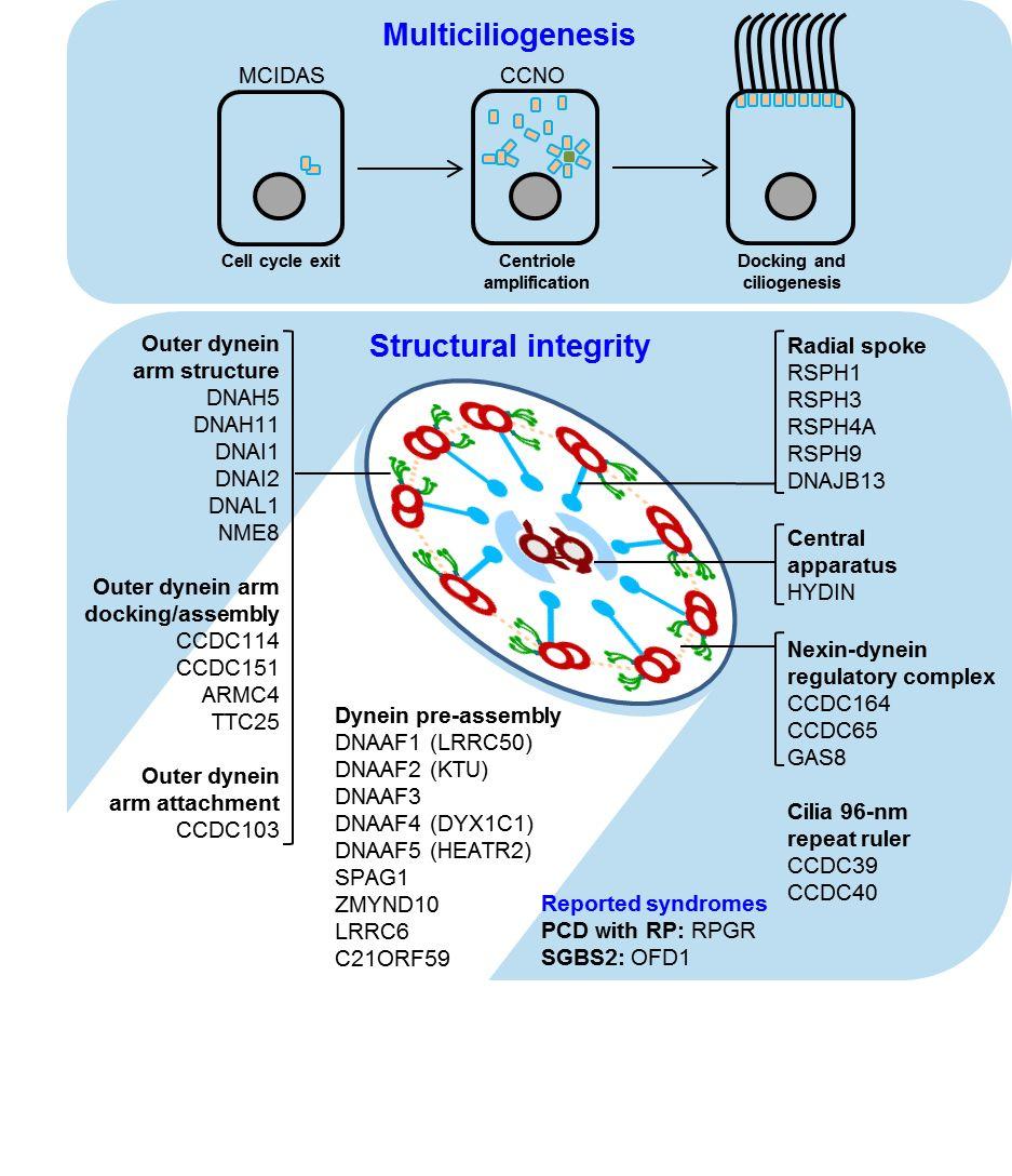
48

49

50
CilioPathy panel. 3-Jul-2018 (102 genen) Centrum voor Medische Genetica Gent. versie. OMIM gene ID
 Centrum voor Medische Genetica Gent. versie. OMIM gene ID") versie 3-Jul-2018 (102 genen) CilioPathy panel Centrum voor Medische Genetica Gent Gene OMIM gene ID Associated phenotype, OMIM phenotype ID, phenotype mapping key and inheritance pattern AHI1 608894 Joubert
versie 3-Jul-2018 (102 genen) CilioPathy panel Centrum voor Medische Genetica Gent Gene OMIM gene ID Associated phenotype, OMIM phenotype ID, phenotype mapping key and inheritance pattern AHI1 608894 Joubert
Utilization of the MiSeq in a clinical lab. Tony Krentz, PhD PreventionGenetics
 Utilization of the MiSeq in a clinical lab Tony Krentz, PhD PreventionGenetics PreventionGenetics Founded in 2004 in Marshfield, Wisconsin by James Weber ~90 employees Largest test menu in US Vision: Disease
Tubulointerstitial Renal Disease. Anna Vinnikova, MD Division of Nephrology
 Tubulointerstitial Renal Disease Anna Vinnikova, MD Division of Nephrology Part I: Cystic Renal Disease www.pathguy.com Simple cysts Simple cysts May be multiple Usually 1 5cm, may be bigger Translucent,
Anastasia Konstantinidou
 XXXVIIth IPPA Advanced Course 2015 Fontainebleau, France CILIOPATHIES and SKELETAL DYSPLASIA Anastasia Konstantinidou National University of Athens, Greece Birmingham Women s Hospital, U.K. Ciliary Skeletal
Renal Cystic Disease. Dr H Bierman
 Renal Cystic Disease Dr H Bierman Objectives Be able to diagnose renal cystic disease Genetic / non-genetic Be able to describe patterns of various renal cystic disease on routine imaging studies Be able
Centrosomes and Cilia
 Centrosomes and Cilia SMC6052/BIM6028/EXMD604 William Y. Tsang Research Unit Director, Cell Division and Centrosome Biology, IRCM Chercheur Adjoint, Faculté de medécine, Université de Montréal Adjunct
ANALYSIS OF THE CILIARY GENES GAS8 AND MKS6 REVEAL CONSERVED ROLES IN CILIA MOTILITY AND TRANSITION ZONE FUNCTION. by: WESLEY R.
 ANALYSIS OF THE CILIARY GENES GAS8 AND MKS6 REVEAL CONSERVED ROLES IN CILIA MOTILITY AND TRANSITION ZONE FUNCTION by: WESLEY R. LEWIS JAMES F. COLLAWN: COMMITTEE CHAIR BRADLEY K. YODER: MENTOR JOHN M.
a) They are the most common cause of pediatric kidney failure. b) They are always symptomatic. c) They can be asymmetric.
 They are the most common cause of pediatric kidney failure. b) They are always symptomatic. c) They can be asymmetric.") Practice questions: 1. The paraxial mesoderm gives rise to somites. The structure of the somite a) is a loose mesenchymal sheet that will migrate toward the notochord. b) is an epithelial rosette with
Practice questions: 1. The paraxial mesoderm gives rise to somites. The structure of the somite a) is a loose mesenchymal sheet that will migrate toward the notochord. b) is an epithelial rosette with
Primary Ciliary Dyskinesia Clinical Presentation and Diagnosis. Douglas Conrad California Thoracic Society January, 2018
 Primary Ciliary Dyskinesia Clinical Presentation and Diagnosis Douglas Conrad California Thoracic Society January, 2018 Disclosures Current Funding: NIH RO1 National CF Foundation Gilead Sciences Potential
Cystic Renal Disease, for USMLE Step One. Howard J. Sachs, MD
 Cystic Renal Disease, for USMLE Step One Howard J. Sachs, MD www.12daysinmarch.com The Major Players Medullary Sponge Kidney (MSK) Polycystic Kidney Disease (PKD) Autosomal Recessive: Childhood Autosomal
Case Report Joubert Syndrome Presenting with Motor Delay and Oculomotor Apraxia
 Case Reports in Pediatrics Volume 2011, Article ID 262641, 5 pages doi:10.1155/2011/262641 Case Report Joubert Syndrome Presenting with Motor Delay and Oculomotor Apraxia Harjinder Gill, 1 Brinda Muthusamy,
Axis Formation and Mesoderm Induction
 Developmental Biology Biology 4361 Axis Formation and Mesoderm Induction October 27, 2005 Amphibian anteroposterior specification polarized eggs animal/vegetal pigment yolk v. clear cytoplasm mitochondrial
Histology = the study of tissues. Tissue = a complex of cells that have a common function
 { EPITHELIAL TISSUE Histology = the study of tissues Tissue = a complex of cells that have a common function The Four Primary Tissue Types: Epithelium (epithelial tissue) covers body surfaces, lines body
Epithelium-1. Hanan Jafar BDS.MSc.PhD
 Epithelium-1 Hanan Jafar BDS.MSc.PhD General features Epithelium is an avascular tissue composed of cells that cover the exterior body surfaces and line internal closed cavities and tubes. It also forms
Structures in Cells. Lecture 5, EH1008: Biology for Public Health, Biomolecules.
 Structures in Cells Lecture 5, EH1008: Biology for Public Health, Biomolecules Limian.zheng@ucc.ie 1 Cytoplasm Nucleus Centrioles Cytoskeleton Cilia Microvilli 2 Cytoplasm Cellular material outside nucleus
Genetics in Nephrology. Saeid Morovvati Associate Professor of BMSU Director of Biogene Laboratory
 Genetics in Nephrology Saeid Morovvati Associate Professor of BMSU Director of Biogene Laboratory Genetics in: A. Congenital Anomalies of the Kidney and Urinary Tract B. Cystic Diseases of the Kidney C.
DIAGNOSIS OF PRIMARY CILIARY DYSKINESIA
 DIAGNOSIS OF PRIMARY CILIARY DYSKINESIA Kris De Boeck, MD PHD Pediatric Pulmonology, University of Leuven, Belgium Introduction Cause Congenital dysfunction of motile cilia Absence/dysfunction of one of
Human height. Length of some nerve and muscle cells. Chicken egg. Frog egg. Most plant and animal cells Nucleus Most bacteria Mitochondrion
 10 m 1 m 0.1 m 1 cm Human height Length of some nerve and muscle cells Chicken egg Unaided eye 1 mm Frog egg 100 µm 10 µm 1 µm 100 nm 10 nm Most plant and animal cells Nucleus Most bacteria Mitochondrion
By Sally Pobojewski Photos by Scott Galvin. Secrets of the. Cilia. { long-overlooked organelles hold some hefty keys to human health }
 By Sally Pobojewski Photos by Scott Galvin Secrets of the Cilia { long-overlooked organelles hold some hefty keys to human health } Photo: Dr. G. Moscoso/Photo Researchers, Inc. 20 Fall 2007 Medicine at
Primary Ciliary Dyskinesia (PCD) Angelo Barbato Department of Pediatrics Padova University/General Hospital Padova-Italy
 Angelo Barbato Department of Pediatrics Padova University/General Hospital Padova-Italy") Primary Ciliary Dyskinesia (PCD) Angelo Barbato Department of Pediatrics Padova University/General Hospital Padova-Italy barbato@pediatria.unipd.it PCD IS A RARE DISEASE THAT BEGINS IN CHILDHOOD AND CONTINUES
Primary Ciliary Dyskinesia (PCD) Angelo Barbato Department of Pediatrics Padova University/General Hospital Padova-Italy barbato@pediatria.unipd.it PCD IS A RARE DISEASE THAT BEGINS IN CHILDHOOD AND CONTINUES
Primary Ciliary Dyskinesia Pathogenesis, diagnosis and treatment DR CLAIRE HOGG DEPT. RESPIRATORY PAEDIATRICS ROYAL BROMPTON HOSPITAL LONDON.
 Primary Ciliary Dyskinesia Pathogenesis, diagnosis and treatment DR CLAIRE HOGG DEPT. RESPIRATORY PAEDIATRICS ROYAL BROMPTON HOSPITAL LONDON. Pathogenesis of PCD Autosomal recessive Heterogeneous Defect
(a) TEM of a plasma. Fimbriae. Nucleoid. Ribosomes. Plasma membrane. Cell wall Capsule. Bacterial chromosome
 TEM of a plasma. Fimbriae. Nucleoid. Ribosomes. Plasma membrane. Cell wall Capsule. Bacterial chromosome") 0 m m 0. m cm mm 00 µm 0 µm 00 nm 0 nm Human height Length of some nerve and muscle cells Chicken egg Frog egg Most plant and animal cells Most bacteria Smallest bacteria Viruses Proteins Unaided eye Light
0 m m 0. m cm mm 00 µm 0 µm 00 nm 0 nm Human height Length of some nerve and muscle cells Chicken egg Frog egg Most plant and animal cells Most bacteria Smallest bacteria Viruses Proteins Unaided eye Light
Structures in Cells. Cytoplasm. Lecture 5, EH1008: Biology for Public Health, Biomolecules
 Structures in Cells Lecture 5, EH1008: Biology for Public Health, Biomolecules Limian.zheng@ucc.ie 1 Cytoplasm Nucleus Centrioles Cytoskeleton Cilia Microvilli 2 Cytoplasm Cellular material outside nucleus
DISEASES WITH ABNORMAL MATRIX
 DISEASES WITH ABNORMAL MATRIX MSK-1 FOR 2 ND YEAR MEDICAL STUDENTS Dr. Nisreen Abu Shahin CONGENITAL DISEASES WITH ABNORMAL MATRIX OSTEOGENESIS IMPERFECTA (OI): also known as "brittle bone disease" a group
Department of Cognitive Science UCSD
 Department of Cognitive Science UCSD Verse 1: Neocortex, frontal lobe, Brain stem, brain stem, Hippocampus, neural node, Right hemisphere, Pons and cortex visual, Brain stem, brain stem, Sylvian fissure,
Cells. 1. Smallest living structures. 2. Basic structural and functional units of the body. 3. Derived from pre-existing cells. 4. Homeostasis.
 Cells The Cell The human body has about 75 trillion cells All tissues and organs are made up of cells Smallest functional unit of life Cytology Histology Cytology Epithelial cells Fibroblasts Erythrocytes
CNS Embryology 5th Menstrual Week (Dorsal View)
") Imaging of the Fetal Brain; Normal & Abnormal Alfred Abuhamad, M.D. Eastern Virginia Medical School CNS Embryology 5th Menstrual Week (Dorsal View) Day 20 from fertilization Neural plate formed in ectoderm
Imaging of the Fetal Brain; Normal & Abnormal Alfred Abuhamad, M.D. Eastern Virginia Medical School CNS Embryology 5th Menstrual Week (Dorsal View) Day 20 from fertilization Neural plate formed in ectoderm
NIH Public Access Author Manuscript Kidney Int. Author manuscript; available in PMC 2011 September 1.
 NIH Public Access Author Manuscript Published in final edited form as: Kidney Int. 2011 March ; 79(6): 691 692. doi:10.1038/ki.2010.514. The case: Familial occurrence of retinitis pigmentosa, deafness
Joubert syndrome: congenital cerebellar ataxia with the molar tooth
 Joubert syndrome: congenital cerebellar ataxia with the molar tooth Marta Romani, Alessia Micalizzi, Enza Maria Valente Lancet Neurol 2013; 12: 894 905 Published Online July 17, 2013 http://dx.doi.org/10.1016/
Nicholas Katsanis, Ph.D.
 Ciliopathies and Oligogenic Phenomena Prof. Center for Human Disease Modeling Duke University 1 Some key questions in human genetics What variants cause disease? What variants are associated with disease
Dysmorphology And The Paediatric Eye. Jill Clayton-Smith Manchester Centre For Genomic Medicine
 Dysmorphology And The Paediatric Eye Jill Clayton-Smith Manchester Centre For Genomic Medicine Why Make A Syndrome Diagnosis? Why did it happen? What does the future hold? How can you treat/manage it?
MULTIPLE CHOICE. Choose the one alternative that best completes the statement or answers the question.
 Exam Name MULTIPLE CHOICE. Choose the one alternative that best completes the statement or answers the question. 1) All of the following are synthesized along various sites of the endoplasmic reticulum
Test Information Sheet
 Prenatal Joubert Syndrome and Related Disorders (JSRD) Panel Sequence Analysis and Exon-Level Deletion/Duplication Testing of 29 Genes Panel Gene List: AHI1, ARL13B, B9D1, B9D2, C5orf42, CC2D2A, CEP41,
Epithelial Tissue. Functions include: 1. Protection 4. Absorption 2. Secretion 5. Filtration 3. Sensory reception
 Tissues There are 4 primary tissue types in the human body: 1. Epithelial (covering/lining) 2. Connective (support) 3. Muscle (movement) 4. Nervous (control) Epithelium Epithelial Tissue Covers the surface
Next Generation Sequencing Panel for Joubert and Meckel Gruber Syndrome
 Next Generation Sequencing Panel for Joubert and Meckel Gruber Syndrome Joubert syndrome Joubert syndrome (JBTS) is characterized by hypotonia, oculomotor apraxia, nystagmus, and intellectual disability.
Development of the Nervous System 1 st month
 Development of the Nervous System 1 st month day 1 - fertilization of egg day 6 - uterine implantation day 18 - trilaminar (3-layered) disc (blastoderm, embryo) ectoderm (dorsal) - nervous system and skin
High prevalence of Primary Ciliary Dyskinesia in a British Asian population
 High prevalence of Primary Ciliary Dyskinesia in a British Asian population Chris O Callaghan 1, Phil Chetcuti 2, Eduardo Moya 3 1. University of Leicester, Leicester, United Kingdom 2. Leeds General Infirmary,
Development of the Pharyngeal Arches
 Development of the Pharyngeal Arches Thomas A. Marino, Ph.D. Temple University School of Medicine Competencies: Upon completion of this section of the course, the student must be able to: 1. Recall the
CHAPTER II PDL 101 HUMAN ANATOMY & PHYSIOLOGY. Ms. K. GOWRI. M.Pharm., Lecturer.
 CHAPTER II PDL 101 HUMAN ANATOMY & PHYSIOLOGY Ms. K. GOWRI. M.Pharm., Lecturer. Structure of cell: Human body develops from a single cell zygote which results from fusion of the ovum andd the spermatozoan.
Inner ear development Nervous system development
 Upcoming Sessions April 22: Nervous System Development Lecture April 24: Reviews of Axonal Pathfinding in Sensory Systems April 29: Inner Ear Development Lecture May 1: May 6: May 8: Auditory System Pathfinding
Primary Ciliary Dyskinesia (PCD) Angelo Barbato Department of Pediatrics Padova University/General Hospital Padova-Italy
 Angelo Barbato Department of Pediatrics Padova University/General Hospital Padova-Italy") Thank you for viewing this presentation. We would like to remind you that this material is the property of the author. It is provided to you by the ERS for your personal use only, as submitted by the author.
Thank you for viewing this presentation. We would like to remind you that this material is the property of the author. It is provided to you by the ERS for your personal use only, as submitted by the author.
Neuroanatomy. Assistant Professor of Anatomy Faculty of Medicine The University of Jordan Dr Maha ELBeltagy
 Neuroanatomy Dr. Maha ELBeltagy Assistant Professor of Anatomy Faculty of Medicine The University of Jordan 2018 Development of the Central Nervous System Development of the nervous system Development
MIDTERM EXAM 1 COGNITIVE SCIENCE 107A
 MIDTERM EXAM 1 COGNITIVE SCIENCE 107A FALL 2011 Name: Points: / 100 PID: I. SHORT ANSWERS (6 points each for a total of 30 points) 1. Describe two contributions made by Ramon y Cajal (1852-1934) in terms
Cell Communication and Cell Signaling
 Cell Communication and Cell Signaling Why is cell signaling important? Why is cell signaling important? Allows cells to communicate and coordinate functions/activities of the organism Usually involves
Situs inversus. Dr praveena pulmonology- final year post graduate
 Situs inversus Dr praveena pulmonology- final year post graduate Definiton History Types Cause Clinical features Diagnosis Treatment Definition The term situs inversus is a short form of the latin phrase
Cellular Physiology (PHSI3009) Contents:
 Contents:") Cellular Physiology (PHSI3009) Contents: Cell membranes and communication 2 nd messenger systems G-coupled protein signalling Calcium signalling Small G-protein signalling o RAS o MAPK o PI3K RHO GTPases
Cellular Physiology (PHSI3009) Contents: Cell membranes and communication 2 nd messenger systems G-coupled protein signalling Calcium signalling Small G-protein signalling o RAS o MAPK o PI3K RHO GTPases
Neuropathology Specialty Conference
 Neuropathology Specialty Conference March 22, 2010 Case 2 Rebecca Folkerth, MD Brigham and Women s Hospital Children s Hospital Harvard Medical School Clinical History 18-gestational-week fetus found on
Ciliary Biology: Understanding the Cellular and Genetic Basis of Human Ciliopathies
 A R T I C L E American Journal of Medical Genetics Part C (Seminars in Medical Genetics) 151C:263 280 (2009) Ciliary Biology: Understanding the Cellular and Genetic Basis of Human Ciliopathies MAGDALENA
Lecture 6 9/17 Dr. Hirsh Organization of Cells, continued
 Cell structure of Eukaryotic cells Lecture 6 9/17 Dr. Hirsh Organization of Cells, continued Lots of double-membraned organelles Existence of an Endo-membrane system separation of areas of cell, transport
ORGANELLES OF THE ENDOMEMBRANE SYSTEM
 Membranes compartmentalize the interior of the cell and facilitate a variety of metabolic activities. Chloroplasts and a rigid cell wall are what distinguish a plant cell from an animal cell. A typical
Cellular components of CNS
 Cellular components of CNS Cellular components of CNS Neurons Glial cells: Astrocytes (including radial glia), oligodendrocytes, microglia, ependymal cells Epithelial cells of choroid plexus Endothelial
Central Nervous System (CNS) -> brain and spinal cord. Major Divisions of the nervous system:
 -> brain and spinal cord. Major Divisions of the nervous system:") Central Nervous System (CNS) -> brain and spinal cord Major Divisions of the nervous system: Afferent (sensory input) -> cell bodies outside of the central nervous system (CNS), carry info into the CNS
Central Nervous System (CNS) -> brain and spinal cord Major Divisions of the nervous system: Afferent (sensory input) -> cell bodies outside of the central nervous system (CNS), carry info into the CNS
Cell Birth and Death. Chapter Three
 Cell Birth and Death Chapter Three Neurogenesis All neurons and glial cells begin in the neural tube Differentiated into neurons rather than ectoderm based on factors we have already discussed If these
Biology 218 Human Anatomy
 Chapter 17 Adapted form Tortora 10 th ed. LECTURE OUTLINE A. Overview of the Nervous System (p. 537) 1. The nervous system and the endocrine system are the body s major control and integrating centers.
A Primer to Cystic Kidney Diseases and Ciliopathies
 A Primer to Cystic Kidney Diseases and Ciliopathies ERKNet Webinar Max Liebau 04.09.2018 Liebau Pediatric Nephrology, Center for chronically ill children, Center for Molecular Medicine WG CAKUT and Ciliopathies
Posterior fossa malformations
 ANDREA ROSSI, MD Head, Department of Pediatric Neuroradiology G. Gaslini Children s Research Hospital Genoa Italy andrearossi@ospedale-gaslini.ge.it Posterior fossa malformations Cerebellar ataxia Hypotonia
Neurodevelopment II Structure Formation. Reading: BCP Chapter 23
 Neurodevelopment II Structure Formation Reading: BCP Chapter 23 Phases of Development Ovum + Sperm = Zygote Cell division (multiplication) Neurogenesis Induction of the neural plate Neural proliferation
cell movement and neuronal migration
 cell movement and neuronal migration Paul Letourneau letou001@umn.edu Chapter 16; The Cytoskeleton; Molecular Biology of the Cell, Alberts et al. 1 Cell migration Cell migration in 3 steps; protrusion,
STEIN IN-TERM EXAM -- BIOLOGY APRIL 19, PAGE
 STEIN IN-TERM EXAM -- BIOLOGY 3058 -- APRIL 19, 2018 -- PAGE 1 of 9 There are 25 questions in this Biology 3058 exam. All questions are "A, B, C, D, E, F, G, H" questions worth one point each. There is
Muscle and Neuromuscular Junction. Peter Takizawa Department of Cell Biology
 Muscle and Neuromuscular Junction Peter Takizawa Department of Cell Biology Types and structure of muscle cells Structural basis of contraction Triggering muscle contraction Skeletal muscle consists of
Evaluation of Patients with Diffuse Bronchiectasis
 Evaluation of Patients with Diffuse Bronchiectasis Dr. Patricia Eshaghian, MD Assistant Clinical Professor of Medicine Director, UCLA Adult Cystic Fibrosis Affiliate Program UCLA Division of Pulmonary
Cystic Renal Disease of Childhood
 Acta Radiológica Portuguesa, Vol.XIX, nº 74, pág. 90-107, Abr.-Jun., 2007 Cystic Renal Disease of Childhood Ellen Chung Armed Forces Institute of Pathology Terminology Cyst Polycystic kidney disease ARPKD
Analysis on the mechanism of reduced nephron number and the pathological progression of chronic renal failure in Astrin deficient rats
 Analysis on the mechanism of reduced nephron number and the pathological progression of chronic renal failure in Astrin deficient rats Summary of Doctoral Thesis Hidenori Yasuda Graduate School of Veterinary
STEIN IN-TERM EXAM -- BIOLOGY APRIL 18, PAGE
 STEIN IN-TERM EXAM -- BIOLOGY 3058 -- APRIL 18, 2019 -- PAGE 1 of 9 There are 25 questions in this Biology 3058 exam. All questions are "A, B, C, D, E, F, G, H" questions worth one point each. There is
The global epidemic of obesity
 The global epidemic of obesity Obesity is a genetic disorder Heterogeneity of the genetics of obesity ENVIRONMENT GENE MONOGENIC FORMS -> Early-onset, severe obesity -> Syndromic forms of obesity POLYGENIC
Systematizing in vivo modeling of pediatric disorders
 Systematizing in vivo modeling of pediatric disorders Nicholas Katsanis, Ph.D. Duke University Medical Center Center for Human Disease Modeling Rescindo Therapeutics www.dukegenes.org Task Force for
Joubert syndrome: report of 11 cases
 The Turkish Journal of Pediatrics 2012; 54: 605-611 Original Joubert syndrome: report of 11 cases Faruk İncecik 1, M. Özlem Hergüner 1, Şakir Altunbaşak 1, Joseph G. Gleeson 2 1 Department of Pediatric
Radiological and pathologic findings of fetal renal cystic diseases and associated fetal syndromes: A pictorial review
 Radiological and pathologic findings of fetal renal cystic diseases and associated fetal syndromes: A pictorial review Poster No.: C-2835 Congress: ECR 2010 Type: Educational Exhibit Topic: Pediatric Authors:
V. CENTRAL NERVOUS SYSTEM TRAUMA
 V. CENTRAL NERVOUS SYSTEM TRAUMA I. Concussion - Is a clinical syndrome of altered consiousness secondary to head injury - Brought by a change in the momentum of the head when a moving head suddenly arrested
Protein Trafficking in the Secretory and Endocytic Pathways
 Protein Trafficking in the Secretory and Endocytic Pathways The compartmentalization of eukaryotic cells has considerable functional advantages for the cell, but requires elaborate mechanisms to ensure
CYSTIC DISEASES of THE KIDNEY. Dr. Nisreen Abu Shahin
 CYSTIC DISEASES of THE KIDNEY Dr. Nisreen Abu Shahin 1 Types of cysts 1-Simple Cysts 2-Dialysis-associated acquired cysts 3-Autosomal Dominant (Adult) Polycystic Kidney Disease 4-Autosomal Recessive (Childhood)
SWISS SOCIETY OF NEONATOLOGY. Kartagener s syndrome: Neonatal respiratory distress as initial symptom of primary ciliary dyskinesia
 SWISS SOCIETY OF NEONATOLOGY Kartagener s syndrome: Neonatal respiratory distress as initial symptom of primary ciliary dyskinesia MAY 2005 2 Christen M, and Berger TM, Neonatal and Pediatric Intensive
Muscle Tissue. General concepts. Classification of muscle. I. Functional classification is based on the type of neural control.
 Muscle Tissue LEARNING OBJECTIVES 1. Identify the three types of muscle tissue at the light microscopic level. 2. List and compare the structural and functional features of each of the three muscle fiber
CASE REPORT. 1. Assistant Professor. Department of Paediatrics, Vinayaka Missions Medical College, Karaikal
 A CASE OF KARTAGENER SYNDROME Pagadpally Srinivas 1. Assistant Professor. Department of Paediatrics, Vinayaka Missions Medical College, Karaikal CORRESPONDING AUTHOR: Pagadpally Srinivas, 72, Vellai Pillaiyar
Polarity and Segmentation. Chapter Two
 Polarity and Segmentation Chapter Two Polarization Entire body plan is polarized One end is different than the other Head vs. Tail Anterior vs. Posterior Front vs. Back Ventral vs. Dorsal Majority of neural
Supplementary Online Content
 Supplementary Online Content Honein MA, Dawson AL, Petersen E, et al; US Zika Pregnancy Registry Collaboration. Birth Defects Among Fetuses and Infants of US Women With Laboratory Evidence of Possible
Development: is the growth of an individual organism from a simple to a more complex or mature level. A slow process of progressive change
 1. Define the following terms (use your own words): development, growth, differentiation, histogenesis, organogenesis, morphogenesis, reproduction, tissue, organ, organ system, and organism. Development:
Central nervous system (CNS): brain and spinal cord Collections of cell body and dendrites (grey matter) are called nuclei/nucleus Nucleus can also
: brain and spinal cord Collections of cell body and dendrites (grey matter) are called nuclei/nucleus Nucleus can also") Chapter 3 Part 1 Orientation Directions in the nervous system are described relatively to the neuraxis An imaginary line drawn through the center of the length of the central nervous system, from the bottom
Chapter 3 Part 1 Orientation Directions in the nervous system are described relatively to the neuraxis An imaginary line drawn through the center of the length of the central nervous system, from the bottom
Organelles of the Cell & How They Work Together. Packet #5
 Organelles of the Cell & How They Work Together Packet #5 Developed by Mr. Barrow 2018 1 Introduction Organization of cells is basically similar in all cells. Additionally, most cells are tiny Ranging
The Complexity of Simple Genetics
 The Complexity of Simple Genetics? The ciliopathies: a journey into variable penetrance and expressivity Bardet-Biedl Syndrome Allelism at a single locus is insufficient to explain phenotypic variability
BIOH111. o Cell Module o Tissue Module o Integumentary system o Skeletal system o Muscle system o Nervous system o Endocrine system
 BIOH111 o Cell Module o Tissue Module o Integumentary system o Skeletal system o Muscle system o Nervous system o Endocrine system Endeavour College of Natural Health endeavour.edu.au 1 TEXTBOOK AND REQUIRED/RECOMMENDED
Cell Structure Review
 Cell Structure Review By: Ms. Latner s Awesome Class Yeah Ms. Latner! Yeah Science! Human Organelles cell compartment that carries out a specific function. most organelles are membrane bound nucleus, golgi,
Development of the Nervous System. Leah Militello, class of 2018
 Development of the Nervous System Leah Militello, class of 2018 Learning Objectives 1. Describe the formation and fate of the neural tube and neural crest including timing and germ layer involved. 2. Describe
International Journal of Pharma and Bio Sciences. Meckel-Gruber Syndrome Associated with CNS Malformations A Case Report
 International Journal of Pharma and Bio Sciences RESEARCH ARTICLE PATHOLOGY Meckel-Gruber Syndrome Associated with CNS Malformations A Case Report Corresponding Author DR. N. HIMA BINDU Assistant Professor,
Organelles of the Cell & How They Work Together. Packet #7
 Organelles of the Cell & How They Work Together Packet #7 Introduction Introduction Organization of cells is basically similar in all cells. Additionally, most cells are tiny Ranging from 1 1000 cubic
Major Structures of the Nervous System. Brain, cranial nerves, spinal cord, spinal nerves, ganglia, enteric plexuses and sensory receptors
 Major Structures of the Nervous System Brain, cranial nerves, spinal cord, spinal nerves, ganglia, enteric plexuses and sensory receptors Nervous System Divisions Central Nervous System (CNS) consists
Title: Chapter 5 Recorded Lecture. Speaker: Amit Dhingra Created by: (remove if same as speaker) online.wsu.edu
 online.wsu.edu") Title: Chapter 5 Recorded Lecture Speaker: Title: What Anthony is the title Berger/Angela of this lecture? Williams Speaker: Amit Dhingra Created by: (remove if same as speaker) online.wsu.edu Chapter
Title: Chapter 5 Recorded Lecture Speaker: Title: What Anthony is the title Berger/Angela of this lecture? Williams Speaker: Amit Dhingra Created by: (remove if same as speaker) online.wsu.edu Chapter
Biology Developmental Biology Spring Quarter Midterm 1 Version A
 Biology 411 - Developmental Biology Spring Quarter 2013 Midterm 1 Version A 75 Total Points Open Book Choose 15 out the 20 questions to answer (5 pts each). Only the first 15 questions that are answered
A study of cilia density in sea urchin embryos during gastrulation
 A study of cilia density in sea urchin embryos during gastrulation Steve Das Independent Research Project Report Bio 254 Developmental Biology May 3, 2012 Introduction Embryo growth and development is
Chapter 11 Development and Diseases
 Chapter 11 Development and Diseases Fertilization Development of the Nervous System Cleavage (Blastula, Gastrula) Neuronal Induction- Neuroblast Formation Cell Migration Axon Growth/Target innervation
Development of the Liver and Pancreas
 Development of the Liver and Pancreas Professor Alfred Cuschieri Department of Anatomy University of Malta Three glandular buds arise from the distal end of the foregut during the fourth week Day 22 -The
Ciliopathies 00-Kenny-Prelims.indd i 22/07/13 11:15 AM
 Ciliopathies This page intentionally left blank Ciliopathies A reference for clinicians Edited by Thomas D. Kenny National Institute for Health Research Evaluations, Trials and Studies Coordinating Centre,
Human Histology The Nervous System. Dr. Rawaa Salim Hameed
 Human Histology The Nervous System Dr. Rawaa Salim Hameed The organization of the nervous system Anatomically, the nervous system is divided into:- Neurohistology Structurally, nerve tissue consists of
Review from Biology A
 Chapter 4 Review from Biology A The Cell Theory All organisms are made of cells Cells come from pre-existing cells The cell is the simplest collection of matter that can live Scientists whose work you
Clinical Commissioning Policy Proposition: Dornase alfa inhaled therapy for primary ciliary dyskinesia (all ages)
") Clinical Commissioning Policy Proposition: Dornase alfa inhaled therapy for primary ciliary dyskinesia (all ages) Reference: NHS England E03X05/01 Information Reader Box (IRB) to be inserted on inside
Clinical Commissioning Policy Proposition: Dornase alfa inhaled therapy for primary ciliary dyskinesia (all ages) Reference: NHS England E03X05/01 Information Reader Box (IRB) to be inserted on inside
Announcement. Danny to schedule a time if you are interested.
 Announcement If you need more experiments to participate in, contact Danny Sanchez (dsanchez@ucsd.edu) make sure to tell him that you are from LIGN171, so he will let me know about your credit (1 point).
18. PANCREATIC FUNCTION AND METABOLISM. Pancreatic secretions ISLETS OF LANGERHANS. Insulin
 18. PANCREATIC FUNCTION AND METABOLISM ISLETS OF LANGERHANS Some pancreatic functions have already been discussed in the digestion section. In this one, the emphasis will be placed on the endocrine function
Collin County Community College BIOL Week 5. Nervous System. Nervous System
 Collin County Community College BIOL 2401 Week 5 Nervous System 1 Nervous System The process of homeostasis makes sure that the activities that occur in the body are maintained within normal physiological
Brain Meninges, Ventricles and CSF
 Brain Meninges, Ventricles and CSF Lecture Objectives Describe the arrangement of the meninges and their relationship to brain and spinal cord. Explain the occurrence of epidural, subdural and subarachnoid
Zool 3200: Cell Biology Exam 4 Part I 2/3/15
 Name: Key Trask Zool 3200: Cell Biology Exam 4 Part I 2/3/15 Answer each of the following questions in the space provided, explaining your answers when asked to do so; circle the correct answer or answers