The Abdomen. Surface Anatomy, Vessels, Muscles, and Peritoneum
|
|
|
- Shon Thomas
- 5 years ago
- Views:
Transcription
1 The Abdomen Surface Anatomy, Vessels, Muscles, and Peritoneum
 Tendinous line Extends from xiphoid process to pubic")
2 Surface Anatomy Anterior abdominal wall extends from costal margin to inferior boundaries: Iliac crest Anterior superior iliac spine Inguinal ligament Pubic crest Superior boundary Diaphragm Central landmark Umbilicus Linea alba (white line) Tendinous line Extends from xiphoid process to pubic symphysis

3 Abdominal Quadrants 9 regions 4 quadrants Draw line through navel Right upper quadrant Left upper quadrant Left lower quadrant Right lower quadrant
4 Muscles Function: Help contain abdominal organs Move trunk Forced breathing Increase intraabdominal pressure Abdominal wall Anterior (4) Innervated by intercostal nerves Continuous with layers of intercostal muscles Fibers of layers run in different directions for strength Ends in aponeurosis which contains rectus abdominis muscle Posterior (3)
5
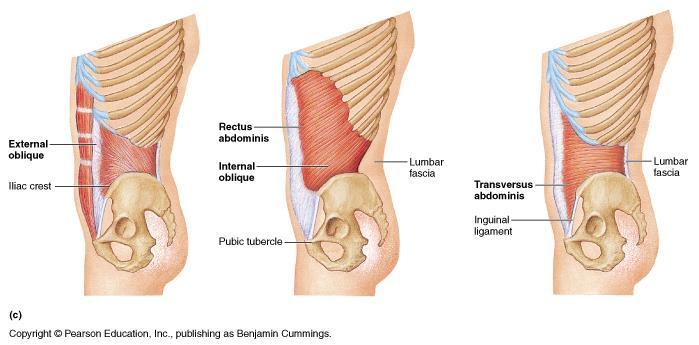
6 Anterior Abdominal Wall Muscles Rectus Abdominis Origin Pubic crest, symphysis Insertion Xiphoid process, costal cartilages of ribs 5-7 Function Flex, rotate trunk, fix and depress ribs, stabilize pelvis, compress abdomen
7 Anterior Abdominal Wall External oblique ( hands-in-pocket ) Origin Lower 8 ribs Insertion Aponeurosis to linea alba, pubic and iliac crest Function Flex trunk, compress abdominal wall (together), rotate trunk (separate sides) Internal oblique Origin Lumbar fascia, iliac crest, inguinal ligament Insertion Linea alba, pubic crest, last 3-4 ribs, costal margin Function Same for external obliques

8 Transversus abdominis Origin Inguinal ligament, lumbar fascia, cartilage of last 6 ribs, iliac crest Insertion Linea alba, pubic crest Function Compress abdominal contents
9 Levels of Rectus Abdominis Muscle Sheath Structures
")
10 ANTERIOR ABDOMINAL WALL Cord of connective tissue LINEA ALBA Extend sternum (xyphoid process) symphysis, pubic bones Aponeurotic parts of oblique muscles attache to the linea alba at the midline One of the surgical approaches to the peritoneal cavity (midline incision)

11 Weak Places of the Anterior Abdominal Wall


12 Layer Structure of the Anterior Abdominal Wall
13 Abdominal Incisions Must locate nearest to the organ. Must have sufficient length for surgeon activities. Must be atraumatic.

14 Skin Incisions of the Ventral Abdominal Wall

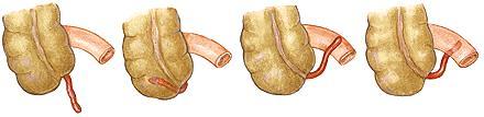
15 Male Inguinal Canal

16 Female Inguinal Canal
17 Superficial Inguinal Ring

18 Internal Inguinal Ring

19 Anterior Abdominal Wall (lower internal view)

20 Posterior Abdominal Wall Iliopsoas Psoas major Origin Lumbar vertebrae, T 12 Insertion Lesser trochanter of femur via iliopsoas tendon Function Thigh flexion, trunk flexion, lateral flexion Innervation Ventral rami L1-L3 Iliacus Origin Iliac fossa, ala of sacrum Insertion Lesser trochanter of femur via iliopsoas tendon Function Thigh flexion, trunk flexion Innervation Femoral nerve (L2 and L3) Psoas minor variable (40-60% do not have)

21 Posterior Abdominal Wall Quadratus lumborum Origin Iliac crest and lumbar fascia Insertion Transverse process of upper lumbar vertebrae, lower margin of rib 12 Function Flex vertebral column, maintains upright posture, assists in inspiration Innervation: T12 and upper lumbar spinal nerves (ventral rami)

22 Abdominopelvic Cavity Ventral body cavity Thoracic Abdominopelvic Abdominopelvic Abdominal Liver Stomach Kidneys Pelvic cavity Bladder Some reproductive organs Rectum


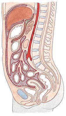
23 Abdominal cavity The space bounded by: Anterolateral abdominal wall Posterior abdominal wall Diaphragm Pelvic walls and pelvic floor. Subdivided into: True abdominal cavity (from diaphragm to linea terminalis) Pelvic cavity (below linea terminalis).
24 Peritoneum and peritoneal compartment Peritoneum is a continuous serous membrane, composed of two layers: Parietal peritoneum, lines abdominal and pelvic wall Visceral peritoneum, lines abdominal and pelvic organs. Peritoneal compartment is part of the abdominal cavity enclosed within the parietal peritoneum. Contains organs covered with peritoneum and peritoneal structures. Outside the parietal peritoneum is the extraperitoneal compartment of the abdominal cavity.
25 Peritoneal cavity Peritoneal cavity (PC) - the space between the two peritoneal layers, is a potential space, into which the organs are tightly packed against each other. PC contains thin layer of fluid, which lubricates the peritoneal surfaces and allows movement of the organs without friction. PC is closed in males, but communicates with the external environment in females through the uterine tubes, uterus and vagina. Peritoneum, peritoneal cavity and all the organs are situated in the abdominal cavity.

26 Development of the peritoneum
27 Relationship between the organs and peritoneum Due to intraembryonal processes the organs have different relationship with the peritoneum. 1. Intraperitoneal organs are entirely covered with peritoneum. They are connected to the abdominal wall with ligaments or meso, which ensures greater mobility. 2. Extraperitoneal organs are partially or entirely devoid of peritoneum. They are slightly movable or immovable. According to their position these are: а) retroperitoneal on the posterior abdominal wall b) subperitoneal in the lesser pelvis c) preperitoneal at the anterior abdominal wall.

28 Vertical layout of the peritoneum
29 Horizontal layout of the peritoneum

30 Passage of the parietal into visceral peritoneum
31 Peritoneal structures 1. Mesentery double peritoneal layer, representing elongation of the visceral peritoneum. М. connects the corresponding organ with the abdominal wall (e.g., mesentery of the small intestine). М. contains connective tissue in which are embedded blood vessels, nerves and lymph nodes. М. ensures mobility of the organs.
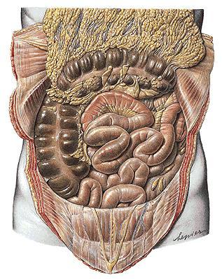
 connects the lesser curvature of the stomach and intitial portion of pars superior duodeni")
 descends from the greater curvature of the stomach and intitial portion of pars superior")
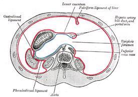
32 2. Omentum double layered structure of visceral peritoneal, extending from the stomach to neighbouring organs. Lesser omentum (оmentum minus) connects the lesser curvature of the stomach and intitial portion of pars superior duodeni with liver. Greater omentum (оmentum majus) descends from the greater curvature of the stomach and intitial portion of pars superior duodeni, covers the intestines, and then ascends back to attache to the transverse colon. Contains great amount of fat tissue.
. 4. Peritoneal folds (plicae) formed over underlying structures (e.g., plica iliocecalis superior, plica umbilicalis mediana). 5.")
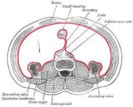
33 3. Peritoneal ligaments double layered structures of visceral peritoneum, between neighbouring organs or between organ and abdominal wall (e.g., lig. falciforme, lig. gastrophrenicum, lig. gastrolienale, lig. gastrocolicum). 4. Peritoneal folds (plicae) formed over underlying structures (e.g., plica iliocecalis superior, plica umbilicalis mediana). 5. Peritoneal recessuses spaces in the peritoneal cavity заградени between peritoneal structures and abdominal organs or abdominal wall (e.g., bursa omentalis, recessus subphrenicus, fossa retrocecalis).
34 Divisions of the peritoneal cavity By mesocolon transversum the peritoneal compartment divites into: 1. Supracolic compartment between diaphragm and mesocolon transversum with its mesentery. 2. Infracolic compartment - between mesocolon transversum and linea terminalis. 3. Pelvic compartment - below linea terminalis in the pelvi cavity.
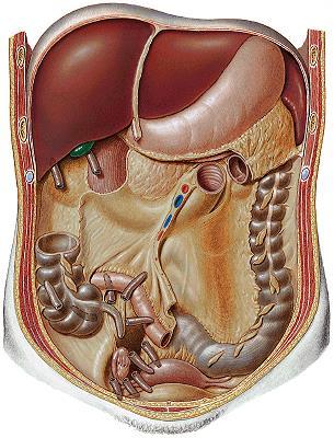
35 Supracolic compartment Organs: 1. Esophagus, pars abdominalis - intraperitoneal 2. Stomach - intraperitoneal 3. Liver - intraperitoneal 4. Gall bladder - intraperitoneal 5. Spleen - intraperitoneal


36 Supracolic compartment. Projections of organs
 area nuda 3. Lig.")
37 Supracolic compartment Peritoneal structures: 1. Lig. falciforme hepatis lig. teres hepatis 2. Lig. coronarium hepatis (dextum et sinistrum) area nuda 3. Lig. triangulare (dextum et sinistrum)

38 Supracolic compartment 4. Omentum minus lig. hepatogastricum lig. hepatoduodenale 5. Omentum majus lig. gastrocolicum lig. gastrolienale lig. gastrophrenicum 6. Lig. phrenicolienale

39 Supracolic compartment Peritoneal spaces: 1. Recessus subphrenicus dexter - bursa hepatica 2. Recessus subphrenicus sinister - bursa pregastrica 3. Perilienal space 4. Recessus subhepaticus а) anterior part b) posterior part - recessus hepatorenalis 5. Bursa omentalis


40 Supracolic compartment Bursa omentalis. Opened thru lig. hepatogastricum Bursa omentalis. Opened thru lig. gastrocolicum
41 Infracolic compartment Organs: 1. Small intestine duodenum (pars superior, descendens, horizontalis, ascendens) - retroperitoneal, pars superior intraperitoneal Jejunum and ileum - intraperitoneal 2. Large intestine cecum - intraperitoneal appendix vermiformis - intraperitoneal colon (ascendens, transversum, descendens, sigmoideum) - intraperitoneal /mesoperitoneal rectum most extraperitoneal

42 Organs and projections


43 Peritoneal structures 1. Omentum majus - pars libera 2. Mesenterium 3. Mesocolon transversum 4. Mesocolon sigmoideum 5. Mesoappendix


44 Peritoneal structures 1. Plicae duodenalis superior/inferior - recessus duodenalis superior/inferior 2. Plicae ileocecalis superior/inferior - recessus ileocecalis superior/inferior


45 Peritoneal spaces 1. Canalis lateralis dexter 2. Sinus mesentericus dexter 3. Sinus mesentericus sinister 4. Canalis lateralis sinister 5. Recessus intersigmoideus 6. Recessus retrocecalis
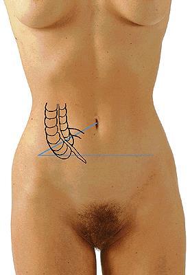
46 Appendix vermiformis

 - a. gastrica dextra 3. A.")
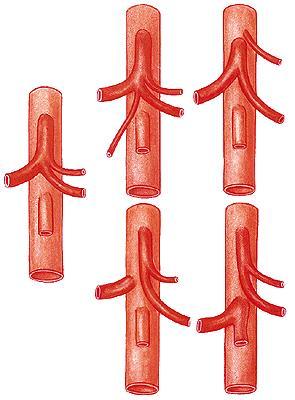
47 Supracolic compartment. Blood supply Truncus celiacus 1. A. gastrica sisnistra - r. esophageus 2. A. hepatica communis - a. hepatica propria - a. hepatica dextra/sinistra - a. gastroduodenalis - a. gastroepiploica dextra - aa. pancreaticoduodenales superiores (anterior/posterior) - a. gastrica dextra 3. A. lienalis - aa. gastricae breves - a. gastroepiploica sinistra
48 Supracolic compartment. Blood supply

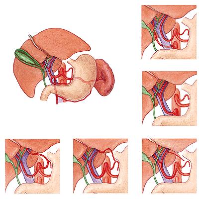
49 Arteriogram of truncus celiacus

 3. A. iliocolica 4. A. colica dextra 5.")
50 Infracolic compartment. Blood supply A. mesenterica superior 1. A. pancreaticoduodenalis inferior 2. Aa. intestinales (15-18) 3. A. iliocolica 4. A. colica dextra 5. A. colica media


51 Infracolic compartment. Blood supply A. mesenterica inferior 1. A. colica sinistra 2. Aa. sigmoideae (3-4) 3. A. rectalis superior
Abdomen: Introduction. Prof. Oluwadiya KS
 Abdomen: Introduction Prof. Oluwadiya KS www.oluwadiya.com Abdominopelvic Cavity Abdominal Cavity Pelvic Cavity Extends from the inferior margin of the thorax to the superior margin of the pelvis and the
ABDOMINAL WALL & RECTUS SHEATH
 ABDOMINAL WALL & RECTUS SHEATH Learning Objectives Describe the anatomy, innervation and functions of the muscles of the anterior, lateral and posterior abdominal walls. Discuss their functional relations
The posterior abdominal wall. Prof. Oluwadiya KS
 The posterior abdominal wall Prof. Oluwadiya KS www.oluwadiya.sitesled.com Posterior Abdominal Wall Lumbar vertebrae and discs. Muscles opsoas, quadratus lumborum, iliacus, transverse, abdominal wall
Biology Human Anatomy Abdominal and Pelvic Cavities
 Biology 351 - Human Anatomy Abdominal and Pelvic Cavities Please place your name and I.D. number on the back of the last page of this exam. You must answer all questions on this exam. Because statistics
Lab 9 Abdomen MUSCLES
 Lab 9 Abdomen MUSCLES External abdominal oblique continuous with the external intercostal muscle; its fibers point in a caudal direction as it moves anteriorly until it inserts on the linea alba via its
Anatomy of the Large Intestine
 Large intestine Anatomy of the Large Intestine 2 Large Intestine Extends from ileocecal valve to anus Length = 1.5-2.5m = 5 feet Regions Cecum = 2.5-3 inch Appendix= 3-5 inch Colon Ascending= 5 inch Transverse=
Digestive system (Systema digestorium/ alimentarium) Lecture #1
 Lecture #1") Digestive system (Systema digestorium/ alimentarium) Lecture #1 Internal organs are grouped into 1. System - Have the same functions and development Digestive system Respiratory system 2. Apparatus - Have
Digestive system (Systema digestorium/ alimentarium) Lecture #1 Internal organs are grouped into 1. System - Have the same functions and development Digestive system Respiratory system 2. Apparatus - Have
The peritoneum. Prof. Oluwadiya KS, MBBS, FMCS(Orthop) Website:
 Website:") The peritoneum Prof. Oluwadiya KS, MBBS, FMCS(Orthop) Website: http://oluwadiya.com The peritoneum Serous membrane that lines the abdominopelvic cavity and invests the viscera The largest serous membrane
The peritoneum Prof. Oluwadiya KS, MBBS, FMCS(Orthop) Website: http://oluwadiya.com The peritoneum Serous membrane that lines the abdominopelvic cavity and invests the viscera The largest serous membrane
Abdominal muscles. Subinguinal hiatus and ingiunal canal. Femoral and adductor canals. Neurovascular system of the lower limb. Sándor Katz M.D.,Ph.D.
 Abdominal muscles. Subinguinal hiatus and ingiunal canal. Femoral and adductor canals. Neurovascular system of the lower limb. Sándor Katz M.D.,Ph.D. External oblique muscle Origin: outer surface of the
Abdomen. Retroperitoneal space
 Abdomen. Retroperitoneal space Abdominal cavity The space bounded by: Anterolateral abdominal wall Posterior abdominal wall Diaphragm Pelvic walls and pelvic floor. Subdivided into: True abdominal cavity
The Human Body: An Overview of Anatomy. Anatomy. Physiology. Anatomy - Study of internal and external body structures
 C H A P T E R 1 The Human Body: An Orientation An Overview of Anatomy Anatomy The study of the structure of the human body Physiology The study of body function Anatomy - Study of internal and external
Exploring Anatomy: the Human Abdomen
 Exploring Anatomy: the Human Abdomen PERITONEUM AND PERITONEAL CAVITY PERITONEUM The peritoneum is a thin serous membrane that lines the abdominal cavity and covers, in variable amounts, the viscera within
Borders of the Abdomen
 Abdominal wall Borders of the Abdomen Abdomen is the region of the trunk that lies between the diaphragm above and the inlet of the pelvis below Borders Superior: Costal cartilages 7-12. Xiphoid process:
GI module Lecture: 9 د. عصام طارق. Objectives:
 GI module Lecture: 9 د. عصام طارق Objectives: To list structures forming posterior abdominal wall. To follow aorta & its main branches. To describe IVC & its main tributaries. To list nerves of posterior
BLOCK IV: OFFICIAL BODY PARTS LIST FOR ANTERIOR ABDOMINAL WALL AND ABDOMINAL CONTENTS
 BLOCK IV: OFFICIAL BODY PARTS LIST FOR ANTERIOR ABDOMINAL WALL AND ABDOMINAL CONTENTS External oblique muscle Muscular portion Aponeurotic portion Superficial inguinal ring Lateral (inferior) crus Medial
The Anterolateral Abdominal Wall By Prof. Dr. Muhammad Imran Qureshi
 1 P age The Anterolateral Abdominal Wall By Prof. Dr. Muhammad Imran Qureshi Introduction The abdomen is the region of the trunk located between the thorax and the pelvis. It includes the anterolateral
Netter's Anatomy Flash Cards Section 4 List 4 th Edition
 Netter's Anatomy Flash Cards Section 4 List 4 th Edition https://www.memrise.com/course/1577335/ Section 4 Abdomen (31 cards) Plate 4-1 Bony Framework of Abdomen 1.1 Costal cartilages 1.2 Iliac crest 1.3
Thoracolumbar Anatomy Eric Shamus Catherine Patla Objectives
 1 2 Thoracolumbar Anatomy Eric Shamus Catherine Patla Objectives List the muscular and ligamentous attachments of the thoracic and lumbar spine Describe how the muscles affect the spine and upper extremity
Biology Human Anatomy Abdominal and Pelvic Cavities
 Biology 351 - Human Anatomy Abdominal and Pelvic Cavities You must answer all questions on this exam. Because statistics demonstrate that, on average, between 2-5 questions on every 100-point exam are
Mousa Salah. Dr. Mohammad Al. Mohtasib. 1 P a g e
 8 Mousa Salah Dr. Mohammad Al. Mohtasib 1 P a g e In the previous lecture we talked about the peritoneum, and we said that the peritonium is a serous sac, and it consists of two layers, visceral and parietal.
Anatomy: Know Your Abdomen
 Anatomy: Know Your Abdomen Glossary Abdomen - part of the body below the thorax (chest cavity); separated by the diaphragm. Anterior - towards the front of the body. For example, the umbilicus is anterior
In the name ofgod. Abdomen 3. Dr. Zahiri
 In the name ofgod Abdomen 3 Dr. Zahiri Peritoneum Peritoneum It is the serous membrane(a type of loose connective tissue and is covered by mesothelium) that lines the abdominal cavity. Extensions of the
Peritoneum: Def. : It is a thin serous membrane that lines the walls of the abdominal and pelvic cavities and clothes the viscera.
 Peritoneum: Def. : It is a thin serous membrane that lines the walls of the abdominal and pelvic cavities and clothes the viscera. Layers of the peritoneum: 1. Outer Layer ( Parietal Peritoneum) : lines
Muscles of the Core. PSK 4U Mr. S. Kelly North Grenville DHS
 Muscles of the Core PSK 4U Mr. S. Kelly North Grenville DHS Intercostal Muscles Run between the ribs Provide shape and movement for chest wall External intercostals: aid in both quiet (passive) and forced
Introduction to The Human Body
 1 Introduction to The Human Body FOCUS: The human organism is often examined at seven structural levels: chemical, organelle, cell, tissue, organ, organ system, and the organism. Anatomy examines the structure
STRUCTURAL BASIS OF MEDICAL PRACTICE EXAMINATION 3. October 16, 2015
 STRUCTURAL BASIS OF MEDICAL PRACTICE EXAMINATION 3 October 16, 2015 PART l. Answer in the space provided. (12 pts) 1. Identify the structures. (2 pts) A. B. A B C. D. C D 2. Identify the structures. (2
Anatomical Considerations for Lab Practical II
 Anatomical Considerations for Lab Practical II For each of the following please be prepared to provide: Identification System Organ(s) or ducts to Function(s) location which it is attached Use your lecture
STRUCTURAL BASIS OF MEDICAL PRACTICE EXAMINATION 3. October 17, 2014
 STRUCTURAL BASIS OF MEDICAL PRACTICE EXAMINATION 3 October 17, 2014 PART l. Answer in the space provided. (12 pts) 1. Identify the structures. (2 pts) A. B. A B C. D. C D 2. Identify the structures. (2
This presentation will discuss the anatomy of the anterior abdominal wall as it pertains to gynaecological and obstetric surgery.
 This presentation will discuss the anatomy of the anterior abdominal wall as it pertains to gynaecological and obstetric surgery. 1 The border of the anterior abdominal wall is defined superiorly by the
Duodenum retroperitoneal
 Duodenum retroperitoneal C shaped Initial region out of stomach into small intestine RETROperitoneal viscus Superior 1 st part duodenal cap ; moves upwards and backwards to lie on the R crura medial to
Internal abdominal wall and inguinal region. Mathew Wedel, 2015
 Internal abdominal wall and inguinal region Mathew Wedel, 2015 gut tube umbilicus gut tube dorsal mesentery visceral peritoneum gut tube FOREGUT dorsal mesentery parietal peritoneum MIDGUT & HINDGUT gut
GI anatomy Lecture: 2 د. عصام طارق
 GI anatomy Lecture: 2 د. عصام طارق Objectives: To define rectus sheath. To describe anatomy of inguinal canal. To relates types of inguinal hernia to the region. To explore spermatic cord. Rectus Abdominis
Gross Anatomy ABDOMEN/SESSION 1 Dr. Firas M. Ghazi
 Anterior Abdominal Wall Structure, muscles and surface anatomy Curricular Objectives By the end of this session students are expected to: Practical 1. Identify the hip and distinguish the three bones forming
The Human Body: An Orientation
 The Human Body: An Orientation Body standing upright Anatomical Position feet slightly apart palms facing forward thumbs point away from body Directional Terms Superior and inferior toward and away from
Anatomy of the renal system. Professor Nawfal K. Al-Hadithi
 Anatomy of the renal system Professor Nawfal K. Al-Hadithi Objectives To describe the posterior abdominal wall To identify the main anatomical landmarks of the kidneys & ureters To describe the suprarenal
musculoskeletal system anatomy nerves of the lower limb 1 done by: dina sawadha & mohammad abukabeer
 musculoskeletal system anatomy nerves of the lower limb 1 done by: dina sawadha & mohammad abukabeer What is the importance of plexuses? plexuses provides us the advantage of a phenomenon called convergence
1 Right & left Hepatic ducts Gastric Impression of spleen
 Pancreatic Model 1 Right & left Hepatic ducts 14 Gastric Impression of spleen 2 Common hepatic duct 15 Renal Impression of spleen 3 Cystic Duct 16 Colic Impression of spleen 4 Common Bile Duct 17 Splenic
_Ch04_Drake 4/14/04 3:28 PM Page 217. Abdomen. Conceptual overview 218. Regional anatomy 240. Surface anatomy 342. Clinical cases 351
 217-360_Ch04_Drake 4/14/04 3:28 PM Page 217 4 Conceptual overview 218 Regional anatomy 240 Surface anatomy 342 Clinical cases 351 217-360_Ch04_Drake 4/14/04 3:28 PM Page 218 Conceptual overview GENERAL
Lecture 08 THIGH MUSCLES ANTERIOR COMPARTMENT. Dr Farooq Khan Aurakzai. Dated:
 Lecture 08 THIGH MUSCLES ANTERIOR COMPARTMENT BY Dr Farooq Khan Aurakzai Dated: 11.02.2017 INTRODUCTION to the thigh Muscles. The musculature of the thigh can be split into three sections by intermuscular
Accessory Glands of Digestive System
 Accessory Glands of Digestive System The liver The liver is soft and pliable and occupies the upper part of the abdominal cavity just beneath the diaphragm. The greater part of the liver is situated under
Peritoneal cavity. Infracolic compartment. Assoc. prof. dr. S. Delchev, MD, PhD
 Peritoneal cavity. Infracolic compartment Assoc. prof. dr. S. Delchev, MD, PhD Infracolic compartment The infracolic compartment lies inferior to the transverse mesocolon and posterior to the greater omentum
Lab Activity 11: Group I
 Lab Activity 11: Group I Muscles Martini Chapter 11 Portland Community College BI 231 Origin and Insertion Origin: The place where the fixed end attaches to a bone, cartilage, or connective tissue. Insertion:
Structure and Function of the Vertebral Column
 Structure and Function of the Vertebral Column Posture Vertebral Alignment Does it really matter? Yes it does! Postural Curves The vertebral column has a series of counterbalancing curves posterior anterior
Anatomy - Reconnect with your Spine Muscles by NFPT Idea World 2016 : Session 449 Friday July 15th 9:40-11:30am Beverly Hosford, MA
 Anatomy - Reconnect with your Spine Muscles by NFPT Idea World 2016 : Session 449 Friday July 15th 9:40-11:30am Beverly Hosford, MA Posture Core Anatomy Awareness Action 1. Anatomy *Know the muscle attachments.
Breasts (mammae) In female breast:
 In female breast:") اهداف جلسه ا شناي ی با ساختمان پستان عضلات قفسه سينه ا شناي ی با ديافراگم ا شناي ی با عضلات شکم ا شناي ي با Breasts (mammae) In female breast: Modified sweat glands a secondary sexual Source of nutrition
اهداف جلسه ا شناي ی با ساختمان پستان عضلات قفسه سينه ا شناي ی با ديافراگم ا شناي ی با عضلات شکم ا شناي ي با Breasts (mammae) In female breast: Modified sweat glands a secondary sexual Source of nutrition
THE THORACIC WALL. Boundaries Posteriorly by the thoracic part of the vertebral column. Anteriorly by the sternum and costal cartilages
 THE THORACIC WALL Boundaries Posteriorly by the thoracic part of the vertebral column Anteriorly by the sternum and costal cartilages Laterally by the ribs and intercostal spaces Superiorly by the suprapleural
Sports Medicine Part II : ANATOMY OF THE SPINE, ABDOMEN AND SHOULDER COMPLEX
 Sports Medicine 25 1.1 Part II : ANATOMY OF THE SPINE, ABDOMEN AND SHOULDER COMPLEX c.w.p. Wagner High School, Sports Medicine, A. Morgan, T. Morgan & A. Eastlake, 2008 Muscles of the Upper Limbs In this
Thoracic Muscles Origin Insertion Action Innervation
 MUSCLES OF THE THORAX, BACK & ABDOMEN Muscles of the Thorax Thoracic Muscles Origin Insertion Action Innervation M. pectoralis major pars clavicularis clavicula (medial ½ ) M. pectoralis major pars sternocostalis
Control center! (set point)! (Change is compared! to the set point.)!
! (Change is compared! to the set point.)!") Fig01.05 Control center! (set point)! Receptors (Change is compared! to the set point.)! Effectors! (muscles or glands)! Stimulus! (Change occurs! in internal! environment.)! Response! (Change is corrected.)
Fig01.05 Control center! (set point)! Receptors (Change is compared! to the set point.)! Effectors! (muscles or glands)! Stimulus! (Change occurs! in internal! environment.)! Response! (Change is corrected.)
-2 ة يمجع وبأ اه م - - Dr Muhtaseb Al - 1
 -2 م ها أبو عجمي ة - - Dr Al - Muhtaseb 1 Refer to Snell for clinical notes (as the doctor said in his first lecture O_O) and to the slides for illustrations. This sheet is about abdomen, there are anterior
The thigh. Prof. Oluwadiya KS
 The thigh Prof. Oluwadiya KS www.oluwadiya.com The Thigh: Boundaries The thigh is the region of the lower limb that is approximately between the hip and knee joints Anteriorly, it is separated from the
In-Depth Foundations: Anatomy Terms to Know
 Be familiar with / able to identify and define all the following parts. The Spine Cranium Vertebrae Cervical, Thoracic, Lumbar Sacrum Coccyx Bones of Upper Body Cranium Mastoid process; Occipital condyle,
Clarification of Terms
 Clarification of Terms The Spine, Spinal Column, and Vertebral Column are synonymous terms referring to the bony components housing the spinal cord Spinal Cord = made of nervous tissue Facet = a small,
Clarification of Terms
 Clarification of Terms The Spine, Spinal Column, and Vertebral Column are synonymous terms referring to the bony components housing the spinal cord Spinal Cord = made of nervous tissue Facet = a small,
Anatomy of the Thorax
 Anatomy of the Thorax A) THE THORACIC WALL Boundaries Posteriorly by the thoracic part of the vertebral column Anteriorly by the sternum and costal cartilages Laterally by the ribs and intercostal spaces
#1 - Chapter 1 - Anatomy. General Anatomical Terms The Anatomical Position
 #1 - Chapter 1 - Anatomy General Anatomical Terms The Anatomical Position The anatomical position is a stance in which a person stands erect with the feet flat on the floor and close together, arms at
The Thoracic wall including the diaphragm. Prof Oluwadiya KS
 The Thoracic wall including the diaphragm Prof Oluwadiya KS www.oluwadiya.com Components of the thoracic wall Skin Superficial fascia Chest wall muscles (see upper limb slides) Skeletal framework Intercostal
rotation of the hip Flexion of the knee Iliac fossa of iliac Lesser trochanter Femoral nerve Flexion of the thigh at the hip shaft of tibia
 Anatomy of the lower limb Anterior & medial compartments of the thigh Dr. Hayder The fascia lata encloses the entire thigh like a sleeve/stocking. Three intramuscular fascial septa (lateral, medial, and
ANATYOMY OF The thigh
 ANATYOMY OF The thigh 1- Lateral cutaneous nerve of the thigh Ι) Skin of the thigh Anterior view 2- Femoral branch of the genitofemoral nerve 5- Intermediate cutaneous nerve of the thigh 1, 2 and 3 are
LIVING ANATOMY: IMPLICATIONS OF RESPIRATION CONVOCATION MARCH 16, 2019 PAMELA L. WILSON, D.O.
 LIVING ANATOMY: IMPLICATIONS OF RESPIRATION CONVOCATION MARCH 16, 2019 PAMELA L. WILSON, D.O. I believe you are taught anatomy in our school more thoroughly than any other school to date, because we want
3 Movements of the Trunk. Flexion Rotation Extension
 3 Movements of the Trunk Flexion Rotation Extension 1 TRUNK FLEXION 2 TRUNK FLEXION: Rectus Abdominalis O: Crest of Pubis & ligaments covering front of symphysis pubis. I: By «3 portions into cartilages
Bushra Arafa Zayed & Hanan Jamal. - Dana AF
 - 10 - Bushra Arafa Zayed & Hanan Jamal - Dana AF - Mohammad Al Muhtaseb Notes: This sheet was written in the same order as the slides, and everything in the slides is mentioned in this sheet. Pictures
Identify the muscles associated with the medial compartment of the thigh. Identify the attachment points of the medial thigh muscles.
 L 8 A B O R A T O R Y Thigh MEDIAL THIGH Identify the muscles associated with the medial compartment of the thigh. Identify the attachment points of the medial thigh muscles. Identify the actions of these
Ex. 1 :Language of Anatomy
 Collin College BIOL 2401 : Human Anatomy & Physiology Ex. 1 :Language of Anatomy The Anatomical Position Used as a reference point when referring to specific areas of the human body Body erect Head and
Lecture 01 Internal surface of anterolateral abdominal wall. BY Dr Farooq Khan Aurakzai
 Lecture 01 Internal surface of anterolateral abdominal wall BY Dr Farooq Khan Aurakzai Dated: 21.12.2017 Internal surface of the anterolateral abdominal wall The internal ( posterior ) surface of the anterolateral
Clarification of Terms
 Clarification of Terms The Spine, Spinal Column, and Vertebral Column are synonymous terms referring to the bony components housing the spinal cord Spinal Cord = made of nervous tissue Facet = a small,
Axial Muscles of the Abdominal Wall, and Thorax *
 OpenStax-CNX module: m46485 1 Axial Muscles of the Abdominal Wall, and Thorax * OpenStax This work is produced by OpenStax-CNX and licensed under the Creative Commons Attribution License 4.0 By the end
lower limb Anterior Compartment: lecture 3 The deep fascia ( fascia lata) divides the thigh into 3 compartments:
 divides the thigh into 3 compartments:") lower limb lecture 3 The deep fascia ( fascia lata) divides the thigh into 3 compartments: 1. Anterior Extensor compartment 2. Medial Adductor compartment 3. Posterior Flexor compartment Anterior Compartment:
lower limb lecture 3 The deep fascia ( fascia lata) divides the thigh into 3 compartments: 1. Anterior Extensor compartment 2. Medial Adductor compartment 3. Posterior Flexor compartment Anterior Compartment:
Diaphragm and intercostal muscles. Dr. Heba Kalbouneh Associate Professor of Anatomy and Histology
 Diaphragm and intercostal muscles Dr. Heba Kalbouneh Associate Professor of Anatomy and Histology Skeletal System Adult Human contains 206 Bones 2 parts: Axial skeleton (axis): Skull, Vertebral column,
CHAPTER 2 Terms Pertaining to the Body as a Whole
 CHAPTER 2 Terms Pertaining to the Body as a Whole OBJECTIVES 1. Define terms that apply to the structural organization of the body. 2. Identify the body cavities and the organs contained within the cavities.
Inferior Pelvic Border
 Pelvis + Perineum Pelvic Cavity Enclosed by bony, ligamentous and muscular wall Contains the urinary bladder, ureters, pelvic genital organs, rectum, blood vessels, lymphatics and nerves Pelvic inlet (superior
Baraa Ayed حسام أبو عوض. Ahmad Salman. 1 P a g e
 4 Baraa Ayed حسام أبو عوض Ahmad Salman 1 P a g e Today we are going to cover these concepts: Iliotibial tract Anterior compartment of the thigh and the hip Medial compartment of the thigh Femoral triangle
The abdominal Esophagus, Stomach and the Duodenum. Prof. Oluwadiya KS
 The abdominal Esophagus, Stomach and the Duodenum Prof. Oluwadiya KS www.oluwadiya.com Viscera of the abdomen Abdominal esophagus: Terminal part of the esophagus The stomach Intestines: Small and Large
The Trunk and Spinal Column Kinesiology Cuneyt Mirzanli Istanbul Gelisim University
 The Trunk and Spinal Column Kinesiology Cuneyt Mirzanli Istanbul Gelisim University The Trunk and Spinal Column Vertebral column 24 articulating vertebrae 31 pairs of spinal nerves Abdominal muscles some
STERNUM. Lies in the midline of the anterior chest wall It is a flat bone Divides into three parts:
 STERNUM Lies in the midline of the anterior chest wall It is a flat bone Divides into three parts: 1-Manubrium sterni 2-Body of the sternum 3- Xiphoid process The body of the sternum articulates above
Basic Body Structure
 Basic Body Structure The Cell All life consists of microscopic living structures called cells. They perform various functions throughout the body. All cells are similar in structure, but not identical.
Scapula Spine Lateral edge of clavicle. Medial border Scapula. Medial border of Scapula, between superior angle and root of spine. Scapula.
 Muscle attachments and actions answer sheet Muscle Origins insertions Movements Joints crossed Trapezius Base of skull Spinous process of C7 Thoracic Spine Lateral edge of clavicle Elevation Retraction
Block 3: DISSECTION 2 CELIAC TRUNK, JEJUNUM/ILEUM, LARGE INTESTINE, DUODENUM, PANCREAS, PORTAL VEIN; MOBILIZATION OF THE LIVER
 1 Block 3: DISSECTION 2 CELIAC TRUNK, JEJUNUM/ILEUM, LARGE INTESTINE, DUODENUM, PANCREAS, PORTAL VEIN; MOBILIZATION OF THE LIVER Attempt to complete as much as you can of the dissection explained in the
Copy Right- Hongqi ZHANG-Department of Anatomy-Fudan University. Systematic Anatomy. Locomotor system - Part 6
 Systematic Anatomy Locomotor system - Part 6 Muscles of abdomen Muscles of the upper limb Dr.Hongqi Zhang ( 张红旗 ) Email: zhanghq58@126.com 1 Muscles of abdomen Muscles of the upper limb Muscles of abdomen
Lecture 02 Anatomy of the LIVER
 Lecture 02 Anatomy of the LIVER BY Dr Farooq Khan Aurakzai Dated: 02.01.2018 Introduction to Liver Largest gland in the body. 2 nd largest organ of the body. Weight approximately 1500 gm, and is roughly
Chapter 8: The abdomen and perineum
 Chapter 8: The abdomen and perineum The abdomen is that part of the trunk between the diaphragm and pelvis. For descriptive purposes the anterior abdominal wall is divided into nine regions by two horizontal
Intercostal Muscles LO4
 Intercostal Muscles LO4 4 List the structures, from superficial to deep, in an intercostal space. Describe their relationships to each other, to the associated neurovascular bundle and to the pleural cavity.
Omran Saeed. Mohammad Al-muhtaseb. 1 P a g e
 13 Omran Saeed Mohammad Al-muhtaseb 1 P a g e Posterior abdominal wall - The diaphragm separates between thoracic cavity and abdominal cavity. Structures of posterior abdominal wall: (below diaphragm)
Anatomy & Physiology Pelvic Girdles 10.1 General Information
 Anatomy & Physiology Pelvic Girdles 10.1 General Information ICan2Ed, Inc. In human anatomy, the pelvis (plural pelves or pelvises) is the lower part of. The area of the body that is between the abdomen
SUBJECTS 2nd year, 1st semester I. 1. Primitive gut - limits, derivatives 2. Foregut -limits, evolution, derivatives 3. Midgut -limits, evolution,
 SUBJECTS 2nd year, 1st semester I. 1. Primitive gut - limits, derivatives 2. Foregut -limits, evolution, derivatives 3. Midgut -limits, evolution, derivatives 4. Hindgut- limits, evolution, derivatives
A Frame of Reference for Anatomical Study. Anatomy and Physiology Mr. Knowles Chapter 1 Liberty Senior High School
 A Frame of Reference for Anatomical Study Anatomy and Physiology Mr. Knowles Chapter 1 Liberty Senior High School Anatomical Terms of Direction and Position Created for communicating the direction and
Date Lab Pd. Lecture Notes (57)
") Name SECTION OBJECTIVES Describe the locations of the major body cavities List the organs located in each major body cavity Name the membranes associated with the thoracic and abdominopelvic cavities Name
Name SECTION OBJECTIVES Describe the locations of the major body cavities List the organs located in each major body cavity Name the membranes associated with the thoracic and abdominopelvic cavities Name
The Hip (Iliofemoral) Joint. Presented by: Rob, Rachel, Alina and Lisa
 Joint. Presented by: Rob, Rachel, Alina and Lisa") The Hip (Iliofemoral) Joint Presented by: Rob, Rachel, Alina and Lisa Surface Anatomy: Posterior Surface Anatomy: Anterior Bones: Os Coxae Consists of 3 Portions: Ilium Ischium Pubis Bones: Pubis Portion
The Hip (Iliofemoral) Joint Presented by: Rob, Rachel, Alina and Lisa Surface Anatomy: Posterior Surface Anatomy: Anterior Bones: Os Coxae Consists of 3 Portions: Ilium Ischium Pubis Bones: Pubis Portion
Anatomy. Contents Brain (Questions)
") Anatomy 12 Contents 12.1 Brain (Questions).................................................... 683 12.2 Head and Neck (Questions)............................................. 685 12.3 Thorax (Questions)...................................................
Anatomy 12 Contents 12.1 Brain (Questions).................................................... 683 12.2 Head and Neck (Questions)............................................. 685 12.3 Thorax (Questions)...................................................
1TRUNK: BODY WALL AND SPINE
 TRUNK: BODY WALL AND SPINE SURFACE ANATOMY SKELETON JOINTS & LIGAMENTS MUSCLES VASCULATURE NERVES SPINAL CORD & VERTEBRAL CANAL ANTERIOR BODY WALL & MAMMARY GLAND LATERAL BODY WALL INGUINAL REGION SUPERFICIAL
ANATYOMY OF The thigh
 ANATYOMY OF The thigh 1- Lateral cutaneous nerve of the thigh Ι) Skin of the thigh Anterior view 2- Femoral branch of the genitofemoral nerve 5- Intermediate cutaneous nerve of the thigh 1, 2 and 3 are
ANATOMY OF THE SMALL & LARGE INTESTINES. Semester 1, 2011 A. Mwakikunga
 ANATOMY OF THE SMALL & LARGE INTESTINES Semester 1, 2011 A. Mwakikunga LEARNING OBJECTIVES 1. List the parts and anatomical regions of the small and large intestines 2. State anatomical relations of the
Essentials of Anatomy and Physiology 6th Edition Scanlon Sanders Test Bank
 Essentials of Anatomy and Physiology 6th Edition Scanlon Sanders Test Bank Link full download: http://testbankcollection.com/download/essentials-of-anatomy-and-physiology- 6th-edition-scanlon-sanders-test-bank
Main Menu. Trunk and Spinal Column click here. The Power is in Your Hands
 1 The Trunk and Spinal Column click here Main Menu K.9 http://www.handsonlineeducation.com/classes/k9/k9entry.htm[3/27/18, 2:00:55 PM] The Trunk and Spinal Column Vertebral column complex 24 intricate
ANATYOMY OF The thigh
 ANATYOMY OF The thigh 1- Lateral cutaneous nerve of the thigh Ι) Skin of the thigh Anterior view 2- Femoral branch of the genitofemoral nerve 1, 2 and 3 are From the lumber plexus 5- Intermediate cutaneous
CHAPTER 5. Abdomen GENERAL TERMINOLOGY
 114 CHAPTER 5 Abdomen GENERAL TERMINOLOGY WALLS OF THE ABDOMINAL CAVITY Subcutaneous Layer of the Anterolateral Abdominal Wall Bony Components Muscular Components Anterolateral Muscles Rectus Abdominis
Dissection Lab Manuals: Required Content
 Dissection Lab Manuals: Required Content 1. Introduction a. Basic terminology (directions) b. External features of the cat c. Adaptations to predatory niche d. How to skin a cat e. How to make the incisions
Dana Alrafaiah. - Amani Nofal. - Ahmad Alsalman. 1 P a g e
 - 2 - Dana Alrafaiah - Amani Nofal - Ahmad Alsalman 1 P a g e This lecture will discuss five topics as follows: 1- Arrangement of pelvic viscera. 2- Muscles of Pelvis. 3- Blood Supply of pelvis. 4- Nerve
Test Bank for Essentials of Anatomy and Physiology 6th edition by Valerie C. Scanlon and Tina Sanders
 Test Bank for Essentials of Anatomy and Physiology 6th edition by Valerie C. Scanlon and Tina Sanders Link download full: https://digitalcontentmarket.org/download/test-bank-foressentials-of-anatomy-and-physiology-6th-edition-by-scanlon-and-sanders/
CHAPTER 5. Abdomen, Pelvis and Perineum
 106 CHAPTER 5 Abdomen, Pelvis and Perineum GENERAL TERMINOLOGY WALLS OF THE ABDOMINAL CAVITY Bony Components Muscular Components Psoas Major and Iliacus Quadratus Lumborum More About the Abdominal Diaphragm