PROCEDURE A EXTERNAL ANATOMY
|
|
|
- Olivia Arnold
- 5 years ago
- Views:
Transcription

1 Fetal Pig Dissection OBJECTIVES Know the external anatomy of the fetal pig. Dissect the digestive, respiratory, circulatory, excretory, and reproductive systems of the fetal pig. Identify and know the functions of the structures associated with digestive, respiratory, circulatory, reproductive and excretory systems of the fetal pig. Key structures you should know for the final are underlined and bold. It is highly recommended that one student create a list of these terms and their respective functions. Use this list to prepare for the 30-question lab test for your final exam, on the fetal pig. There is no write-up associated with this lab! Use the following web site to identify the unlabeled pictures associated with this lab: MATERIALS Preserved fetal pig Dissecting tray Sharpie and 2 rubber bands 2 pieces of string or twine, 40-cm long 2 gal & 1-quart zip lock bag Dissection Tool Kit: Probe (1 metal) Forceps tweezers (2) Dissecting scissors (1) scalpel with fairly clean blade (1) PROCEDURE A EXTERNAL ANATOMY 1. Obtain a fetal pig, a lab manual, a dissecting kit (should match your lab station and/or group #), dissecting tray, a 1-quart size and a 2-gallon size plastic Ziploc bag. 2. DO NOT THROW AWAY THE PRESERVATIVE FROM THE BAG!!!! You will need it to re-soak your pig every day so the pig won't decompose over the next week. If during the coarse of doing the dissection, you find the formalin fumes are too irritating for your eyes, then rinse your pig with tap water to remove excess preservative from the pig's body. Remember to re-soak it like you would do with Shake-n-Bake chicken at the end of each lab. Figure 1 3. Place the pig in a dissecting tray. Locate the following external structures on the pig's body (use below figure 1 as a reference or page 7 of pig manual): umbilical cord (umbilicus), pinna (ears), tail, hind and fore limbs, anus and rooter (it s foremost rim of tough tissue on the upper margin of the snout; used for digging for food in dirt.) 4. Sexing your pig: Find the double row of nipples (mammary papillae) on the pig's ventral surface, (both sexes have these), but male pigs have a slight swelling or loose tissue, which is their scrotal sac at the posterior end, while females have a genital papilla or vulva (small opening with a little Page 1 of 12


2 nipple-like structure; see Figure 2 or refer to Male page 10 of the pig manual). Be sure you Female can determine the sex of any fetal pig you will be tested on it. Male PROCEDURE B PREPARING FOR THE DISSECTION Figure 2 1. Place the pig on its back in the dissecting tray. Tie a piece of string or twine around the wrist of one forelimb. Then, pass the string under the tray, and tie it to the other wrist with a shoestring knot that can be untied and tightened down after the pig is opened. Repeat tying the pig's hind legs as you did the forelimbs. After the pig is opened you will permanently tie off the legs. DO NOT CUT THE STRING; untie and retie your pig each day. 2. CAUTION: Always cut away from yourself when you use dissecting scissors or a scalpel. Pull up on the umbilicus and use scalpel to make a nick (cut) through the skin and muscles on the ventral side as indicated on Figure 3. Proceed cutting as indicated on the picture, using the scissors (angle the tips of the scissors upwards) to cut through the chest bone. For cut 6, cut along the lung side of Page 2 of 12


3 the diaphragm, leaving diaphragm inside and attached to the inner, backside of the pig, see figure 4 on the next page. 3. The only blood vessel that you can cut is the umbilical vein. It is just under the skin at the initial cut and it goes from the umbilicus towards the liver. You need to cut this so you can pull the umbilicus and its arteries down towards the tail as shown in figure 4. Figure 3 4. Remove any lateral (side) flaps of tissues. Cut the single umbilical vein coming from the liver in the abdominal cavity leading to the umbilical cord. 5. Drain the fluids from abdominal cavity and if there is a lot of loose, brown, clotted blood, then rinse with running water to remove it. This step can be repeated daily to keep organs fresh, but resoak in preservative solution when you return the pig to its bag. 6. Any muscles, skin or rib cage removed should be thrown away in the garbage. Your group will loose points if your station is dirty or anything is left in and around the sink. 7. Cut and remove all necessary tissue in order to make your pig resemble the picture below. Locate all the organs listed on the picture. (NOTE: As you proceed through the dissection, do not remove any organs unless you are directed to do so.) The respiratory system consists of the trachea (carries O2 to lungs), the lungs (place of gas exchange) and the diaphragm, the thin sheet of muscle that separates the abdominal organs from the chest cavity whose function is to allow air to enter and push air out of the lung tissue. The thymus and thyroid glands are part of the endocrine system. The thymus makes immune cells, while the thyroid regulates our body s metabolism. The rest of the organs will be covered in their respective systems during the rest of the lab. Figure 4 Page 3 of 12


4 PROCEDURE C: DIGESTIVE SYSTEM EXPOSING THE CHEEK AND LOCATING THE SALIVARY GLANDS: 1. The pigs have cuts in their throats in order to inject the plastic dye in their arteries and veins. Turn the pig's head to one side, and carefully remove the skin from the side of its face, above the neck region as shown in figure 5. Pull on an edge of a tear in the throat and run your scalpel parallel to its face, lightly cutting the connective tissue of the skin. 2. You should know that the nerves and salivary ducts are located very close to the surface, so be sure to just to remove the outer skin the face i.e. no striated or globular tissue in the flap of skin that you remove. Also be careful because nerves and ducts aren't injected with rubber dye, so are very fragile and tear easily. 3. The three masses of globular tissue are salivary glands. Note that each gland secretes saliva into a duct that crosses over the masseter muscle (1-chewing muscle) of the jaw to the mouth. Identify the parotid (3), and submaxillary salivary glands (2). Saliva contains an enzyme that helps digest the pig's food. 4. Expose the muscles, glands and their respective ducts, by removing any facial connective tissue as illustrated on Figure 6. EXAMINING THE INSIDE OF THE MOUTH: 5. FIRST, pry the mouth open. Then, insert your scissors into one of the corners of the mouth and cut through the skin, muscles, and bones. Repeat this procedure with the other comer of the pig's mouth. The lower jaw can now be pulled open easily so that you can examine the inside of the mouth, as shown in Figure Notice the ridges on the hard palate (1) on the roof of the pig's mouth. Food is rolled against this hard surface and formed into a ball that is easily swallowed. Behind the hard palate is the soft palate (4), which prevents food from entering the upper nasal passageway. Figure 5 Figure 6 Page 4 of 12
5 7. Use a probe to locate the epiglottis (2), a flap of cartilage found at the back of the mouth. The epiglottis prevents food from going into the trachea as the pig swallows. 8. At the rear of the mouth, locate the entrance to the esophagus. Separate the trachea (a ribbed tube that sits on top) from the esophagus (just below it) by breaking the connective tissue. Feel the muscular walls of the esophagus with your finger. The esophagus moves food down to the stomach. REMOVE THE DIGESTIVE ORGANS IN ORDER TO TRACE THE ORGAN PLACEMENT. 9. Slide your scissors between the diaphragm and the liver and cut through the thick, dark blue inferior vena cava entering the top of the liver, (see page 63 of lab manual). You will also have to cut towards the back of the pig until you cut through the esophagus, which is lying right behind the vena cava entering the stomach. 10. Move to the anal end of the pig and cut through the large intestine, right before it enters the pelvis (approximately spot 37 on the diagram above. 11. Use your probe or forceps to break any of the connective tissue holding the intestines down. Tear only clear mesentery, do not tear out any glands and don't tear the intestines (small are lighter in color and connected by many pink blood vessels; large appear as a tightly coiled, green roll). Roll the intestinal mass in your hands and move it one way, then the other, from front to back until you free up the GI tract. You should have the liver (with gallbladder attached), stomach, spleen (loosely attached to the bottom side of the stomach), pancreas (tightly connected to small intestine leading from the stomach), and intestines in one ball of quivering flesh. SEPARATE THE LIVER FROM THE REST OF THE GI TRACT. 12. The liver is the largest internal organ and should be dark brown in color, located at the top of the visceral mass that you just removed. Separate the gallbladder from behind the right central lobe of the liver, so it is dangling by the common bile duct to the duodenum. The gallbladder (71 on Figure 7) concentrates and stores bile made by the liver in order to breakdown (emulsify) fat. The liver detoxifies our blood. Locate the duct that leads from the liver and gall bladder to the duodenum (small intestine). 13. The esophagus leads into the large, soft, bag-like, J-shaped stomach (32 on Figure 7) that secretes enzymes and HCl acid to break down proteins. 14. Locate the glandular, light-colored pancreas (42 in Figure 7) behind and slightly posterior to the stomach. It is held onto the stomach/duodenum by mesentery (clear connective tissue). Its function is to secrete digestive enzymes into the duodenum. 15. The (spleen 42 on Figure 7) is a pinkish, glandular organ, attached to the lower, outside portion of the stomach. Its function is to remove worn out red blood cells. STRINGING OUT THE SMALL INTESTINE. 16. This will take two people; one person pulls on the small intestine, exposing the connective mesentery, while the other cuts the mesentery and arteries it with a scalpel. The mesentery and arteries form a fan-like structure, which you can throw away. Don't cut or tear the small intestine; it should be attached to the coiled, large intestine at one end and the stomach at the other end, with the gallbladder hanging off of it by the common bile duct. 17. Although the small intestine is one long tube it has 3 different divisions. The duodenum is the first several inches coming off of the stomach, in which most of the chemical digestion occurs. The middle intestine is the jejunum where most of the absorption of digested food occurs. Finally the ileum is the last couple of inches of small intestine leading to the coiled large intestine (colon.) In order for you to get full points on the lab practical you have to give their exact name or respective functions. Page 5 of 12


6 Figure Where the small intestine connects to the large intestine, there is a small pouch-like structure forking off of the large intestines called the cecum as shown in figure 7. Remove the connective tissue holding the cecum to the large intestine. 19. DO NOT UNCOIL THE LARGE INTESTINE! Notice how the large intestine spirals through the abdominal cavity, loops through the upper portion of the small intestine, and then forms a straight tube, the rectum. PROCEDURE D CIRCULATORY SYSTEM 1. Continue the dissection by opening the chest cavity as described below. If you haven't already cut through the chest cavity, make incision as shown in the diagram on page 2. Use your dissecting scissors to cut through the ribs, breastbone, and collarbone. (NOTE: In order to avoid puncturing the delicate lung and heart tissues pull up on your scissors to keep them tightly pressed against the inside of the rib cage.) Remove the flaps of skin and chest bones. 2. Free the diaphragm from the body wall by clipping the diaphragm around the edges where it joins the ribcage. Do not completely remove the diaphragm! Page 6 of 12

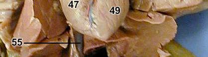
 and body (left ventricle). GROUPS 1-5 5. Remove the heart from the chest cavity.")
, left atrium (#48 on Figure 8b & 8a), right ventricle (#47 on Figure on Figure 8a & b) and the left ventricle (#49 on Figure 8a &")


 and internal jugulars (#88 on figure 9), precava (# 54 on Figure 9b) and postcava, (#55 on Figure 9b) and aorta (#50 on Figure 8b). See Figure 9 on the next page.")
7 3. Locate the heart in the middle of the chest cavity between the lungs. Note that the heart is enclosed in a transparent membrane, called the pericardium. The heart may be partially covered by a portion of the thymus gland. This gland is a dull white color and may resemble a mass of fatty tissue. 4. Mammalian hearts are divided into four chambers. The top chambers the atria collect blood as it comes into the heart (right atrium gets blood from the body, while left atrium gets blood from the lungs). The bottom chambers the ventricles pump blood to the lungs (right ventricle) and body (left ventricle). GROUPS Remove the heart from the chest cavity. Once you remove the heart from the chest cavity, bring it to your instructor to determine your next cut. Make a longitudinal cut of the heart along the midline from top to bottom of the heart. You should be able to locate the right atrium (#46 on Figure 8a & 8b), left atrium (#48 on Figure 8b & 8a), right ventricle (#47 on Figure on Figure 8a & b) and the left ventricle (#49 on Figure 8a & b). Clean out any excess blue or red plastic dye from the chambers, but leave any dye in the blood vessels. Your will loose points if I can't see at least 3 out of 4 of the chambers. GROUPS 6-12 will leave their hearts inside the pig, but should clean up the chest/neck/heart area so it resembles 9a or 9b of this writeup. This means I should be able to see the blood vessels leaving the heart towards the head and lower extremities. You will loose 1 point for every vessel that is not cleaned up, i.e. carotid arteries (#82 on Figure 9a), external (#87 on figure 9a) and internal jugulars (#88 on figure 9), precava (# 54 on Figure 9b) and postcava, (#55 on Figure 9b) and aorta (#50 on Figure 8b). See Figure 9 on the next page. Figure 8a above Figure 8b below 6. Notice the blood vessels leading to and from the heart. The precava enters the right atrium from above and the postcava enters the right atrium from below. The aorta, located underneath the pulmonary artery, carries the blood from the left ventricle to the rest of the body. Locate the branches of the aorta. Locate as many of the labelled arteries and veins illustrated in figure The chambers and major vessels that lead to and away from the heart as well as the blood flow through the heart is required knowledge for everyone taking the final exam. Page 7 of 12


8 Figure 9a Figure 9b Page 8 of 12

. Use a probe to trace the umbilical arteries and vein to and from the heart.")


9 8. Locate the blood vessels in the umbilical cord. The umbilical arteries supplies blood rich in oxygen and nutrients from the placenta to the fetus. The umbilical vein carrys away waste products from the fetal pig's body (See Figure 4 on page 3 of this writeup). Use a probe to trace the umbilical arteries and vein to and from the heart. PROCEDURE F EXCRETOR SYSTEM 1. CLEANING UP THE ABDOMINAL CAVITY: There is a membrane that encloses the abdominal cavity, called the peritoneum. Under the peritoneum, on either side of the spine are two large, dark red, bean-shaped kidneys (109 on Figure 10). The kidneys filter wastes from the blood. 2. Notice the whitish, crescentshaped organs attached to the anterior edge of each of the kidneys. This pair of adrenal glands (#44 on figure 10) is not part of the excretory system, but the endocrine system. The adrenal glands produce adrenaline that affects the behavior and metabolism the animal. GROUPS 6-10: 3. Use your probe or your forceps to tear the connective tissue holding a kidney to the body wall. Then, cut the blood vessels and ureter that lead from this kidney and remove the kidney. Cut the kidney longitudinally from the lateral to the medial surface. Notice that the kidney is divided into three sections. The light-colored renal cortex is the outer section of the kidney (#114 of Figure 10b). The renal medulla is darker in color (#115 on Figure 10b) and is made of many tiny tubules that project into the renal pelvis. The renal pelvis (# 116 on figure 10b) drains the incoming and outgoing fluids. Figure Figure 10b Page 9 of 12

 that carries the urine from the kidney to the urinary bladder.")


10 4. You must remove the peritoneum (semi-transparent covering) from the surface of the renal arteries, renal veins and adrenal glands. You must remove any connective tissue from the descending aorta and postcava, thereby separating the two major vessels. Also clean away any connective tissue covering the iliac arteries and umbilical arteries. 5. LOCATING STRUCTURES OF THE EXCRETORY SYSTEM: Each kidney has a white tube called a ureter (#111 on Figure 10a) that carries the urine from the kidney to the urinary bladder. Remove any connective tissue on the ureters all the way to the urinary bladder (#112 on Figure 10a). The urinary bladder is located on the inside of the ventral flap that contains the umbilical cord. Urinary bladder is responsible for storing urine to be removed from the body. 6. LOCATING LOWER STRUCTURES OF THE EXCRETORY SYSTEM: After birth urine leaves the body through the urethra. You must make an incision down the middle of the hips, but not too deep. To location of the urethra varies slightly depending on the sex of your pig. In male pigs it goes from the bladder loops back towards the umbilical cord in the penis. In females you must make a deep incision with your scalpel down the middle of the hips and spread the hind legs as far apart as you can until the pelvis snaps. Probe with your finger until you find the cartilage that makes up the pelvic bone. Cut through the cartilage and expose the urethra. The urethra leads to the urogenital opening through which urine passes out of the body. PROCEDURE G REPRODUCTIVE SYSTEM Follow the directions for dissecting the reproductive system of your pig. You will be expected to answer questions on both male and female urogenital structures; therefore I recommend you examine other groups pigs. Female Reproductive System 1. Below the kidneys, and above the rectum, locate the small, off-white, almond-shaped ovaries (#101 on Figure 11) which are about 0.5 cm in diameter. Ovaries produce eggs and estrogen. 2. Use a probe to locate the thin, threadlike oviducts or Fallopian tubes (#103 on Figure 11), that loop behind the ovaries. In a mature female pig, eggs break through the ovary walls and pass into the oviduct. Tiny cilia line this passage, sweeping the egg through the coiled tube. If fertilization occurs, it usually takes place in one of the oviducts. Page 10 of 12 Figure 11
11 3. The uterus is broken down into three regions: the Horns of the uterus (#104, Figure 11) where the coiled fallopian tubes (left and right) come together, the uterus (#105, figure 11) and at the end of the tube is the vagina (#106, Figure 11). 4. Notice how the urethra (#113, figure 11) merges with the vagina near the urogenital opening. This common tube is called the urogenital sinus. Locate the urogenital sinus of your fetal pig. Male Reproductive System 1. Locate the scrotum on either side of the anus. Refer to figure at the top of page 2. If you haven't already cut posterior to the umbilicus, continue the posterior incision through the scrotum (structure 9, Figure 2) on one side of the anus. You may need to cut through the cartilage of the pelvic bones. Sometimes prying open the pelvis with your hands is more effective, see page 110 of the lab manual. 2. Cut through the lining of the scrotum, and determine if the testis is present. A male has two testes Figure 12 Page 11 of 12


, a tube that passes through the inguinal canal, loops over the ureters, and joins the urethra near")
.")
12 small, hard oval masses about 0.5 cm in length. In a young male fetus the testes may not yet have descended into the scrotal sacs from the abdominal cavity. You may have to cut through the region above the scrotal sac called the inguinal canal to locate the descending testis (notice the difference between 90 and 91 remove the connective tissue coverings so they both look like 91, Figure 12). Testes produce sperm and testosterone. 3. You will have to remove the connective covering of the testes to observe the following structures. You will be graded on this step. Along one side of the testis, find the coiled mass of tiny tubes the epididymis (#92, Figure 12) that store the sperm. The epididymis leads into the vas deferens (#94, Figure 12), a tube that passes through the inguinal canal, loops over the ureters, and joins the urethra near the entrance to the urinary bladder. From this point the excretory and reproductive systems share the same passageway, the urethra. 4. Trace the urethra posterior until it enters the cord like cylinder under the skin and anterior to the umbilical cord, which becomes the penis (#96 Figure 12b). Notice that the urogenital opening from the urethra in the penis is located just posterior to the umbilical cord. Figure 12b Page 12 of 12
Name: Fetal Pig Dissection Internal Anatomy
 In this lab exercise you will open the thoracic cavities and abdominal pelvic of a fetal pig and identify its major organs. Remember you are dissecting not butchering. The goal is for you to identify all
7 weeks: 28 mm 8 weeks: 40 mm 15 weeks: 220 mm 17 weeks: 300 mm
 Fetal Pig Dissection Background Pigs are placental mammals and show the distinguishing characteristics of that group. In studying the anatomy of the fetal, or unborn, pig, you will see that its various
Fetal Pig Dissection Materials Introduction A. Preparing the Fetal Pig Anterior Posterior Dorsal Ventral length gender mammary papillae
 Fetal Pig Dissection Materials Preserved Fetal Pig Cotton String Dissection tray Dissection pins Disposable Gloves Paper towels Dissection scissors Label pins Lab apron Re-sealable bag Dissection probe
OVARIES URETER FALLOPIAN TUBES BLADDER UROGENITAL OPENINGS (BOTH SEXES) PENIS VAGINA UTERUS
 PENIS VAGINA UTERUS") URETER OVARIES FALLOPIAN TUBES BLADDER UROGENITAL OPENINGS (BOTH SEXES) PENIS VAGINA UTERUS REPRODUCTIVE PRODUCE FEMALE HORMONES EXCRETORY FROM KIDNEY TO BLADDER EXCRETORY STORES URINE REPRODUCTIVE TRANSPORTS
URETER OVARIES FALLOPIAN TUBES BLADDER UROGENITAL OPENINGS (BOTH SEXES) PENIS VAGINA UTERUS REPRODUCTIVE PRODUCE FEMALE HORMONES EXCRETORY FROM KIDNEY TO BLADDER EXCRETORY STORES URINE REPRODUCTIVE TRANSPORTS
Fetal Pig Dissection: External Anatomy
 Name Fetal Pig Dissection: External Anatomy External Anatomy 1. Determine the sex of your pig by looking for the urogenital opening. On females, this opening is located near the anus. On males, the opening
Fetal Pig Dissection Packet
 Fetal Pig Dissection Packet Name Period * Each person will turn in his/her own packet You may use the Virtual Fetal Pig Dissection website from Whitman College as a visual reference for all stages of dissection.
Vertebrate Anatomy Study Guide
 Vertebrate Anatomy Study Guide 1. Anatomical terms: dorsal, ventral, anterior, posterior 2. Body sections: head, neck, thorax, abdomen, tail 3. Body Cavities: oral, thoracic, abdominal, cranial 4. Know
Fetal Pig Dissection Packet (2019)
") Name Period [Each person will turn in his/her own packet] [ Final Score out of 166 points: ] You may use the Virtual Fetal Pig Dissection website from Whitman College as a visual reference for all stages
Name Period [Each person will turn in his/her own packet] [ Final Score out of 166 points: ] You may use the Virtual Fetal Pig Dissection website from Whitman College as a visual reference for all stages
Fetal Pig Dissection:
 Fetal Pig Dissection: REMEMBER: Dissection involves disassembling and observing something to determine its internal structure and develop an understanding of the relationship of those structures to function.
Step 1: Salivary Structures
 (Slide1) Step 1: Salivary Structures Remove the skin, fat and connective fascia to view the salivary glands and ducts. The submaxillary salivary gland is just behind the masseter muscle and pretty easy
Fetal Pig Dissection. preserved fetal pig dissecting tray and paper towels length of string plastic storage bag and twist tie
 Name Date Class LAB PROGRAM INQUIRY SKILLS B29 Fetal Pig Dissection Skills Objectives using dissection instruments and techniques Describe the appearance of various organs found in a fetal pig. Name the
Fetal Pig Dissection Labs Dr. J. Lim
 Fetal Pig Dissection Labs Dr. J. Lim Objective: In this exercise you will examine the organization of the many body systems studied this semester in the context of a single specimen, the fetal pig. Be
Dissection Lab Manuals: Required Content
 Dissection Lab Manuals: Required Content 1. Introduction a. Basic terminology (directions) b. External features of the cat c. Adaptations to predatory niche d. How to skin a cat e. How to make the incisions
#8 - Ventral Body Cavity Organs
 #8 - Objectives: Use a cat dissection to study the organs of the ventral body cavity; Use virtual human dissection software and a human model to observe the organs of the Respiratory, Digestive, Urinary,
MATERIALS: preserved fetal pig, dissecting pan, dissecting kit, dissecting pins, string, plastic bag, metric ruler, paper towels
 ANATOMY & PHYSIOLOGY 12 ROCKRIDGE Fetal Pig Dissection NAME: BLOCK: DATE: Partner: BACKGROUND: Mammals are vertebrates having hair on their body and mammary glands to nourish their young. The majority
BIOLOGY 30S: Fetal Pig Dissection Worksheet
 BIOLOGY 30S: Fetal Pig Dissection Worksheet Name: Part A: External Anatomy & Oral Cavity 1. How long (metric) is your fetal pig? 2. What is the age of your fetal pig? 3. What sense organs are located on
Pig Dissection for Dummies
 Pig Dissection for Dummies External Anatomy 1. Say hello to your pig. To determine the sex of your pig so that you can give him/her an appropriate name, look for a little hole DIRECTLY posterior (toward
Mammalian Dissection: Fetal Pig Integrated Science 4 Honors
 Mammalian Dissection: Fetal Pig Integrated Science 4 Honors Name Per. Introduction Organisms are classified based on similarities and differences to: 1) make sense of the millions of organisms on record,
DISSECTION 8: URINARY AND REPRODUCTIVE SYSTEMS
 8546d_c01_1-42 6/25/02 4:32 PM Page 38 mac48 Mac 48: 420_kec: 38 Cat Dissection DISSECTION 8: URINARY AND REPRODUCTIVE SYSTEMS Typically, the urinary and reproductive systems are studied together, because
SESSION 2: THE MOUTH AND PHARYNX
 SESSION 2: THE MOUTH AND PHARYNX 9 In the pig s digestive tract, food flows in only one direction from mouth to anus.this allows for greatly specialized sections that can act independently of each other.
The$Pig$ $ Dissection$Manual$ (you%only%get%one,%don t%lose%it!)%! DIGENIS!CLASSES!!2016!!!!!!!!!!!!!!! Name:!!!!! Block:!!
%! DIGENIS!CLASSES!!2016!!!!!!!!!!!!!!! Name:!!!!! Block:!!") The$Pig$ $ Dissection$Manual$ (you%only%get%one,%don t%lose%it)% DIGENISCLASSES2016 Name: Block: Fetal Pig Lab Check off each organ/structure as you locate it on your pig. Answer questions where indicated.
The$Pig$ $ Dissection$Manual$ (you%only%get%one,%don t%lose%it)% DIGENISCLASSES2016 Name: Block: Fetal Pig Lab Check off each organ/structure as you locate it on your pig. Answer questions where indicated.
Fetal Pig Dissection Day 2 Circulatory and Respiratory Systems
 Name: Date: Period: Fetal Pig Dissection Day 2 Circulatory and Respiratory Systems Dissection Roles (choose a different role from Day 1): Recorder reads directions out loud to group and records answers
Fetal Pig Dissection Guide
 Fetal Pig Dissection Guide Day 1 - External Anatomy 1. Obtain a fetal pig and rinse off the excess preservative by holding it under running water. Lay the pig on its side in the dissecting pan and locate
This booklet belongs to: Spring Page 1 of 10
 This booklet belongs to: Spring 2017 Page 1 of 10 Frog Dissection Background Amphibians are studied in science for a variety of reasons. Amphibians are unique in many ways because their anatomy allows
Mammalian Dissection: Fetal Pig 11/06
 Mammalian Dissection: Fetal Pig 11/06 Integrated Science 4 Name Per. Introduction Organisms are classified based on similarities and differences to: 1) make sense of the millions of organisms on record,
Frog Dissection. Name: Block:
 Name: Block: Frog Dissection Background: As members of the class Amphibia, frogs may live some of their adult lives on land, but they must return to water to reproduce. Eggs are laid and fertilized in
Dissection: The Fetal Pig
 Lab Exercise Dissection: The Fetal Pig Objectives - To learn some of anatomical structures of the fetal pig. - To be able to make contrasts and comparisons of structures between different animal phyla
NORTH CENTRAL HIGH SCHOOL NOTE & STUDY GUIDE. X Biology II
 Unit 2-5, Animal Biology & Organ Systems, FETAL PIG DISSECTION MANUAL X Biology II, Mr. Doc Miller, M.Ed. North Central High School Name: ID#: NORTH CENTRAL HIGH SCHOOL NOTE & STUDY GUIDE X Biology II
Contents. Pig Dissection. Contents. External Features Sex Determination Mouth and Maxillary Nerve Muscles Index Internal Systems Index
 Pig Dissection External Features Sex Determination Mouth and Maxillary Nerve Muscles Index Internal Systems Index External features Sex determination Male Female Male to External anatomy 1. Pinna 2. External
Student Guide to the Frog Dissection
 Student Guide to the Frog Dissection Dissection Instructions 1. Place the frog in the dissecting pan ventral side up. 2. Use scissors to life the abdominal muscles away from the body cavity. Cut along
Fetal Pig Visual Dissection Guide
 Fetal Pig Visual Dissection Guide WARD470156-776 Orientation Cranial Anterior Sagittal plane Frontal plane Ventral Dorsal Transverse plane Caudal Posterior 1 Incisions 1 Gender Key Male Female Both 4 3
Fetal Pig Dissection Guide
 Fetal Pig Dissection Guide Background: Mammals are vertebrates having hair on their body and mammary glands to nourish their young. The majority are placental mammals in which the developing young, or
This booklet belongs to: Spring Page 1 of 10
 This booklet belongs to: Spring 2013 Page 1 of 10 Frog Dissection Background Amphibians are studied in science for a variety of reasons. Amphibians are unique in many ways because their anatomy allows
Frog Dissection-Skin Vista Murrieta High School-- Biomedical Science. Mr. Diaz
 Frog Dissection-Skin Vista Murrieta High School-- Biomedical Science Mr. Diaz Background: Please read entire lab As members of the class Amphibia, frogs may live some of their adult lives on land, but
Frog Dissection SNC2P Grade 10 Science Applied Biology Tissues, Organs and Systems of Living Things
 Frog Dissection SNC2P Grade 10 Science Applied Biology Tissues, Organs and Systems of Living Things Purpose To identify and examine the external and internal structures of the frog and compare them with
Name Date Per. HANDOUT Frog Dissection Lab
 Name Date Per UNIT 6 HANDOUT Frog Dissection Lab Purpose: To observe the anatomy of an amphibian To discover characteristics of complex vertebrates To compare anatomy of the frog to that of other organisms
FROG DISSECTION GUIDE
 FROG DISSECTION GUIDE I. Introduction Frogs belong to the class amphibian. Although many differences exist between humans and frogs, the basic body plans are similar. Humans and frogs both belong to the
"...AND THIS LITTLE PIG..."
 London and District Science Olympics Annual Competition "...AND THIS LITTLE PIG..." Senior Biology "...AND THIS LITTLE PIG..." RULES Biology Grades 9-12 Changes to the rules are marked by a vertical line
Day 5 Respiratory & Cardiovascular: Respiratory System
 Day 5 Respiratory & Cardiovascular: Respiratory System Be very careful not to damage the heart and lungs while separating the ribs! Analysis Questions-Respiratory & Cardiovascular Log into QUIA using your
Biology Overview Dissection Assignment
 Biology Overview Dissection Assignment What is this assignment about? After learning several major systems of the human body, this overview dissection assignment is for you to review all of those major
Fetal Pigs and You BIO 171 WEEK 10
 Fetal Pigs and You BIO 171 WEEK 10 The Domestic Pig: Sus scrofa Kingdom: Animalia Phylum: Chordata Class: Mammalia - Skin covered in hair or fur; Milk-producing glands (mammary glands) in the female to
 Name: Date: Period: The Dissection 1. Place your specimen dorsal side down. Make your incisions following the diagram below. Make sure to make shallow cuts with the scissors. DO NOT CUT TOO DEEP! You will
Fetal Pig Dissection. Introduction:
 Fetal Pig Dissection Introduction: Today, we begin a new chapter in our study of biology. In the first half of the year we looked at how the smallest units of life work, reproduce and pass on their genes.
MINK DISSECTION LAB DAY 1 NECK
 MINK DISSECTION LAB PURPOSE: This lab dissection is designed to give you first hand experience with the organs (or their mink counterparts) that we have learned about all year long. After seeing the organs,
Cells Tissues Organs Organ Systems Organism. Cells: the smallest unit of life.
 Cells Tissues Organs Organ Systems Organism Cells: the smallest unit of life. The Circulatory Systems brings oxygen, nutrients and hormones to cells; fights infections; removes cell wastes; regulates
PIG DIGESTIVE SYSTEM
 ~Date PIG DIGESTIVE SYSTEM It is not easy to study the digestive organs of a human. However, anatomy of the human digestive system can be studied by examining the digestive system of a pig, an animal similar
Function Alimentary Canal
 THE DIGESTIVE SYSTEM Function: to help convert food into simpler molecules that can be absorbed and used by the cells of the body. Alimentary Canala one way tube that passes through the body. (found in
#5 Cardiovascular II Blood Vessels
 #5 Cardiovascular II Blood Vessels Objectives: Identify a list of human arteries and veins using a virtual human dissection and a human model Dissect and identify a list of arteries and veins in the cat
Fetal Pig Dissection
 Fetal Pig Dissection The fetal pig is a mammal like us so it has many structures that are similar to ours. The anatomy class will analyze the structure and function of the external and internal parts of
Violation of these rules, failure to participate, or unsafe behavior will result in the loss of participation points. THERE WILL BE NO WARNINGS!
 BIOLOGY II MINK DISSECTION ACTIVITY #1 NAME DATE HOUR MINK DISSECTION PART 1: CLASSROOM RULES 1. No food or drink in the lab. 2. Follow directions the first time they are given. 3. No unauthorized dissections.
Fetal Pig Dissection Honors Biology
 Fetal Pig Dissection Honors Biology Introduction In this lab, you will study the external and internal anatomy of a fetal pig, relate its structures to those of other mammals, and determine differences
Body Regions Review. Anatomical Position. Anatomical Planes. Supine versus Prone 9/9/2009
 Body Regions Review The fundamental divisions of the human body Christine Sparks Anatomy / Physiology I Sept. 9, 2009 Anatomical Position Universal terms are used to describe the body accurately and result
Lab 9 Abdomen MUSCLES
 Lab 9 Abdomen MUSCLES External abdominal oblique continuous with the external intercostal muscle; its fibers point in a caudal direction as it moves anteriorly until it inserts on the linea alba via its
LAB: Sheep or Pig Heart Dissection
 Biology 12 Name: Circulatory System Per: Date: Observation: External Anatomy LAB: Sheep or Pig Heart Dissection 1. Line a dissecting tray with paper towel for easy clean up as the heart is fatty and will
Dissection on Demand: Fetal Pig
 TEACHER GUIDE Dissection on Demand: Fetal Pig 60-Minute Health & Life Science Lesson Interactive Video Conferencing Grades: 6-12 Dissection on Demand: Fetal Pig Description Observe the dissection of a
Heart Dissection. 5. Locate the tip of the heart or the apex. Only the left ventricle extends all the way to the apex.
 Heart Dissection Page 1 of 6 Background: The heart is a four-chambered, hollow organ composed primarily of cardiac muscle tissue. It is located in the center of the chest in between the lungs. It is the
NORTH CENTRAL HIGH SCHOOL NOTE & STUDY GUIDE. X Biology II
 Unit 2-5, Animal Biology & Organ Systems, FETAL PIG DISSECTION MANUAL X Biology II, Mr. Doc Miller, M.Ed. North Central High School Name: ID#: NORTH CENTRAL HIGH SCHOOL NOTE & STUDY GUIDE X Biology II
The Human Body. Lesson Goal. Lesson Objectives 9/10/2012. Provide a brief overview of body systems, anatomy, physiology, and topographic anatomy
 The Human Body Lesson Goal Provide a brief overview of body systems, anatomy, physiology, and topographic anatomy Medial Lateral Proximal Distal Superior Inferior Anterior Lesson Objectives Explain the
3. There are three pairs of salivary glands that have three important functions. These are: a)
") Reference: 1. Use the human systems in your textbook.. 2. Pig instruction packet. DIGESTIVE SYSTEM 1. What is the process of digestion? 2. List three major glands involved in this process? 3. There are
Reference: 1. Use the human systems in your textbook.. 2. Pig instruction packet. DIGESTIVE SYSTEM 1. What is the process of digestion? 2. List three major glands involved in this process? 3. There are
This lab activity is aligned with Visible Body s Human Anatomy Atlas app. Learn more at visiblebody.com/professors
 1 This lab activity is aligned with Visible Body s Human Anatomy Atlas app. Learn more at visiblebody.com/professors 2 A. Digestive System Overview To Start: Go to the Views menu and scroll down to the
Honors Biology: Rat Dissection ONLINE ASSIGNMENT
 Name: Honors Biology: Rat Dissection ONLINE ASSIGNMENT You and your group members will use the Honors Biology WIKI to create an online dissection manual. The point of this assignment is to illustrate what
Organ Systems Overview
 Laboratory Manual for Anatomy and Physiology 6th Edition Marieb SOLUTIONS MANUAL Exercise 2 5 Full download at: https://testbankreal.com/download/laboratory-manual-for-anatomy-and-physiology-6th-edition-marieb-solutionsmanual/
The Digestive System. Prepares food for use by all body cells.
 The Digestive System Prepares food for use by all body cells. Digestion The chemical breakdown of complex biological molecules into their component parts. Lipids to fatty acids Proteins to individual amino
In this lab, you will observe the external structures of a crayfish and dissect it to study its internal structures and systems.
 Crayfish Dissection Objectives: Describe the appearance of various organs found in a crayfish. Name the organs that make up systems of the crayfish. Materials: safety goggles, gloves, magnifying glass,
Fetal Pig Dissection Lab
 Fetal Pig Dissection Lab Introduction: In this lab you will be examining many characteristics of an unborn mammal--the fetal pig. Dissection will help you to get a 3-dimensional picture of how all the
Human Body Systems. Human Body Project Notes
 Human Body Systems Human Body Project Notes Human Body Organ Systems for the Project Big Idea: Organ systems are composed of organs that are made of more than one type of tissue. Tissues are made of one
Basic Body Structure
 Basic Body Structure The Cell All life consists of microscopic living structures called cells. They perform various functions throughout the body. All cells are similar in structure, but not identical.
Name Date: Block: Honors Marine Biology Mr. Conlan - Squid Dissection Lab Objective: Can you identify a squid s structures and their functions?
 Name Date: Block: Honors Marine Biology Mr. Conlan - Squid Dissection Lab Objective: Can you identify a squid s structures and their functions? Helpful Hints Everyone must wear safety goggles during the
Lab Schedule for Rest of Semester
 Laboratory 9 Cat Dissection II Respiratory Urinary/Reproductive Systems Lab Schedule for Rest of Semester Cat dissection labs Dissection II (today) Respiratory (Ex. 57 in Hole) Human Reproductive Systems
Cat Dissection. Muscular Labs
 Cat Dissection Muscular Labs Tibialis anterior External oblique Pectroalis minor Gastrocnemius Sartorius Pectoralis major Gastrocnemius Semitendinosis Sartorius External oblique Trapezius Latissimus dorsi
Group B: Organ systems (digestive, respiratory, urinary, genital system, heart, glands and skin) green
 green") Group B: Organ systems (digestive, respiratory, urinary, genital system, heart, glands and skin) green Digestive system 1. Teeth Main points: external and internal structure of a tooth, fixation of a tooth
Group B: Organ systems (digestive, respiratory, urinary, genital system, heart, glands and skin) green Digestive system 1. Teeth Main points: external and internal structure of a tooth, fixation of a tooth
Overview of Anatomy & Physiology
 Overview of Anatomy & Physiology Anatomy the study of the structure of body parts and their relationships to one another Gross or macroscopic Microscopic Developmental Physiology the study of the function
The Digestive System
 The Digestive System Identify the Structure and Function. Mesentery of the Large Intestine The mesentery functions to connect the visceral organs to the abdominal wall. Identify the Structure. Nasal Cavity
Nervous System. Skeletal System. Muscular System. Reproductive System. Circulatory System. Endocrine System. Respiratory System. Integumentary System
 The Human Body Skeletal System Muscular System Circulatory System Respiratory System Digestive System Nervous System Reproductive System Endocrine System Integumentary System Excretory System Lymphatic/Immune
Laboratory Investigation 24A Chapter 24A: Human Skin
 Name Class Date Station # Laboratory Investigation 24A Chapter 24A: Human Skin Human Anatomy & Physiology: Integumentary System You may refer to pages 415-421 in your textbook for a general discussion
Homework Packet. The branch of biological science that studies and describes how body parts. The study of the shape and structure of body parts
 Anatomy & Physiology Chap. 1: The Human Body Name Block: P/W Homework Packet ANATOMY & PHYSIOLOGY DISTINCTIONS 1. Match the term on the right to the appropriate description on the left. Enter the correct
Duodenum retroperitoneal
 Duodenum retroperitoneal C shaped Initial region out of stomach into small intestine RETROperitoneal viscus Superior 1 st part duodenal cap ; moves upwards and backwards to lie on the R crura medial to
Human Body Systems. Long narrow tube mixes enzymes with food Small nutrient molecules diffuse into blood
 Human Body Systems Living Environment AIS Mr. DuRoss Digestive System : Break down large food molecules into smaller parts that the body can use Mouth Esophagus Stomach Small intestine Large intestine
Crayfish Dissection. Objectives: Describe the appearance of various organs found in a crayfish. Name the organs that make up systems of the crayfish.
 Crayfish Dissection Objectives: Describe the appearance of various organs found in a crayfish. Name the organs that make up systems of the crayfish. Background: Like all crustaceans, a crayfish has a fairly
STREAM. Human Body Project Pages Website QR Code body project/
 STREAM Human Body Project Pages 1 16 Website QR Code https://sites.google.com/a/wyckoffschools.org/human body project/ Project Checklist Did you include Head a brain that can open to show the inside as
3 Circulatory Pathways
 40 Chapter 3 Circulatory Pathways Systemic Arteries -Arteries carry blood away from the heart to the various organs of the body. -The aorta is the longest artery in the body; it branches to give rise to
1. Three Main Functions. Chapter 19: 2. Two Groups of digestive organs. 2. Two Groups of digestive organs 6/1/2015. The Wall of the Digestive Tract
 1. Three Main Functions Chapter 19: General Structure and Function of the Digestive System Digestion-breakdown of food into small particles for transport to blood Absorption- into bloodstream to take to
#5 Cardiovascular II Blood Vessels
 Page1 #5 Cardiovascular II Blood Vessels Objectives: Observe slide of artery and vein cross-section Identify a list of human arteries and veins using a virtual human dissection Dissect and identify a list
Rattus norvegicus Dissection Guide
 Adult Rat Dissection Introduction: Rattus norvegicus (Berkenhout, 1769), the Norway rat, belongs to the family Muridae, a large group of rodents that includes the house mouse, gerbil, and hamster. It is
Biol 111 Comparative & Human Anatomy Lab 5: Digestive, Respiratory, and Urogenital Systems of the Shark Spring 2014
 Biol 111 Comparative & Human Anatomy Lab 5: Digestive, Respiratory, and Urogenital Systems of the Shark Spring 2014 Philip J. Bergmann Lab Objectives 1. To learn the component parts of the shark digestive,
Time Allotment: 1½ hours (rat dissection 1 hour; human torso model ½ hour).
.") Human Anatomy Laboratory Manual with Cat Dissections 8th Edition Marieb SOLUTIONS MANUAL Full download at: https://testbankreal.com/download/human-anatomy-laboratory-manual-with-catdissections-8th-edition-marieb-solutions-manual/
Human Anatomy Laboratory Manual with Cat Dissections 8th Edition Marieb SOLUTIONS MANUAL Full download at: https://testbankreal.com/download/human-anatomy-laboratory-manual-with-catdissections-8th-edition-marieb-solutions-manual/
The Human Body. Mrs. Green
 The Human Body Mrs. Green Bell Work Which of the following helps the body to cool down? a) Shivering b) Sweating c) Running a fever d) Taking a deep breath Which of the following is a function of the digestive
Growing bones. Baby s s bones are made from cartilage Babies have 300 bones Adults have 206. bones
 Body Systems Objectives Students should be able to list the different body systems Students should be able to give a general function of each of the body systems Students should be able to tell the differences
Anatomy: Know Your Abdomen
 Anatomy: Know Your Abdomen Glossary Abdomen - part of the body below the thorax (chest cavity); separated by the diaphragm. Anterior - towards the front of the body. For example, the umbilicus is anterior
1. Three Main Functions. Chapter 19: 2. Two Groups of digestive organs. 2. Two Groups of digestive organs. 1. The Teeth 5/18/2015
 1. Three Main Functions Chapter 19: General Structure and Function of the Digestive System Digestion-breakdown of food into small particles for transport to blood Absorption- into bloodstream to take to
CJ Shuster A&P2 Lab Addenum Beef Heart Dissection 1. Heart Dissection. (taken from Johnson, Weipz and Savage Lab Book)
") CJ Shuster A&P2 Lab Addenum Beef Heart Dissection 1 Heart Dissection. (taken from Johnson, Weipz and Savage Lab Book) Introduction When you have finished examining the model, you are ready to begin your
CJ Shuster A&P2 Lab Addenum Beef Heart Dissection 1 Heart Dissection. (taken from Johnson, Weipz and Savage Lab Book) Introduction When you have finished examining the model, you are ready to begin your
Digestive System 7/15/2015. Outline Digestive System. Digestive System
 Digestive System Biology 105 Lecture 18 Chapter 15 Outline Digestive System I. Functions II. Layers of the GI tract III. Major parts: mouth, pharynx, esophagus, stomach, small intestine, large intestine,
Name Partner(s) Name. Name your rat. Rat Dissection Lab
 Name. Name your rat. Rat Dissection Lab") Name Partner(s) Name Name your rat Rat Dissection Lab!!CAUTION!! You must follow all safety instructions as outlined in this lab manual and by your teacher. There are several sharp objects being used during
Name Partner(s) Name Name your rat Rat Dissection Lab!!CAUTION!! You must follow all safety instructions as outlined in this lab manual and by your teacher. There are several sharp objects being used during
Male Reproductive System
 Male Reproductive System The male reproductive system consists of a number of sex organs that are part of the reproductive process. The following sections describe the function of each part of the male
Name Score. The Neck Bone s Connected to the Head Bone
 Name Score The Neck Bone s Connected to the Head Bone The Function and Interdependence of Organs and Tissues Main Idea 1. We are made of cells. Supporting Information 1. A group of specialized cells form
Pig Dissection Manual
 Great Falls High School Honors Human Biology 5-6 Pig Dissection Manual Student Name School Year Condition 1 Fetal Pig Dissection Lab External Fetal Pig Anatomy Introduction Fetal pigs are readily available,
Warm Up Where in a flower would you find xylem and phloem? 2. Where in a flower would you find palisade cells?
 Body Systems Warm Up 4-4-16 1. Where in a flower would you find xylem and phloem? 2. Where in a flower would you find palisade cells? 3. Where in a flower would you find root hair cells? 4. What organelle
Crayfish Observation and Dissection
 Name Period Date Crayfish Observation and Dissection Purpose: In this lab, you will observe the external structures of a crayfish and dissect it to study its internal structures and systems. Materials:
Internal Morphology. 1.Cut the legs and wings (if present) off your specimen. 5.Use forceps to pull skeleton apart, exposing internal systems.
 off your specimen. 5.Use forceps to pull skeleton apart, exposing internal systems.") Internal Morphology Insect Dissections Often the best approach to understanding internal morphology is by way of a dissection. For this reason, the entire chapter should be treated as a laboratory activity.
Internal Morphology Insect Dissections Often the best approach to understanding internal morphology is by way of a dissection. For this reason, the entire chapter should be treated as a laboratory activity.