CERVICAL SPINE: Radiographs and MRI Cases
|
|
|
- Brendan Henry
- 5 years ago
- Views:
Transcription


1 Radiology reports with recommendations & clinical information - $30 per region, x-ray - $50 per MRI - Medpay Monthly Newsletter 700 East Redlands Blvd, Redlands CA jpedley299@yahoo.com CERVICAL SPINE: Radiographs and MRI Cases Jennifer Pedley, CSCS, MS, DC, CCSP, DACBR 1

2 Cervical Spine Views 3 Views: APOM, APLC, and neutral lateral performed first Followed by extended and flexed lateral views. Oblique views are helpful APOM kvp 70 FFD 40 Film size 8 x 10 CR uvula Grid Yes Film Landscape Collimate 5x5 Image Double mas from APLC 2

 Grid Collimate")
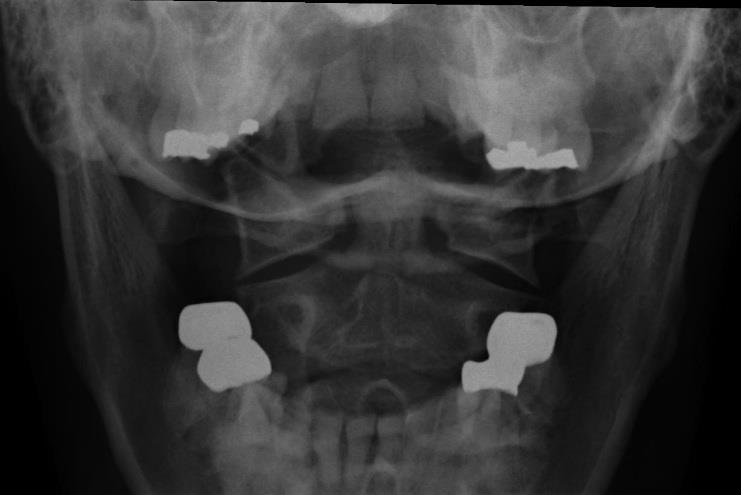
3 AP OPEN MOUTH Structures Visualized: Dens C1 lateral masses Occipital Condyles C2 body C2 SP APLC kvp 70 FFD 40-3 Tube Tilt 15 cephalad Film 8 x 10 CR C3/4 (thyroid cartilage) Grid Collimate 7x10 yes 3


4 AP LOWER CERVICAL Structures Visualized: Vertebral Bodies TP s SP s Upper Ribs Upper Lung Fields Uncinate Processes Tracheal Air Shadow kvp 70 FFD 72 Film size 8x10 CR C3 Grid No Utilize the air gap technique. Collimate 7X10 NEUTRAL LATERAL 4

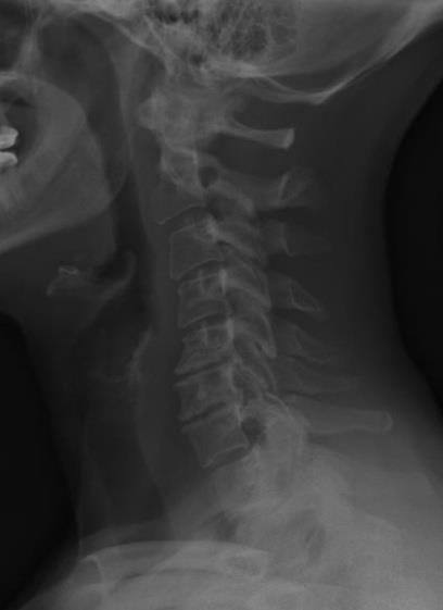
5 LATERAL CERVICAL Structures Visualized: Vertebral bodies C2-T1 Disc Spaces ADI SP s, Lamina, Pedicles, Articular Pillars and Facets Tracheal Air Shadow George s Line & Spino-laminar line Sella Turcica C1 Arches LATERAL EXTENDED kvp 70 FFD 72 Film size 8x10 CR C3 Grid None Collimate 8x10 May need to be landscape in patients with greater range of motion 5

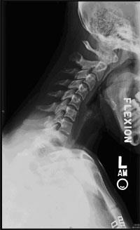
6 LATERAL FLEXED kvp 70 FFD 72 Film size 8x10 CR C3 Grid None Collimate 8x10 May need to be landscape in patients with great range of motion Posterior vs. Anterior Obliques Posterior Visualize the opposite IVF s Marker goes under the mandible Anterior Visualize the same side IVF s Marker goes behind the spine 6


7 LEFT ANTERIOR OBLIQUE kvp 70 FFD 72 Film size 8x10 CR C3 Grid None Tube tilt 15 caudad** Collimate 7x10 LEFT POSTERIOR OBLIQUE kvp 70 FFD 72-3 Film size 8x10 CR C3 Grid No Tube tilt 15 cephalad Collimate 7x10 7


8 CERVICAL OBLIQUE Structures Visualized IVF s Vertebral Bodies C1 arches Ribs SP s Facets Radiographic Signs of Instability Vertebral body displacement >2mm; *3.5mm Widened interlaminar & interspinous space Widened facet joints Widened interpediculate distance (AP view) Atlanto-dental interspace>3mm adults; >5mm in children These findings indicate skeletal, ligamentous and articular disruption. *AMA Guides, 5 th ed. Resnick D. Diagnosis of Bone and Joint Disorders, 4 th ed. 2002;

9 Normal lateral cervical spine Alignment Prevertebral soft tissue swelling (within 3 days of injury) Retropharyngeal: >7.0-mm Retrotracheal: >22.0- mm Cervical MRI Sequences: T1, T2, PD, STIR Anatomy Disc Herniation Types Description; Location; Complications Degenerative Disc Disease and Degenerative Joint Disease Case Presentations Various DDD, DJD, Tumor, etc 9


10 Normal Sagittal T1 & T2 Weighted Images Axial T2 Weighted Image 10

11 Location of Disc Abnormalities Central Paracentral Subarticular Foraminal Extraforaminal Anterior Disc Nomenclature Herniation is non-specific Diffuse disc bulge Asymmetrical disc bulge Broad-based protrusion Focal disc protrusion Disc extrusion Disc sequestration 11
12 Diffuse Disc Bulge Symmetric and circumferential bulge more than 2 mm in all directions Also called a diffuse annular or circumferential disc bulge This is considered a normal finding in the aging spine Asymmetrical Disc Bulge Asymmetric bulge involving more than 50% of the disc circumference 12
13 Broad-based Protrusion 25% to 50% of the disc circumference. Base of the disc protrusion is wider than the AP diameter. Aka broad-based disc bulge Focal Disc Protrusion 25% or less of the disc circumference Focal, asymmetric extension of disc The base is broader than the AP diameter These are contained by the PLL 13
14 Disc Extrusion Less than 25% of the disc circumference AP diameter is greater than base Maintains contact with parent disc Not contained by the PLL Confused with Extruded disc!! Disc Sequestration Loss of continuity between extruded disc and parent disc Free fragment might be a better term 14
15 X-ray Nomenclature Spondylophyte/ Osteophyte/ Spondylosis Bone only Spondylosis deformans Spondylophyte formation with little or no disc narrowing MRI Nomenclature Disc-osteophyte complex bulge or Diffuse disc bulge with osteophytic changes Degenerative Disc Disease Endplate degeneration Modic type I pain generator Modic type II Modic type III 15


16 Modic Type I & II Rahme and Moussa. AJNR May : Modic type III-subchondral sclerosis Rahme and Moussa. AJNR May :
17 Case Hydroxyappatite Deposition Disease of the longus colli tendon Aka calcific tendinitis S/S: px, stiff neck, muscle spasms, painful swallowing Occurs due to trauma Self limiting 1-2 weeks 17

 elbow flexion and wrist")
18 Sagittal Reformatted CT and Sagittal T2 weighted MR images Case 1 Patient experiences numbness and tingling down the right arm and thumb Motor test: 3/5 (able to move but not full ROM) elbow flexion and wrist extension 18


19 Case


20 Sagittal T2 & T1 Weighted Images 20

21 Axial GRE Weighted Image Broad-based right paracentral disc protrusion at C5-C6 Diagnosis Compression of the spinal cord and of the right C6 nerve root 21


22 Case 2 Spastic gait Upper extremity numbness Loss of fine motor control in the hands Chronic and gradually worsening Sagittal & Axial T2 Weighted Images 22
23 Diagnosis Ventral cord compression from broad-based right paracentral disc protrusion C4-C5 and C5-C6; Myelomalacia of spinal cord Compression of the C5 and C6 nerve roots. Degenerative Disc Disease C3 to C6; Facet arthrosis Reversal of the cervical lordosis; Degenerative retrolisthesis at C4 and C5 Complication Compression of cord due to osteophytes or disc>>>stenosis Result Compresses the blood supply to the cord and nerve roots (venous congestion and edema) Demyelination or axonal destruction Tse, V. Spinal Metastasis and Related Structures 23

24 Case 3 Radiculopathy: right fingers with numbness and tingling. Mild weakness of the right bicep, tricep and shoulder abduction. Sagittal T2 Weighted Image 24

25 Diagnosis Multiple disc bulges and protrusion Degenerative disc disease Myelomalacia of spinal cord Straightening and reversal of the cervical lordosis; Degenerative retrolisthesis, C5; Modic endplate changes Result: Spinal canal stenosis Case 4 Radiculopathy down the right arm with severe lower neck pain Car accident a few months ago 25


26 26

27 X-ray Findings Straightening of the cervical spine, apexing at C4-C5. Sagittal T2 Weighted Image 27
28 Diagnosis Disc protrusion at C4-C5 and C5-C6 compressing the cord Degenerative retrolisthesis at C4 and C5. Case 5 Pain and decreased ROM in bilateral lateral flexion and rotation. Constant hypertonicity of the upper trapezius 28


29 X-ray Findings Reversal lordotic curve, apex at C4-C5 with anterior head carriage Posture: tilting to the left; right shoulder low Osteopenia Anterolisthesis at C3, C4 and C6 Disc wedging at C4 Degenerative disc disease at C5-C6 and C6-C7 Facet arthrosis, primarily on the left 29


30 Sagittal T1 & T2 Weighted Image Axial T1 Weighted Image 30


31 Axial T2 Weighted Axial T1 Weighted Image 31
32 Diagnosis C3-C4, C4-C5 facet hypertrophy on the left C4-C5 focal central disc protrusion C5-C6 and C6-C7 spondylosis and broad-based central disc protrusions with compression and displacement of the spinal cord Other: Degenerative anterolisthesis; Mild to moderate uncovertebral arthrosis; C6-C7, Modic type II endplate changes (fatty infiltration) Case 6 Frequent numbness and tingling of both hands/fingers Neck stiffness and getting worse Headaches 32


33 33


34 X-ray Findings Posture: low occiput on the right; spine tilting to the right with low right shoulder; Reversal of cervical lordosis, apex at C5 Disc narrowing at C3-C4 and C4-C5 Degenerative disc disease at C5-C6 and C6-C7 Degenerative retrolisthesis at C5 Moderate facet and uncovertebral arthrosis Sagittal T1 & T2 Weighted Images 34


35 Axial T2 Weighted Image Sagittal & Axial T2 Weighted Image 35

36 Diagnosis Arnold Chiari malformation with syrinx (syringomyelia) Multilevel diffuse disc bulges with osteophytic changes, C5-C6 and C6-C7 Facet and uncovertebral arthrosis Degenerative retrolisthesis, C5 and C6; Reversal of lordosis Case 7 70 year-old male with chronic mid and low back pain 36

37 Sagittal T1 Weighted Image Differential Diagnosis Multiple Myeloma or Metastatic Disease DX: Mets Prostate Lung Breast Renal CA Gastric CA 37


38 Case 8 46 yom with neck pain. The pain travels down both arms. 38
 May or may not be symptomatic Sx s")

39 Diagnosis Meningocele (possible neural tissue within the sac; subcutaneous tissue intact) May or may not be symptomatic Sx s when located in cervical and thoracic MC in lumbar; cervical & thoracic spine Dorsal defect Other DDX: bone tumor, sarcoma 39
40 Case 9 Numbness and tingling in the hands and feet Chronic neck pain Case donated by the patient None given X-rays 40


41 Sagittal Flexed & Extended T2 Weighted Images Axial T2 Weighted Image 41

42 Sagittal T1 FLAIR DDX Syrinx Arachnoid Cyst Spinal cord tumor 42
43 Syrinx What is it? A cyst in the cord that may elongate and expand Potential to cause myelopathy or demyelination of the cord Commonly seen in the cervical and thoracic cord Sx: Pain, weakness and stiffness of the arms, legs, shoulders (cape like) and back. Other: Headaches, loss of temperature & loss of bowel and Causes Previous trauma to the cord and manifests later in life Congenital developmental problems with brain and cord Chiari malformation (herniation of the cerebellum & brain stem) Referral Neurologist MRI with contrast to rule out tumor & of the entire spine THE END 43
Back To Chiropractic CE Seminars Back to Basics: X-Ray ~ 5 Hours
 Back To Chiropractic CE Seminars Back to Basics: X-Ray ~ 5 Hours Welcome to Back To Chiropractic Online CE exams: This course counts toward your California Board of Chiropractic Examiners CE & your Radiography
Spine. Neuroradiology. Spine. Spine Pathology. Distribution of fractures. Radiological algorithm. Role of radiology 18/11/2015
 Spine Neuroradiology Spine Prof.Dr.Nail Bulakbaşı X Ray: AP/L/Oblique Vertebra & disc spaces CT & CTA Vertebra, discs, vessels MRI & MRA Vertebra, disc, vessels, meninges Spinal cord & nerves Myelography
Spinal Imaging. ssregypt.com. Mamdouh Mahfouz MD
 Spinal Imaging Degenerative diseases ssregypt.com Mamdouh Mahfouz MD mamdouh.m5@gmail.com MRI Open MRI Closed Extremity MRI Dynamic MRI Dynamic MRI The bed rotates from Upright to Recumbent, stopping at
Degenerative Disease of the Spine
 Degenerative Disease of the Spine Introduction: I. Anatomy Talk Overview II. Overview of Disease Processes: A. Spondylosis B. Intervertebral Disc Disease III. Diagnosis IV. Therapy Introduction: Myelopathy
Digital Motion X-ray Cervical Spine
 NAME OF PATIENT: CASE STUDY 4 DATE OF REPORT: DATE OF EXAMINATION: REFERRING PHYSICIAN: TESTING FACILITY: Digital Motion X-ray Cervical Spine 1. In the neutral lateral projection: Shows reversal of the
Radiology of Cervical Spine Trauma. Cervical Spine Trauma. Imaging Standards. Canadian C. Spine Rule 11/28/2016
 Radiology of Cervical Spine Trauma Dr. Steven J. Gould, D.C. Board Certified Chiropractic Radiologist Cleveland Chiropractic College, KC. MO. Radiology Residency at CCC, KC Cervical Spine Trauma Vertebral
Case Report: CASE REPORT OF FACET ARTHROPATHY INDUCED NERVE ROOT COMPRESSION RESULTING IN MOTOR WEAKNESS AND PAIN
 Cox Technic Case Report #100 published at www.coxtechnic.com (sent October 2011 on 10/11/11 ) 1 Case Report: CASE REPORT OF FACET ARTHROPATHY INDUCED NERVE ROOT COMPRESSION RESULTING IN MOTOR WEAKNESS
Daniel J. Blizzard, MD, MS
 Daniel J. Blizzard, MD, MS None Common degenerative (usually) condition caused by compression on the spinal cord that is characterized by clumsiness and difficulty with fine motor tasks in the hands and
Imaging of Cervical Spine Trauma Tudor H Hughes, M.D.
 Imaging of Cervical Spine Trauma Tudor H Hughes, M.D. General Considerations Most spinal fractures are due to a single episode of major trauma. Fatigue fractures of the spine are unusual except in the
Cervical Spine: Pearls and Pitfalls
 Cervical Spine: Pearls and Pitfalls Presenters Dr. Rob Donkin Functional Anatomy Current research Cervical Radiculopathy Dr. Gert Ferreira Red flags Case Study Kinesio Taping Chris Neethling Gonstead adjusting
EVALUATION AND MANAGEMENT OF CERVICAL SPINE DISORDERS
 CERVICAL SPINE EVALUATION AND MANAGEMENT OF CERVICAL SPINE DISORDERS Gregory M Yoshida MD Supports the skull Allows movement of the head Houses the spinal cord CERVICAL SPINE Unique anatomy Upper C spine
eck and Low ack pain: ddressing he Surgical valuation
 eck and Low ack pain: ddressing he Surgical valuation KI FOX, DO T WORTH BRAIN & SPINE Goals Review anatomy Identify sources of pain Imaging: the good, the bad, and the ugly PE: findings to determine source
Hidayatullah Hamidi. MD Consultant Radiologist. Lumbar Spine MR Imaging Interpretation
 Hidayatullah Hamidi. MD Consultant Radiologist Lumbar Spine MR Imaging Interpretation 13/12/2018 Presenter Hidayatullah Hamidi Consultant Radiologist, Radiology PGME program director, FMIC, Kabul, Afghanistan
ESSENTIALS OF PLAIN FILM INTERPRETATION: SPINE DR ASIF SAIFUDDIN
 ESSENTIALS OF PLAIN FILM INTERPRETATION: SPINE DR ASIF SAIFUDDIN Consultant Musculoskeletal Radiologist Royal National Orthopaedic Hospital Stanmore,UK. INTRODUCTION 2 INTRODUCTION 3 INTRODUCTION Spinal
CERVICAL SPONDYLOSIS AND CERVICAL SPONDYLOTIC MYELOPATHY
 CERVICAL SPONDYLOSIS AND CERVICAL SPONDYLOTIC MYELOPATHY A NEUROSURGEON S VIEW A Preventable Journey to a wheelchair bound-life Dr H. BOODHOO F.C.S (Neurosurgery) Cervical Spondylosis Spinal Osteoarthritis
CERVICAL SPONDYLOSIS & CERVICAL DISC DISEASE
 CERVICAL SPONDYLOSIS & CERVICAL DISC DISEASE Cervical spondylosis l Cervical osteophytosis l Most common progressive disease in the aging cervical spine l Seen in 95% of the people by 65 years Pathophysiology
Introduction to Neuroimaging spine. John J. McCormick MD
 Introduction to Neuroimaging spine John J. McCormick MD Neuroanatomy Netter drawings Radiographic Anatomy Cervical Spine Cervical Spine Oblique View Cervical Spine Dens View Thoracic Spine Lumbar Spine
SUBAXIAL CERVICAL SPINE TRAUMA- DIAGNOSIS AND MANAGEMENT
 SUBAXIAL CERVICAL SPINE TRAUMA- DIAGNOSIS AND MANAGEMENT 1 Anatomy 3 columns- Anterior, middle and Posterior Anterior- ALL, Anterior 2/3 rd body & disc. Middle- Posterior 1/3 rd of body & disc, PLL Posterior-
POSTERIOR CERVICAL FUSION
 AN INTRODUCTION TO PCF POSTERIOR CERVICAL FUSION This booklet provides general information on the Posterior Cervical Fusion (PCF) surgical procedure for you to discuss with your physician. It is not meant
Cervical Degenerative Disease - Surgical Approaches to CSM 가톨릭의대인천성모병원척추센터 김종태
 KNS Main Topic Session Spine Surgery : Case-Based Lecture of Spinal Disease Cervical Degenerative Disease - Surgical Approaches to CSM 가톨릭의대인천성모병원척추센터 김종태 Cervical Spondylotic Myelopathy ( CSM ) (1984,
HISTORY AND CHIEF COMPLAINT:
 submitted by Keith M. Bartley, D.C. Jasper, IN 07/21/11 presented at Cox Seminar in Nashville, TN, on October 8 9, 2011 HISTORY AND CHIEF COMPLAINT: 01/21/11 55 year old male press operator for Jasper
PREPARED FOR. Marsha Eichhorn DATE OF INJURY : N/A DATE OF ANALYSIS : 12/14/2016 DATE OF IMAGES : 12/8/2016. REFERRING DOCTOR : Dr.
 Accent on Health Chiropractic 405 Firemans Ave PREPARED FOR Marsha Eichhorn DATE OF INJURY : N/A DATE OF ANALYSIS : 12/14/2016 DATE OF IMAGES : 12/8/2016 REFERRING DOCTOR : Dr. David Bohn This report contains
SPINAL MAGNETIC RESONANCE IMAGING INTERPRETATION
 CLINICAL VIGNETTE 2017; 3:2 SPINAL MAGNETIC RESONANCE IMAGING INTERPRETATION Editor-in-Chief: Idowu, Olufemi E. Neurological surgery Division, Department of Surgery, LASUCOM/LASUTH, Ikeja, Lagos, Nigeria.
Restraints to Movement... 4 Restraints to flexion... 4 Primary restraint into Extension... 4
 CERVICAL SPINE... 4 Neck Pain Categories... 4 Kinematics... Error! Bookmark not defined. Ranges of Motion C2-7... 4 Coupled Movements... 4 Ranges of Motion C0-2... 4 Coupled Movements... 4 Restraints to
Diagnosis of Neck & Upper Extremity Pain
 Diagnosis of Neck & Upper Extremity Pain David B. Bumpass, MD Assistant Professor, Spine Surgery UAMS Depts. of Orthopaedic Surgery & Neurosurgery May 12, 2018 Disclosures Medtronic Spine speaking fees
Imaging the Degenerative Diseases of the Lumbar Spine
 221 Imaging the Degenerative Diseases of the Lumbar Spine David Malfair, MD a, Douglas P. Beall, MD b,c, * MAGNETIC RESONANCE IMAGING CLINICS Magn Reson Imaging Clin N Am 15 (2007) 221 238 - Degenerative
LUMBAR SPINAL STENOSIS
 LUMBAR SPINAL STENOSIS Always occurs in the mobile segment. Factors play role in Stenosis Pre existing congenital or developmental narrowing of the lumbar spinal canal Translation of one anatomic segment
Upper Cervical Spine - Occult Injury and Trigger for CT Exam
 Upper Cervical Spine - Occult Injury and Trigger for CT Exam Main Menu Introduction Clinical clearance of C-SpineC Radiographic evaluation Norms for C-spineC Triggers for CT exam: Odontoid Lateral view
Bony framework of the vertebral column Structure of the vertebral column
 5.1: Vertebral column & back. Overview. Bones o vertebral column. o typical vertebra. o vertebral canal. o spinal nerves. Joints o Intervertebral disc. o Zygapophyseal (facet) joint. Muscles o 2 compartments:
NMH happens when there is an abnormal reflex interaction between the heart and the brain, although both are structurally normal.
 Neurally mediated hypotension: is also known as: the fainting reflex, neurocardiogenic syncope, vasodepressor syncope, the vaso-vagal reflex, and autonomic dysfunction. (Hypotension= low blood pressure,
WORKPLACE SAFETY AND INSURANCE APPEALS TRIBUNAL DECISION NO. 2192/16
 WORKPLACE SAFETY AND INSURANCE APPEALS TRIBUNAL DECISION NO. 2192/16 BEFORE: E. Kosmidis: Vice-Chair HEARING: August 30, 2016 at Toronto Written DATE OF DECISION: October 25, 2016 NEUTRAL CITATION: 2016
REVIEW QUESTIONS ON VERTEBRAE, SPINAL CORD, SPINAL NERVES
 REVIEW QUESTIONS ON VERTEBRAE, SPINAL CORD, SPINAL NERVES 1. A 28-year-old-women presented to the hospital emergency room with intense lower back spasms in the context of coughing during an upper respiratory
RADICULOPATHY AN INTRODUCTION TO
 AN INTRODUCTION TO RADICULOPATHY This booklet provides general information on radiculopathy. It is not meant to replace any personal conversations that you might wish to have with your physician or other
RETROLISTHESIS. Retrolisthesis. is found mainly in the cervical spine and lumbar region but can also be often seen in the thoracic spine
 RETROLISTHESIS A retrolisthesis is a posterior displacement of one vertebral body with respect to adjacent vertebrae Typically a vertebra is to be in retrolisthesis position when it translates backward
Kinematic Cervical Spine Magnetic Resonance Imaging in Low-Impact Trauma Assessment
 Kinematic Cervical Spine Magnetic Resonance Imaging in Low-Impact Trauma Assessment 1 Seminars in Ultrasound, CT, and MRI June 2009; Volume 30; Number 3; pp. 168-173 Vincenzo Giuliano, MD, Antonio Pinto,
MUSCULOSKELETAL IMAGING FOR PHYSICAL THERAPISTS. COMBINED SECTIONS MEETING 2006 San Diego, CA February 1-5, 2006
 MUSCULOSKELETAL IMAGING FOR PHYSICAL THERAPISTS COMBINED SECTIONS MEETING 2006 San Diego, CA February 1-5, 2006 John Meyer, DPT, OCS University of Southern California Department of Athletic Medicine Los
NECK AND BACK PAIN AN INTRODUCTION TO
 AN INTRODUCTION TO NECK AND BACK PAIN This booklet provides general information on neck and back pain. It is not meant to replace any personal conversations that you might wish to have with your physician
Anatomy & Physiology II. Trunk
 Anatomy & Physiology II Trunk Bones and Landmarks of the Vertebral column 24 vertebrae Sacrum - consists of 5 vertebrae that fuse into one bone Median sacral crest Sacral hiatus 4 sacral foramina Coccyx
3/3/2017. Acute spine disorder (< 4weeks duration) Subacute spine disorder (4-12 weeks duration) Chronic spine disorder (>12 weeks duration)
 Subacute spine disorder (4-12 weeks duration) Chronic spine disorder (>12 weeks duration)") William Hsu BSc DC DACBR March 4, 2017 Acute spine disorder (< 4weeks duration) Subacute spine disorder (4-12 weeks duration) Chronic spine disorder (>12 weeks duration) Neurologic symptoms and signs pain
William Hsu BSc DC DACBR March 4, 2017 Acute spine disorder (< 4weeks duration) Subacute spine disorder (4-12 weeks duration) Chronic spine disorder (>12 weeks duration) Neurologic symptoms and signs pain
Outline. Epidemiology Indications for C-spine imaging Modalities Interpretation Types of fractures
 C-Spine Plain Films Outline Epidemiology Indications for C-spine imaging Modalities Interpretation Types of fractures Epidemiology 7000-10000 c-spine injuries treated each year Additional 5000 die at the
Cervical Spine Anatomy and Biomechanics. Typical Cervical Vertebra C3 6. Typical Cervical Vertebra Anterior 10/5/2017
 Cervical Spine Anatomy and Biomechanics Typical Cervical Vertebra C3 6 Small, relatively broad body Bifid SpinousProcess Long and narrow laminae Spinal Canal: large, triangular; remarkably consistent dimensions
Orthopadic cors. Topic : -Cervical spondylitis. -Development disorders(spondylolysis and Spodylolsithesis)
") Orthopadic cors Topic : -Cervical spondylitis. -Development disorders(spondylolysis and Spodylolsithesis) Cervical spondylitis. Definition : - a painful condition of the cervical spine resulting from the
Orthopadic cors Topic : -Cervical spondylitis. -Development disorders(spondylolysis and Spodylolsithesis) Cervical spondylitis. Definition : - a painful condition of the cervical spine resulting from the
Comprehension of the common spine disorder.
 Objectives Comprehension of the common spine disorder. Disc degeneration/hernia. Spinal stenosis. Common spinal deformity (Spondylolisthesis, Scoliosis). Osteoporotic fracture. Anatomy Anatomy Anatomy
Revised Dec Spine MR Protocols
 Spine MR Protocols Sp 1: Cervical spine MRI without contrast Sp 2: Pre- and post-contrast cervical spine MRI Sp 3: Pre- and post-contrast cervical spine MRI (multiple sclerosis protocol) Sp 4: Thoracic
Objectives. Comprehension of the common spine disorder
 Objectives Comprehension of the common spine disorder Disc degeneration/hernia Spinal stenosis Common spinal deformity (Spondylolisthesis, Scoliosis) Osteoporotic fracture Destructive spinal lesions Anatomy
River North Pain Management Consultants, S.C., Axel Vargas, M.D., Regional Anesthesiology and Interventional Pain Management.
 River North Pain Management Consultants, S.C., Axel Vargas, M.D., Regional Anesthesiology and Interventional Pain Management. Chicago, Illinois, 60611 Phone: (888) 951-6471 Fax: (888) 961-6471 Clinical
Epidemiology of Low back pain
 Low Back Pain Definition Pain felt in your lower back may come from the spine, muscles, nerves, or other structures in that region. It may also radiate from other areas like the mid or upper back, a inguinal
Subaxial Cervical Spine Trauma
 Subaxial Cervical Spine Trauma Pooria Salari, MD Assistant Professor Of Orthopaedics Department of Orthopaedic Surgery St. Louis University School of Medicine St. Louis, Missouri, USA Initial Evaluation
Key Primary CPT Codes: Refer to pages: 7-9 Last Review Date: October 2016 Medical Coverage Guideline Number:
 National Imaging Associates, Inc. Clinical guidelines CERVICAL SPINE SURGERY: ANTERI CERVICAL DECOMPRESSION WITH FUSION CERVICAL POSTERI DECOMPRESSION WITH FUSION CERVICAL ARTIFICIAL DISC CERVICAL POSTERI
Gillian Wooldridge, DO Houston Methodist Willowbrook Hospital Primary Care Sports Medicine Fellowship May 3, 2018
 Gillian Wooldridge, DO Houston Methodist Willowbrook Hospital Primary Care Sports Medicine Fellowship May 3, 2018 Disclosures Neither I nor any family members have financial disclosures Special thanks
Misdiagnosis in cervical spondylosis myelopathy.
 Journal of the International Society of Head and Neck Trauma (ISHANT) Case report Misdiagnosis in cervical spondylosis myelopathy. Dr. Reinel A. Junco Martin. Neurosurgeon. Assistant professor Miguel Enriquez
Thoracic Spine Applied Anatomy. Jason Zafereo, PT, OCS, FAAOMPT
 Thoracic Spine Applied Anatomy Jason Zafereo, PT, OCS, FAAOMPT Clinical i l Orthopedic Rehabilitation ti Education Objectives Discuss concepts relevant to thoracic pain of red flag origin Discuss concepts
Take Pride in Performance
 2017 Take Pride in Performance Knee: Meniscal Tear FSE PD - Sagittal FSE PD - Coronal FSTIR - Coronal Knee: ACL Tibial Avulsion 3D SHARC ISO - Sagittal FSE PD - Sagittal FSTIR - Coronal Knee: Subchondral
Ligaments of the vertebral column:
 In the last lecture we started talking about the joints in the vertebral column, and we said that there are two types of joints between adjacent vertebrae: 1. Between the bodies of the vertebrae; which
ACDF. Anterior Cervical Discectomy and Fusion. An introduction to
 An introduction to ACDF Anterior Cervical Discectomy and Fusion This booklet provides general information on ACDF. It is not meant to replace any personal conversations that you might wish to have with
Thoracic Spine Applied Anatomy. Jason Zafereo, PT, OCS, FAAOMPT
 Thoracic Spine Applied Anatomy Jason Zafereo, PT, OCS, FAAOMPT Clinical i l Orthopedic Rehabilitation ti Education 1 Objectives Discuss red flag signs for the thoracic region Apply key concepts from the
Degenerative Disc Disease. Nafi Aygun, MD. Associate Professor of Radiology
 Degenerative Disc Disease Nafi Aygun, MD. Associate Professor of Radiology Big Problem Great majority of adults suffer from at least one episode of acute low back pain during life time Disc degeneration
1 Normal Anatomy and Variants
 1 Normal Anatomy and Variants 1.1 Normal Anatomy MR Technique. e standard MR protocol for a routine evaluation of the spine always comprises imaging in sagittal and axial planes, while coronal images are
SpineFAQs. Neck Pain Diagnosis and Treatment
 SpineFAQs Neck Pain Diagnosis and Treatment Neck pain is a common reason people visit their doctor. Neck pain typically doesn't start from a single injury. Instead, the problem usually develops over time
Diagnostic Imaging Exams
 Guide for Chiropractors Diagnostic Imaging Exams CREATED FOR OUR CHIROPRACTIC PARTNERS This document has been prepared by the specialized, board-certified radiologists who interpret patient exams for Center
The Positive Findings In Neck Injuries. American Journal of Orthopedics. August-September, 1964, pp
 The Positive Findings In Neck Injuries 1 American Journal of Orthopedics August-September, 1964, pp. 178-187 Ruth Jackson, MD This author analyzed 5,000 patients with disorders and found the following:
Evaluation and Management of Spinal Cord Emergency and Cervical Spondylotic Myelopathy
 Evaluation and Management of Spinal Cord Emergency and Cervical Spondylotic Myelopathy James J. Lehman, DC, MBA, FACO Associate Professor of Clinical Sciences University of Bridgeport College of Chiropractic
3D imaging reformation was obtained. The 3D color imaging reformation was reviewed in a different high resolution setting.
 POST OPERATIVE SPINE WITH CONTRAST CLINICAL INDICATION: Low back pain, Patient is post operative status for L4/5 diskectomy TECHNIQUE: MRI of the lumbosacral spine was performed with multiplanar imaging
Cervical spondylarthrotic myelopathy with early onset in Down's syndrome: five cases and a review of the literature
 283 Journal of Intellectual Disability Research VOLUME 43 PART 4 pp 283±288 AUGUST 1999 Cervical spondylarthrotic myelopathy with early onset in Down's syndrome: five cases and a review of the literature
Chapter 2 Diagnostic Algorithms. 4 Traumatic Neck Pain Algorithm
 Chapter 2 Diagnostic Algorithms 4 Traumatic Neck Pain Algorithm Patient presents with a traumatic onset of neck pain. In general, radiographs should be ordered with a history of recent, significant trauma.
Cox Technic Case Report #169 published at (sent 5/9/17) 1
 1") Cox Technic Case Report #169 published at www.coxtechnic.com (sent 5/9/17) 1 Management of Lumbar Radiculopathy Associated with an Extruded L4 L5 disc and concurrent L5 S1 Spondylolytic Spondylolisthesis
Cox Technic Case Report #169 published at www.coxtechnic.com (sent 5/9/17) 1 Management of Lumbar Radiculopathy Associated with an Extruded L4 L5 disc and concurrent L5 S1 Spondylolytic Spondylolisthesis
DIAGNOSTIC VIDEOFLUOROSCOPY IMPRESSIONS and BIOMECHANICS REPORT
 P.O. Box 6743 New Albany, IN 47151-6743 (812) 945-5515 (812) 945-5632 Fax WWW.KMX.CC DIAGNOSTIC VIDEOFLUOROSCOPY IMPRESSIONS and BIOMECHANICS REPORT Patient Name: Lubna Ibriham Date of Digitization and
L-SPINE. At C1-2 --> C2 exits At C7-T1 --> C8 exits At T1-T2 -->T1 exits At T12-L1 -->T12 exits At L1-2-->L1 exits At L5-S1-->L5 exits
 L-SPINE CERVICAL SPINE: C1 exits above C1 vert body THORACIC & LUMBAR SPINE: T1 nerve exits below T1 at T1-T2 and similarly, L1 nerve exists below L1 at L1-2; at L3-4 level exiting L3 nerve root within
Recommended reading Christopher M Norris Back Stability edition two
 1 2 CHERRY BAKER 2017 3 Recommended reading http://www.physology.co.uk/james-tyrell-nestor/ james@physology.co.uk Christopher M Norris Back Stability edition two 4 Welcome Anatomy of the spine Vertebra
Cox Technic Case Report #173 published at (sent 9/12/17) 1
 1") Cox Technic Case Report #173 published at www.coxtechnic.com (sent 9/12/17) 1 CASE REPORT: CLINICAL PRESENTATION OF A THORACIC DISC PROTRUSION, CERVICAL SPONDYLOSIS, AND SCOLIOSIS IN A 50 YEAR OLD PROFESSIONAL
Cox Technic Case Report #173 published at www.coxtechnic.com (sent 9/12/17) 1 CASE REPORT: CLINICAL PRESENTATION OF A THORACIC DISC PROTRUSION, CERVICAL SPONDYLOSIS, AND SCOLIOSIS IN A 50 YEAR OLD PROFESSIONAL
Common Conditions. Visit our homepage for more info >> TABLE OF CONTENTS. Bulging/Herniated Disc... PAGE 2. Cervical (Neck) Pain...
 Pain...") Common Conditions TABLE OF CONTENTS Bulging/Herniated Disc... PAGE 2 Cervical (Neck) Pain... PAGE 3 Degenerative Disc Disease... PAGE 4 Sciatica...PAGE 5 Spinal Stenosis... PAGE 6 Spondylolisthesis...
Common Conditions TABLE OF CONTENTS Bulging/Herniated Disc... PAGE 2 Cervical (Neck) Pain... PAGE 3 Degenerative Disc Disease... PAGE 4 Sciatica...PAGE 5 Spinal Stenosis... PAGE 6 Spondylolisthesis...
SPINAL CORD DISEASE IN DOGS PART TWO: MOST LIKELY CAUSES
 Vet Times The website for the veterinary profession https://www.vettimes.co.uk SPINAL CORD DISEASE IN DOGS PART TWO: MOST LIKELY CAUSES Author : RITA GONÇALVES Categories : Vets Date : April 7, 2014 RITA
The vault bones Frontal Parietals Occiput Temporals Sphenoid Ethmoid
 The Vertebral Column Head, Neck and Spine Bones of the head Some consider the bones of the head in terms of the vault bones and the facial bones hanging off the front of them The vault bones Frontal Parietals
Case Studies, Impairment of the Spine in Washington State
 Case Studies, Impairment of the Spine in Washington State NAOEM at Skamania, 2015 25 Sep, 2015 Tim Gilmore, MD Several Slides from this Presentation Borrowed with permission from the Washington State Department
Neck Pain: Help! Eric M. Massicotte, MD, MSc, MBA, FRCSC Associate Professor University of Toronto
 Neck Pain: Help! Eric M. Massicotte, MD, MSc, MBA, FRCSC Associate Professor University of Toronto Copyright 2017 by Sea Courses Inc. All rights reserved. No part of this document may be reproduced, copied,
WORKPLACE SAFETY AND INSURANCE APPEALS TRIBUNAL DECISION NO. 1068/15
 WORKPLACE SAFETY AND INSURANCE APPEALS TRIBUNAL DECISION NO. 1068/15 BEFORE: J. Frenschkowski : Vice-Chair B. M. Young : Member Representative of Employers R. J. Lebert : Member Representative of Workers
Common fracture & dislocation of the cervical spine. Theerachai Apivatthakakul Department of Orthopaedic Chiangmai University
 Common fracture & dislocation of the cervical spine Theerachai Apivatthakakul Department of Orthopaedic Chiangmai University Objective Anatomy Mechanism and type of injury PE.and radiographic evaluation
L5 S1 Extruded Disc Relieved with Cox Technic Decompression Spinal Adjusting
 1 L5 S1 Extruded Disc Relieved with Cox Technic Decompression Spinal Adjusting submitted by Joseph d'angiolillo DC 11 Clyde Road, Suite 103 Somerset, NJ 08873 (732) 873 2222 This is a case study of a patient
Clarification of Terms
 Clarification of Terms The Spine, Spinal Column, and Vertebral Column are synonymous terms referring to the bony components housing the spinal cord Spinal Cord = made of nervous tissue Facet = a small,
Cervical Motion Preservation
 Spinal Disorders D. Pelinkovic, M. D. M&M Orthopaedics 1259 Rickert Drive Naperville, IL 1900 Ogden Ave Aurora, IL Cervical Motion Preservation Neck Pain Symptoms Trapezius myalgia ( Phosphates Bengston
Objectives. Identify and differentiate appropriate surgical cases. Good Surgical Outcomes
 ECHO February 5 th, 2015 Surgical Selection for Low Back Pain Objectives Identify and differentiate appropriate surgical cases Disclosures Medical director for UHN Rehabilitations Solution Back and Neck
FILED: RICHMOND COUNTY CLERK 01/17/ :45 PM INDEX NO /2015 NYSCEF DOC. NO. 65 RECEIVED NYSCEF: 01/17/2019
 BROOKLYN, N.Y. 11234 Kalant & Roytblat, PLLC January 3, 2019 9131 Queens Blyd., Ste. 313 Elmhurst, NY 11373 Attention: Alexander Roytblat, Esq. Re: Eran Taussi Date of Accident: January 30, 2014 Dear Mr.
Francine M. Pulver, MD, Clinical Assistant Professor Department of Physical Medicine & Rehabilitation Ohio State University Medical Center
 Oh My Aching Back! Francine M. Pulver, MD, Clinical Assistant Professor Department of Physical Medicine & Rehabilitation Ohio State University Medical Center Epidemiology 90% of episodes of LBP resolves
Clarification of Terms
 Clarification of Terms The Spine, Spinal Column, and Vertebral Column are synonymous terms referring to the bony components housing the spinal cord Spinal Cord = made of nervous tissue Facet = a small,
EVALUATE, TREAT AND WHEN TO REFER RED FLAGS Mid Atlantic Occupational Regional Conference and Environmental Medicine October 6, 2018
 EVALUATE, TREAT AND WHEN TO REFER RED FLAGS Mid Atlantic Occupational Regional Conference and Environmental Medicine October 6, 2018 Marc J. Levine, MD Rothman Institute Director Spine Surgery Program
Clarification of Terms
 Clarification of Terms The Spine, Spinal Column, and Vertebral Column are synonymous terms referring to the bony components housing the spinal cord Spinal Cord = made of nervous tissue Facet = a small,
Soccer causes degenerative changes in the cervical spine. European Spine Journal, February 2004, 13(1):76-82
:76-82") Soccer causes degenerative changes in the cervical spine European Spine Journal, February 2004, 13(1):76-82 Alparslan Kartal, Brahim Yldran, Alparslan Enköylü and Feza Korkusuz FROM ABSTRACT: Background
Soccer causes degenerative changes in the cervical spine European Spine Journal, February 2004, 13(1):76-82 Alparslan Kartal, Brahim Yldran, Alparslan Enköylü and Feza Korkusuz FROM ABSTRACT: Background
Inferior view of the skull showing foramina (Atlas of Human Anatomy, 5th edition, Plate 12)
") Section 1 Head and Neck Skull, Basal View Incisive foramen Choanae Foramen ovale Foramen lacerum Foramen spinosum Carotid canal Jugular fossa Mastoid process Inferior view of the skull showing foramina
Section 1 Head and Neck Skull, Basal View Incisive foramen Choanae Foramen ovale Foramen lacerum Foramen spinosum Carotid canal Jugular fossa Mastoid process Inferior view of the skull showing foramina
Facet Joint Arthrosis Disc Degeneration and Lumbago. Dr.Ruchira Sethi Dr. Vishram Singh Department of Anatomy Santosh University, India
 Facet Joint Arthrosis Disc Degeneration and Lumbago Dr.Ruchira Sethi Dr. Vishram Singh Department of Anatomy Santosh University, India INTRODUCTION The initial division of spine into three columns for
Subaxial Cervical Spine Trauma Dr Hesarikia BUMS
 Subaxial Cervical Spine Trauma Dr. Hesarikia BUMS Subaxial Cervical Spine From C3-C7 ROM Majority of cervical flexion Lateral bending Approximately 50% rotation Ligamentous Anatomy Anterior ALL, PLL, intervertebral
Subaxial Cervical Spine Trauma. Introduction. Anatomic Considerations 7/23/2018
 Subaxial Cervical Spine Trauma Sheyan J. Armaghani, MD Florida Orthopedic Institute Assistant Professor USF Dept of Orthopedics Introduction Trauma to the cervical spine accounts for 5 of all spine injuries
RECIPES FOR RATINGS !!! A. FIBROMYALGIA: 0% WPI P. 569 B. THORACIC OUTLET SYNDROME 0% WPI P. 569 C. MYOFASCIAL PAIN SYNDROME 0% WPI P.
 RECIPES FOR RATINGS 1. THE "0% WPI" RATINGS A. FIBROMYALGIA: 0% WPI P. 569 B. THORACIC OUTLET SYNDROME 0% WPI P. 569 C. MYOFASCIAL PAIN SYNDROME 0% WPI P. 569 D. TENDINITIS OF UPPER EXTREMITY 0% WPI P.
102 Results RESULTS. Age Mean=S.D Range 42= years -84 years Number % <30 years years >50 years
 102 Results RESULTS A total of 50 cases were studied 39 males and 11females.Their age ranged between 16 years and 84 years (mean 42years). T1 and T2WI were acquired for all cases in sagittal and axial
A Patient s Guide to Diffuse Idiopathic Skeletal Hyperostosis (DISH)
") A Patient s Guide to Diffuse Idiopathic Skeletal Hyperostosis (DISH) 6565 Fannin Street Houston, TX 77030 Phone: 713-790-3333 DISCLAIMER: The information in this booklet is compiled from a variety of sources.
A Patient s Guide to Diffuse Idiopathic Skeletal Hyperostosis (DISH) 6565 Fannin Street Houston, TX 77030 Phone: 713-790-3333 DISCLAIMER: The information in this booklet is compiled from a variety of sources.
Spectrum of magnetic resonance imaging findings in chronic low back pain
 Original article: Spectrum of magnetic resonance imaging findings in chronic low back pain Dr Sanjeev Sharma (1), Dr Monika Sharma (2), DR Bhardwaj (3), MD; Dr Asha Negi, (4) Department of Radiodiagnosis,
Cox Technic Decompression Spinal Manipulation Resolves Symptoms Associated with a 17mm L3-4 Disc Extrusion
 Cox Technic Case Report #142 published at www.coxtechnic.com (sent 4/4/15) 1 Cox Technic Decompression Spinal Manipulation Resolves Symptoms Associated with a 17mm L3-4 Disc Extrusion submitted by Sara
Properties of Purdue. Anatomy. Positioning AXIAL SKELETAL RADIOLOGY FOR PRIVATE PRACTITIONERS 11/30/2018
 AXIAL SKELETAL RADIOLOGY FOR PRIVATE PRACTITIONERS Anatomy Complex Text book is needed Species Contrast Positioning Painful/ non cooperative Sedation General anesthesia Species Contrast 1 Slightly oblique
Stand-Alone Technology. Reginald Davis, M.D., FAANS, FACS Director of Clinical Research
 Stand-Alone Technology Reginald Davis, M.D., FAANS, FACS Director of Clinical Research Disclosures Stand-Alone Devices Optio-C Stalif C Coalition Prevail ROI-C Technology Descriptions Optio-C A no profile,
Medicare Regulations for Chiropractors. Presented by Clinic Pro Software Inc. Marilyn K. Gard. CEO, MBA
 Medicare Regulations for Chiropractors Presented by Clinic Pro Software Inc. Marilyn K. Gard. CEO, MBA Use AT modifier which means active treatment. Claims submitted for Chiropractic manipulative treatment
Lumbar Spine Applied Anatomy. Jason Zafereo, PT, OCS, FAAOMPT
 Lumbar Spine Applied Anatomy Jason Zafereo, PT, OCS, FAAOMPT Clinical i l Orthopedic Rehabilitation ti Education 1 Objectives Apply key concepts from the cervical anatomy/kinesiology self-study to aid