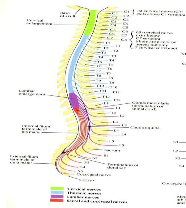
L-SPINE. At C1-2 --> C2 exits At C7-T1 --> C8 exits At T1-T2 -->T1 exits At T12-L1 -->T12 exits At L1-2-->L1 exits At L5-S1-->L5 exits
|
|
|
- Norman Barrett
- 6 years ago
- Views:
Transcription

 -Disc bulge -NF narrowing -Pars defect -Cong canal stenosis or short pedicle (10mm AP for L-spine; 12mm for C-spine) Sag T2 STIR -Conus medullaris (terminates")
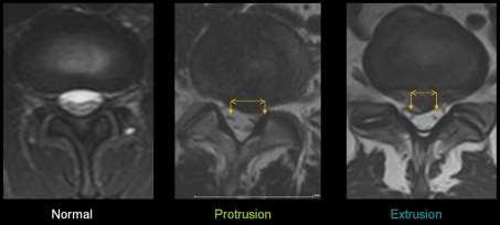
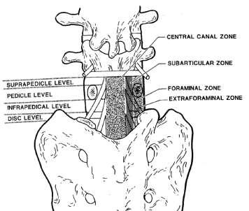
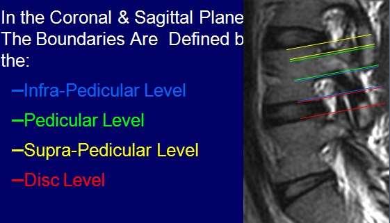
1 L-SPINE CERVICAL SPINE: C1 exits above C1 vert body THORACIC & LUMBAR SPINE: T1 nerve exits below T1 at T1-T2 and similarly, L1 nerve exists below L1 at L1-2; at L3-4 level exiting L3 nerve root within NF and originating L4 nerve root within lateral recess. At C1-2 --> C2 exits At C7-T1 --> C8 exits At T1-T2 -->T1 exits At T12-L1 -->T12 exits At L1-2-->L1 exits At L5-S1-->L5 exits Sag T1 (L R) -Alignment (listhesis: ant/post/lat) -Loss of intervertebral height -Marrow abnl (compare w/ muscle) -Disc bulge -NF narrowing -Pars defect -Cong canal stenosis or short pedicle (10mm AP for L-spine; 12mm for C-spine) Sag T2 STIR -Conus medullaris (terminates at approp level; above L3 at 3mos) -Cord signal (confirm on axial T2) -Disc dessication -Marrow abnl -Annular tear/fissure Axial T2 -Disc pathology (mild <1/3, moderate 1/2-2/3, severe >2/3) -BULGE: diffuse bulge ( deg) beyond edges of ring apophyses -symmetric vs asymmetric -HERNIATION: -BROAD: broad-based protusion (90-180deg) -FOCAL (best seen on sag): -focal protusion (<90deg) triangular (best assessed on sag view) -extrusion (neck) rounded -may migrate (sup/inf) -may have sequestered free disc frag (cranio/caudal ant epidural space) LOCATION: central, paracentral, lateral/foraminal, far-lateral (extraforaminal) -Facet joint (sclerosis, hypertrophy, cystic degen) -Lig flavum hypertrophy -Effect on nerve root w/in lateral recess (originating) and NF (exiting): effaces, abuts, flattens/deforms/displaces, impinges/compresses -Effect on ventral thecal sac and central canal -Annular tear/fissure (high T2) -Arachnoditis (clumped or adhered to dural sac): 3 patterns=empty thecal sac; clumped nerves; cord-like -Synovial cyst (assoc with facet arthropathy)
2 -Epidural lipomatosis (narrows/compresses thecal sac) MISC aorta/iliacs, kidneys, SI joints, pelvis organs POSTOP FAILED BACK spine: Gad with fat sat (alternative=ct myelography); >6wks-6mos; fibrosis/scar enhances, disk only has rim-enhancement. DDX: -epidural scar/fibrosis (enhances) -recurrent/residual/new disk prolapse (does not enhance but peripheral enhancement due to surrounding fibrosis) -inadequate decompression of nerve root -archnoiditis (clumped centrally vs adherent peripherally to wall w/ empty sac appearance vs cord-like central mass) -radiculitis (enhancing nerve root) -hematoma/seroma at laminectomy site (assess for thecal sac compression or extension into site of bone graft at facets which may impair proper fusion) -epidural abscess (if immediate post-op) -pseudomeningocele (due to dural tear) -hardware failure -mechanical instability aka subluxation (if not fused) VERT FX VS TUMOR: fat, air, post-el involvement, multifocal, ST component, post bulge; sup-end plate depressed fx w/ localized edema; chronic compression fx has preserved vert fat but acute is hypointense no T1 TUMOR: Look for focal/circumferential tumor extension with paraspinal or anterior epidural component; degree of spinal canal compromise (thecal sac deformation vs cord contact vs cord compression vs partial/complete effacement of CSF) and spinal instability which may need surgical decompression and hardware prior to XRT; look for impending or current fracture SAMPLE: Alignment is normal without listhesis. No abnormal marrow signal. Conus medullaris terminates at approp level. Thecal sac and contents are normal. No disc herniation, nerve root impingement/compression, or neuroforaminal/cc stenosis. No facet arthropathy or LF hypertrophy. Disc height loss or disc space narrowing. 3mm Grade I anterolisthesis of L3 on L4. Degenerative Modic end-late changes/signal. Diffuse disc bulge (eccentric to left); Central/left paracentral disc protrusion; Focal disc extrusion measuring 5mm and extending cranially/caudally; which along with LF hypertrophy and facet arthropathy results in mild/mod/sig CC stenosis and mild to moderate bilateral neuroforaminal stenosis and right lateral recess stenosis. For purpose of this dictation, the inferior most level of fully formed intervertebral disk will be referred to as L5/S1 or L6/S1.
3 L1-2 Disc Dessic, Disc space narrowing, Listhesis, Hemangioma Bulge/herniation Location/type Annular Fissure Lig Flavum Hypertr Facet Hypertr/ Arthropathy Effect on ventral thecal sac Effect on lateral recess (orig NR) Effect on NF (exit NR) Central Canal Stenosis L2-3 L3-4 L4-5 L5-S1 -Alignment (listhesis) -Pars defect -Marrow signal (modic changes/hemangioma/schmorl) -Disc space narrowing (disc height loss) -Disc dessication -Annular tear/fissure -Conus medullaris and cauda equina -Arachanoditis -Misc: Aorta/iliacs, Kidneys, SI joints, Pelvis organs Disc dessication. [severity] disk space narrowing +/- endplate spondylosis [Central/paracentral/lateral or foraminal/far-lateral or extraforaminal] [diffuse disk bulge] vs [broad-based herniation/ protusion//extrusion (neck) +/-migration/sequestered or free disk fragment located cranio/caudal ant epidural space] Lig flavum hypertrophy Facet arthropathy [abuts/flattens, effaces/displaces ventral thecal sac] [abuts/effaces lateral recess resulting in originating NR impingement/compression] [results in neuroforaminal narrowing with exiting NR impingement/compression; but NR exits freely] [central canal stenosis] NORMAL VARIANTS: -epidural lipomatosis (encases thecal sac with loss of CSF signal around cauda equine) -fatty filum terminale -terminal ventricle (5 th vent; dilated central canal extends below tip of conus into filum) -congenitally short pedicles (narrowed spinal canal) -dilated nerve sheath (tarlov cyst) -paraspinal muscle atrophy -synovial cyst (facet arthropathy) -pars defect
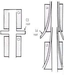
4 -yellow marrow around basivertebral vein (baxton s plexus) -heterogenous marrow w/ intermixed red and yellow marrow -hemangioma (T1 bright; may enhance) -intraosseous lipoma C-SPINE 8 cervical nerves, 7 exit above pedicle of respective vertebra, 8 th exits below level of it vertebra (i.e. T1); Brachial plexus C5 C8/T1. C1 exits below occiput; C4-5 herniated disc will involve C5 nerve root (not so in L-spine) Axial GRE are good for eval of discogenic ridging (GRE often able to distinguish between disc which is relatively bright vs osteophyte which is dark). Cong canal stenosis or short pedicle (10mm AP for L-spine; 12mm for C-spine) C2-3 Diskosteophyte Complex Uncovertebral Hypertrophy; facet arthopathy Ventral thecal sac effacement Cord abuts/ displaces/flattens/ deforms/ compresses Cord signal (myelopathy; myelomalacia) Central canal stenosis NF narrowing C3-4 C4-5 C5-6 C6-7 C7-T1 -Alignment: spondylolithesis -Marrow signal: -Disc height: -Cord signal alteration: -Prevertebral ST: -Paraspinal ST: -Misc: Posterior fossa (tonsils), Pituitary, Thyroid, Airways, Lung apex Vertebral alignment is normal w/o lithesis. BM signal is unremarkable. Atlantoaxial DJD with thickened and post bulging PLL. [severity] disk space narrowing +/- endplate spondylosis Uncovertebral hypertrophy; [severity][diffuse/central /paracentral] disk-osteophyte complex or disk protusion [abuts/effaces] ventral thecal sac and results in [severity] CC stenosis (w/ residual AP diameter of x mm) and [left/right/bilat] [severity] NF narrowing. No cord compression or abnormal cord signal. Effaces ventral thecal sac Abuts ventral cord Displaces cord Flattens ventral cord Compresses/Deforms cord Don t talk about nerve root impingement. NORMAL VARIANT: -atlanto-occ sublux -unfused ant/post arch of C1 -odontoid cleft -os odontoideum
5 -cong fusion of vert body or facet jt -nerve root sleeve cyst -CSF pulsation (flow artifact) -intradural lipoma -extradural arachnoid cyst (remodels osseous str) CORD SIGNAL: -cord edema -compressive myelopathy (cord compression) -myelomalacia (chronic compression; usually with atrophy) -neoplasm (astrocytoma vs ependymoma) vs demyelination vs transverse myelitis DEGREE OF CORD COMPRESSION: -mild 8-10mm AP central canal -mod 6-8mm AP central canal -severe <6mm AP central canal Radiculopathy vs Myelopathy? Radiculopathy: a) Young patients get herniation (acute onset; pain with sitting) b) Older patients get spondylosis w/ lateral recess stenosis (insidious onset; pain better w/ sitting) Myelopathy: a) Cervical (hyperreflexia, spasticity) b) Lumbar (parasthesia, pain, +/-weakness; worse w/ walking mimics vascular claudication) Neurogenic claudication (due to central canal stenosis): bilat LE pain (prox thighs), pain at rest, worse with walking/standing, better w/ sitting/bending forward (flexes spine), worse with spine extension Sciatica: shoots down distal leg, worse with sitting or flexion Vascular claudication: at rest sx stop immediately, sxs more distal, cold foot w/ decreased pulses
6 Myotomal pattern of cervical radiculoapthy? C5: deltoid C6: biceps (decreased biceps DTR) C7: triceps (decreased triceps DTR) C8-T1: interossei, abductor digiti minimi Myotomal pattern of lumbar radiculopathy? L2, L3: Iliopsoas L4: Quadriceps (decreased patellar DTR) L4: extensor hallucis longus S1: gastroc-soleus (decreased ankle DTR)
7 MARROW T1 T2 STIR TYPE I hypo hyper hyper May enhance; painful; can revert to normal or convert to type2 (Inflammation) TYPE II hyper hyper hypo (fat) Fatty marrow TYPE III hypo hypo hypo Discogenic sclerosis Marrow conversion from red to yellow: -extremity axial -epiphysis diaphysis metaphysis -marrow conversion generally completed by mid20s What is normal red marrow: -amount and distribution is symmetric -red marrow still has higher signal on T1 than surrounding muscle -red marrow may enhance after Gad Processes that cause abnormal marrow signal: -Too much red marrow: delayed conversion (obesity, smokers, athletes) or reconversion (anemia or chronic illness) -Infiltration of marrow: cells (tumor, infx, marrow-packing d/o) or edema (trauma or transient) -Osteonecrosis
8
9

10


11
12


13


14

15

16


17
Degenerative Disease of the Spine
 Degenerative Disease of the Spine Introduction: I. Anatomy Talk Overview II. Overview of Disease Processes: A. Spondylosis B. Intervertebral Disc Disease III. Diagnosis IV. Therapy Introduction: Myelopathy
Spine. Neuroradiology. Spine. Spine Pathology. Distribution of fractures. Radiological algorithm. Role of radiology 18/11/2015
 Spine Neuroradiology Spine Prof.Dr.Nail Bulakbaşı X Ray: AP/L/Oblique Vertebra & disc spaces CT & CTA Vertebra, discs, vessels MRI & MRA Vertebra, disc, vessels, meninges Spinal cord & nerves Myelography
CERVICAL SPINE: Radiographs and MRI Cases
 www.jprad.com Radiology reports with recommendations & clinical information - $30 per region, x-ray - $50 per MRI - Medpay Monthly Newsletter 700 East Redlands Blvd, Redlands CA 92373 909.353.9348 jpedley299@yahoo.com
Hidayatullah Hamidi. MD Consultant Radiologist. Lumbar Spine MR Imaging Interpretation
 Hidayatullah Hamidi. MD Consultant Radiologist Lumbar Spine MR Imaging Interpretation 13/12/2018 Presenter Hidayatullah Hamidi Consultant Radiologist, Radiology PGME program director, FMIC, Kabul, Afghanistan
Lumbar spinal canal stenosis Degenerative diseases F 08
 What is lumbar spinal canal stenosis? This condition involves the narrowing of the spinal canal, and of the lateral recesses (recesssus laterales) and exit openings (foramina intervertebralia) for the
3D imaging reformation was obtained. The 3D color imaging reformation was reviewed in a different high resolution setting.
 POST OPERATIVE SPINE WITH CONTRAST CLINICAL INDICATION: Low back pain, Patient is post operative status for L4/5 diskectomy TECHNIQUE: MRI of the lumbosacral spine was performed with multiplanar imaging
SPINAL MAGNETIC RESONANCE IMAGING INTERPRETATION
 CLINICAL VIGNETTE 2017; 3:2 SPINAL MAGNETIC RESONANCE IMAGING INTERPRETATION Editor-in-Chief: Idowu, Olufemi E. Neurological surgery Division, Department of Surgery, LASUCOM/LASUTH, Ikeja, Lagos, Nigeria.
1 Normal Anatomy and Variants
 1 Normal Anatomy and Variants 1.1 Normal Anatomy MR Technique. e standard MR protocol for a routine evaluation of the spine always comprises imaging in sagittal and axial planes, while coronal images are
Essentials of Clinical MR, 2 nd edition. 51. Primary Neoplasms
 51. Primary Neoplasms As with spinal central canal neoplasms in other regions, those of the lumbar spine may be classified as extradural, intradural extramedullary, and medullary. If an extradural lesion
The ABC s of LUMBAR SPINE DISEASE
 The ABC s of LUMBAR SPINE DISEASE Susan O. Smith ANP-BC University of Rochester Department of Neurological Surgery Diagnosis/Imaging/Surgery of Lumbar Spine Disorders Objectives Identify the most common
The ABC s of LUMBAR SPINE DISEASE
 The ABC s of LUMBAR SPINE DISEASE Susan O. Smith ANP-BC University of Rochester Department of Neurological Surgery URMC Neurosurgery APP s Objectives Identify the most common pathology that leads to spine
Peggers Super Summaries: The Aging Spine
 Aging Spine: AGING PROCESS Osteopenia 10% of 50 year old males and 25% of 50 year females Disc dehydration Facet degeneration Soft tissue hypertrophy 2 0 deformity Leg pain worse than back pain from nerve
Epidemiology of Low back pain
 Low Back Pain Definition Pain felt in your lower back may come from the spine, muscles, nerves, or other structures in that region. It may also radiate from other areas like the mid or upper back, a inguinal
Imaging the Degenerative Diseases of the Lumbar Spine
 221 Imaging the Degenerative Diseases of the Lumbar Spine David Malfair, MD a, Douglas P. Beall, MD b,c, * MAGNETIC RESONANCE IMAGING CLINICS Magn Reson Imaging Clin N Am 15 (2007) 221 238 - Degenerative
LUMBAR SPINAL STENOSIS
 LUMBAR SPINAL STENOSIS Always occurs in the mobile segment. Factors play role in Stenosis Pre existing congenital or developmental narrowing of the lumbar spinal canal Translation of one anatomic segment
Spinal Imaging. ssregypt.com. Mamdouh Mahfouz MD
 Spinal Imaging Degenerative diseases ssregypt.com Mamdouh Mahfouz MD mamdouh.m5@gmail.com MRI Open MRI Closed Extremity MRI Dynamic MRI Dynamic MRI The bed rotates from Upright to Recumbent, stopping at
Daniel J. Blizzard, MD, MS
 Daniel J. Blizzard, MD, MS None Common degenerative (usually) condition caused by compression on the spinal cord that is characterized by clumsiness and difficulty with fine motor tasks in the hands and
Comprehension of the common spine disorder.
 Objectives Comprehension of the common spine disorder. Disc degeneration/hernia. Spinal stenosis. Common spinal deformity (Spondylolisthesis, Scoliosis). Osteoporotic fracture. Anatomy Anatomy Anatomy
Module: #15 Lumbar Spine Fusion. Author(s): Jenni Buckley, PhD. Date Created: March 27 th, Last Updated:
: Jenni Buckley, PhD. Date Created: March 27 th, Last Updated:") Module: #15 Lumbar Spine Fusion Author(s): Jenni Buckley, PhD Date Created: March 27 th, 2011 Last Updated: Summary: Students will perform a single level lumbar spine fusion to treat lumbar spinal stenosis.
Module: #15 Lumbar Spine Fusion Author(s): Jenni Buckley, PhD Date Created: March 27 th, 2011 Last Updated: Summary: Students will perform a single level lumbar spine fusion to treat lumbar spinal stenosis.
CERVICAL SPONDYLOSIS & CERVICAL DISC DISEASE
 CERVICAL SPONDYLOSIS & CERVICAL DISC DISEASE Cervical spondylosis l Cervical osteophytosis l Most common progressive disease in the aging cervical spine l Seen in 95% of the people by 65 years Pathophysiology
Spinal canal stenosis Degenerative diseases F 06
 What is spinal canal stenosis? The condition known as spinal canal stenosis is a narrowing (stenosis) of the spinal canal that in most cases develops due to the degenerative (wear-induced) deformation
MR Imaging of the Degenerative Lumbar Spine. Acknowledgements 3/3/2016 MRI
 MR Imaging of the Degenerative Lumbar Spine Gina A. Ciavarra Assistant Professor of Radiology NYU-Langone Medical Center 4/1/2016 Acknowledgements Thank you to Leon Rybak, M.D. and Michael Mechlin, M.D.,
Ligaments of the vertebral column:
 In the last lecture we started talking about the joints in the vertebral column, and we said that there are two types of joints between adjacent vertebrae: 1. Between the bodies of the vertebrae; which
8/4/2012. Causes and Cures. Nucleus pulposus. Annulus fibrosis. Vertebral end plate % water. Deforms under pressure
 Causes and Cures Intervertebral discs Facet (zygopophyseal) joints Inter body joints Spinal nerve roots Nerve compression Pathological conditions Video Causes of back pain Nucleus pulposus Annulus fibrosis
DEGENERATIVE SPINAL DISEASE PRABIN SHRESTHA ANISH M SINGH B&B HOSPITAL
 SPINAL CHAPTER, NESON DEGENERATIVE SPINAL DISEASE PRABIN SHRESTHA ANISH M SINGH B&B HOSPITAL INTRODUCTION DEGENERATIVE SPINAL DISEASE Gradual loss of normal structure and function of spine with time Also
Common fracture & dislocation of the cervical spine. Theerachai Apivatthakakul Department of Orthopaedic Chiangmai University
 Common fracture & dislocation of the cervical spine Theerachai Apivatthakakul Department of Orthopaedic Chiangmai University Objective Anatomy Mechanism and type of injury PE.and radiographic evaluation
University of Jordan. Professor Freih Abuhassan -
 Freih Odeh Abu Hassan F.R.C.S.(Eng.), F.R.C.S.(Tr.& Orth.). Professor of Orthopedics University of Jordan 1 A. Sacroiliitis History Trauma is very common Repetitive LS motion--lumbar rotation or axial
Objectives. Comprehension of the common spine disorder
 Objectives Comprehension of the common spine disorder Disc degeneration/hernia Spinal stenosis Common spinal deformity (Spondylolisthesis, Scoliosis) Osteoporotic fracture Destructive spinal lesions Anatomy
Kathleen R. Fink, MD Virginia Mason Medical Center. 6 th Nordic Emergency Radiology Course 2017
 Kathleen R. Fink, MD Virginia Mason Medical Center 6 th Nordic Emergency Radiology Course 2017 Disclosure My spouse receives research salary support from: Guerbet Outline Acute neck and back pain Acute
SPONDYLOSIS Spin13 (1)
") SPONDYLOSIS Spin13 (1) Spondylosis Last updated: September 5, 2017 ETIOPATHOPHYSIOLOGY... 1 Mechanisms of damage / irritation to neural structures... 2 EPIDEMIOLOGY... 2 Cervical Spondylosis... 3 CLINICAL
SPONDYLOSIS Spin13 (1) Spondylosis Last updated: September 5, 2017 ETIOPATHOPHYSIOLOGY... 1 Mechanisms of damage / irritation to neural structures... 2 EPIDEMIOLOGY... 2 Cervical Spondylosis... 3 CLINICAL
MUSCULOSKELETAL IMAGING FOR PHYSICAL THERAPISTS. COMBINED SECTIONS MEETING 2006 San Diego, CA February 1-5, 2006
 MUSCULOSKELETAL IMAGING FOR PHYSICAL THERAPISTS COMBINED SECTIONS MEETING 2006 San Diego, CA February 1-5, 2006 John Meyer, DPT, OCS University of Southern California Department of Athletic Medicine Los
11 May Disclosure. + Outline: Acute Spine Emergencies
 Kathleen R. Fink, MD University of Washington 5 th Nordic Emergency Radiology Course May 21, 2015 Disclosure My spouse receives research salary support from: Bracco BayerHealthcare Guerbet K Fink Nordic
MR imaging the post operative spine - What to expect!
 MR imaging the post operative spine - What to expect! Poster No.: C-2334 Congress: ECR 2012 Type: Educational Exhibit Authors: A. Jain, M. Paravasthu, M. Bhojak, K. Das ; Warrington/UK, 1 1 1 2 1 2 Liverpool/UK
Introduction to Neuroimaging spine. John J. McCormick MD
 Introduction to Neuroimaging spine John J. McCormick MD Neuroanatomy Netter drawings Radiographic Anatomy Cervical Spine Cervical Spine Oblique View Cervical Spine Dens View Thoracic Spine Lumbar Spine
102 Results RESULTS. Age Mean=S.D Range 42= years -84 years Number % <30 years years >50 years
 102 Results RESULTS A total of 50 cases were studied 39 males and 11females.Their age ranged between 16 years and 84 years (mean 42years). T1 and T2WI were acquired for all cases in sagittal and axial
Cox Technic Case Report #124 published at ( sent October 2013 ) 1
 1") Cox Technic Case Report #124 published at www.coxtechnic.com ( sent October 2013 ) 1 5 th Lumbar Disc Herniation with Spondylolisthesis Treated with Cox Technic Flexion Distraction by Travis Cross BS,
Cox Technic Case Report #124 published at www.coxtechnic.com ( sent October 2013 ) 1 5 th Lumbar Disc Herniation with Spondylolisthesis Treated with Cox Technic Flexion Distraction by Travis Cross BS,
NEURORADIOLOGY. Part III. Angela Csomor University of Szeged Department of Radiology
 NEURORADIOLOGY Part III Angela Csomor University of Szeged Department of Radiology DISEASES OF SPINE AND SPINAL CORD I. Non-tumourous diseases developmental anomalies vascular disorders inflammatory processes
Degenerative Disc Disease. Nafi Aygun, MD. Associate Professor of Radiology
 Degenerative Disc Disease Nafi Aygun, MD. Associate Professor of Radiology Big Problem Great majority of adults suffer from at least one episode of acute low back pain during life time Disc degeneration
Gillian Wooldridge, DO Houston Methodist Willowbrook Hospital Primary Care Sports Medicine Fellowship May 3, 2018
 Gillian Wooldridge, DO Houston Methodist Willowbrook Hospital Primary Care Sports Medicine Fellowship May 3, 2018 Disclosures Neither I nor any family members have financial disclosures Special thanks
Degenerative Spinal Disorders. Gábor Nagy MD PhD Zoltán Papp MD
 Degenerative Spinal Disorders Gábor Nagy MD PhD Zoltán Papp MD Neurosurgery Surgical treatment of diseases of central and peripheral nervous system Surgery of nerves (brain, spine, peripheral nerves) Surgery
Key Primary CPT Codes: Refer to pages: 7-9 Last Review Date: October 2016 Medical Coverage Guideline Number:
 National Imaging Associates, Inc. Clinical guidelines CERVICAL SPINE SURGERY: ANTERI CERVICAL DECOMPRESSION WITH FUSION CERVICAL POSTERI DECOMPRESSION WITH FUSION CERVICAL ARTIFICIAL DISC CERVICAL POSTERI
Spine Trauma- Part B
 Spine Trauma- Part B Cervical Spine Injuries Atlanto- Occipital Dislocation Hyperextension and distraction mechanism Down s syndrome, RA more susceptible Asymmetric lateral masses on odontoid view Widened
eck and Low ack pain: ddressing he Surgical valuation
 eck and Low ack pain: ddressing he Surgical valuation KI FOX, DO T WORTH BRAIN & SPINE Goals Review anatomy Identify sources of pain Imaging: the good, the bad, and the ugly PE: findings to determine source
Current Spine Procedures
 SPINE BOOT CAMP: WHAT YOU DON T KNOW MAY COST YOU! David Abraham, M.D. The Reading Neck and Spine Center Reading, PA Current Spine Procedures Epidural/Transforaminal Injections Lumbar Procedures Laminectomy
2. The vertebral arch is composed of pedicles (projecting from the body) and laminae (uniting arch posteriorly).
 and laminae (uniting arch posteriorly).") VERTEBRAL COLUMN 2018zillmusom I. VERTEBRAL COLUMN - functions to support weight of body and protect spinal cord while permitting movements of trunk and providing for muscle attachments. A. Typical vertebra
VERTEBRAL COLUMN 2018zillmusom I. VERTEBRAL COLUMN - functions to support weight of body and protect spinal cord while permitting movements of trunk and providing for muscle attachments. A. Typical vertebra
Spinal Neoplasms. First Things First!! Localize the Lesion!! Ependymomas. Common Intramedullary Lesions
 Acta Radiológica Portuguesa, Vol.XXIII, nº 90, pág. 101-114, Abr.-Jun., 2011 Spinal Neoplasms Bruno A Policeni University of Iowa Hospitals and Clinics Assistant Professor of Radiology Disclosure of Commercial
A Journey Down The Canal
 A Journey Down The Canal Radiological Assessment of Spinal Cord Masses John Berry-Candelario HMS III Gillian Lieberman, MD BIDMC Objectives Patient review Anatomy of the spine Imaging techniques Classification
PARADIGM SPINE. Patient Information. Treatment of a Narrow Lumbar Spinal Canal
 PARADIGM SPINE Patient Information Treatment of a Narrow Lumbar Spinal Canal Dear Patient, This brochure is intended to inform you of a possible treatment option for narrowing of the spinal canal, often
Evaluation of Patient with Spine Symptoms. Kenneth Nguyen, DO Providence Physiatry
 Evaluation of Patient with Spine Symptoms Kenneth Nguyen, DO Providence Physiatry Epidemiology of Low Back Pain (LBP) Lifetime prevalence of 84% Chronic symptoms in 10-15% 80-90% of economic resources
MRI of LEFT KNEE. There is a fluid collection seen anterior to and inferior to the superiorly displaced patella.
 MRI of LEFT KNEE Protocol: Multiplanar MRI of the left knee joint performed in the sagittal, coronal and transverse planes using T1 weighted spin echo, T2 and proton-density weighted fast spin echo, fatsaturated
Common Thoraco- Lumbar Problems in the Mature Athlete
 Common Thoraco- Lumbar Problems in the Mature Athlete Diana Heiman, MD Associate Professor, Family Medicine Residency Director East Tennessee State University Objectives Review the pathophysiology of the
Cox Technic Flexion Distraction and Decompression Relieves Right Lower Extremity Radiculopathy and Low Back Pain Post Laminectomy
 Cox Technic Case Report #94 published at www.coxtechnic.com ( sent April 2011 on 4/9/11 ) 1 Cox Technic Flexion Distraction and Decompression Relieves Right Lower Extremity Radiculopathy and Low Back Pain
Imaging of Cervical Spine Trauma Tudor H Hughes, M.D.
 Imaging of Cervical Spine Trauma Tudor H Hughes, M.D. General Considerations Most spinal fractures are due to a single episode of major trauma. Fatigue fractures of the spine are unusual except in the
Bony framework of the vertebral column Structure of the vertebral column
 5.1: Vertebral column & back. Overview. Bones o vertebral column. o typical vertebra. o vertebral canal. o spinal nerves. Joints o Intervertebral disc. o Zygapophyseal (facet) joint. Muscles o 2 compartments:
Case Studies, Impairment of the Spine in Washington State
 Case Studies, Impairment of the Spine in Washington State NAOEM at Skamania, 2015 25 Sep, 2015 Tim Gilmore, MD Several Slides from this Presentation Borrowed with permission from the Washington State Department
Pathophysiology of lumbar disc degeneration: a review of the literature. Neurosurg Focus 13 (2): August, 2002
: August, 2002") Pathophysiology of lumbar disc degeneration: a review of the literature Neurosurg Focus 13 (2): August, 2002 MICHAEL D. MARTIN, M.D., CHRISTOPHER M. BOXELL, M.D., F.A.C.S., AND DAVID G. MALONE, M.D. FROM
Pathophysiology of lumbar disc degeneration: a review of the literature Neurosurg Focus 13 (2): August, 2002 MICHAEL D. MARTIN, M.D., CHRISTOPHER M. BOXELL, M.D., F.A.C.S., AND DAVID G. MALONE, M.D. FROM
BACK PAIN. Disclaimer. Integrated web marketing. Multimedia Health Education
 BACK PAIN Disclaimer This movie is an educational resource only and should not be used to make a decision on. All decisions about surgery must be made in conjunction with your surgeon or a licensed healthcare
Imaging of Trauma to the Spine. Orthopedic Diplomate Program University of Bridgeport College of Chiropractic
 Imaging of Trauma to the Spine Orthopedic Diplomate Program University of Bridgeport College of Chiropractic Jefferson Fracture Yee, LL: The Jefferson Fracture, Radiology Cases in Pediatric Emergency Medicine.
Interlaminar Decompression & Stabilization. Reginald Davis, M.D., FAANS, FACS Director of Clinical Research
 Interlaminar Decompression & Stabilization Reginald Davis, M.D., FAANS, FACS Director of Clinical Research Disclosures Background Device meant to stabilize the spine without fusion following decompression
VERTEBRAL COLUMN ANATOMY IN CNS COURSE
 VERTEBRAL COLUMN ANATOMY IN CNS COURSE Vertebral body Sections of the spine Atlas (C1) Axis (C2) What type of joint is formed between atlas and axis? Pivot joint What name is given to a fracture of both
Spine Pain Management Program
 Spine Pain Management Program Please complete the following information: Patient Name: Patient ID Number: Patient DOB: The procedure being requested: Epidural Adhesiolysis Please check the indication (reason)
Spinal Cord Injuries: The Basics. Kadre Sneddon POS Rounds October 1, 2003
 Spinal Cord Injuries: The Basics Kadre Sneddon POS Rounds October 1, 2003 Anatomy Dorsal columntouch, vibration Corticospinal tract- UMN Anterior horn-lmn Spinothalamic tractpain, temperature (contralateral)
Case Report: CASE REPORT OF FACET ARTHROPATHY INDUCED NERVE ROOT COMPRESSION RESULTING IN MOTOR WEAKNESS AND PAIN
 Cox Technic Case Report #100 published at www.coxtechnic.com (sent October 2011 on 10/11/11 ) 1 Case Report: CASE REPORT OF FACET ARTHROPATHY INDUCED NERVE ROOT COMPRESSION RESULTING IN MOTOR WEAKNESS
Radiography of the Spine
 Radiography of the Spine Radiography of the Spine Attila ARANY-TóTH, DVM Complex anatomy Vertebrae: 7 cervical, 13 thoracal, 7 lumbal, 3 sacral, n caudal Thorough neurological examination - localization!!!
HERNIATED DISCS AN INTRODUCTION TO
 AN INTRODUCTION TO HERNIATED S This booklet provides general information on herniated discs. It is not meant to replace any personal conversations that you might wish to have with your physician or other
HISTORY AND CHIEF COMPLAINT:
 submitted by Keith M. Bartley, D.C. Jasper, IN 07/21/11 presented at Cox Seminar in Nashville, TN, on October 8 9, 2011 HISTORY AND CHIEF COMPLAINT: 01/21/11 55 year old male press operator for Jasper
Discal herniation and spondylosis
 III.8.4.6 Degenerative disorders of the spine Introduction the frequency of locomotor disorders increases with age Low back pain is a very common disorder. According to medical literature, it is the second
River North Pain Management Consultants, S.C., Axel Vargas, M.D., Regional Anesthesiology and Interventional Pain Management.
 River North Pain Management Consultants, S.C., Axel Vargas, M.D., Regional Anesthesiology and Interventional Pain Management. Chicago, Illinois, 60611 Phone: (888) 951-6471 Fax: (888) 961-6471 Clinical
Disclosures 5/27/2016. Narrowing of the spinal canal or neuroforamina causing a symptomatic compression of the neural element.
 Radiculopathy Saggital MRI view Contained disc extrusion uplifting PLL from bone Terrence Julien, MD, FACS, (blue FAANS arrows) Associate Professor of Neurosurgery System Director, Surgical Neuro-Oncology
Osteoarthrosis, unspecified whether generalized or localized, lower leg. Osteoarthrosis, localized, not specified whether primary or secondary, pelvic
 Page 1 Appendix TABLE E-1 Codes (and Definitions) in Humana Database Used for Study Inclusion and Exclusion of Patients Who Underwent,, or 1 to 2-Level Inclusion ICD-9-P-8154 Total knee replacement ICD-9-D-71596
Cervical Spine: Pearls and Pitfalls
 Cervical Spine: Pearls and Pitfalls Presenters Dr. Rob Donkin Functional Anatomy Current research Cervical Radiculopathy Dr. Gert Ferreira Red flags Case Study Kinesio Taping Chris Neethling Gonstead adjusting
Neurological manifestations of thoracic myelopathy in 203 patients
 Neurological manifestations of thoracic myelopathy in 203 patients Shota Takenaka* a, MD; Takashi Kaito b, MD; Noboru Hosono a, MD; Toshitada Miwa c, MD; Takenori Oda d, MD; Shinya Okuda d, MD; Tomoya
Lumbar Spinal Epidural Lipomatosis: Prevalence and Patterns Abstract: Keywords: Introduction II. Materials And Methods
 IOSR Journal of Dental and Medical Sciences (IOSR-JDMS) e-issn: 2279-0853, p-issn: 2279-0861.Volume 15, Issue 10 Ver. VII (October. 2016), PP 27-32 www.iosrjournals.org Lumbar Spinal Epidural Lipomatosis:
ABCs of the degenerative spine
 Insights into Imaging (2018) 9:253 274 https://doi.org/10.1007/s13244-017-0584-z PICTORIAL REVIEW ABCs of the degenerative spine Sergiy V. Kushchayev 1 & Tetiana Glushko 1 & Mohamed Jarraya 1 & Karl H.
Spinal Column. Anatomy Of The Spine
 Anatomy Of The Spine The spine is a flexible column, composed of a stack of individual bones. Each bone is called a vertebra. There are seven vertebrae in the neck (cervical vertebrae) twelve in the thoracic
Clinical Study. Overview
 Clinical Study David A. Mayer, MD, JD, FICS Associate Professor of Clinical Surgery New York Medical College Chairman of Surgery Emeritus Overview A practice based prospective study of 31 consecutive patients
Objectives. Identify and differentiate appropriate surgical cases. Good Surgical Outcomes
 ECHO February 5 th, 2015 Surgical Selection for Low Back Pain Objectives Identify and differentiate appropriate surgical cases Disclosures Medical director for UHN Rehabilitations Solution Back and Neck
WORKPLACE SAFETY AND INSURANCE APPEALS TRIBUNAL DECISION NO. 2257/13
 WORKPLACE SAFETY AND INSURANCE APPEALS TRIBUNAL DECISION NO. 2257/13 BEFORE: B. Alexander : Vice-Chair A.D.G. Purdy : Member Representative of Employers M. Ferrari : Member Representative of Workers HEARING:
Patient Information MIS LLIF. Lateral Lumbar Interbody Fusion Using Minimally Invasive Surgical Techniques
 Patient Information MIS LLIF Lateral Lumbar Interbody Fusion Using Minimally Invasive Surgical Techniques Table of Contents Anatomy of Spine....2 General Conditions of the Spine....4 What is Spondylolisthesis....5
Spine Pain Management Program
 Spine Pain Management Program Please complete the following information: Patient Name: Patient ID Number: Patient DOB: The procedure being requested: Epidural Injection Please check the indication (reason)
SUBAXIAL CERVICAL SPINE TRAUMA- DIAGNOSIS AND MANAGEMENT
 SUBAXIAL CERVICAL SPINE TRAUMA- DIAGNOSIS AND MANAGEMENT 1 Anatomy 3 columns- Anterior, middle and Posterior Anterior- ALL, Anterior 2/3 rd body & disc. Middle- Posterior 1/3 rd of body & disc, PLL Posterior-
A Patient s Guide to Artificial Cervical Disc Replacement
 A Patient s Guide to Artificial Cervical Disc Replacement Each year, hundreds of thousands of adults are diagnosed with Cervical Disc Degeneration, an upper spine condition that can cause pain and numbness
POSTERIOR CERVICAL FUSION
 AN INTRODUCTION TO PCF POSTERIOR CERVICAL FUSION This booklet provides general information on the Posterior Cervical Fusion (PCF) surgical procedure for you to discuss with your physician. It is not meant
SPINAL CORD DISEASE IN DOGS PART TWO: MOST LIKELY CAUSES
 Vet Times The website for the veterinary profession https://www.vettimes.co.uk SPINAL CORD DISEASE IN DOGS PART TWO: MOST LIKELY CAUSES Author : RITA GONÇALVES Categories : Vets Date : April 7, 2014 RITA
Neuroimaging. spine / spinal cord
 Neuroimaging spine / spinal cord Spine & spinal cord imaging methodology Plain x-ray of spine Computed tomography CT - traditional ( normal CT) - reconstructions - myelo-ct Magnetic resonance MR - standard
Patient Chart Quotes. Spine Mythology and Evidence- Based Management of Back Pain. Patient Chart Quotes. Patient Chart Quotes
 Spine Mythology and Evidence- Based Management of Back Pain John Engstrom, MD Professor of Neurology August 11, 2009 Patient Chart Quotes The patient was in his usual state of good health until his airplane
 www.fisiokinesiterapia.biz NOTE : THIS PRESENTATION DOES NOT REPLACE ATTENDANCE OR INFORMATION GIVEN IN THE LECTURE.IT IS INTENDED AS A HIGHLIGHT FOR THE TOPIC INTRODUCTION 60-80% of people will have LBP
Orthopadic cors. Topic : -Cervical spondylitis. -Development disorders(spondylolysis and Spodylolsithesis)
") Orthopadic cors Topic : -Cervical spondylitis. -Development disorders(spondylolysis and Spodylolsithesis) Cervical spondylitis. Definition : - a painful condition of the cervical spine resulting from the
Orthopadic cors Topic : -Cervical spondylitis. -Development disorders(spondylolysis and Spodylolsithesis) Cervical spondylitis. Definition : - a painful condition of the cervical spine resulting from the
DEGENERATIVE SPONDYLOLISTHESIS
 AN INTRODUCTION TO DEGENERATIVE SPONDYLOLISTHESIS This booklet is designed to inform you about lumbar degenerative spondylolisthesis. It is not meant to replace any personal conversations that you might
Anatomy of the Nervous System. Brain Components
 Anatomy of the Nervous System Brain Components NERVOUS SYSTEM INTRODUCTION Is the master system of human body, controlling the functions of rest of the body systems Nervous System CLASSIFICATION A. Anatomical
Coventry Pain Clinic - Spianl Pain - Sciatica and Brachalgia
 Coventry Pain Clinic - Spianl Pain - Sciatica and Brachalgia Copyright 2002-2005 Dr. Richard S. Walker Introduction Spinal Nerve Root Pain (Radiculopathy) can arise from the problems affecting the:- C4
Cox Technic Case Report #169 published at (sent 5/9/17) 1
 1") Cox Technic Case Report #169 published at www.coxtechnic.com (sent 5/9/17) 1 Management of Lumbar Radiculopathy Associated with an Extruded L4 L5 disc and concurrent L5 S1 Spondylolytic Spondylolisthesis
Cox Technic Case Report #169 published at www.coxtechnic.com (sent 5/9/17) 1 Management of Lumbar Radiculopathy Associated with an Extruded L4 L5 disc and concurrent L5 S1 Spondylolytic Spondylolisthesis
Diagnosis of Neck & Upper Extremity Pain
 Diagnosis of Neck & Upper Extremity Pain David B. Bumpass, MD Assistant Professor, Spine Surgery UAMS Depts. of Orthopaedic Surgery & Neurosurgery May 12, 2018 Disclosures Medtronic Spine speaking fees
INTERVERTEBRAL FORAMEN STUDIES
 INTERVERTEBRAL FORAMEN STUDIES I. FORAMEN ENCROACHMENT ASSOCIATED WITH DISC HERNIATION* LEE A. HADLEY, M.D. t Syracuse, New York (Received for publication November ] 8, 1949) T HESE studies are the outgrowth
Persistent Terminal Ventricle
 Persistent Terminal Ventricle Ventriculus Terminalis Incomplete regression of TV of 2 neurulation, continuity with central canal small cavity PTV vs terminal myelocystocele (?severe manifestation from
Nursing review section of Surgical Neurology International: Part 1 lumbar disc disease
 SNI: Neurosurgical Nursing OPEN ACCESS For entire Editorial Board visit : http://www.surgicalneurologyint.com Editor: Nancy E. Epstein, MD Winthrop Hospital, Mineola, NY, USA Review Article Nursing review
DAUGHERTY & ASSOCIATES, LLC
 DAUGHERTY & ASSOCIATES, LLC August 19, 2008 Mr. RA A Law Firm 111 East Ohio Street, Anywhere, Indiana 46204 Dear Mr. A, Thank you for giving me the opportunity to review the file of DD. As you are aware,
Alan H Daniels, MD. Spine Division, Department of Orthopaedics Warren Alpert School of Medicine of Brown University
 Spinal and Orthopaedic Surgery in the Elderly Alan H Daniels, MD Spine Division, Department of Orthopaedics Warren Alpert School of Medicine of Brown University As the population ages, and patients remain
ESSENTIALS OF PLAIN FILM INTERPRETATION: SPINE DR ASIF SAIFUDDIN
 ESSENTIALS OF PLAIN FILM INTERPRETATION: SPINE DR ASIF SAIFUDDIN Consultant Musculoskeletal Radiologist Royal National Orthopaedic Hospital Stanmore,UK. INTRODUCTION 2 INTRODUCTION 3 INTRODUCTION Spinal
ACDF. Anterior Cervical Discectomy and Fusion. An introduction to
 An introduction to ACDF Anterior Cervical Discectomy and Fusion This booklet provides general information on ACDF. It is not meant to replace any personal conversations that you might wish to have with