* Articular system II
|
|
|
- Kristina Dickerson
- 5 years ago
- Views:
Transcription
1 Articular system II


2 Hyaline Fibrous Elastic - Has an intercellular matrix rich in hyaluronic acid and mucopolysaccharides, which are natural lubricants - Forms the articular cartilage in most joints - Provides the anlage for long bone development - Has an intercellular matrix rich in mucopolysaccharides and bundles of collagenous fibers - Mucoplysaccharides provide high water content - Especially resilient and durable form of cartilage - Forms most symphyses and joint discs - Has an intercellular matrix rich in mucopolysaccharides and bundles of elastic fibers, which provide a strong, yet flexible, support - Forms the skeletal structure of external ear and the tip of the nose
:")
Synostosis 2.")
3 Classification of the joints 1.Synarthrosis (immovable joint): 1)Syndesmosis 2)Synchondrosis 3)Synostosis 2. Diarthrosis (synovial joint, joint)


4 Complexity of the joints Simple joint Complex joint - two articular surfaces - more than two articular surfaces


5 Synovial joints Combined joint - Two anatomically isolated joints move together at the same time
 Symphysis / synchondrosis Syndesmosis")
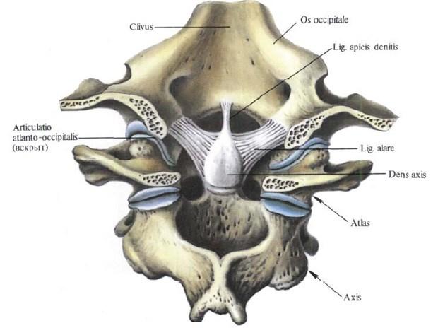
6 Joints of the vertebral column Zygapophysial joint (simple plane combined synovial joint) Symphysis / synchondrosis Syndesmosis (Ligaments)

7 Intervertebral disc - Each disc composed of an outer fibrocartilaginous portion (annulus fibrosus) and an inner gelatinous central portion (nucleus pulposus) - Nucleus pulposus remnant of notochord Functions of intervertebral disc: - Permit limited movement between adjacent vertebrae - Nucleus pulposus compressible but deformable pad, that distribute forces over the entire surface of the vertebra
8 * The anterior longitudinal ligament (lig. longitudinale anterius) * The posterior longitudinal ligament (lig. longitudinale posterius) * The yellow ligaments (ligg. flava) elastic fibers! * The interspinous ligaments (ligg. interspinalia * The supraspinous ligament (lig. supraspinale * The intertransverse ligaments (ligg. intertransversaria)
9 *Flexion and extension along frontal axis
10 *Lateral flexion along sagittal axis *Rotation along vertical axis


11 Lordosis Kyphosis Lordosis Kyphosis

12

13 *Adduction and abduction along sagittal axis

14 *Elevation and depression along transverse axis


15 Articular surfaces: *The head of the humerus the glenoid cavity of the scapula. *The glenoid labrum (labrum glenoidale) is on the circumference of the glenoid cavity. It increases its depth.

16

17

18 *Simple spheroidal joint.
19 *Flexion and extension along frontal axis
20 *Adduction and abduction along sagittal axis
21 *Rotation and circumduction along vertical axis
 Articular capsule embraces the olecranon, radial and coronoid fossae but leaves the epicondyles free.")
22 Three articulating bones form three joints invested in a common capsule: 1) humero-ulnar joint (hinge joint) 2) humeroradial joint (spheroidal joint) 3) proximal radio-ulnar joint (cylindrical=pivot joint) Articular capsule embraces the olecranon, radial and coronoid fossae but leaves the epicondyles free.

23 Elbow joint
24 *Flexion and extension along frontal axis
25 *Pronation and supination along vertical axis *Combined movement in proximal and distal radio-ulnar joint
 scaphoid, lunate and triquetral bones. Complex ellipsoid joint.")
26 Articular surfaces The carpal articular surface of the radius and the articular disc (the distal radio-ulnar joint) scaphoid, lunate and triquetral bones. Complex ellipsoid joint.

27
28 *Flexion and extension along frontal axis
29 *Adduction and abduction along sagittal axis
30

31 The hip joint (articulatio coxae)

32 The hip joint (articulatio coxae)
 Lig.")
 Zona orbicularis")
33 The hip joint Ligaments: 1) Lig. iliofemorale 2) Lig. ischiofemorale 3) Lig. pubofemorale 4) Zona orbicularis 5) Lig. capitis femoris
34 *Flexion and extension along frontal axis
35 *Adduction and abduction along sagittal axis

36 *Rotation along vertical axis


37 Complex bicondylar joint


38 Intra-articular ligaments


39 Extra-articular ligaments


40 Anterolateral ligament of the knee joint Hypothesized function - control internal tibial rotation, stabilize internal rotation
 223, pp321--328")
41 Origin on the prominence of the lateral femoral epicondyle Insertion - the body of the ALL ran an oblique course to the anterolateral side of the proximal tibia. Anatomy of the ALL, S. Claes et al. J. Anat. (2013) 223, pp

42 Fig. 4 Anatomic drawing considering the ALL and its relationship with wellknown anatomical landmarks on the lateral aspect of the human knee. (A) Knee in full extension. (B) Knee in 90 of flexion. ALL, anterolateral ligament; LCL, lateral collateral ligament; GT, Gerdy s tubercle; LFE, lateral femoral epicondyle; PT, popliteus tendon; PFL, popliteo-fibular ligament. Anatomy of the ALL, S. Claes et al. J. Anat. (2013) 223, pp

43 1879, years before the discovery of X-rays, Dr. Paul Segond described a remarkably constant avulsion fracture pattern at the anterolateral proximal tibia as a result of forced internal rotation at the knee (Segond fracture)

44 Lateral and medial menisci of the knee joint (viscoelastic soft tissue) Functions of the menisci: - adapt articular surfaces of femur and tibia, increase their congruence, hence the stresses on tibial cartilage are reduced - to distribute loads and therefore reduce the stresses on the tibia, - joint stabilisation; - shock absorption; - joint lubrication; - cartilage protection and prevention of osteoarthrosis
.")
45 Human meniscus Fig. 1. Human meniscus. (a) Right human knee joint viewed from above (the femur has been removed); the tibial tuberosity is on top. The medial and lateral menisci are connected by a transverse ligament (TL). 1 - anterior insertional ligament of the medial meniscus; 2 - posterior insertional ligament of the medial meniscus; 3 - anterior insertional ligament of the lateral meniscus; 4 - posterior insertional ligament of the lateral meniscus; ACL - cross section of the anterior cruciate ligament; PCL cross section of the posterior cruciate ligament. Karola Messner and Jizong Gao. The menisci of the knee joint. Anatomical and functional characteristics, and a rationale for clinical treatment. Review. J. Anat. (1998) 193, pp. 161±178
46 Karola Messner and Jizong Gao. The menisci of the knee joint. Anatomical and functional characteristics, and a rationale for clinical treatment. Review. J. Anat. (1998) 193, pp. 161±178 Fig. 2. Diagram demonstrating the importance of intact meniscal entheses for the load distribution function of the meniscus. (a) With intact entheses the load (thick arrows) is transmitted via the menisci and articular cartilage through a large contact area (left hand side of figure; small arrows). Part of the load is transformed to hoop stresses (right hand side of figure; long arrows). (b) When the insertional ligaments are transected (right hand side of figure; arrowheads), the meniscus will extrude from the knee joint during loading, and the load (left hand side of figure; thick arrows) is mainly transmitted via articular cartilage through a reduced contact area (small arrows).


47
48 *Flexion and extension along frontal axis
49 Rotation becomes possible when the knee is flexed!


50 Articulatio talocruralis Complex saddle joint

51 Ankle joint ligaments Medial ligaments
52 *Plantar flexion and dorsi flexion along frontal axis

53 *Inversion and eversion along vertical axis
, Transverse arch (tarsal), in formation of this arch navicular, cuboid and cuneiform bones take part (the highest points of all the longitudinal")
54 3 support points: Tuber calcanei Caput ossis metatarsi I Caput ossis metatarsi V Five longitudinal arches: from tuber calcanei to the heads of metatarsal bones (The longest arch is the second arch), Transverse arch (tarsal), in formation of this arch navicular, cuboid and cuneiform bones take part (the highest points of all the longitudinal arches)


55 - Weight distribution, amortization - Adequate blood supply of foot
56 *Simple bicondylar combined joint
 the disc acts as a shock absorber when the joint is subjected to impact loading. *The surfaces are complemented by a fibrous articular disc (discus articularis) located between them.")
57 Disc is made out of fibrocartilage with markedly anteroposterior alignment. Functions: 1) to diminish the effects of incongruence between the articular surfaces. 2) the disc acts as a shock absorber when the joint is subjected to impact loading. *The surfaces are complemented by a fibrous articular disc (discus articularis) located between them. The edges of the disc are joined to the articular capsule as a result of which the articular cavity is separated into two isolated compartments.

58 Articular capsule is attached along the borders of articular surfaces. The mandibular neck is within the articular cavity.

59

60
61 A B *When the mandible moves laterally, one condyle (B) moves forward and inward, while the other condyle (A) will shift slightly in a lateroposterior (or rotate in vertical axis) direction


62
63 Consequence of the answer: 1.Name of the joint (English and Latin) 2.Classification of the joint (simple, combined, complex) 3.Description of the essential elements of the joint (articular surface, type of cartilage, cavity and capsule) 4.Description of the ligaments. 5.Special features (bursa) 6.Movements
64 Which of the joint has intraarticular ligaments: 1) Shoulder joint 2) Hip joint 3) Elbow joint 4) Knee joint 5) TMJ 6) Wrist joint
65 Hyaline cartilage covers the surfaces of the joints: 1) Shoulder joint 2) Hip joint 3) Sternoclavicular joint 4) Knee joint 5) TMJ 6) Wrist joint

66
67

68

69

70
 combines two joints: - Calcaneocuboid joint - Talonavicular joint Ligg.")
71 Ligamentum bifurcatum: - lig. calcaneonaviculare - lig. calcaneocuboideum - key of Chopart`s joint Articulatio tarsi transversa (Chopart`s joint) combines two joints: - Calcaneocuboid joint - Talonavicular joint Ligg. cuneometatarsalia interossea: Articulatio tarsimetatarsales (Lisfrank`s joint) The key of Lisfrank`s joint cuneometatarsal interosseus ligament between medial cuneiform bone and second metatarsal bone
Chapter 9 Articulations Articulations joints where two bones interconnect. Two classification methods are used to categorize joints:
 Chapter 9 Articulations Articulations joints where two bones interconnect Two classification methods are used to categorize joints: Functional classification Structural classification Functional classification
CLASSIFICATION OF JOINTS STRUCTURAL VS FUNCTIONAL
 CHAPTER 8 JOINTS CLASSIFICATION OF JOINTS STRUCTURAL VS FUNCTIONAL The most moveable type of joint is a 1) Synarthrosis 2) Amphiarthrosis 3) Diarthrosis FIBROUS JOINTS Figure 8.1 Fibrous joints. (a) Suture
Biology 325 Fall 2003
 Name: pre-lab exercise due at beginning of your lab session Matching a. fibrous joints b. cartilaginous joints c. synovial joints 1. exhibit a joint cavity 2. types are sutures and syndesmoses 3. bones
UNIT 2 - CHAPTER 8: JOINTS OF THE SKELETAL SYSTEM LEARNING OUTCOMES:
 LEARNING OUTCOMES: 8.1 Types of Joints 1. Explain how joints can be classified according to the type of tissue that binds the bones together and the degree of movement possible at the joint. (p. 268) 2.
UNIT 2 - CHAPTER 8: JOINTS OF THE SKELETAL SYSTEM LEARNING OUTCOMES:
 LEARNING OUTCOMES: 8.1 Introduction 1. List the functions of joints. 2. Explain how joints can be classified according to the type of tissue that binds the bones together and the degree of movement possible
8.2: Fibrous Joints. There are three (3) types of fibrous joints (synarthroses): Syndesmosis Suture Gomphosis. Interosseus membrane of leg.
 types of fibrous joints (synarthroses): Syndesmosis Suture Gomphosis. Interosseus membrane of leg.") 8.1: Introduction Are known as articulations Functional junctions between bones Bind parts of skeletal system together Make bone growth possible Permit parts of the skeleton to change shape during childbirth
8.1: Introduction Are known as articulations Functional junctions between bones Bind parts of skeletal system together Make bone growth possible Permit parts of the skeleton to change shape during childbirth
Introduction. Fibrous Joints. 8.1: Types of Joints. Cartilaginous Joints. Fibrous Joints 12/14/2016. Chapter 08 Lecture Outline
 Introduction Chapter 08 Lecture Outline See separate PowerPoint slides for all figures and tables preinserted into PowerPoint without notes. Joints (Articulations): Functional junctions between bones Bind
Exercise 13. Articulations and Body Movements
 Exercise 13 Articulations and Body Movements Articulations Articulations, or joints, are points where a bone is connected to one or more other bones. Articulations hold the skeleton together. Articulations
Amy Warenda Czura, Ph.D. 1 SCCC BIO130 Lab 7 Appendicular Skeleton & Articulations
 The Skeletal System II: Appendicular Skeleton and Articulations Exercises 11, 13 (begins: page 145 in 9 th and 10 th editions) Exercises 10, 11 (begins: page 147 in 11 th edition, page 149 in 12 th edition)
Anatomy and Physiology 1 Chapter 9 self quiz Pro, Dima Darwish,MD.
 Anatomy and Physiology 1 Chapter 9 self quiz Pro, Dima Darwish,MD. 1) Joints can be classified structurally as A) bony. B) fibrous. C) cartilaginous. D) synovial. E) All of the answers are correct. 2)
Joints Dr. Ali Ebneshahidi
 Joints Dr. Ali Ebneshahidi Function of Joints 1. Serve as functional junctions between bones. 2. Bind bones, strokes, and other related tissues together. 3. Allow bone growth to occur. 4. Permit certain
I. Introduction. Unit Two. of the Skeletal System. II. Classification of Joints. URLs for this chapter:
 8 URLs for this chapter: http://www.vh.org/adult/provider/radiology/joint Fluoro/JointFluoroHP.html of the Skeletal System Karen Webb Smith Unit Two http://www.science.ubc.ca/~biomania/tutorial/bonejt/
* Articular system I
 *Articular system I *Articular system=syndesmology (Systema articulare) System of joints Joint occurs, where 2 bones meet Combine bones of skeleton into a single unit Provide mobility *Classification
Pelvic Girdle
 ARTICULATIONS OF LOWER EXTREMITY Pages 429-437 Pelvic Girdle formed by connection of the hip bones and the sacrum Sacroiliac Joints compound joints synovial joint - anterior, between the auricular surfaces
PowerPoint Lecture Slides prepared by Janice Meeking, Mount Royal College C H A P T E R. Joints: Part A. Copyright 2010 Pearson Education, Inc.
 PowerPoint Lecture Slides prepared by Janice Meeking, Mount Royal College C H A P T E R 8 Joints: Part A Warm Up 11/28/16 Happy Thanksgiving welcome back! J (be ready to share something fun you did over
9.1 Joints. Objectives Describe the structural and functional classifications of joints
 Joints 9.1 Joints Describe the structural and functional classifications of joints Joints have both structural and functional classifications: The criteria for classifying joints structurally are anatomical
Skeletal System Joints, Relationship with other systems
 Skeletal System Joints, Relationship with other systems Review the Types of Bones Articulations Classification of Joints (Articulations) Joint Where two bones interact Three functional classes of joint
Articulations. Articulation. Joint between bones. Does not mean movement! Some joints are immovable; sutures.
 Articulations Joint between bones Articulation Does not mean movement Some joints are immovable; sutures. Classification of joints Two questions about joints: 1- How does it move? - functional 2- How is
The Dance Hall by Vincent van Gogh,1888
 The Dance Hall by Vincent van Gogh,1888 Articulations of the pelvic girdle Lumbosacral joints, sacroiliac joints & pubic symphysis The remaining joints of the lower limb Hip joint Knee joint Tibiofibular
Joints of the upper limb II
 Joints of the upper limb II Prof. Abdulameer Al-Nuaimi E-mail: a.al-nuaimi@sheffield.ac.uk E. mail: abdulameerh@yahoo.com Elbow joint The elbow joint is connecting the upper arm to the forearm. It is classed
Joints. Judi Laprade. Illustrations from: Essential Clinical Anatomy 3 rd ed. (ECA3) Moore, K. and Agur, A. Lippincott Williams and Wilkins, 2007
 Moore, K. and Agur, A. Lippincott Williams and Wilkins, 2007") Slide 1 Joints Judi Laprade Illustrations from: Essential Clinical Anatomy 3 rd ed. (ECA3) Moore, K. and Agur, A. Lippincott Williams and Wilkins, 2007 Grant s Atlas of Anatomy 12 th ed. (GA12) Agur, A.
Slide 1 Joints Judi Laprade Illustrations from: Essential Clinical Anatomy 3 rd ed. (ECA3) Moore, K. and Agur, A. Lippincott Williams and Wilkins, 2007 Grant s Atlas of Anatomy 12 th ed. (GA12) Agur, A.
Ch. 8 Joints of the Skeletal System
 Ch. 8 Joints of the Skeletal System Part 1: Classifying Joints & Joint Movements Interactive pages 269-278 Types of Joints (AKA: Articulations) Structural Classification (type of tissue that binds the
Lecture 9: Arthrology
 Lecture 9: Arthrology M/O Chapter 9 45. Classify joints based on the degree of movement allowed and give examples of each classification. 46. Classify joints based on anatomical structure and give examples
Student Objectives. When you have completed the exercises in this chapter, you will have accomplished the following objectives:
 Student Objectives When you have completed the exercises in this chapter, you will have accomplished the following objectives: Classification of Joints 1. Define joint or articulation. 2. Classify joints
and medial) circumduction supination pronation eversion Tibial
 circumduction supination pronation eversion Tibial") T igure l8.l Anterior view of right knee (patella removed). emur Posterior cruciate Anterior cruciate meniscus meniscus ibular----collateral tji,l-+;jli your own body to demonstrate the follon-ing ioint
T igure l8.l Anterior view of right knee (patella removed). emur Posterior cruciate Anterior cruciate meniscus meniscus ibular----collateral tji,l-+;jli your own body to demonstrate the follon-ing ioint
Answers to Pre-Lab Quiz (p. 171) Answers to Activity Questions
 Answers to Activity Questions") Answers to Pre-Lab Quiz (p. 171) 1. Holds bones together; allows the rigid skeleton some flexibility so that gross body movements can occur 2. c, amount of movement allowed by the joint 3. synovial 4.
Answers to Pre-Lab Quiz (p. 171) 1. Holds bones together; allows the rigid skeleton some flexibility so that gross body movements can occur 2. c, amount of movement allowed by the joint 3. synovial 4.
Anatomy. Anatomy deals with the structure of the human body, and includes a precise language on body positions and relationships between body parts.
 Anatomy deals with the structure of the human body, and includes a precise language on body positions and relationships between body parts. Proper instruction on safe and efficient exercise technique requires
Definition: A joint or articulation is a place in the body where two bones come together.
 Definition: A joint or articulation is a place in the body where two bones come together. CLASSES OF JOINTS. 1. Joints are classified according to how the bones are held together. 2. The three types of
Human Anatomy & Physiology I Dr. Sullivan Unit IX Arthrology (joints) - Chapter 9
 - Chapter 9") Human Anatomy & Physiology I Dr. Sullivan Unit IX Arthrology (joints) - Chapter 9 I. Joints: aka Articulations a) Joints are points of contact between two or more bones. Joints may be moveable or may not
Human Anatomy & Physiology I Dr. Sullivan Unit IX Arthrology (joints) - Chapter 9 I. Joints: aka Articulations a) Joints are points of contact between two or more bones. Joints may be moveable or may not
Joints. Agenda. Joints. Structural and Functional Classification of Articulations
 Joints Structural and Functional Classification of Articulations Agenda Joint Basics Classification Structural Joint Details Joint Stability Movements of Synovial Joints Shape Classification of Synovial
Anatomy of the Musculoskeletal System
 Anatomy of the Musculoskeletal System Kyle E. Rarey, Ph.D. Department of Anatomy & Cell Biology and Otolaryngology University of Florida College of Medicine Outline of Presentation Vertebral Column Upper
LEVEL 3 DIPLOMA IN AROMATHERAPY MODULE 10 KNOWLEDGE OF ANATOMY, PHYSIOLOGY & PATHOLOGY FOR COMPLEMENTARY THERAPIES THE ARTICULAR SYSTEM COURSE MANUAL
 LEVEL 3 DIPLOMA IN AROMATHERAPY MODULE 10 KNOWLEDGE OF ANATOMY, PHYSIOLOGY & PATHOLOGY FOR COMPLEMENTARY THERAPIES THE ARTICULAR SYSTEM COURSE MANUAL CHRISTINA LYNE christina@aromalyne.com 1 THE ARTICULAR
Skeletal System. Supplementary Information
 Skeletal System Supplementary Information COMMON ANATOMICAL TERMS Planes run through the body side to side and front to back eg. median plane Surfaces of the body are also named eg. anterior surface This
The Articular System OBJECTIVES ACTIVITIES. A. Completion
 C H A P T E R 8 The Articular System OBJECTIVES After studying this chapter, you should be able to: 1. Name and describe the three types of joints. 2. Name the two types of synarthroses joints. 3. Name
Functions of Joints (Articulations) Lecture Overview. Marieb s Human Anatomy and Physiology. Chapter 8 Joints Lecture 15. Functions of joints
 Lecture Overview. Marieb s Human Anatomy and Physiology. Chapter 8 Joints Lecture 15. Functions of joints") Marieb s Human Anatomy and Physiology Marieb Hoehn Chapter 8 Joints Lecture 15 1 Lecture Overview Functions of joints Classification of joints Types of joints Types of joint movements Some representative
Marieb s Human Anatomy and Physiology Marieb Hoehn Chapter 8 Joints Lecture 15 1 Lecture Overview Functions of joints Classification of joints Types of joints Types of joint movements Some representative
KEY TO OBJECTIVES CHAPTER 8: JOINTS OF THE SKELETAL SYSTEM
 1. Define the term articulation. A joint (articulation) is the site where two bones come together. 2. Distinguish between the structural and functional classification of joints, and relate the terms that
Anatomy and Physiology 2016
 Anatomy and Physiology 2016 O = Temporal line I = coronoid process (Mandible) A = elevates mandible (chewing) O = galea aponeurotica (layer of dense fibrous tissue which covers the upper part of the cranium)
Pelvic cavity. Gross anatomy of the lower limb. Walking. Sándor Katz M.D.,Ph.D.
 Pelvic cavity. Gross anatomy of the lower limb. Walking. Sándor Katz M.D.,Ph.D. Lower limb Pelvic girdle Free lower extremity Hip bone Definitive fusion of the Y- shaped growth plate occurs 16th -18th
Biology 218 Human Anatomy
 Chapter 9 Adapted form Tortora 10 th ed. LECTURE OUTLINE A. Introduction (p. 229) 1. A joint or articulation or arthrosis is a point of contact between neighboring bones, between cartilage and bones, or
Joints of the Lower Limb II
 Joints of the Lower Limb II Lecture Objectives Describe the components of the knee and ankle joint. List the ligaments associated with these joints and their attachments. List the muscles acting on these
Knee Joint Anatomy 101
 Knee Joint Anatomy 101 Bone Basics There are three bones at the knee joint femur, tibia and patella commonly referred to as the thighbone, shinbone and kneecap. The fibula is not typically associated with
Lab Activity 9. Appendicular Skeleton Martini Chapter 8. Portland Community College BI 231
 Lab Activity 9 Appendicular Skeleton Martini Chapter 8 Portland Community College BI 231 Appendicular Skeleton Upper & Lower extremities Shoulder Girdle Pelvic Girdle 2 Humerus 3 Humerus: Proximal End
Joints Outline 8.1 Joints are classified into three structural and three functional categories (p. 251; Table 8.1) A. Joints are classified by
 A. Joints are classified by") Joints Outline 8.1 Joints are classified into three structural and three functional categories (p. 251; Table 8.1) A. Joints are classified by structure and by function: Structural classification focuses
Joints Outline 8.1 Joints are classified into three structural and three functional categories (p. 251; Table 8.1) A. Joints are classified by structure and by function: Structural classification focuses
CHAPTER 8: JOINTS OF THE SKELETAL SYSTEM (M.C. FLATH, Ph.D.)
") CHAPTER 8: JOINTS OF THE SKELETAL SYSTEM (M.C. FLATH, Ph.D.) KEY TO OBJECTIVES: 1. Define the term articulation. A joint (articulation) is the site where two bones come together. 2. Distinguish between
CHAPTER 8: JOINTS OF THE SKELETAL SYSTEM (M.C. FLATH, Ph.D.) KEY TO OBJECTIVES: 1. Define the term articulation. A joint (articulation) is the site where two bones come together. 2. Distinguish between
Muscle Tissue. Isometric Contraction. Isotonic Contractions 11/22/2016. Muscles. Anatomy Two Joints And Movements
 Muscles Anatomy Two Joints And Movements Structure of a Muscle Organ Copyright 2008 by Saunders Muscle Tissue Highly elastic and vascularized, produces movement through elongation and contraction Types
Important Parts of Bones
 Important Parts of Bones For 2015 Know: Humerus (posterior) Clavical Femur (Anterior) Foot Hand Mandible Os Coxa Scapula Skull (Anterior, Inferior, Lateral) Sternum Humerus (posterior) A. olecranon fossa
Dr.Israa H. Mohsen. Lecture 5. The vertebral column
 Anatomy Lecture 5 Dr.Israa H. Mohsen The vertebral column The vertebral column a flexible structure consisting of 33 vertebrae holds the head and torso upright, serves as an attachment point for the legs,
CHAPTER 9 LECTURE OUTLINE INTRODUCTION
 CHAPTER 9 LECTURE OUTLINE INTRODUCTION I. A joint (articulation or arthrosis) is a point of contact between two or more bones, between cartilage and bones, or between teeth and bones. A. The scientific
Chapter 8 Joints & Skeletal Movement
 Chapter 8 Joints & Skeletal Movement Classification of joints is by functional group (the amount of movement possible), and structural group (how the bones are held together). Functional Group Structural
Chapter 09 Articulations Pearson Education, Inc.
 Chapter 09 Articulations An Introduction to Articulations Articulations Body movement occurs at joints (articulations) where two bones connect Joint Structure Determines direction and distance of movement
Chapter 8. Articulations & Movement. AP1 Chapter 8 1
 Chapter 8 Articulations & Movement AP1 Chapter 8 1 Chapter 8 Outline I. Naming joints II. Classes of joints III. Types of movement IV. Range of motion V. Description of selected joints VI. Effects of aging
THE SHORT DESCRIPTION OF THE JOINTS 1. THE UPPER LIMB (Dr. Dóra Reglődi*, version )
") THE SHORT DESCRIPTION OF THE JOINTS 1. THE UPPER LIMB (Dr. Dóra Reglődi*, version 02-2007) Shoulder girdle The shoulder girdle consists of the clavicle and scapula on both sides. The two sides are connected
THE SHORT DESCRIPTION OF THE JOINTS 1. THE UPPER LIMB (Dr. Dóra Reglődi*, version 02-2007) Shoulder girdle The shoulder girdle consists of the clavicle and scapula on both sides. The two sides are connected
Joints. Articulations Arthroses
 Joints Articulations Arthroses 1 Joints, defined Points of contact between Two bones Bone and teeth Joint classification: 2 schemes Functional classification degree of movement permitted Structural classification
By Dr.Sanaa Alshaarawy
 By Dr.Sanaa Alshaarawy OBJECTIVES By the end of the lecture, students should be able to: Define the term Joint. Describe the classification of the 3 types of joints & give an example of each. Describe
Hip joint Type: Articulating bones:
 Ana (242 ) Hip joint Type: Synovial, ball & socket Articulating bones: Formed between head of femur and lunate surface of acetabulum of hip bone. Capsule: it is a strong fibrous sleeve connecting the articulating
Chapter 7 Skeletal System. Skeletal System: Bone Functions: Describe the role the skeletal system plays in each of the following functions.
 Chapter 7 Skeletal System Skeletal System: Bone Functions: Describe the role the skeletal system plays in each of the following functions. support protection muscle attachment - movement blood production
Chapter 9 Joints. Classification of Joints. Fibrous Joints. Structural classification based upon: Functional classification based upon movement:
 Chapter 9 Joints Joints hold bones together but permit movement Point of contact between 2 bones between cartilage and bone between teeth and bones Arthrology = study of joints Kinesiology = study of motion
Joints. Lecture Presentation by Lori Garrett Pearson Education, Inc.
 8 Joints Lecture Presentation by Lori Garrett Section 1: Joint Structure and Movement Learning Outcomes 8.1 Contrast the major categories of joints, and explain the relationship between structure and function
Arthrology the study of joint structure, function and dysfunction. Sentenced to Life in the Joint
 Arthrology Arthrology the study of joint structure, function and dysfunction Sentenced to Life in the Joint Kinesiology study of musculo-skeletal movement Articulations any point where two bones meet (joint)
Anatomy I: Lesson 8. Articulations, Ligaments and Joints Part I
 Anatomy I: Lesson 8 Articulations, Ligaments and Joints Part I Objective: Students will examine the various types of anatomical articulations in order to differentiate between these joints and amongst
Joints (Ar5cula5ons) Func5onal Classifica5on of Joints. Structural Classifica5on of Joints 10/26/14
 Func5onal Classifica5on of Joints. Structural Classifica5on of Joints 10/26/14") Joints (Ar5cula5ons) 8 Joints: Part A site where two or more bones meet Func5ons of joints: Give skeleton Hold skeleton together Func5onal Classifica5on of Joints Based on amount of allowed by the joint
Joints (Ar5cula5ons) 8 Joints: Part A site where two or more bones meet Func5ons of joints: Give skeleton Hold skeleton together Func5onal Classifica5on of Joints Based on amount of allowed by the joint
CHAPTER 3 What Is Anatomy?
 CHAPTER 3 What Is Anatomy? Kinesiology Books Publisher 1 TABLE OF CONTENTS The Language of Anatomy Anatomical Position Directional Terms Body Planes Movements Musculoskeletal System Human Skeleton Types
Human Anatomy Laboratory Manual with Cat Dissections Marieb Mitchell Smith Seventh Edition
 Human Anatomy Laboratory Manual with Cat Dissections Marieb Mitchell Smith Seventh Edition Pearson Education Limited Edinburgh Gate Harlow Essex CM20 2JE England and Associated Companies throughout the
Joints (Ar5cula5ons) Func5onal Classifica5on of Joints. Structural Classifica5on of Joints. Fibrous Joints. Fibrous Joints: Sutures 10/26/14
 Func5onal Classifica5on of Joints. Structural Classifica5on of Joints. Fibrous Joints. Fibrous Joints: Sutures 10/26/14") Joints (Ar5cula5ons) 8 Joints: Part A site where two or more bones meet Func5ons of joints: Give skeleton Hold skeleton together Func5onal Classifica5on of Joints Based on amount of allowed by the joint
Joints (Ar5cula5ons) 8 Joints: Part A site where two or more bones meet Func5ons of joints: Give skeleton Hold skeleton together Func5onal Classifica5on of Joints Based on amount of allowed by the joint
Joints. Vi Michelle Austin
 Joints Vi Michelle Austin Joints Overview A joint, otherwise known as an articulation, is a point at which points connect. They are constructed to allow movement (except for skull bones) and provide mechanical
The Skeletal System: Articulations Pearson Education, Inc.
 8 The Skeletal System: Articulations Introduction The body cannot move without joints Movements are linked to the range of joint action Joints (arthroses) are connections between bones that may or may
Unit I Problem 5 Anatomy: Types of Movements and Joints
 Unit I Problem 5 Anatomy: Types of Movements and Joints - Anatomical position: The person is standing erect, with the upper limbs by the sides and the face and palms of the hands directed forward. - Imaginary
Skeletal System. Chapter 7.1. Objective- Read 7.1 and understand that bones are alive and multifunctional. Introduction:
 Chapter 7.1 Skeletal System Objective- Read 7.1 and understand that bones are alive and multifunctional. Introduction: A. Bones are very active tissues B. Each bone is made up of several types of tissues
NHS Training for Physiotherapy Support Workers. Workbook 11 The articular system
 NHS Training for Physiotherapy Support Workers Workbook 11 The articular system Contents Workbook 11 The articular system 1 11.1 Aim 3 11.2 Learning outcomes 3 11.3 The articular system 4 11.4 Individual
Forbush High School Anatomy and Physiology presents: Joints and Movements
 Forbush High School Anatomy and Physiology presents: Joints and Movements. Joints Joints and their classification bony joints fibrous joints cartilaginous joints Synovial joints Anatomy of selected diarthroses
Biology 218 Human Anatomy. Adapted from Martini Human Anatomy 7th ed. Chapter 7 The Skeletal System Appendicular Division
 Adapted from Martini Human Anatomy 7th ed. Chapter 7 The Skeletal System Appendicular Division Introduction The appendicular skeleton includes: Pectoral girdle Shoulder bones Upper limbs Pelvic girdle
Connects arm to thorax 3 joints. Glenohumeral joint Acromioclavicular joint Sternoclavicular joint
 Connects arm to thorax 3 joints Glenohumeral joint Acromioclavicular joint Sternoclavicular joint Scapula Elevation Depression Protraction (abduction) Retraction (adduction) Downward Rotation Upward Rotation
Articulations Chapter 9
 Articulations Chapter 9 Biology 210 Instructor: John McGill Original PowerPoint: Jack Bagwell Supplemental Notes: Beth Wyatt Last updated: October 2, 2007 INTRODUCTION TO ARTICULATIONS DEFINITION Articulations
Pectoral (Shoulder) Girdle
 Girdle") Chapter 8 Skeletal System: Appendicular Skeleton Pectoral girdle Pelvic girdle Upper limbs Lower limbs 8-1 Pectoral (Shoulder) Girdle Consists of scapula and clavicle Clavicle articulates with sternum
Chapter 8 Skeletal System: Appendicular Skeleton Pectoral girdle Pelvic girdle Upper limbs Lower limbs 8-1 Pectoral (Shoulder) Girdle Consists of scapula and clavicle Clavicle articulates with sternum
Chapter 8 The Skeletal System: The Appendicular Skeleton. Copyright 2009 John Wiley & Sons, Inc.
 Chapter 8 The Skeletal System: The Appendicular Skeleton Appendicular Skeleton It includes bones of the upper and lower limbs Girdles attach the limbs to the axial skeleton The pectoral girdle consists
BIOL 2010 Human Anatomy & Physiology I -- Exam 3 -Form A- Name:
 BIOL 2010 Human Anatomy & Physiology I -- Exam 3 -Form A- Name: 1. Another name for the bones of the hand is A. phalanges. B. metacarpals. C. tarsals. D. carpals. 2. If you stand on tiptoes to reach something
The study of the internal workings of the human body and how it moves. A user s guide
 DEFINITION The study of the internal workings of the human body and how it moves. A user s guide OUR FOCUS Bones: structure, protection, levers Joints: allow for movement Muscles: cause movement Anatomical
To classify the joints relative to structure & shape
 To classify the joints relative to structure & shape To describe the anatomy of the hip joint To describe the ankle joint To memorize their blood & nerve supply JOINTS: Joints are sites where skeletal
Types of Body Movements
 Types of Body Movements Bởi: OpenStaxCollege Synovial joints allow the body a tremendous range of movements. Each movement at a synovial joint results from the contraction or relaxation of the muscles
10/12/2010. Upper Extremity. Pectoral (Shoulder) Girdle. Clavicle (collarbone) Skeletal System: Appendicular Skeleton
 Girdle. Clavicle (collarbone) Skeletal System: Appendicular Skeleton") Skeletal System: Appendicular Skeleton Pectoral girdle Pelvic girdle Upper limbs Lower limbs 8-1 Pectoral (Shoulder) Girdle Consists of scapula and clavicle Clavicle articulates with sternum (Sternoclavicular
Skeletal System: Appendicular Skeleton Pectoral girdle Pelvic girdle Upper limbs Lower limbs 8-1 Pectoral (Shoulder) Girdle Consists of scapula and clavicle Clavicle articulates with sternum (Sternoclavicular
Microanatomy, Physiology of Bone & Joints
 Microanatomy, Physiology of Bone & Joints The Skeleton There are 206 bones in the human body. The bones that are required in this syllabus are the cranium, mandible, clavicle, sternum, scapula, ribs, humerous,
Joints: Part B 10/30/14. Classification of Synovial Joints. Six types, based on shape of articular surfaces: Plane Joints
 PowerPoint Lecture Slides prepared by Janice Meeking, Mount Royal College C H A P T E R 8 Joints: Part B Classification of Synovial Joints Six types, based on shape of articular surfaces: Plane Hinge Pivot
PRELIMINARY HSC PDHPE. CQ1 How do the musculoskeletal and cardiorespiratory systems of the body influence and respond to movement?
 PRELIMINARY HSC PDHPE CQ1 How do the musculoskeletal and cardiorespiratory systems of the body influence and respond to movement? How do the musculoskeletal and cardiorespiratory systems of the body influence
The Elbow and the cubital fossa. Prof Oluwadiya Kehinde
 The Elbow and the cubital fossa Prof Oluwadiya Kehinde www.oluwadiya.com Elbow and Forearm Anatomy The elbow joint is formed by the humerus, radius, and the ulna Bony anatomy of the elbow Distal Humerus
Schedule. Quiz 3: Lower Extremity Bones (10pts) Assignment 3 was due New Material: Joints and Ligaments Assignment 4 and EC 2 Posted
 Assignment 3 was due New Material: Joints and Ligaments Assignment 4 and EC 2 Posted") Schedule Quiz 3: Lower Extremity Bones (10pts) Assignment 3 was due New Material: Joints and Ligaments Assignment 4 and EC 2 Posted JOINTS & LIGAMENTS What is a joint? AKA Articulations Definition: Union
Schedule Quiz 3: Lower Extremity Bones (10pts) Assignment 3 was due New Material: Joints and Ligaments Assignment 4 and EC 2 Posted JOINTS & LIGAMENTS What is a joint? AKA Articulations Definition: Union
Skeletal Considerations for Movement. Kinesiology RHS 341 Lecture 2 Dr. Einas Al-Eisa
 Skeletal Considerations for Movement Kinesiology RHS 341 Lecture 2 Dr. Einas Al-Eisa The Skeletal System Bones, cartilage, ligaments, & joints Consists of approximately 20% of total body weight Bone constitutes
Introduction. Physiology. Classification of Bones. Anatomy of a Long Bone. Anatomy of a Long Bone. Skeletal System and Joint Movements.
 Chapter 13 Skeletal System and Joint Movements Susan G. Salvo Introduction Skeletal system is composed of bones, cartilage, ligaments, and joints 206 bones in the body Bone is living tissue Skeletal system
ARTICULATIONS and MUSCULAR SYSTEM
 ARTICULATIONS and MUSCULAR SYSTEM PART #1 ARTICULATIONS 1. Introduction A. Articulation C. Kinesiology B. Arthrology D. Rheumatology 2. Structural Classifications for Joints A. Fibrous Joints i. Suture
Introduction. The primary function of the ankle and foot is to absorb shock and impart thrust to the body during walking.
 The ankle 1 Introduction The primary function of the ankle and foot is to absorb shock and impart thrust to the body during walking. OSTEOLOGRY The term ankle refers primarily to the talocrural joint,
Non Synovial: JOINTS Synovial or Non Synovial (Fibrous or Cartilaginous) Characteristics Fibrous Cartilaginous
 Characteristics Fibrous Cartilaginous") Joints part 2 JOINTS Synovial or Non Synovial (Fibrous or Cartilaginous) Non Synovial: Characteristics Fibrous Cartilaginous Designed for Suture Jts of Skull No motion Vert. Body w/ disc Stability protects
Joints part 2 JOINTS Synovial or Non Synovial (Fibrous or Cartilaginous) Non Synovial: Characteristics Fibrous Cartilaginous Designed for Suture Jts of Skull No motion Vert. Body w/ disc Stability protects
JOINTS STRUCTURE AND FUNCTION
 JOINTS STRUCTURE AND FUNCTION Axial Skeleton The Axial Skeleton makes up the central bony axis of the body and is composed of: the skull hyoid bone sternum ribs vertebral column sacrum coccyx Appendicular
Articulations. PowerPoint Lecture Presentations prepared by Jason LaPres. Lone Star College North Harris Pearson Education, Inc.
 9 Articulations PowerPoint Lecture Presentations prepared by Jason LaPres Lone Star College North Harris An Introduction to Articulations Learning Outcomes 9-1 Contrast the major categories of joints,
Principles of Anatomy and Physiology
 Principles of Anatomy and Physiology 14 th Edition CHAPTER 8 The Skeletal System: The Appendicular Skeleton The Appendicular Skeleton The 126 bones of the appendicular skeleton are primarily concerned
Chapter 7 - Joints. Think About It: Describe the two structural classifications of joints. Describe the three functional classifications of joints.
 Name Date Physiology & Anatomy Chapter 7 - Joints Think About It: Describe the two structural classifications of joints. a. b. Describe the three functional classifications of joints. a. b. c. Checkpoint
Assignment 2: Human Anatomy
 Assignment 2: Human Anatomy Chapter 2 Quiz: How Much Do You Know About Anatomy? 1. Which of the following is not a feature of the anatomical position: A) The body stands erect. B) The body is facing forward.
The Appendicular Skeleton
 8 The Appendicular Skeleton PowerPoint Lecture Presentations prepared by Jason LaPres Lone Star College North Harris 8-1 The Pectoral Girdle The Pectoral Girdle Also called shoulder girdle Connects the
Lab Exercise #04 The Skeletal System Student Performance Objectives
 Lab Exercise #04 The Skeletal System Student Performance Objectives The material that you are required to learn in this exercise can be found in either the lecture text or the supplemental materials provided
INJURY ASSESSMENT AND MANAGEMENT
 INJURY ASSESSMENT AND MANAGEMENT UNIT THREE Despite the certified athletic trainer s best efforts toward injury prevention, injuries do still occur. It is important for the ATC to be able to recognize,
SKELETAL SYSTEM 206. AXIAL SKELETON 80 APPENDICULAR SKELETON 126 (see Figure 6.1) Clavicle. Clavicle. Pectoral girdles. Scapula. Scapula.
 Clavicle. Clavicle. Pectoral girdles. Scapula. Scapula.") SKELETAL SYSTEM 206 AXIAL SKELETON 80 APPENDICULAR SKELETON 126 (see Figure 6.1) Pectoral girdles 4 Clavicle Scapula 2 2 Clavicle Scapula Humerus 2 Humerus Upper limbs 60 Radius 2 Ulna Carpal bones Metacarpal
SKELETAL SYSTEM 206 AXIAL SKELETON 80 APPENDICULAR SKELETON 126 (see Figure 6.1) Pectoral girdles 4 Clavicle Scapula 2 2 Clavicle Scapula Humerus 2 Humerus Upper limbs 60 Radius 2 Ulna Carpal bones Metacarpal
Exercise Science Section 4: Joint Mechanics and Joint Injuries
 Exercise Science Section 4: Joint Mechanics and Joint Injuries An Introduction to Health and Physical Education Ted Temertzoglou Paul Challen ISBN 1-55077-132-9 Types of Joints Fibrous joint Cartilaginous