Hip and Knee Approaches
|
|
|
- Zoe Ryan
- 5 years ago
- Views:
Transcription
1 Hip and Knee Approaches Professor VLADIMIR STAVREV, MD, PhD, DMSc GEORGI MINEV MD DEPARTMENT OF ORTHOPEDICS AND TRAUMATOLOGY, MEDICAL UNIVERSITY -PLOVDIV

2 Anterior View Hip Joint

3 Posterior View - Hip Joint

4 X-ray view Hip Joint

5 Oblique view - Pelvis

6 Muscles - Regio Glutea

7 Muscles regio femoris anterior

8 Joint Capsule Anterior View

9 Joint Capsule Posterior View
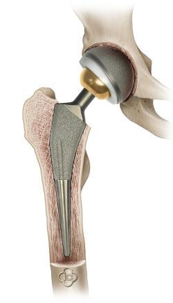
10 Total Hip Arthroplasty
11 SURGICAL APPROACHES TO THE HIP

12 LATERAL APPROACH TO THE HIP (WATSON-JONES) Begin an incision 2.5 cm distal and lateral to the anterior superior iliac spine and curve it distally and posteriorly over the lateral aspect of the greater trochanter and lateral surface of the femoral shaft to 5 cm distal to the base of the trochanter. Locate the interval between the gluteus medius and tensor fasciae latae. The delineation of this interval often is difficult. Brackett pointed out that it can be done more easily by beginning the separation midway between the anterior superior spine and the greater trochanter, before the tensor fasciae latae blends with its fascial insertion. The coarse grain and the direction of the fibers of the gluteus medius help to distinguish them from the finer structure of the tensor fasciae latae muscle.

13 LATERAL APPROACH TO THE HIP (WATSON-JONES) Carry the dissection proximally to expose the inferior branch of the superior gluteal nerve, which innervates the tensor fasciae latae muscle. Incise the capsule of the joint longitudinally along the anterosuperior surface of the femoral neck. In the distal part of the incision, the origin of the vastus lateralis may be reflected distally or split longitudinally to expose the base of the trochanter and proximal part of the femoral shaft.
14 LATERAL APPROACH TO THE HIP (WATSON-JONES) If a wider field is desired, detach the anterior fibers of the gluteus medius tendon from the trochanter or reflect the anterosuperior part of the greater trochanter proximally with an osteotome, together with the insertion of the gluteus medius muscle. This preserves the insertion of the gluteus medius in such a way that it can be easily reat tached later.


15 LATERAL APPROACH TO THE HIP PRESERVING THE GLUTEUS MEDIUS (MCFARLAND AND OSBORNE) Make a midlateral skin incision centered over the greater trochanter; its length depends on the amount of subcutaneous fat. Expose the gluteal fascia and the iliotibial band, and divide them in a straight midlateral line along the entire length of the skin incision. Retract the gluteus maximus posteriorly and the tensor fasciae latae anteriorly. Expose the gluteus medius, and separate it from the piri formis and gluteus minimus by blunt dissection.
16 LATERAL APPROACH TO THE HIP PRESERVING THE GLUTEUS MEDIUS (MCFARLAND AND OSBORNE) Identify the prominent posterior border of the gluteus medius where it joins the posterior edge of the greater trochanter. From this point, make an incision down to the bone through the periosteum and fascia obliquely and distally across the greater trochanter to the middle of the lateral aspect of the femur; continue it further distally in the vastus lateralis to the distal end of the skin incision.
17 LATERAL APPROACH TO THE HIP PRESERVING THE GLUTEUS MEDIUS (MCFARLAND AND OSBORNE) With a knife or a sharp chisel, peel from the bone, in one piece, the attachment of the gluteus medius, the perios teum, the tendinous junction of the gluteus medius and vastus lateralis, and the origin of the vastus lateralis. The portion of the vastus lateralis peeled off includes that attached to the proximal part of the linea aspera, the distal border of the greater trochanter, and part of the shaft of the femur. Anteriorly retract the whole combined muscle mass, con sisting of the gluteus medius and vastus lateralis with their tendinous junction.

18 LATERAL APPROACH TO THE HIP PRESERVING THE GLUTEUS MEDIUS (MCFARLAND AND OSBORNE) Split, divide, and proximally retract the tendon of the gluteus minimus to expose the capsule of the hip joint. Incise the capsule as desired. During closure, suture the capsule and gluteus minimus as one structure. Abduct the hip, return the gluteus medius and vastus lateralis to their original position, and suture them to the undisturbed part of the vastus latera lis, to the deep insertion of the gluteus maximus, and to the proximal part of the quadratus femoris.

19 LATERAL TRANSGLUTEAL APPROACH TO THE HIP (HARDINGE) Place the patient supine with the greater trochanter at the edge of the table and the muscles of the buttocks freed from the edge. Make a posteriorly directed lazy J incision centered over the greater trochanter. Divide the fascia lata in line with the skin incision and centered over the greater trochanter.

20 LATERAL TRANSGLUTEAL APPROACH TO THE HIP (HARDINGE) Retract the tensor fasciae latae anteriorly and the gluteus maximus posteriorly, exposing the origin of the vastus lateralis and the insertion of the gluteus medius. Incise the tendon of the gluteus medius obliquely across the greater trochanter, leaving the posterior half still attached to the trochanter. Carry the incision proximally in line with the fibers of the gluteus medius at the junction of the middle and posterior thirds of the muscle. This gluteus medius split should be no farther than 4 to 5 cm from the tip of the greater trochanter to avoid damage to the superior gluteal nerve and artery. Distally, carry the incision anteriorly in line with the fibers of the vastus lateralis down to bone along the anterolateral surface of the femur.
21 LATERAL TRANSGLUTEAL APPROACH TO THE HIP (HARDINGE) Elevate the tendinous insertions of the anterior portions of the gluteus minimus and vastus lateralis muscles. Abduction of the thigh exposes the anterior capsule of the hip joint. Incise the capsule as desired. During closure, repair the tendon of the gluteus medius with nonabsorbable braided sutures.

22 POSTERIOR APPROACH TO THE HIP (OSBORNE) Begin the incision 4.5 cm distal and lateral to the posterior superior iliac spine, and continue it laterally and distally, remaining parallel with the fibers of the gluteus maximus muscle, to the posterosuperior angle of the greater trochanter, and distally along the posterior border of the trochanter for 5 cm.

23 POSTERIOR APPROACH TO THE HIP (OSBORNE) Separate the fibers of the gluteus maximus parallel with the line of incision, no more than 7 cm to protect the branches of the inferior gluteal artery and nerve. Divide the insertion of the gluteus maximus into the fascia lata for 5 cm, corresponding to the longitudinal limb of the incision.

24 POSTERIOR APPROACH TO THE HIP (OSBORNE) Rotate the thigh internally, detach the tendons of the piriformis and gemelli muscles near their insertions into the trochanter, and retract the muscles medially. The gemelli protect the sciatic nerve. The capsule of the joint is now in view and may be incised longitudinally to expose the posterior surface of the femoral neck and posterior border of the acetabulum.
25 Operation Total Hip Arthroplasty
26 SURGICAL APPROACES TO THE KNEE

27 Knee Anatomy

28 Anterior View - PS

29 Posterior View - PS

30 Coronal view -PS

31 Axis of the Lower Limb

32 Medial View - AS

33 Lateral view - AS

34 Disorder of the axis
35 Knee Prosthesis


36 ANTEROMEDIAL PARAPATELLAR APPROACH (VON LANGENBECK) Begin the incision at the medial border of the quadriceps tendon 7 to 10 cm proximal to the patella, curve it around the medial border of the patella and back toward the midline, and end it at or distal to the tibial tuberosity. Divide and retract the fascia. Deepen the dissection between the vastus medialis muscle and the medial border of the quadriceps tendon, and incise the capsule and synovium along this medial border and along the medial border of the patella and patellar tendon

 dividing the medial alar fold and adjacent")
37 ANTEROMEDIAL PARAPATELLAR APPROACH (VON LANGENBECK) Retract the patella laterally and flex the knee to gain a good view of the anterior compartment of the joint and the suprapatellar bursa. Divide the ligamentum mucosa if necessary Attain wider access to the joint in the following ways: (1) extending the incision proximally, (2) extending the proximal part of the incision obliquely medially and separating the fibers of the vastus medialis, (3) dividing the medial alar fold and adjacent fat pad longitudinally, and (4) mobilizing the medial part of the insertion of the patellar tendon subperiosteally..


38 SUBVASTUS (SOUTHERN) ANTEROMEDIAL APPROACH TO THE KNEE Exsanguinate the limb and inflate the tourniquet with the knee flexed to at least 90 degrees to prevent tenodesis of the extensor mechanism. Make a straight anterior skin incision, beginning 8 cm above the patella, carrying it distally just medial and 2 cm distal to the tibial tubercle. Incise the superficial fascia slightly medial to the patella, and bluntly dissect it off the vastus medialis muscle fascia down to the muscle insertion.


39 SUBVASTUS (SOUTHERN) ANTEROMEDIAL APPROACH TO THE KNEE Identify the inferior edge of the vastus medialis, and bluntly dissect it off the periosteum and intermuscular septum for a distance of 10 cm proximal to the adductor tubercle. Identify the tendinous insertion of the muscle on the medial patellar retinaculum and lift the vastus medialis muscle anteriorly and perform an L-shaped arthrotomy beginning medially through the vastus insertion on the medial patellar retinaculum and carrying it along the medial edge of the patella. Partially release the medial edge of the patellar tendon and evert the patella laterally with the knee extended

40 ANTEROLATERAL APPROACH TO THE KNEE Usually the anterolateral approach is not as satisfactory as the anteromedial one, primarily because it is more difficult to displace the patella medially than laterally. It also requires a longer incision, and often the patellar tendon must be partially freed subperiosteally or subcortically. The iliotibial band can be released or lengthened, and the tight posterolateral corner can be released easily. The fibular head can be resected through the same incision to decompress the peroneal nerve if necessary.


41 ANTEROLATERAL APPROACH TO THE KNEE (KOCHER) Begin the incision 7.5 cm proximal to the patella at the insertion of the vastus lateralis muscle into the quadriceps tendon; continue it distally along the lateral border of this tendon, the patella, and the patellar tendon; and end it 2.5 cm distal to the tibial tuberosity. Deepen the dissection through the joint capsule. Retract the patella medially, with the tendons attached to it, and expose the articular surface of the joint.

42 DIRECT POSTERIOR APPROACH TO THE KNEE Make a curvilinear incision 10 to 15 cm long over the popliteal space with the proximal limb following the tendon of the semitendinosus muscle distally to the level of the joint Curve it laterally across the posterior aspect of the joint for about 5 cm and distally over the lateral head of the gastrocnemius muscle Reflect the skin and subcutaneous tissues to expose the popliteal fascia

43 DIRECT POSTERIOR APPROACH TO THE KNEE Identify the posterior cutaneous nerve of the calf (the medial sural cutaneous nerve) lying beneath the fascia and between the two heads of the gastrocnemius muscle because it is the clue to the dissection. Lateral to it, the short saphenous vein perforates the popliteal fascia to join the popliteal vein at the middle of the fossa Trace the posterior cutaneous nerve of the calf (the medial sural cutaneous nerve) proximally to its origin from the tibial nerve, because the contents of the fossa can be dissected accurately and safely once this nerve is located
44 DIRECT POSTERIOR APPROACH TO THE KNEE Trace the tibial nerve distally and expose its branches to the heads of the gastrocnemius, the plantaris, and the soleus muscles; these branches are accompanied by arteries and veins Follow the tibial nerve proximally to the apex of the fossa where it joins the common peroneal nerve Dissect the common peroneal nerve distally along the medial border of the biceps muscle and tendon, and protect the lateral cutaneous nerve of the calf and the anastomotic peroneal nerve.

45 DIRECT POSTERIOR APPROACH TO THE KNEE Expose the popliteal artery and vein, which lie directly anterior and medial to the tibial nerve. Gently retract the artery and vein and locate and trace the superolateral and superomedial genicular vessels passing beneath the hamstring muscles on either side just proximal to the heads of origin of the gastrocnemius

46 DIRECT POSTERIOR APPROACH TO THE KNEE Greater access can be achieved by ligating one or more genicular vessels If the posterolateral aspect of the joint is to be exposed, elevate the lateral head of the gastroc nemius muscle from the femur and approach the lateral compartment between the tendon of the biceps femoris and the lateral head of the gastrocnemius muscle. When closing the wound, place interrupted sutures in the capsule, the deep fascia and the skin. The popliteal fascia is best closed by placing all sutures before drawing them tight. Tie the sutures one by one

47 MEDIAL APPROACH TO THE KNEE (HOPPENFELD AND DEBOER) With the patient supine and the affected knee flexed about 60 degrees, place the foot on the opposite shin, and abduct and externally rotate the hip. Begin the incision 2 cm proximal to the adductor tubercle of the femur, curve it anteroinferiorly about 3 cm medial to the medial border of the patella, and end it 6 cm distal to the joint line on the anteromedial aspect of the tibia.

48 MEDIAL APPROACH TO THE KNEE (HOPPENFELD AND DEBOER) Retract the skin flaps to expose the fascia of the knee, and extend the exposure from the midline anteriorly to the posteromedial corner of the knee. Cut the infrapatellar branch of the saphenous nerve and bury its end in fat; preserve the saphenous nerve itself and the long saphenous vein. Longitudinally incise the fascia along the anterior border of the sartorius, starting at the tibial attachment of the muscle and extending it to 5 cm proximal to the joint line.


49 MEDIAL APPROACH TO THE KNEE (HOPPENFELD AND DEBOER) Flex the knee further, and allow the sartorius to retract posteriorly, exposing the semitendinosus and gracilis muscles Retract all three components of the pes anserinus posteriorly, and expose the tibial attachment of the tibial collateral ligament, which inserts 6 to 7 cm distal to the joint line. To open the joint anteriorly, make a longitudinal medial parapatellar incision through the retinaculum and synovium.


50 MEDIAL APPROACH TO THE KNEE (HOPPENFELD AND DEBOER) To expose the posterior third of the medial meniscus and the posteromedial corner of the knee, retract the three components of the pes anserinus posteriorly and separate the medial head of the gastrocnemius muscle from the posterior capsule of the knee almost to the midline by blunt dissection. To open the joint posteriorly, make an incision through the capsule posterior to the tibial collateral ligament.

51 LATERAL APPROACH TO THE KNEE Place the patient supine with a sandbag beneath the ipsilateral buttock, and flex the knee 90 degrees. Begin the incision 3 cm lateral to the middle of the patella, extend it distally over the Gerdy tubercle on the tibia, and end it 4 to 5 cm distal to the joint line. Complete the incision proximally by curving it along the line of the femur.

52 LATERAL APPROACH TO THE KNEE Widely mobilize the skin flaps anteriorly and posteriorly. Incise the fascia between the iliotibial band and biceps femoris, carefully avoiding the common peroneal nerve on the posterior aspect of the biceps tendon. Retract the iliotibial band anteriorly and the biceps femoris and common peroneal nerve posteriorly to expose the fibular collateral ligament and the posterolateral corner of the knee capsule.

53 LATERAL APPROACH TO THE KNEE To expose the lateral meniscus, make a separate lateral parapatellar incision through the fascia and joint capsule. To avoid cutting the meniscus, begin the arthrotomy 2 cm proximal to the joint line. To expose the posterior horn of the lateral meniscus, locate the origin of the lateral head of the gastrocnemius muscle on the posterior surface of the lateral femoral condyle.
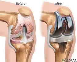
54 Total Knee Replacement
55 THANK YOU FOR ATTENTION!
The thigh. Prof. Oluwadiya KS
 The thigh Prof. Oluwadiya KS www.oluwadiya.com The Thigh: Boundaries The thigh is the region of the lower limb that is approximately between the hip and knee joints Anteriorly, it is separated from the
Gluteal region DR. GITANJALI KHORWAL
 Gluteal region DR. GITANJALI KHORWAL Gluteal region The transitional area between the trunk and the lower extremity. The gluteal region includes the rounded, posterior buttocks and the laterally placed
Human Anatomy Biology 351
 Human Anatomy Biology 351 Lower Limb Please place your name on the back of the last page of this exam. You must answer all questions on this exam. Because statistics demonstrate that, on average, between
The Hip (Iliofemoral) Joint. Presented by: Rob, Rachel, Alina and Lisa
 Joint. Presented by: Rob, Rachel, Alina and Lisa") The Hip (Iliofemoral) Joint Presented by: Rob, Rachel, Alina and Lisa Surface Anatomy: Posterior Surface Anatomy: Anterior Bones: Os Coxae Consists of 3 Portions: Ilium Ischium Pubis Bones: Pubis Portion
The Hip (Iliofemoral) Joint Presented by: Rob, Rachel, Alina and Lisa Surface Anatomy: Posterior Surface Anatomy: Anterior Bones: Os Coxae Consists of 3 Portions: Ilium Ischium Pubis Bones: Pubis Portion
Human Anatomy Biology 351
 Human Anatomy Biology 351 Lower Limb Please place your name on the back of the last page of this exam. You must answer all questions on this exam. Because statistics demonstrate that, on average, between
Mohammad Ashraf. Abdulrahman Al-Hanbali. Ahmad Salman. 1 P a g e
 - 7 Mohammad Ashraf Abdulrahman Al-Hanbali Ahmad Salman 1 P a g e Structures under the cover of Gluteus Maximus: 1-Bones: Ileum, Femur (Head, greater trochanter and gluteal tuberosity), Ischium (ischial
Muscles of the lower extremities. Dr. Nabil khouri MD, MSc, Ph.D
 Muscles of the lower extremities Dr. Nabil khouri MD, MSc, Ph.D Posterior leg Popliteal fossa Boundaries Biceps femoris (superior-lateral) Semitendinosis and semimembranosis (superior-medial) Gastrocnemius
rotation of the hip Flexion of the knee Iliac fossa of iliac Lesser trochanter Femoral nerve Flexion of the thigh at the hip shaft of tibia
 Anatomy of the lower limb Anterior & medial compartments of the thigh Dr. Hayder The fascia lata encloses the entire thigh like a sleeve/stocking. Three intramuscular fascial septa (lateral, medial, and
DISSECTION SCHEDULE. Session I - Hip (Front) & Thigh (Superficial)
 & Thigh (Superficial)") DISSECTION SCHEDULE Session I - Hip (Front) & Thigh (Superficial) Surface anatomy Inguinal region Gluteal region Thigh Leg Foot bones Hip bone Femur Superficial fascia Great saphenous vein Superficial
DISSECTION SCHEDULE Session I - Hip (Front) & Thigh (Superficial) Surface anatomy Inguinal region Gluteal region Thigh Leg Foot bones Hip bone Femur Superficial fascia Great saphenous vein Superficial
Contents of the Posterior Fascial Compartment of the Thigh
 Contents of the Posterior Fascial Compartment of the Thigh 1-Muscles: B i c e p s f e m o r i s S e m i t e n d i n o s u s S e m i m e m b r a n o s u s a small part of the adductor magnus (h a m s t
1-Muscles: 2-Blood supply: Branches of the profunda femoris artery. 3-Nerve supply: Sciatic nerve
 1-Muscles: B i c e p s f e m o r i s S e m i t e n d i n o s u s S e m i m e m b r a n o s u s a small part of the adductor magnus (h a m s t r i n g p a r t o r i s c h i a l p a r t ) 2-Blood supply:
Sports Medicine 15. Unit I: Anatomy. The knee, Thigh, Hip and Groin. Part 4 Anatomies of the Lower Limbs
 Sports Medicine 15 Unit I: Anatomy Part 4 Anatomies of the Lower Limbs The knee, Thigh, Hip and Groin Anatomy of the lower limbs In Part 3 of this section we focused upon 11 of the 12 extrinsic muscles
Muscles of Lesson Five. Muscular Nomenclature and Kinesiology - Two. Muscles of Lesson Five, cont. Chapter 16
 Chapter 16 Muscular Nomenclature and Kinesiology - Two Lessons 5-6 Muscles of Lesson Five Iliopsoas (psoas major, iliacus) Hip outward rotators (piriformis, gemellus superior, gemellus inferior, obturator
Muscles of the Hip 1. Tensor Fasciae Latae O: iliac crest I: lateral femoral condyle Action: abducts the thigh Nerve: gluteal nerve
 Muscles of the Hip 1. Tensor Fasciae Latae O: iliac crest I: lateral femoral condyle Action: abducts the thigh Nerve: gluteal nerve 2. Gluteus Maximus O: ilium I: femur Action: abduct the thigh Nerve:
Distal Femoral Resection
 Distal Femoral Resection Annie Arteau, Bruno Fuchs Introduction This text is a general description of a distal femoral resection. Focus is on anatomical structures and muscle resection. Each femoral resection
lesser trochanter of femur lesser trochanter of femur iliotibial tract (connective tissue) medial surface of proximal tibia
 medial surface of proximal tibia") LOWER LIMB MUSCLES OF THE APPENDICULAR SKELETON The muscles that act on the lower limb fall into three groups: those that move the thigh, those that move the lower leg, and those that move the ankle, foot,
LOWER LIMB MUSCLES OF THE APPENDICULAR SKELETON The muscles that act on the lower limb fall into three groups: those that move the thigh, those that move the lower leg, and those that move the ankle, foot,
Anterior and Medial compartments of the thigh. Dr. Heba Kalbouneh Associate Professor of Anatomy and Histology
 Anterior and Medial compartments of the thigh Dr. Heba Kalbouneh Associate Professor of Anatomy and Histology Terms Related to Movements Movement Flexion Extension Abduction Adduction Medial (internal)
Main Menu. Joint and Pelvic Girdle click here. The Power is in Your Hands
 1 Hip Joint and Pelvic Girdle click here Main Menu K.6 http://www.handsonlineeducation.com/classes//k6entry.htm[3/23/18, 2:01:12 PM] Hip Joint (acetabular femoral) Relatively stable due to : Bony architecture
Lecture 08 THIGH MUSCLES ANTERIOR COMPARTMENT. Dr Farooq Khan Aurakzai. Dated:
 Lecture 08 THIGH MUSCLES ANTERIOR COMPARTMENT BY Dr Farooq Khan Aurakzai Dated: 11.02.2017 INTRODUCTION to the thigh Muscles. The musculature of the thigh can be split into three sections by intermuscular
ANATYOMY OF The thigh
 ANATYOMY OF The thigh 1- Lateral cutaneous nerve of the thigh Ι) Skin of the thigh Anterior view 2- Femoral branch of the genitofemoral nerve 5- Intermediate cutaneous nerve of the thigh 1, 2 and 3 are
SURGICAL EXPOSURES SURGERY OF THE HIP
 1 of 24 11/19/03 1:11 PM SURGICAL EXPOSURES SURGERY OF THE HIP by R. CALANDRUCCIO In: Atlas of Orthopaedic Surgery Volume 3 Lower Extremity; Editors: Laurin, CA, Riley Jr. LH, Roy-Camille R Reprinted with
Lower limb summary. Anterior compartment of the thigh. Done By: Laith Qashou. Doctor_2016
 Lower limb summary Done By: Laith Qashou Doctor_2016 Anterior compartment of the thigh Sartorius Anterior superior iliac spine Upper medial surface of shaft of tibia 1. Flexes, abducts, laterally rotates
The University Of Jordan Faculty Of Medicine THE LOWER LIMB. Dr.Ahmed Salman Assistant Prof. of Anatomy. The University Of Jordan
 The University Of Jordan Faculty Of Medicine THE LOWER LIMB Dr.Ahmed Salman Assistant Prof. of Anatomy. The University Of Jordan Gluteal Region Cutaneous nerve supply of (Gluteal region) 1. Lateral cutaneous
Lower Limb Nerves. Clinical Anatomy
 Lower Limb Nerves Clinical Anatomy Lumbar Plexus Ventral rami L1 L4 Supplies: Abdominal wall External genitalia Anteromedial thigh Major nerves.. Lumbar Plexus Nerves relation to psoas m. : Obturator n.
ANATYOMY OF The thigh
 ANATYOMY OF The thigh 1- Lateral cutaneous nerve of the thigh Ι) Skin of the thigh Anterior view 2- Femoral branch of the genitofemoral nerve 1, 2 and 3 are From the lumber plexus 5- Intermediate cutaneous
Muscles of the Thigh. 6.1 Identify, describe the attachments of and deduce the actions of the muscles of the thigh: Anterior group
 Muscles of the Thigh 6.1 Identify, describe the attachments of and deduce the actions of the muscles of the thigh: Anterior group Sartorius: This is a long strap like muscle with flattened tendons at each
Lectures of Human Anatomy
 Lectures of Human Anatomy Lower Limb Gluteal Region and Hip Joint By DR. ABDEL-MONEM AWAD HEGAZY M.B. with honor 1983, Dipl."Gynecology and Obstetrics "1989, Master "Anatomy and Embryology" 1994, M.D.
Joints of the Lower Limb II
 Joints of the Lower Limb II Lecture Objectives Describe the components of the knee and ankle joint. List the ligaments associated with these joints and their attachments. List the muscles acting on these
The Knee. Clarification of Terms. Osteology of the Knee 7/28/2013. The knee consists of: The tibiofemoral joint Patellofemoral joint
 The Knee Clarification of Terms The knee consists of: The tibiofemoral joint Patellofemoral joint Mansfield, p273 Osteology of the Knee Distal Femur Proximal tibia and fibula Patella 1 Osteology of the
Human Anatomy Biology 255
 Human Anatomy Biology 255 Exam #4 Please place your name and I.D. number on the back of the last page of this exam. You must answer all questions on this exam. Because statistics demonstrate that, on average,
Anatomage Table Instructors Guide- Lower Limb
 The Lower Limb Anatomage Table Instructors Guide- Lower Limb Table of Contents Lower Limb 1- The Skeletal System...3 1: Hip Bone...3 2: Hip Joint and Femur...4 3: Patella and Knee Joint...7 4: Tibia, Fibula,
The Muscular System. Chapter 10 Part D. PowerPoint Lecture Slides prepared by Karen Dunbar Kareiva Ivy Tech Community College
 Chapter 10 Part D The Muscular System Annie Leibovitz/Contact Press Images PowerPoint Lecture Slides prepared by Karen Dunbar Kareiva Ivy Tech Community College Table 10.14: Muscles Crossing the Hip and
Surgical Anatomy of the Hip. Joseph H. Dimon
 Surgical Anatomy of the Hip Joseph H. Dimon The hip joint is a deep joint surrounded by large and powerful muscles necessary for its proper function. Essential neurovascular structures lie in front and
Posterior compartment of the thigh. Dr. Heba Kalbouneh Associate Professor of Anatomy and Histology
 Posterior compartment of the thigh Dr. Heba Kalbouneh Associate Professor of Anatomy and Histology Posterior compartment of the thigh 1-Muscles: Biceps femoris Semitendinosus Semimembranosus Adductor magnus
MUSCULOSKELETAL LOWER LIMB
 MUSCULOSKELETAL LOWER LIMB Spinal Cord Lumbar and Sacral Regions Spinal cord Dorsal root ganglion Conus medullaris Cauda equina Dorsal root ganglion of the fifth lumbar nerve End of subarachnoid space
musculoskeletal system anatomy nerves of the lower limb 2 done by: Dina sawadha & mohammad abukabeer
 musculoskeletal system anatomy nerves of the lower limb 2 done by: Dina sawadha & mohammad abukabeer #Sacral plexus : emerges from the ventral rami of the spinal segments L4 - S4 and provides motor and
ANATYOMY OF The thigh
 ANATYOMY OF The thigh 1- Lateral cutaneous nerve of the thigh Ι) Skin of the thigh Anterior view 2- Femoral branch of the genitofemoral nerve 5- Intermediate cutaneous nerve of the thigh 1, 2 and 3 are
The Lower Limb II. Anatomy RHS 241 Lecture 3 Dr. Einas Al-Eisa
 The Lower Limb II Anatomy RHS 241 Lecture 3 Dr. Einas Al-Eisa Tibia The larger & medial bone of the leg Functions: Attachment of muscles Transfer of weight from femur to skeleton of the foot Articulations
Myology of the Knee. PTA 105 Kinesiology
 Myology of the Knee PTA 105 Kinesiology Objectives Describe the planes of motion and axes of rotation of the knee joint Visualize the origins and insertions of the muscles about the knee List the innervations
Where should you palpate the pulse of different arteries in the lower limb?
 Where should you palpate the pulse of different arteries in the lower limb? The femoral artery In the femoral triangle, its pulse is easily felt just inferior to the inguinal ligament midway between the
ANATOMY TEAM GLUTEAL REGION & BACK OF THIGH
 ANATOMY TEAM GLUTEAL REGION & BACK OF THIGH OBJECTIVES By the end of this lecture, the student should be able to identify and discuss: Contents of gluteal region: Groups of Glutei muscles and small muscles
5.1 Identify, describe the attachments of and deduce the actions of the muscles of the thigh:
 5.1 Identify, describe the attachments of and deduce the actions of the muscles of the thigh: Anterior group Proximal attachment Distal attachment Sartorius ASIS» Upper part of shaft tibia (middle surface)»
Anatomy & Physiology. Muscles of the Lower Limbs.
 Anatomy & Physiology Muscles of the Lower Limbs http://www.ishapeup.com/musclecharts.html Muscles of the Lower Limbs Among the strongest muscles in the body. Because pelvic girdle is composed of heavy,
Muscles of Gluteal Region
 1 The Gluteal Region In the gluteal region the skin is tough with many layers underneath. Directly under it is the superficial fascia followed by the deep fascia then the muscles and the bones of the thigh.
The Knee. Prof. Oluwadiya Kehinde
 The Knee Prof. Oluwadiya Kehinde www.oluwadiya.sitesled.com The Knee: Introduction 3 bones: femur, tibia and patella 2 separate joints: tibiofemoral and patellofemoral. Function: i. Primarily a hinge joint,
Muscle Testing of Knee Extensors. Yasser Moh. Aneis, PhD, MSc., PT. Lecturer of Physical Therapy Basic Sciences Department
 Muscle Testing of Knee Extensors Yasser Moh. Aneis, PhD, MSc., PT. Lecturer of Physical Therapy Basic Sciences Department Muscle Testing of Knee Extensors othe Primary muscle Quadriceps Femoris -Rectus
and K n e e J o i n t Is the most complicated joint in the body!!!!
 K n e e J o i n t K n e e J o i n t Is the most complicated joint in the body!!!! 1-Consists of two condylar joints between: A-The medial and lateral condyles of the femur and The condyles of the tibia
Baraa Ayed حسام أبو عوض. Ahmad Salman. 1 P a g e
 4 Baraa Ayed حسام أبو عوض Ahmad Salman 1 P a g e Today we are going to cover these concepts: Iliotibial tract Anterior compartment of the thigh and the hip Medial compartment of the thigh Femoral triangle
MUSCLES OF THE LOWER LIMBS
 MUSCLES OF THE LOWER LIMBS Naming, location and general function Dr. Nabil khouri ROLES THAT SHOULD NOT BE FORGOTTEN Most anterior compartment muscles of the hip and thigh Flexor of the femur at the hip
Topic 7: Hip and pelvis. Parts of the hip. Parts of the femur
 Topic 7: Hip and pelvis Parts of the hip Parts of the femur Classifying the hip joint Ball and socket Synovial Multiaxial Movements of the hip: Abduction/adduction Flexion/extension Medial/lateral rotation
HUMAN BODY COURSE LOWER LIMB NERVES AND VESSELS
 HUMAN BODY COURSE LOWER LIMB NERVES AND VESSELS October 22, 2010 D. LOWER LIMB MUSCLES 2. Lower limb compartments ANTERIOR THIGH COMPARTMENT General lfunction: Hip flexion, knee extension, other motions
To describe he knee joint, ligaments, structure & To list the main features of other lower limb joints
 To describe he knee joint, ligaments, structure & neurovascular supply To demonstrate the ankle joint anatomy To list the main features of other lower limb joints To list main groups of lymph nodes in
Copyright 2003 Pearson Education, Inc. publishing as Benjamin Cummings. Dr. Nabil Khouri MD, MSc, Ph.D
 Dr. Nabil Khouri MD, MSc, Ph.D Pelvic Girdle (Hip) Organization of the Lower Limb It is divided into: The Gluteal region The thigh The knee The leg The ankle The foot The thigh and the leg have compartments
Leg. Dr. Heba Kalbouneh Associate Professor of Anatomy and Histology
 Leg Dr. Heba Kalbouneh Associate Professor of Anatomy and Histology Skin of the Leg Cutaneous Nerves Medially: The saphenous nerve, a branch of the femoral nerve supplies the skin on the medial surface
LOWER LIMB. As we know the bony part of the body is divided into Axial and Appendicular (upper and lower Limbs)
") LOWER LIMB As we know the bony part of the body is divided into Axial and Appendicular (upper and lower Limbs) Bones of the Lower limb: 1-Pelvic Girdle: composed of: 1. Right hip bone : is formed by 3
LOWER LIMB As we know the bony part of the body is divided into Axial and Appendicular (upper and lower Limbs) Bones of the Lower limb: 1-Pelvic Girdle: composed of: 1. Right hip bone : is formed by 3
Identify the muscles associated with the medial compartment of the thigh. Identify the attachment points of the medial thigh muscles.
 L 8 A B O R A T O R Y Thigh MEDIAL THIGH Identify the muscles associated with the medial compartment of the thigh. Identify the attachment points of the medial thigh muscles. Identify the actions of these
The Leg. Prof. Oluwadiya KS
 The Leg Prof. Oluwadiya KS www.oluwadiya.sitesled.com Compartments of the leg 4 Four Compartments: 1. Anterior compartment Deep fibular nerve Dorsiflexes the foot and toes 2. Lateral Compartment Superficial
Lumbar Plexus. Ventral rami L1 L4 Supplies: Major nerves.. Abdominal wall External genitalia Anteromedial thigh
 Lower Limb Nerves Lectures Objectives Describe the structure and relationships of the plexuses of the lower limb. Describe the course, relationships and structures supplied for the major nerves of the
The Hip Joint. Shenequia Howard David Rivera
 The Hip Joint Shenequia Howard David Rivera Topics Of Discussion Movement Bony Anatomy Ligamentous Anatomy Muscular Anatomy Origin/Insertion/Action/Innervation Common Injuries MOVEMENT Flexion Extension
Practical 1 Worksheet
 Practical 1 Worksheet ANATOMICAL TERMS 1. Use the word bank to fill in the missing words. reference side stand body arms palms anatomical forward All anatomical terms have a(n) point which is called the
Lecture 09. Popliteal Fossa. BY Dr Farooq Khan Aurakzai
 Lecture 09 Popliteal Fossa BY Dr Farooq Khan Aurakzai Dated: 14.02.2018 What is popliteus? Introduction Anything relating to, or near the part of the leg behind the knee. From New Latin popliteus the muscle
The Knee. Tibio-Femoral
 The Knee Tibio-Femoral Osteology Distal Femur with Proximal Tibia Largest Joint Cavity in the Body A modified hinge joint with significant passive rotation Technically, one degree of freedom (Flexion/Extension)
In-Depth Foundations: Anatomy Terms to Know
 Be familiar with / able to identify and define all the following parts. The Spine Cranium Vertebrae Cervical, Thoracic, Lumbar Sacrum Coccyx Bones of Upper Body Cranium Mastoid process; Occipital condyle,
5 Testing the Muscles of the Lower Extremity
 C H A P T E R 5 Testing the Muscles of the Lower Extremity Hip Flexion Hip Flexion, Abduction, and External Rotation with Knee Flexion Hip Extension Hip Abduction Hip Abduction from Flexed Position Hip
The Hay is in the Barn
 Anatomy 1 Practical 1 Review Made by Forrest Allen (nerd) Edited by TJ Williamson (not nerd) The Hay is in the Barn 2019 Thunderbringers Too much to handle https://www.youtube.com/watch?v=glii-kaza d8
lower limb Anterior Compartment: lecture 3 The deep fascia ( fascia lata) divides the thigh into 3 compartments:
 divides the thigh into 3 compartments:") lower limb lecture 3 The deep fascia ( fascia lata) divides the thigh into 3 compartments: 1. Anterior Extensor compartment 2. Medial Adductor compartment 3. Posterior Flexor compartment Anterior Compartment:
lower limb lecture 3 The deep fascia ( fascia lata) divides the thigh into 3 compartments: 1. Anterior Extensor compartment 2. Medial Adductor compartment 3. Posterior Flexor compartment Anterior Compartment:
The Lower Limb. Anatomy RHS 241 Lecture 2 Dr. Einas Al-Eisa
 The Lower Limb Anatomy RHS 241 Lecture 2 Dr. Einas Al-Eisa The bony pelvis Protective osseofibrous ring for the pelvic viscera Transfer of forces to: acetabulum & head of femur (when standing) ischial
Joints of the lower limb
 Joints of the lower limb 1-Type: Hip joint Synovial ball-and-socket joint 2-Articular surfaces: a- head of femur b- lunate surface of acetabulum Which is deepened by the fibrocartilaginous labrum acetabulare
Anatomy MCQs Week 13
 Anatomy MCQs Week 13 1. Posterior to the medial malleolus of the ankle: The neurovascular bundle lies between Tibialis Posterior and Flexor Digitorum Longus The tendon of Tibialis Posterior inserts into
CHAPTER 8: THE BIOMECHANICS OF THE HUMAN LOWER EXTREMITY
 CHAPTER 8: THE BIOMECHANICS OF THE HUMAN LOWER EXTREMITY _ 1. The hip joint is the articulation between the and the. A. femur, acetabulum B. femur, spine C. femur, tibia _ 2. Which of the following is
Bio 113 Anatomy and Physiology The Muscles. Muscles of the Head and Neck. Masseter. Orbicularis occuli. Orbicularis oris. Sternocleidomastoid
 Bio 113 Anatomy and Physiology The Muscles Muscles of the Head and Neck Masseter Orbicularis occuli Orbicularis oris Sternocleidomastoid Temporalis BIO 113 Fall 2011 Muscles Page 1 of 5 Muscles of the
Acland's DVD Atlas of Human Anatomy. Transcript for Volume Robert D Acland
 Acland's DVD Atlas of Human Anatomy Transcript for Volume 2 2007 Robert D Acland This free downloadable pdf file is to be used for individual study only. It is not to be reproduced in any form without
The Knee Joint By Prof. Dr. Muhammad Imran Qureshi
 The Knee Joint By Prof. Dr. Muhammad Imran Qureshi Structurally, it is the Largest and the most complex joint in the body because of the functions that it performs: Allows mobility (flexion/extension)
Lesson 24. A & P Hip
 Lesson 24 A & P Hip 1 Aims of the Session This session will allow candidates to have an understanding of the bony prominences and soft tissues of the hip 2 Learning Outcomes By the end of the lesson the
Gluteal Region and Back of Thigh
 Gluteal Region and Back of Thigh Musculoskeletal block- Anatomy-lecture 14 Editing file Objectives Know contents of gluteal region: 1. Groups of Glutei muscles and small muscles (Lateral Rotators). 2.
The psoas minor is medial to the psoas major. The iliacus is a fan-shaped muscle that when contracted helps bring the swinging leg forward in walking
 1 p.177 2 3 The psoas minor is medial to the psoas major. The iliacus is a fan-shaped muscle that when contracted helps bring the swinging leg forward in walking and running. The iliopsoas and adductor
ANATOMY. Lecturer : Maher Hadidi Done by: ,,Subject : lecture#: ~ Date:
 ANATOMY,,Subject : Lecturer : Maher Hadidi Done by: ~~ lecture#: ~ Date: GftJteal Reaion ' ' v Highest point of iliac cres Safe site for (/M) injection Horizantal line Sciatic nerve Ischial tubrosity
Adductor canal (Subsartorial) or Hunter s canal
 or Hunter s canal") Adductor canal (Subsartorial) or Hunter s canal John Hunter described the exposure and ligation of the femoral artery in this canal for aneurysm of the popliteal artery; this method has the advantage that
Adductor canal (Subsartorial) or Hunter s canal John Hunter described the exposure and ligation of the femoral artery in this canal for aneurysm of the popliteal artery; this method has the advantage that
Hip joint and pelvic girdle. Lower Extremity. Pelvic Girdle 6/5/2017
 Hip joint and pelvic girdle Lower Extremity The relationship between the pelvic girdle and hip is similar to that between the shoulder girdle and shoulder joint. The lower limbs are attached to the axial
Zimmer MIS Mini-Incision THA Anterolateral Approach
 Zimmer MIS Mini-Incision THA Anterolateral Approach Retractor Placement Guide Optimizing exposure and preserving soft tissue during MIS THA Minimally invasive surgery allows you to follow the basic principles
Knee Disarticulation Amputation
 Knee Disarticulation Amputation Pre-Op 64 year old man, previous spinal cord injury, diabetes, renal failure, and a history of spasticity with dynamic knee flexion contracture. He had an open left ankle
ANTERIOR TOTAL HIP ARTHOPLASTY
 ANTERIOR TOTAL HIP ARTHOPLASTY And Other Approaches Bill Rhodes PTA 236 Total Hip Arthoplasty (THA) Background THA, also know as Total Hip Replacement Regarded as the most valued development in orthopedics
THIEME. Anterior and Medial Compartments of the Thigh
 CHAPTER Anterior and Medial Compartments of the Thigh Learning Objectives 2 At the end of the dissection, you should be able to identify the following: Cutaneous nerves innervating the skin of the anterior
Scapula Spine Lateral edge of clavicle. Medial border Scapula. Medial border of Scapula, between superior angle and root of spine. Scapula.
 Muscle attachments and actions answer sheet Muscle Origins insertions Movements Joints crossed Trapezius Base of skull Spinous process of C7 Thoracic Spine Lateral edge of clavicle Elevation Retraction
this makes sense, however this is lower order thinking and does not solve the lower leg
 Functional Knee Valgus in a Barbell Squat 1 One of the most common lower leg dysfunction we see in athletes, particularly general population is functional knee valgus, or better referred to as the knees
Due in Lab weeks because of Thanksgiving Prelab #10. Homework #8. Both sides! Both sides!
 Lab 8 MUSCLES Due in Lab 10 2 weeks because of Thanksgiving Prelab #10 Both sides! Homework #8 Both sides! Refer to Muscles 22-23 Naming of muscles Origin Site of muscle attachment that doesn t move during
17 FibulA FlAP Tor Chiu fibula flap 153
 17 Fibula Flap Tor Chiu Fibula Flap 153 Fibula Flap FLAP TERRITORY This flap includes a segment of the fibular bone with or without the overlying skin island on the peroneal/ lateral aspect of the calf.
In the name of god. Knee. By: Tofigh Bahraminia Graduate Student of the Pathology Sports and corrective actions. Heat: Dr. Babakhani. Nov.
 In the name of god Knee By: Tofigh Bahraminia Graduate Student of the Pathology Sports and corrective actions Heat: Dr. Babakhani Nov. 2014 1 Anatomy-Bones Bones Femur Medial/lateral femoral condyles articulate
Compartment Syndrome
 Compartment Syndrome Chapter 34 Compartment Syndrome Introduction Compartment syndrome may occur with an injury to any fascial compartment. The fascial defect caused by the injury may not be adequate to
THE LOWER LIMB NERVES VESSELS
 THE LOWER LIMB NERVES VESSELS LOWER LIMB: FEMORAL TRIANGLE FEMORAL TRIANGLE LOWER LIMB: FEMORAL TRIANGLE FEMORAL TRIANGLE is a triangular landmark useful in dissection and in understanding relationships
First practical session. Bones of the gluteal region
 First practical session 2017 Bones of the gluteal region The Hip bone The hip bone is made of: 1 The ilium: superior in position 2 The ischium:postero-inferior in position 3 The pubis: antero-inferior
11/15/2018. Temporalis Elevates & retracts mandible. Masseter = Prime mover of jaw closure. Levator scapulae Supraspinatus Clavicle.
 Due in Lab 10 Lab 8 MUSCLES 2 weeks because of Thanksgiving Prelab #10 Both sides! Homework #8 Both sides! Refer to Muscles 22-23 Examples of Origin & Insertion Naming of muscles Origin Site of muscle
Femoral Triangle and Adductor Canal. Dr. Heba Kalbouneh Associate Professor of Anatomy and Histology
 Femoral Triangle and Adductor Canal Dr. Heba Kalbouneh Associate Professor of Anatomy and Histology Femoral Triangle and Adductor Canal Femoral triangle Is a triangular depressed area located in the upper
Applied anatomy of the hip and buttock
 CHAPTER CONTENTS The hip joint e9 Capsule and ligaments e9 s e0 Flexor muscles................... e0 Extensor muscles.................. e Abductor muscles.................. e Adductor muscles..................
Anatomy of the lower limb
 Anatomy of the lower limb 1. Bones of the lower limb Pelvis Hip bone/coxal bone Acetabulum o Acetabular margin o Acetabular fossa o Acetabular notch o Lunate surface Ischiopubic ramus Obturator foramen
Bones of the Lower Limb Bone Structure Description Notes. border of the superior ramus. inferolaterally from the pubic symphysis
 Bones of the Lower Limb Bone Structure Description Notes pubis an angulated bone the forms the anterior part of the pelvis one of three bones that form the os coxae: ilium, ischium, pubis; its forms 1/5
Femoral Artery. Its entrance to the thigh Position Midway between ASIS and pubic symphysis
 Lower Limb Vessels Lecture Objectives Describe the major arteries of the lower limb. Describe the deep and superficial veins of the lower limb. Describe the topographical relationships of the arteries
Chapter 10. The Knee Joint. The Knee Joint. Bones. Bones. Bones. Bones. Knee joint. Manual of Structural Kinesiology R.T. Floyd, EdD, ATC, CSCS
 The Knee Joint Chapter 10 The Knee Joint Manual of Structural Kinesiology R.T. Floyd, EdD, ATC, CSCS 2007 McGraw-Hill Higher Education. All rights reserved. 10-1 Knee joint largest joint in body very complex
RN(EC) ENC(C) GNC(C) MN ACNP *** MECHANISM OF INJURY.. MOST IMPORTANT *** - Useful in determining mechanism of injury / overuse
 ENC(C) GNC(C) MN ACNP *** MECHANISM OF INJURY.. MOST IMPORTANT *** - Useful in determining mechanism of injury / overuse") HISTORY *** MECHANISM OF INJURY.. MOST IMPORTANT *** Age of patient Sport / Occupation - Certain conditions are more prevalent in particular age groups (Osgood Schlaters in youth / Degenerative Joint Disease
HISTORY *** MECHANISM OF INJURY.. MOST IMPORTANT *** Age of patient Sport / Occupation - Certain conditions are more prevalent in particular age groups (Osgood Schlaters in youth / Degenerative Joint Disease
Muscles of the Gluteal Region
 Muscles of the Gluteal Region 1 Some of the most powerful in the body Extend the thigh during forceful extension Stabilize the iliotibial band and thoracolumbar fascia Related to shoulders and arms because