CT of Maxillofacial Injuries
|
|
|
- Stephen Richard
- 5 years ago
- Views:
Transcription


1 CT of Maxillofacial Injuries Stuart E. Mirvis, M.D., FACR Department of Radiology University of Maryland School of Medicine Viking MGS 2001 Technology changes the diagnosis

")
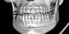
 Le Fort midface patterns")
2 Technologic Evolution Topics to Cover Struts and buttresses Orbital fractures Naso-orbital ethmoid (NOE) Zygomaticomaxillary complex (ZMC) Le Fort midface patterns Ocular Mandible Boom!
, anterior alveolar (5) components.")
 Gentry LR, et al. High-Resolution CT Analysis of Facial Struts in Trauma: 1.")
, middle (2), and inferior (3) parts.")
")

3 Anterior coronal struts consisting of frontal (1 ), zygomaticofrontal (2), nasofrontal (3), anterior maxillary (4), anterior alveolar (5) components. Facial Struts Posterior coronal struts consisting of posterior wall of maxillary sinus (1) and pterygoid plates (2) Gentry LR, et al. High-Resolution CT Analysis of Facial Struts in Trauma: 1. Normal Anatomy. AJR 140: , March 1983 Horizontal struts superior (1), middle (2), and inferior (3) parts. Sagittal struts consisting of median (1 ), parasagittal(2), and lateral (3) parts Concept of Facial Buttresses Major support for facial skeleton to maintain form and function (I beams) Attach directly or indirectly to skull base or cranium 3 vertical and 3 horizontal Buttresses accommodate screw fixation Maintain facial width and height Hopper RA, Salemy S, Sze RW. Diagnosis of Midface Fractures with CT: What the Surgeon Needs to Know. RadioGraphics 2006; 26: Establish functional support (orbits and teeth)
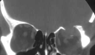
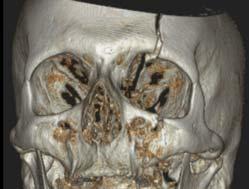
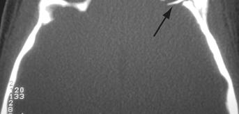
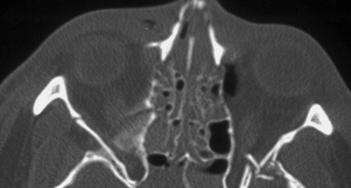
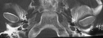
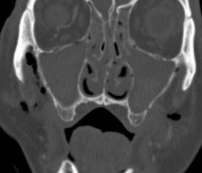
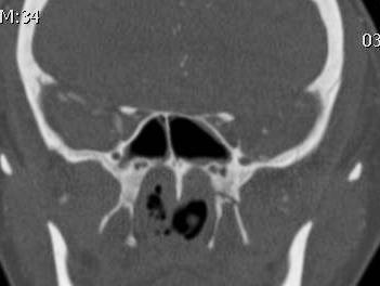
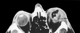
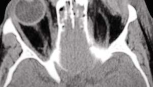
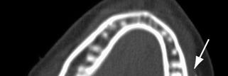
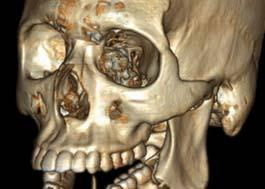
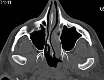
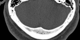
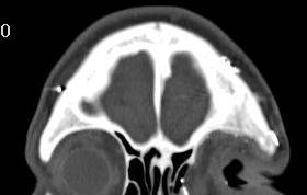
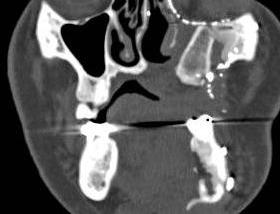
4 Highlights of Facial Anatomy - Orbit Orbital sutures and thin orbital bony plates allow suture diastasis and fractures of thin bone to absorb impacting energy. This mechanism plus orbital fat and muscles cushions the globe and preserves vision in high-energy impacts to the orbit. Orbital Blow-out out Fractures: Significant Imaging Features Eid Evidence of muscle or ft fat entrapment t (position/shape of muscle) Pure or impure fracture (?intact inferior orbit rim) Orbital hematoma (up to 24% orbital injuries) Complications: enopthalmous, diplopia, hypoesthesia Size (area) of floor defect or associated fractures Calculations of blow out fractures ofthe orbital floorby 3D CT and 2D CT method are accurate for assessing the area of fracture and the volume of herniated tissue * * Ploder O, 2D- and 3D-based measurements of orbital floor fractures from CT scans. J Craniomaxillofac Surg. 2002


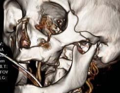
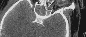
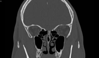
5 Orbital blow-out fracture Orbit Blow-out out Fracture


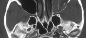
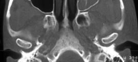
6 Herniated Inferior Rectus Orbit Blow-out Fracture
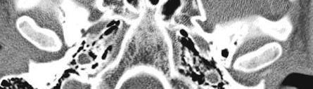
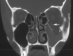
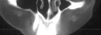
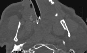
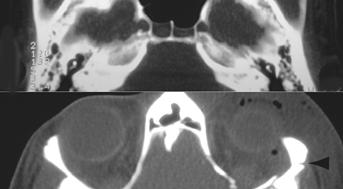
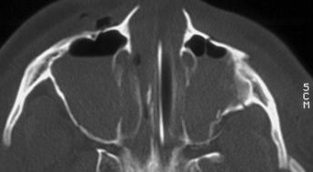
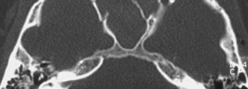
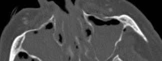
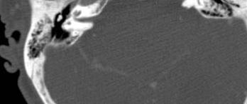
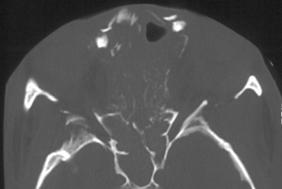
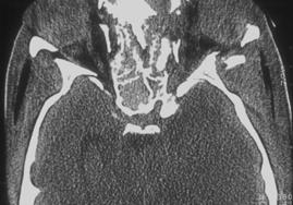
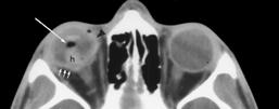
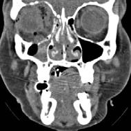
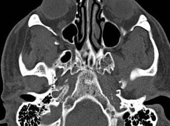
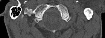
7 Medial orbital wall fracture Isolated or associated 20-40% with floor fracture More common to cause orbital emphysema Rarely surgically repaired Complications: Horizontal gaze palsy, enopthalmous, epistaxis Medial Orbital Blow-out out Fractures


8 Medial wall fracture - entrapment Medial blow-out with herniation


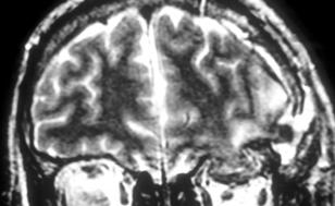
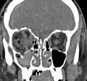
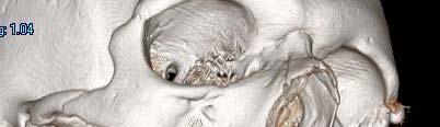
9 Orbital Blow-in fracture Orbital Blow-in fracture: MRI with brain herniation


10 Orbital Blow-up fracture Rare Orbital roof fragments explode into frontal lobe Typical dural tears and CSF leak Frontal sinus involvement common Orbital blow-up fracture


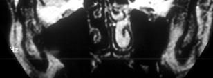
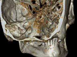
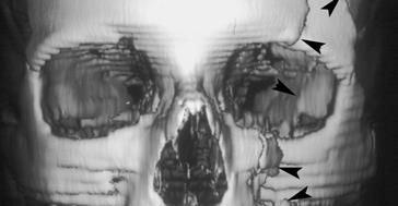
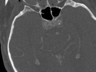
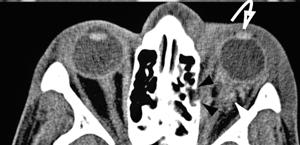
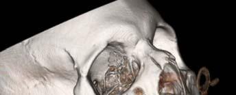
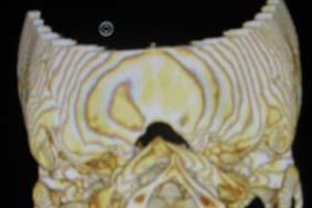
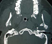
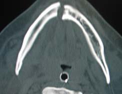
11 Orbital Blow-up fracture Naso-orbital-ethmoid Complex Nasal bridge, lower frontal sinus, medial orbits Comminution, depression, and lateral spread of bones Soft tissue injury; medial canthal ligament, lacrimal drainage, nasofrontal sinus s Usually associated fracture patterns
 Increased nasofrontal angle (28%)")
 Enophthalmos (23%) Facial paralysis (14%)")
12 Naso-orbital Ethmoid Fractures N= 21 Clinical findings: Widened intercanthal distance (71%) Increased nasofrontal angle (28%) Epistaxis (100%) Visual disorder (62%) Cerebrospinal rhinorrhea (33%) Enophthalmos (23%) Facial paralysis (14%) Naso-orbital-ethmoid Complex
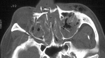
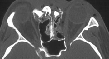
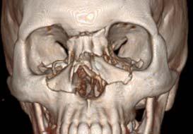
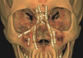
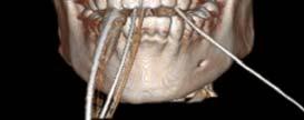
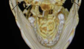
13 Naso-orbital-ethmoid Complex NOE and repair
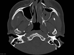
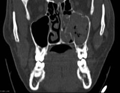
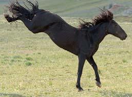
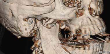
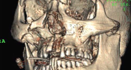
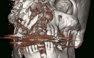
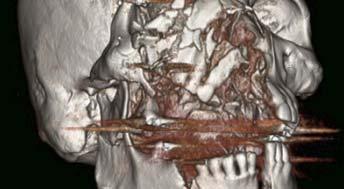
14 Nasomaxillary Fracture Kicked by horse Sagittal mid-face pattern
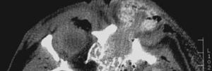
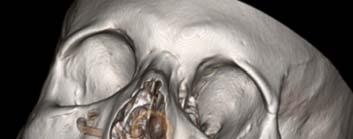
 Impact on malar eminence")


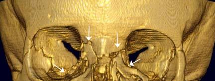
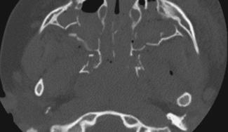
 Always involves orbital")

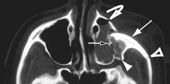
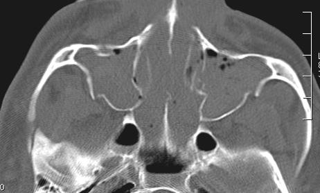
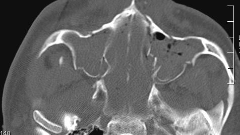
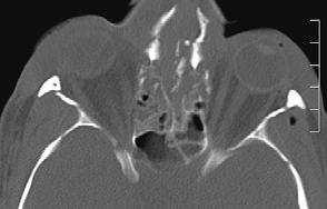
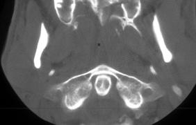
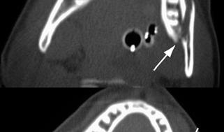
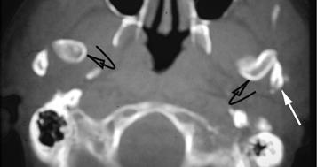
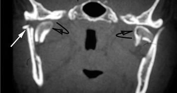
15 Zygomatic-maxillary Complex (ZMC) Impact on malar eminence 4- point fracture Displaces posterior and medially Simple type vs. hi-grade variant Zygomaticmaxillary Complex (ZMC) Always involves orbital floor May involve medial orbit wall Lateral canthal ligamant and inferior orbital nerve Coronoid process impact
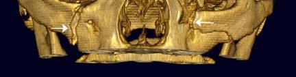
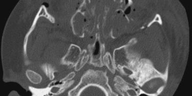
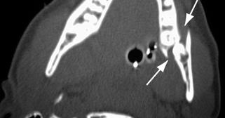
16 Zygomatic-maxillary Complex (ZMC) hi-grade Zygomatic-maxillary Complex (ZMC) Higrade


17 Complex ZMC Orbital Apex Syndrome Optic neuropathy and ophthalmoplegia Loss of cranial nerves II, III, IV, opthalmic division of V, and VI Blindness, fixed dilated pupils, proptosis, ptosis Causes: inflammatory, infectious neoplastic, iatrogenic/traumatic, and vascular conditions


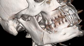
18 LeFort Fracture Patterns Described as symmetric mid face lines of weakness experimental Often asymmetric clinically and combined with ZMC, NOE Always involves pterygoid plate lt fractures Higher energy usually leads to higher grade Any pattern of Lefort 1,2,3 fractures can occur In 1901 the French surgeon, Dr. Rene LeFort ( ), French army surgeon from Lille, published his results from experiments aimed to describe fracture patterns. He performed a number of experiments on cadaver heads. These experiments included blows to the cadaver head at different angles with a wooden club. He also hurtled the head against stationary objects and kicked it in various places in the face


19 LeFort Fracture Patterns LeFort Fracture 1 Fracture all 6 walls of maxillary sinuses Floating palate Typically: nasal septum & maxillary nasal spine Airway compromise - rare


20 LeFort Fracture I + LeFort II Fracture Patterns Mobile nose and maxilla (a portion of the upper transverse maxillary buttress [orbital rim] is involved in mobile segment) Fx. Lateral maxillary sinus, medial orbital floor, nasal bridge, g,pterygoids (pyramidal) Soft tissues: medial orbit, infraorbital nerve Frontomaxillary Zygomaticomaxillary

21 LeFort 2 Lefort II and NOE

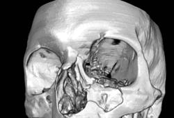
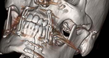
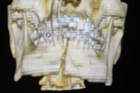
22 LeFort Fracture III Unilateral LeFort 2/3


23 LeFort Fracture III Lefort II/III: highly comminuted


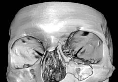
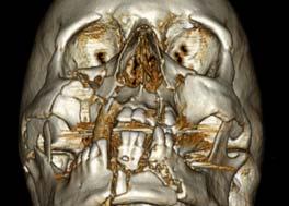
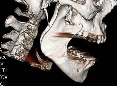
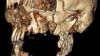
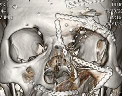
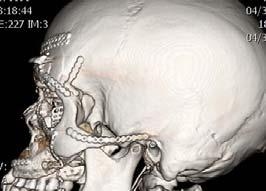
24 Combined LeFort Fracture Pattern - Smash
25


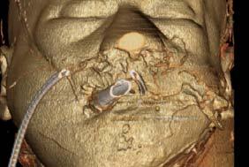
26 GSW through the medial orbit monocular blindness


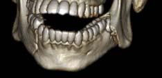
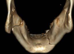
27 Mandibular Fractures Mandibular Fractures Fracture Type Prevalence Body % Angle % Condyle % Symphysis 7-15 % Ramus 3-9 % Alveolarl 2-4% Coronoid process 1-2 %
28 Mandibular Angle Fracture
29
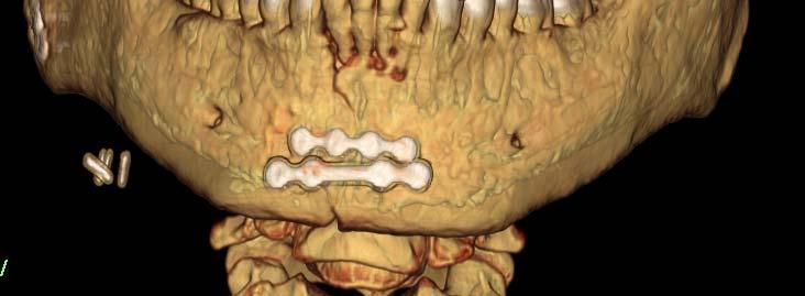
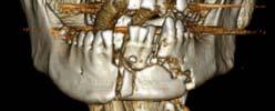
30 ORIF angle/body fx oops! Mandibular Fracture Dislocation
31 Mandible fracturedislocation (sagittal split) Mandibular Fracture - Dislocation

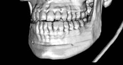
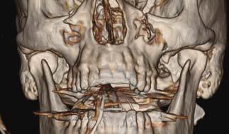
32 Bilateral TMJ dislocation - yawning LeFort I/II/III: Comminuted mandible fractures


33 Attempted suicide: Bit blasting cap GSW: Facial explosion

34 Fibular graft reconstituting maxillary contour
CT of Maxillofacial Fracture Patterns. CT of Maxillofacial Fracture Patterns
 CT of Maxillofacial Fracture Patterns CT of Maxillofacial Fracture Patterns Stuart E. Mirvis, M.D., FACR Department of Radiology University of Maryland School of Medicine Viking 1 1976 MGS 2001 Technology
Imaging Orbit/Periorbital Injury
 Imaging Orbit/Periorbital Injury 9 th Nordic Trauma Radiology Course 2016 Stuart E. Mirvis, M.D., FACR Department of Radiology University of Maryland School of Medicine Fireworks Topics to Cover Struts
Maxillofacial Injuries Practical Tips
 Saturday, October 29, 2016 Maxillofacial Injuries Practical Tips Suyash Mohan MD, PDCC THE ROOTS OF PENN RADIOLOGY RADIOLOGICAL Assistant Professor of Radiology Assistant Professor of Neurosurgery Neuroradiology
Thickened and thinner parts of the skull = important base for understanding of the functional structure of the skull - the transmission of masticatory
 Functional structure of the skull and Fractures of the skull Thickened and thinner parts of the skull = important base for understanding of the functional structure of the skull - the transmission of masticatory
Core Curriculum Syllabus Emergencies in Otolaryngology-Head and Neck Surgery FACIAL FRACTURES
 Core Curriculum Syllabus Emergencies in Otolaryngology-Head and Neck Surgery A. General Considerations FACIAL FRACTURES Look for other fractures like skull and/or cervical spine fractures Test function
Midface fractures; what the radiologist should know.
 Midface fractures; what the radiologist should know. Poster No.: C-1056 Congress: ECR 2013 Type: Educational Exhibit Authors: J. Garcia Villanego, E.-M. Heursen, A. Rodriguez Piñero; Cadiz/ES Keywords:
Facial Trauma ASHNR. Disclosures: Acknowledgments: None. Edward P. Quigley, III, MD PhD University of Utah
 Disclosures: Facial Trauma ASHNR Edward P. Quigley, III, MD PhD University of Utah None Acknowledgments: Dr. Rebecca Cornelius Dr. Ilona M. Schmalfuss Dr. Richard Wiggins III Dr. Yoshimi Anzai Dr. Lindell
North Oaks Trauma Symposium Friday, November 3, 2017
 + Evaluation and Management of Facial Trauma D Antoni Dennis, MD North Oaks ENT an Allergy November 3, 2017 + Financial Disclosure I do not have any conflicts of interest or financial interest to disclose
TRAUMA TO THE FACE AND MOUTH
 Dr.Yahya A. Ali 3/10/2012 F.I.C.M.S TRAUMA TO THE FACE AND MOUTH Bailey & Love s 25 th edition Injuries to the orofacial region are common, but the majority are relatively minor in nature. A few are major
MAXILLOFACIAL TRAUMA. The on-call maxillofacial surgeons can be contacted through the switchboard at the Southern General Hospital
 MAXILLOFACIAL TRAUMA The on-call maxillofacial surgeons can be contacted through the switchboard at the Southern General Hospital Mandibular Injuries Mechanism of injury Assault, falls, RTA-Direct trauma
Diagnosis of Midface Fractures with CT: What the Surgeon Needs to Know 1
 Note: This copy is for your personal non-commercial use only. To order presentation-ready copies for distribution to your colleagues or clients, contact us at www.rsna.org/rsnarights. EDUCATION EXHIBIT
Head and Neck Trauma. Disclosures: Acknowledgments: Introductory case. None
 Head and Neck Trauma None Disclosures: Edward P. Quigley III MD PhD Radiology and Imaging Sciences University of Utah Dr. Richard Wiggins III Dr. Yoshimi Anzai Dr. Lindell Gentry Dr. Blair Winegar Dr.
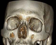
Bones of the skull & face
 Bones of the skull & face Cranium= brain case or helmet Copyright The McGraw-Hill Companies, Inc. Permission required for reproduction or display. The cranium is composed of eight bones : frontal Occipital
Facial and Temporal Bone Trauma Diagnostic imaging and therapeutic challenges in emergency
 Facial and Temporal Bone Trauma Diagnostic imaging and therapeutic challenges in emergency ATTYE A, KRAINIK A Department of Neuroradiology and MRI University Hospital Grenoble / University Grenoble Alpes
Maxillofacial and Ocular Injuries
 Maxillofacial and Ocular Injuries Objectives At the conclusion of this presentation the participant will be able to: Identify the key anatomical structures of the face and eye and the impact of force on
McHenry Western Lake County EMS System Paramedic, EMT-B and PHRN Optional Continuing Education 2019 #1 Facial Trauma
 McHenry Western Lake County EMS System Paramedic, EMT-B and PHRN Optional Continuing Education 2019 #1 Facial Trauma The face is vital to human appearance and function. Facial injuries can impair a patient
Epidemiology 3002). Epidemiology and Pathophysiology
. Epidemiology and Pathophysiology") Epidemiology Maxillofacial trauma or injuries are commonly encountered in the practice of emergency medicine and are presenting one of the most challenging problems to the attending surgeons or physicians
Epidemiology Maxillofacial trauma or injuries are commonly encountered in the practice of emergency medicine and are presenting one of the most challenging problems to the attending surgeons or physicians
Dr. Esam Ahmad Z. Omar BDS, MSc-OMFS, FFDRCSI. Monitor the vital signs. Monitor the vital signs. Complications of Facial Traumas.
 Complications of Facial Traumas 1) Immediate Complications 2) Late Complications Dr. Esam Ahmad Z. Omar BDS, MSc-OMFS, FFDRCSI Assistant Professor Oral & Maxillofacial Surgeon Taibah University Monitor
Cranium Facial bones. Sternum Rib
 Figure 7.1 The human skeleton. Skull Thoracic cage (ribs and sternum) Cranium Facial bones Sternum Rib Bones of pectoral girdle Vertebral column Sacrum Vertebra Bones of pelvic girdle (a) Anterior view
Computed-Tomography of maxillofacial fractures: What do surgeons want to know?
 Computed-Tomography of maxillofacial fractures: What do surgeons want to know? Poster No.: C-0968 Congress: ECR 2016 Type: Educational Exhibit Authors: A. Ammar, M. Jrad, I. KASRAOUI, A. Zoubli, H. Mizouni
Facial skeletal fractures are common,
 CE This symbol indicates that there is more content in the online version of this article. Computed Tomography of Facial Fractures Bryant Furlow, BA Facial skeletal fractures are common, potentially serious,
The diagnostic value of Computed Tomography in evaluation of maxillofacial Trauma
 The diagnostic value of Computed Tomography in evaluation of maxillofacial Trauma Qais H. Muassa FICMS College of Dentistry, Babylon University Ibrahim S. Gataa, BDS, FICMS College of Dentistry, Sulaimania
Chapter 7 Part A The Skeleton
 Chapter 7 Part A The Skeleton Why This Matters Understanding the anatomy of the skeleton enables you to anticipate problems such as pelvic dimensions that may affect labor and delivery The Skeleton The
Anatomy and Physiology. Bones, Sutures, Teeth, Processes and Foramina of the Human Skull
 Anatomy and Physiology Chapter 6 DRO Bones, Sutures, Teeth, Processes and Foramina of the Human Skull Name: Period: Bones of the Human Skull Bones of the Cranium: Frontal bone: forms the forehead and the
MEDICAL CODING FOR FACIAL INJURIES & RECONSTRUCTION
 MEDICAL CODING FOR FACIAL INJURIES & RECONSTRUCTION Tirbod Fattahi, MD, DDS, FACS Chief & Associate Professor Division of Oral & Maxillofacial Surgery University of Florida Health Science Center, Jacksonville
The orbit-1. Dr. Heba Kalbouneh Assistant Professor of Anatomy and Histology
 The orbit-1 Dr. Heba Kalbouneh Assistant Professor of Anatomy and Histology Orbital plate of frontal bone Orbital plate of ethmoid bone Lesser wing of sphenoid Greater wing of sphenoid Lacrimal bone Orbital
Older age, MVC and TBI higher incidence. Facial fractures a distracting injury? Carotid artery injury. Blindness may occur with facial fractures
 Dr Donald C. DeLisi Jr Oral & Maxillofacial Surgeon Multisystem injury 20 50% Nasal and mandibular fractures most common in community ED s Midface and zygomatic injuries most common in Trauma centers 25%
By JOHN MARQUIS CONVERSE, M.D., and DAUBERT TELSEY, D.D.S.
 THE TRIPARTITE OSTEOTOMY OF THE MID-FACE FOR ORBITAL EXPANSION AND CORRECTION OF THE DEFORMITY IN CRANIOSTENOSIS By JOHN MARQUIS CONVERSE, M.D., and DAUBERT TELSEY, D.D.S. Center for Craniofacial Anomalies
AXIAL SKELETON SKULL
 AXIAL SKELETON SKULL CRANIAL BONES (8 total flat bones w/ 2 paired) 1. Frontal forms forehead & upper portion of eyesocket (orbital) 2. Parietal paired bones; form superior & lateral walls of cranium 3.
 www.oralradiologists.com CONE BEAM CT REPORT CASE XXXX Patient information Patient Name: - Referring Doctor: - Patient DOB: - Scan Date: [Start date] Reason for Exam: Maxillary facial pain Doctor Notes:
Facial Trauma. Facial Trauma. Facial Trauma
 Facial Trauma Facial Trauma Brian Bast DMD, MD Department of Oral and Maxillofacial Surgery University of California, San Francisco School of Dentistry Brian Bast DMD, MD Department of Oral and Maxillofacial
Dr. Sami Zaqout, IUG Medical School
 The skull The skull is composed of several separate bones united at immobile joints called sutures. Exceptions? Frontal bone Occipital bone Vault Cranium Sphenoid bone Zygomatic bones Base Ethmoid bone
Oral and Maxillofacial Surgeons and the seriously injured patient. Barts and The London NHS Trust
 Oral and Maxillofacial Surgeons and the seriously injured patient Barts and The London NHS Trust How do you assess this? Primary Survey A B C D E Airway & Cervical Spine Breathing & Ventilation Circulation
Chapter 7. Skeletal System
 Chapter 7 Skeletal System 1 Skull A. The skull is made up of 22 bones: 8 cranial bones, 13 facial bones, and the mandible. B. The Cranium encloses and protects the brain, provides attachments for muscles,
ANATOMY & PHYSIOLOGY I Laboratory Version B Name Section. REVIEW SHEET Exercise 10 Axial Skeleton
 ANATOMY & PHYSIOLOGY I Laboratory Version B Name Section REVIEW SHEET Exercise 10 Axial Skeleton 1 POINT EACH. THE SKULL MULTIPLE CHOICE 1. The major components of the axial skeleton include the 7. The
Extraoral radiography Introduction: Extraoral radiographs (outside the mouth) are taken when large areas of the skull or jaw must be examined or when
 are taken when large areas of the skull or jaw must be examined or when") Extraoral radiography Introduction: Extraoral radiographs (outside the mouth) are taken when large areas of the skull or jaw must be examined or when patients are unable to open their mouths for film placement.
Extraoral radiography Introduction: Extraoral radiographs (outside the mouth) are taken when large areas of the skull or jaw must be examined or when patients are unable to open their mouths for film placement.
Skeletal System -Axial System. Chapter 7 Part A
 Skeletal System -Axial System Chapter 7 Part A Skeleton Learn: Names of the s. Identify specific landmarks that allow: Bones to fit into each other, Organs to fit into the cavities, Muscles to attach,
SKULL BASE AND. Bert De Foer A. Bernaerts, E. Loney +, J. Van Dinther, Th. Somers, E. Offeciers, A. Zarowski, J.W.Casselman / /* FACIAL TRAUMA
 MANDIBULO- Bert De Foer A. Bernaerts, E. Loney +, J. Van Dinther, Th. Somers, E. Offeciers, A. Zarowski, J.W.Casselman / /* DEPARTMENT OF RADIOLOGY, GZA HOSPITALS SINT-AUGUSTINUS, ANTWERP, BELGIUM DEPARTMENT
ORIGINAL ARTICLE. Facial Fracture Classification According to Skeletal Support Mechanisms
 ORIGINAL ARTICLE Facial Fracture Classification According to Skeletal Support Mechanisms Terry L. Donat, MD; Carmen Endress, MD; Robert H. Mathog, MD Objective: To construct, propose, and evaluate the
Structure Location Function
 Frontal Bone Cranium forms the forehead and roof of the orbits Occipital Bone Cranium forms posterior and inferior portions of the cranium Temporal Bone Cranium inferior to the parietal bone forms the
ZYGOMATIC (MALAR) FRACTURES
 FRACTURES") b854_chapter-12.qxd 1/31/2011 9:40 AM Page 129 ZYGOMATIC (MALAR) FRACTURES CHAPTER 12 Anatomical articulations FZ Fronto-zygomatic ZT Zygomaticotemporal ZMB Zygomatico - maxillary buttress IO Infraorbital
b854_chapter-12.qxd 1/31/2011 9:40 AM Page 129 ZYGOMATIC (MALAR) FRACTURES CHAPTER 12 Anatomical articulations FZ Fronto-zygomatic ZT Zygomaticotemporal ZMB Zygomatico - maxillary buttress IO Infraorbital
University of Palestine. Midterm Exam 2013/2014 Total Grade:
 Course No: DNTS2208 Course Title: Head and Neck Anatomy Date: 09/11/2013 No. of Questions: (50) Time: 1hour Using Calculator (No) University of Palestine Midterm Exam 2013/2014 Total Grade: Instructor
MAXILLOFACIAL TRAUMATOLOGY Department of Maxillofacial Surgery Semmelweis University, Budapest. Dr. Huszár Tamás
 MAXILLOFACIAL TRAUMATOLOGY Department of Maxillofacial Surgery Semmelweis University, Budapest Dr. Huszár Tamás Maxillofacial injuries isolated maxillofacial injury multiple injuries polytrauma (injury
Chapter 7: Head & Neck
 Chapter 7: Head & Neck Osteology I. Overview A. Skull The cranium is composed of irregularly shaped bones that are fused together at unique joints called sutures The skull provides durable protection from
Bones Ethmoid bone Inferior nasal concha Lacrimal bone Maxilla Nasal bone Palatine bone Vomer Zygomatic bone Mandible
 splanchnocranium - Consists of part of skull that is derived from branchial arches - The facial bones are the bones of the anterior and lower human skull Bones Ethmoid bone Inferior nasal concha Lacrimal
Current concepts in midface fracture management
 REVIEW C URRENT OPINION Current concepts in midface fracture management AQ1 Alf L. Nastri and Ben Gurney AQ4 Purpose of review Management of midface trauma is complex and challenging and requires a clear
Multidetector computed tomographic evaluation of maxillofacial trauma
 ORIGINAL ARTICLE ASIAN JOURNAL OF MEDICAL SCIENCES Multidetector computed tomographic evaluation of maxillofacial trauma Kaleem Ahmad 1, R. K. Rauniyar 2, Mukesh Kumar Gupta 3, Sajid Ansari 4, Ashok Raj
APPENDICULAR SKELETON 126 AXIAL SKELETON SKELETAL SYSTEM. Cranium. Skull. Face. Skull and associated bones. Auditory ossicles. Associated bones.
 SKELETAL SYSTEM 206 AXIAL SKELETON 80 APPENDICULAR SKELETON 26 Skull Skull and associated s 29 Cranium Face Auditory ossicles 8 4 6 Associated s Hyoid Thoracic cage 25 Sternum Ribs 24 Vertebrae 24 column
Introduction. patterns of injury. The injury pattern produced vanes with. j the object striking the face.
 Dolan et al. Facial fractures I Introduction Facial injury constitutes a frequent finding among emergency room patients. Schultz and Oldham estimate that 54% of such patients will have significant trauma.
Human Anatomy and Physiology - Problem Drill 07: The Skeletal System Axial Skeleton
 Human Anatomy and Physiology - Problem Drill 07: The Skeletal System Axial Skeleton Question No. 1 of 10 Which of the following statements about the axial skeleton is correct? Question #01 A. The axial
Biology 218 Human Anatomy. Adapted from Martini Human Anatomy 7th ed. Chapter 6 The Skeletal System: Axial Division
 Adapted from Martini Human Anatomy 7th ed. Chapter 6 The Skeletal System: Axial Division Introduction The axial skeleton: Composed of bones along the central axis of the body Divided into three regions:
Lesson Plans and Objectives: Review material for article Prep work for article Picture recovery Review for placement on-line.
 Lesson Plans and Objectives: Review material for article Prep work for article Picture recovery Review for placement on-line. After reading the article, the staff will be able to: Define facial trauma
Maxillary and Periorbital Fractures January 2004
 TITLE: Maxillary and Periorbital Fractures SOURCE: Grand Rounds Presentation, UTMB, Dept. of Otolaryngology DATE: January 7, 2004 RESIDENT PHYSICIAN: Gordon Shields, MD FACULTY ADVISOR: Francis B. Quinn,
The sebaceous glands (glands of Zeis) open directly into the eyelash follicles, ciliary glands (glands of Moll) are modified sweat glands that open
 open directly into the eyelash follicles, ciliary glands (glands of Moll) are modified sweat glands that open") The Orbital Region The orbits are a pair of bony cavities that contain the eyeballs; their associated muscles, nerves, vessels, and fat; and most of the lacrimal apparatus upper eyelid is larger and more
The Orbital Region The orbits are a pair of bony cavities that contain the eyeballs; their associated muscles, nerves, vessels, and fat; and most of the lacrimal apparatus upper eyelid is larger and more
Major Anatomic Components of the Orbit
 Major Anatomic Components of the Orbit 1. Osseous Framework 2. Globe 3. Optic nerve and sheath 4. Extraocular muscles Bony Orbit Seven Bones Frontal bone Zygomatic bone Maxillary bone Ethmoid bone Sphenoid
 www.oralradiologists.com CONE BEAM CT REPORT CASE ---- Case Information Referring Doctor: - Patient Name: - Scan Date: December 1, 2015 Patient DOB: - Reason for Exam: - Study Details: icat Flex, 160x160x112
Dr. Sami Zaqout Faculty of Medicine IUG
 The Nose External Nose Nasal Cavity External Nose Blood and Nerve Supplies of the External Nose Blood Supply of the External Nose The skin of the external nose Branches of the ophthalmic and the maxillary
Case Report. Orthognathic Correction of Class II Open Bite. Using the Piezoelectric System and MatrixORTHOGNATHIC Plating System.
 Case Report Orthognathic Correction of Class II Open Bite. Using the Piezoelectric System and MatrixORTHOGNATHIC Plating System. Orthognathic Correction of Class II Open Bite. Using the Piezoelectric System
Bony orbit Roof The orbital plate of the frontal bone Lateral wall: the zygomatic bone and the greater wing of the sphenoid
 Bony orbit Roof: Formed by: The orbital plate of the frontal bone, which separates the orbital cavity from the anterior cranial fossa and the frontal lobe of the cerebral hemisphere Lateral wall: Formed
Nasal Orbital Ethmoid (NOE) Fractures November, 2014
 Fractures November, 2014") TITLE: Nasal Orbital Ethmoid (NOE) Fractures SOURCE: Grand Rounds Presentation, The University of Texas Medical Branch at Galveston, Department of Otolaryngology DATE: November 19, 2014 RESIDENT PHYSICIAN:
TITLE: Nasal Orbital Ethmoid (NOE) Fractures SOURCE: Grand Rounds Presentation, The University of Texas Medical Branch at Galveston, Department of Otolaryngology DATE: November 19, 2014 RESIDENT PHYSICIAN:
Introduction to Local Anesthesia and Review of Anatomy
 5-Sep Introduction and Anatomy Review 12-Sep Neurophysiology and Pain 19-Sep Physiology and Pharmacology part 1 26-Sep Physiology and Pharmacology part 2 Introduction to Local Anesthesia and Review of
PTERYGOPALATINE FOSSA
 PTERYGOPALATINE FOSSA Outline Anatomical Structure and Boundaries Foramina and Communications with other spaces and cavities Contents Pterygopalatine Ganglion Especial emphasis on certain arteries and
Clues of a Ruptured Globe
 Definition any eye that has sustained a full thickness traumatic disruption of the cornea or sclera Overwhelmingly, rupture accidents occur in young men, small children and the elderly Corneal laceration
Temporal region. temporal & infratemporal fossae. Zhou Hong Ying Dept. of Anatomy
 Temporal region temporal & infratemporal fossae Zhou Hong Ying Dept. of Anatomy Temporal region is divided by zygomatic arch into temporal & infratemporal fossae. Temporal Fossa Infratemporal fossa Temporal
Temporal fossa Infratemporal fossa Pterygopalatine fossa Terminal branches of external carotid artery Pterygoid venous plexus
 Outline of content Temporal fossa Infratemporal fossa Pterygopalatine fossa Terminal branches of external carotid artery Pterygoid venous plexus Boundary Content Communication Mandibular division of trigeminal
Management of Craniofacial injuries
 Management of Craniofacial injuries Plastic and Reconstructive Surgery Cirujanos PlástiKos Mundi Cranio-Facial Trauma 1. Introduction Cranio-facial trauma is as old as the human race. What has changed
Nasal region. cartilages: septal cartilage (l); lateral nasal cartilage (2); greater alar cartilages (2); lesser alar cartilages (?
; lateral nasal cartilage (2); greater alar cartilages (2); lesser alar cartilages (?") Nasal region skull bones: nasal and frontal processes of maxilla cartilages: septal cartilage (l); lateral nasal cartilage (2); greater alar cartilages (2); lesser alar cartilages (?) 1 Nasal cavity Roof
Nasal region skull bones: nasal and frontal processes of maxilla cartilages: septal cartilage (l); lateral nasal cartilage (2); greater alar cartilages (2); lesser alar cartilages (?) 1 Nasal cavity Roof
REVIEW OF HEAD AND NECK CRANIAL NERVES AND EVERYTHING ELSE
 REVIEW OF HEAD AND NECK CRANIAL NERVES AND EVERYTHING ELSE OLFACTORY NERVE CN I ANTERIOR CRANIAL FOSSA CRISTA GALLI OF ETHMOID OLFACTORY FORAMINA IN CRIBIFORM PLATE OF ETHMOID BONE CN I OLFACTORY NERVE
Skeletal system. Prof. Abdulameer Al-Nuaimi. E. mail:
 Skeletal system Prof. Abdulameer Al-Nuaimi E-mail: a.al-nuaimi@sheffield.ac.uk E. mail: abdulameerh@yahoo.com Functions of Bone and The Skeletal System Support: The skeleton serves as the structural framework
Chapter 23: Maxillofacial Trauma. Robert B. Stanley, Jr.
 Chapter 23: Maxillofacial Trauma Robert B. Stanley, Jr. Traditionally, fractures of the facial skeleton have been evaluated and treated in a segmentalized fashion, even if complex injuries were obvious
THE SKELETAL SYSTEM. Focus on the Skull
 THE SKELETAL SYSTEM Focus on the Skull Review Anatomical Terms Anterior/Posterior Dorsal/Ventral Medial/Lateral Superior/Inferior Bone Markings - Review Projections for attachment of muscles, ligaments
Dr.Sepideh Falah-kooshki
 Dr.Sepideh Falah-kooshki MAXILLA Premaxillary/median palatal suture (radiolucent). Incisive fossa and foramen (radiolucent). Nasal passages (radiolucent). Nasal septum (radiopaque). Anterior nasal spine
Management Strategies for Communited Fractures of Frontal Skull Base: An Institutional Experience
 80 Original Article THIEME Management Strategies for Communited Fractures of Frontal Skull Base: An Institutional Experience V. Velho 1 Hrushikesh U. Kharosekar 1 Jasmeet S. Thukral 1 Shonali Valsangkar
DR. SAAD AL-MUHAYAWI, M.D., FRCSC. ORL Head & Neck Surgery
 TRAUMA IN ORL DR. SAAD AL-MUHAYAWI, M.D., FRCSC Associate Professor & Consultant ORL Head & Neck Surgery TYPES OF TRAUMA EAR & TEMPORAL BONE TRAUMA NOSE & FACIAL BONES TRAUMA LARYNGEAL TRAUMA NECK TRAUMA
Trigeminal Nerve Worksheets, Distributions Page 1
 Trigeminal Nerve Worksheet #1 Distribution by Nerve Dr. Darren Hoffmann Dental Gross Anatomy, Spring 2013 We have drawn out each of the branches of CN V in lecture and you have an idea now for their basic
Australian Dental Journal
 Australian Dental Journal The official journal of the Australian Dental Association Australian Dental Journal 2018; 63:(1 Suppl): S35 S47 doi: 10.1111/adj.12589 Current and evolving trends in the management
The impact-absorbing effects of facial fractures in closed-head injuries
 J Neurosurg66:542-547, 1987 The impact-absorbing effects of facial fractures in closed-head injuries An analysis of 2 l0 patients K. FRANCIS LEE, M.D., LOUS K. WAGNER~ PH.D., Y. EUGENIA LEE, M.D., JUNG
Oral Surgery Dr. Labeed Sami جامعة تكريت كلية طب االسنان املرحلة اخلامسة م.د. لبيد سامي حسن
 جامعة تكريت كلية طب االسنان جراحة الفم مادة املرحلة اخلامسة م.د. لبيد سامي حسن 6102-6102 1 5 th stage Fracture zygomatic complex As the zygomatic bone is closely associated with the maxilla, frontal and
Anatomy of. External NOSE. By Dr Farooq Aman Ullah Khan PMC
 Anatomy of External NOSE By Dr Farooq Aman Ullah Khan PMC 24 th Nov. 2017 The External Nose Descriptions of the nose always begin with that part of it which is covered by the skin, i.e., the EXPOSED PART
SLLF FOR TMJ CASES IN ADULT DENTITION SEVERE BRACHIFA BRACHIF FACIAL
 SLLF FOR TMJ CASES IN ADULT DENTITION SEVERE BRACHIFAFACIAL TMJ: Severe Postural Imbalance+Severe Myofascial Pain Syndrome, severe soreness Temporalis Tendon RL, Sternocleidomastoideus RL Age:39 years
Case Report Mid Facial Degloving Procedure: Managing A Case of Multiple Mid Face Fractures with Significant External Deformity
 55 Bangladesh J Otorhinolaryngol 2015; 21(1): 51-56 Case Report Mid Facial Degloving Procedure: Managing A Case of Multiple Mid Face Fractures with Significant External Deformity Akhil Chndra Biswas 1,
Infratemporal fossa: Tikrit University college of Dentistry Dr.Ban I.S. head & neck Anatomy 2 nd y.
 Infratemporal fossa: This is a space lying beneath the base of the skull between the lateral wall of the pharynx and the ramus of the mandible. It is also referred to as the parapharyngeal or lateral pharyngeal
1 Eyelids. Lacrimal Apparatus. Orbital Region. 3 The Orbit. The Eye
 1 1 Eyelids Orbital Region 2 Lacrimal Apparatus 3 The Orbit 4 The Eye 2 Eyelids The eyelids protect the eye from injury and excessive light by their closure. The upper eyelid is larger and more mobile
Pictorial review of extraconal and osseous orbital pathology - what can be found 'around' the orbits?
 Pictorial review of extraconal and osseous orbital pathology - what can be found 'around' the orbits? Poster No.: C-2011 Congress: ECR 2013 Type: Educational Exhibit Authors: M. Meissnitzer, T. Meissnitzer,
Complications of Midface Fractures
 557 Kirkland Lozada, MD 1 Sameep Kadakia, MD 1 Manoj T. Abraham, MD 2 Yadranko Ducic, MD, FRCS(C), FACS 3 1 Department of Otolaryngology, New York Eye and Ear Infirmary of MountSinai,NewYork,NewYork 2
Skull basic structures. Neurocranium
 Assoc. Prof. Květuše Lovásová, M.V.D., PhD. Skull basic structures Skull consists of two groups of bones: neurocranium (bones forming the brain box) splanchnocranium (bones forming the facial skeleton)
Skull and Axial Skeleton
 Published on Second Faculty of Medicine, Charles University (http://www.lf2.cuni.cz ) Skull and Axial Skeleton Description of the test The examination of the skull skeleton is in oral format. It consists
Technique Guide. Titanium Wire with Barb and Needle. Surgical Technique Guide for Canthal Tendon Prodecures.
 Technique Guide Titanium Wire with Barb and Needle. Surgical Technique Guide for Canthal Tendon Prodecures. Indications/Features Indications The Synthes Titanium Wire with Barb and straight Needle is
UNIVERSITY OF MEDICINE AND PHARMACY GR. T. POPA - IASI FACULTY OF DENTAL MEDICINE
 UNIVERSITY OF MEDICINE AND PHARMACY GR. T. POPA - IASI FACULTY OF DENTAL MEDICINE ABSTRACT CONTRIBUTIONS OF THREE-DIMENSIONAL IMAGING TO THE DIAGNOSIS AND MANAGEMENT OF CLEFT LIP AND PALATE PhD ADVISOR,
FRONTAL SINUPLASTY P R E P A R E D A N D P R E S E N T E D B Y : D R. Y A H Y A F A G E E H R 4 16/ 12/ 2013
 FRONTAL SINUPLASTY P R E P A R E D A N D P R E S E N T E D B Y : D R. Y A H Y A F A G E E H R 4 16/ 12/ 2013 ANATOMY: FRONTAL SINUS Not present at birth Starts developing at 4 years Radiographically visualized
Research Article Length and Geometric Patterns of the Greater Palatine Canal Observed in Cone Beam Computed Tomography
 International Dentistry Volume 2010, Article ID 292753, 6 pages doi:10.1155/2010/292753 Research Article Length and Geometric Patterns of the Greater Palatine Canal Observed in Cone Beam Computed Tomography
ACTIVITY 3: AXIAL SKELETON AND LONG BONE DISSECTION COW BONE DISSECTION
 ACTIVITY 3: AXIAL SKELETON AND LONG BONE DISSECTION Objectives: 1) How to get ready: Read Chapter 7, McKinley et al., Human Anatomy, 4e. All text references are for this textbook. Learning the meanings
YOU MUST BRING YOUR OWN GLOVES FOR THIS ACTIVITY.
 ACTIVITY 3: AXIAL SKELETON AND LONG BONE DISSECTION Objectives: 1) How to get ready: Read Chapter 7, McKinley et al., Human Anatomy, 5e. All text references are for this textbook. Learning the meanings
Original Research THE USE OF REFORMATTED CONE BEAM CT IMAGES IN ASSESSING MID-FACE TRAUMA, WITH A FOCUS ON THE ORBITAL FLOOR FRACTURES
 DOI: 10.15386/cjmed-601 Original Research THE USE OF REFORMATTED CONE BEAM CT IMAGES IN ASSESSING MID-FACE TRAUMA, WITH A FOCUS ON THE ORBITAL FLOOR FRACTURES RALUCA ROMAN 1, MIHAELA HEDEȘIU 1, FLOAREA
MRI masterfile Part 5 WM Heme Strokes.ppt 1
 Ocular and Orbital Trauma Eye Trauma: Incidence 1.3 million eye injuries in the US per year. 40,000 of these injuries lead to blindness in the US. Patrick Sibony, MD March 23, 2013 Ophthalmic Emergencies
University of Palestine. Midterm Exam 2013/2014 Total Grade:
 [ Course No: DNTS2208 Course Title: Head and Neck Anatomy Date: 17/11/1024 No. of Questions: (52) Time: 2hours Using Calculator (No) University of Palestine Midterm Exam 2013/2014 Total Grade: Instructor
Fracture frontal bone and its management
 From the SelectedWorks of Balasubramanian Thiagarajan March 1, 2013 Fracture frontal bone and its management Balasubramanian Thiagarajan Available at: https://works.bepress.com/drtbalu/14/ ISSN: 2250-0359
SKULL / CRANIUM BONES OF THE NEUROCRANIUM (7) Occipital bone (1) Sphenoid bone (1) Temporal bone (2) Frontal bone (1) Parietal bone (2)
 Occipital bone (1) Sphenoid bone (1) Temporal bone (2) Frontal bone (1) Parietal bone (2)") Important! 1. Memorizing these pages only does not guarantee the succesfull passing of the midterm test or the semifinal exam. 2. The handout has not been supervised, and I can not guarantee, that these
Important! 1. Memorizing these pages only does not guarantee the succesfull passing of the midterm test or the semifinal exam. 2. The handout has not been supervised, and I can not guarantee, that these
AO COIAC COmprehensive Injury Automatic Classifier. Craniomaxillofacial Fracture Classification Module
 AO COIAC COmprehensive Injury Automatic Classifier Software for the classification and documentation of injuries User Manual Version 4.0.0 Craniomaxillofacial Fracture Classification Module Audigé L.,