Older age, MVC and TBI higher incidence. Facial fractures a distracting injury? Carotid artery injury. Blindness may occur with facial fractures
|
|
|
- Blaise Martin
- 5 years ago
- Views:
Transcription


1 Dr Donald C. DeLisi Jr Oral & Maxillofacial Surgeon Multisystem injury 20 50% Nasal and mandibular fractures most common in community ED s Midface and zygomatic injuries most common in Trauma centers 25% of women with facial trauma result of domestic violence Incidence of concomitant cervical spine injuries with facial fractures Older age, MVC and TBI higher incidence Facial fractures a distracting injury? Carotid artery injury Blindness may occur with facial fractures


2 Airway Most urgent complication Airway compromise Simple interventions first No mandible? Flail Mandible Flail Maxilla Bilateral fractures Intubation Avoid nasotracheal intubation Be Prepared and Be Creative Swelling, Bleeding, Suction! Airway Management Options Awake intubation Laryngeal Mask Airway Fiberoptic intubation Lateral or semi prone position Percutaneous transtracheal jet ventilation Retrograde intubation Cricothyroidotomy Hemorrhage Control Rarely develop shock from facial bleeding alone Direct Pressure LeFort Fractures Nasal hemorrhage may require A&P packing History Vision Teeth alignment Abuse Inspection Facial elongation High grade LeFort Fracture Asymmetry Deformities and cranial nerve injury Palpation Tenderness Step offs Facial stability Crepitus Subcutaneous air Cutaneous anesthesia ASSESS FUNCTION!


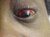
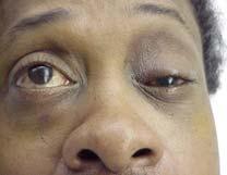
3 Periorbital and Orbital Exam Perform early Periorbital and Orbital Exam Look for exophthalmos or enophthalmos Pupil shape Hyphema Visual acuity Entrapment signs Raccoon sign Bimanual Palpation Test Professional Lid Retractor Penetrating Injuries Occult globe penetration Eyelid lacerations Nose Septal hematoma CSF Rhinorrhea Ears Subperichondral hematoma Hemotympanum Battle sign Oral and Mandibular Exam Mandible deviation Floor of Mouth Ecchymosis Teeth malocclusion Paresthesia Sensory Numbness


4 Head, chest and abdominal trauma takes precedence Plain Films CT Orbital fractures 3D images available Frontal Sinus/Bone Fractures Direct blow Frequent intracranial injuries Posterior Table? Mucopyoceles Consult with NS for treatment, disposition and antibiotics Nasoethmoidal Orbital Injuries Lacrimal apparatus disruption Bimanual palpation if medial canthus pain CT face Orbital Fractures Usually through floor or medial wall Enophthalmos Anesthesia Diplopia Infraorbital stepoff deformity Subcutaneous emphysema Entrapment? Orbital Fissure Syndrome Fracture of the orbital canal Extraocular motor palsies and blindness If significant retrobulbar hemorrhage, may need cantholysis to save vision Zygomatic Fractures Tripod fracture Most serious Lateral subconjunctival hemorrhage Need ORIF Arch fracture Most common Outpatient repair


5 Maxillary Fractures High energy injury Malocclusion Facial lengthening CSF rhinorrhea Periorbital ecchymosis


6 Mandibular Fractures Second most common facial fracture Often multiple Malocclusion Intraoral lacerations Sublingual ecchymosis Nerve injury Plain films Panorex CT Open Fractures Pen G or Cleocin * Body 33 % Angle 23.1 % Condyle 29.3 % Symphysis 8.4 % Ramus 4.8 % Alveolar 1.4 % Coronoid Process 4.8 % Introduction Most common in young males (ages 18-30) Causes: assault, motor vehicle accidents, sports and gunshots wounds Most common Fractures Sites Risks: impacted teeth, osteoporosis, edentulous areas, pathologic, lytic lesions Classification by Site Symphyseal / Parasymphyseal Body Ramus Coronoid Process Condyle Alveolus Angle


7 Classification by Favorability Favorable Unfavorable Anterior Muscles Weaker force Mylohyoid, geniohyoid, genioglossus, platysma, anterior digastric muscles Muscle action depresses and retracts (open mandible)


8 Posterior Muscles: Stronger force Temporalis Muscle Masseter Muscle Medial Pterygoid Muscles Lateral Pterygoid Muscles Classification by Type of Fracture Open versus Closed Fracture Pattern: Communited, oblique, transverse, spiral, greenstick Pathologic: fractures secondary to bone disease (eg, osteogenic tumors, osteoporosis)


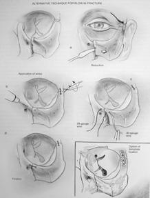
 Closed Reduction")

9 Management Concepts MANAGEMENT Maintain Airway! Goals: restore occlusion, establish bony union& avoid TMJ pathology Repair within first week In general favorable fractures may only need closed reduction Postoperative Care - Maxillo-Mandibular Fixation (MMF) Closed Reduction Indications Methods Requires an intact maxilla Typically MMF may be removed after 4-6 weeks Complications
 Indications")


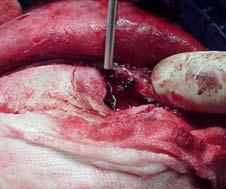
10 Open Reduction &Internal Fixation (ORIF) Indications Approaches: 1. Transoral 2. External Management by Type Coronoid, Greenstick, Unilateral Nondisplaced Fractures: observation with soft diet, analgesics, oral antibiotics and close follow-up, physio-therapy exercises for 3 months (may consider MMF for severely displaced coronoid fractures) Favorable, Minimally Displaced Noncondylar Fractures : may consider closed reduction and 4-6 weeks of MMF


")

 TMJ Ankylosis")
11 Displaced Fractures Symphyseal and Parasymphyseal fractures: tend to be vertically unfavorable Body Fractures : almost always unfavorable Angle fractures in general have the highest complication rate Ramus Fractures: isolated ramus fractures are rare (protected by masseter muscle) Surgical Complications Chin and Lip Numbness Osteomyelitis Malunion Nonunion Plate Exposure Marginal Mandibular Nerve Injury Necrosis of Condylar Head (Aseptic Necrosis) TMJ Ankylosis Dental Injury MAXILLARY FRACTURES
 3. Pterygo-Maxillary (PM) 4.")

12 Classification Buttress System Vertical Buttressess 1. Naso-Maxillary (NM) 2. Zygomatico-Maxillary (ZM) 3. Pterygo-Maxillary (PM) 4. Nasal Septum Horizontal Beams 1. Frontal Bar 2. Inferior Orbital Rims 3. Maxillary Alveolus and Palate 4. Zygomatic Process 5. Greater Wing of the Sphenoid 6. Medial and Lateral Pterygoid Plates 7. Mandible


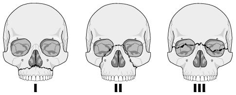
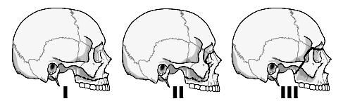
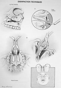
13 Le Fort Classification Based on patterns of fractures (lines of minimal resistance) classified according to the highest level of Injury In many cases Le Fort classification is incomplete for maxillary fractures Le Fort fractures may present in many combinations or on one side (hemi-le Fort) Le Fort I (Low Maxillary) Transverse maxillary fracture Involves anterolateral maxillary wall, medial maxillary wall, pterygoid plates, septum at floor of nose


14 Le Fort II (Pyramidal ) Caused typically from a superiorly directed force against the maxilla. Involves nasofrontal suture, orbital foramen, rim, and floor frontal process of lacrimal bone, zygomaxillary suture, lamina papyracea of ethmoid; pterygoid plate and high septum Le Fort III (Craniofacial Dysjunction) Separates facial skeleton from base of skull, typically caused by high velocity impacts. Involves nasofrontal suture, zygoma and zygomatic arch; pterygoid plates and nasal septum
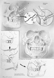
 Interosseous Wire Fixation Bone Grafts Le Fort II: stabilization of the ZM buttress, MMF, nasofrontal process and inferior orbital rim.")
15 Management Principles Goals of Reconstruction Exposure/Approaches Timing Postoperative Care Cont: Management Techniques Management by Le Fort Classification Le Fort I: reduced digitally, MMF, fixation of ZM Plate Fixation (Miniplates) Interosseous Wire Fixation Bone Grafts Le Fort II: stabilization of the ZM buttress, MMF, nasofrontal process and inferior orbital rim. Le Fort III: usually requires coronal flap for adequate exposure for exploration and miniplate fixation
16
 Fractures")

17 Surgical complications ZYGOMATICOMAXILLARY & ORBITAL FRACTURES Malunion, Nonunion, Plate Exposure Palpable or Observable Plates Forehead or Cheek Hypesthesia Zygomaticomaxillary Complex (Trimalar) Fractures Osteomyelitis Dental Injury Zygomaticomaxillary Complex (Trimalar) Fractures Symptom: Introduction Subconjunctival & periorbital ecchymosis Eyelid edema Epistaxis Cheek hypesthesia Diplopia Hypophthalmos Enophthalmos Trismus


18 Four sutures involved in Zygomaticomaxillary Complex Fractures 1. Zygomaticonfrontal Suture 2. Zygomaticomaxillary Suture 3. Zygomaticotemporal Suture 4. Zygomaticosphenoid Suture Management Stabilizing the zygomatic arch Minimum of 2points fixation Closed Reduction Open Reduction Common Approaches to Zygoma Incisions Intraoral approach Coronal, Hemicoronal or Extended Pretragal Approaches Lateral Brow Approach

19 ORBITAL FRACTURES


")

20 Management APPROACHES Indication for Surgical Intervention Contraindications for Surgical Intervention: hyphema, retinal tear, globe perforation Ophthalmological Evaluation retinal edema? Technique Subciliary Incision (Infraciliary) Transconjuctival Incision Lynch Incision (Frontoethmoidal) Brow Incision Subtarsal Incision Caldwell-Luc (Transantral) Approach


21 Surgical Complication FRONTAL SINUS FRACTURE Postoperative Blindness CSF Leak Persistent Enophthalmos and Diplopia Ectropion Entropion Cheek Hypesthesia Extrusion of Grafts Malunion, nonunion, PlateExposure, Osteomyelitis Palpable or Observable Plates


22 FRONTAL SINUS FRACTURE Sign & Symptoms Risk MANAGEMENT


23 Anterior Table Fractures Linear, Minimally Displaced Depressed Fractures Comminuted or Unstable Fractures Posterior Table Fractures Isolated Nondisplaced Psoterior Table Fracture Displaced Posterior Table Fracture Comminuted, Contaminated or through and Through Fractures--Cranialization


24 Surgical Complications Mucocele, Mucopyoceles Sinusitis Forehead Contour Deformity Intracranial Infections Osteomyelitis CSF leak Forehead Hypesthesia Forehead Paralysis

 Lacrimal Collecting System Puncta Canaliculi Lacrimal")
25 NASO-ORBITOETHMOID (NOE)FRACTURES Introduction NOE: frontal process of maxilla, nasal bones, and orbital space Sign& Symptoms Pseudohypertelorism (Traumatic Telecanthus) Anatomy Medial Canthal Ligament (MCL) Lacrimal Collecting System Puncta Canaliculi Lacrimal Sac Lacrimal Duct


 Comminutions are more common in")
26 Management First reconstruct medial orbital wall prior to repair of the MCL Must consider associated injuries May attempt closed reduction if MCL and lacrimal system is intact Telescoping Nasal Bones and Frontal Process of the Maxilla Introduction Nasal Fractures Most common Anterior impacts Lateral impacts Dislocated quadrangular cartilage inferiorly or C-shaped Children usually have dislocated or green stick fractures and have a higher risk of septal hematomas) Comminutions are more common in adults Sign& Symptoms Diagnosis


 Pediatric Nasal Fractures: generally should be")
27 Management Initial Management Preoperative photographs/x-ray may be considered for medicalegal documentation Septal hematomas Open fractures must be cleaned then given antibiotics Cont : Management Surgical Management Generally nasal bone depressed or deviation may undergo closed reduction Open Reduction with Internal Fixation (Septorhinoplasty) Pediatric Nasal Fractures: generally should be treated conservatively


28 Cont : Management Surgical Complications & Associated Injuries Persistent Deformity Nasal Obstruction Septal Hematoma Septal Perforation and Deviations Cribriform Plate Fracture


29 Thank You Bend Oral, Facial, & Implant Surgery Village Office Court Suite 102 Bend, Oregon Office: Cell: my Always feel free to call with any questions or concerns!
Core Curriculum Syllabus Emergencies in Otolaryngology-Head and Neck Surgery FACIAL FRACTURES
 Core Curriculum Syllabus Emergencies in Otolaryngology-Head and Neck Surgery A. General Considerations FACIAL FRACTURES Look for other fractures like skull and/or cervical spine fractures Test function
Maxillofacial and Ocular Injuries
 Maxillofacial and Ocular Injuries Objectives At the conclusion of this presentation the participant will be able to: Identify the key anatomical structures of the face and eye and the impact of force on
North Oaks Trauma Symposium Friday, November 3, 2017
 + Evaluation and Management of Facial Trauma D Antoni Dennis, MD North Oaks ENT an Allergy November 3, 2017 + Financial Disclosure I do not have any conflicts of interest or financial interest to disclose
Maxillofacial Injuries Practical Tips
 Saturday, October 29, 2016 Maxillofacial Injuries Practical Tips Suyash Mohan MD, PDCC THE ROOTS OF PENN RADIOLOGY RADIOLOGICAL Assistant Professor of Radiology Assistant Professor of Neurosurgery Neuroradiology
CT of Maxillofacial Injuries
 CT of Maxillofacial Injuries Stuart E. Mirvis, M.D., FACR Department of Radiology University of Maryland School of Medicine Viking 1 1976 MGS 2001 Technology changes the diagnosis Technologic Evolution
CT of Maxillofacial Fracture Patterns. CT of Maxillofacial Fracture Patterns
 CT of Maxillofacial Fracture Patterns CT of Maxillofacial Fracture Patterns Stuart E. Mirvis, M.D., FACR Department of Radiology University of Maryland School of Medicine Viking 1 1976 MGS 2001 Technology
Thickened and thinner parts of the skull = important base for understanding of the functional structure of the skull - the transmission of masticatory
 Functional structure of the skull and Fractures of the skull Thickened and thinner parts of the skull = important base for understanding of the functional structure of the skull - the transmission of masticatory
TRAUMA TO THE FACE AND MOUTH
 Dr.Yahya A. Ali 3/10/2012 F.I.C.M.S TRAUMA TO THE FACE AND MOUTH Bailey & Love s 25 th edition Injuries to the orofacial region are common, but the majority are relatively minor in nature. A few are major
Dr. Esam Ahmad Z. Omar BDS, MSc-OMFS, FFDRCSI. Monitor the vital signs. Monitor the vital signs. Complications of Facial Traumas.
 Complications of Facial Traumas 1) Immediate Complications 2) Late Complications Dr. Esam Ahmad Z. Omar BDS, MSc-OMFS, FFDRCSI Assistant Professor Oral & Maxillofacial Surgeon Taibah University Monitor
MAXILLOFACIAL TRAUMA. The on-call maxillofacial surgeons can be contacted through the switchboard at the Southern General Hospital
 MAXILLOFACIAL TRAUMA The on-call maxillofacial surgeons can be contacted through the switchboard at the Southern General Hospital Mandibular Injuries Mechanism of injury Assault, falls, RTA-Direct trauma
Lesson Plans and Objectives: Review material for article Prep work for article Picture recovery Review for placement on-line.
 Lesson Plans and Objectives: Review material for article Prep work for article Picture recovery Review for placement on-line. After reading the article, the staff will be able to: Define facial trauma
McHenry Western Lake County EMS System Paramedic, EMT-B and PHRN Optional Continuing Education 2019 #1 Facial Trauma
 McHenry Western Lake County EMS System Paramedic, EMT-B and PHRN Optional Continuing Education 2019 #1 Facial Trauma The face is vital to human appearance and function. Facial injuries can impair a patient
ZYGOMATIC (MALAR) FRACTURES
 FRACTURES") b854_chapter-12.qxd 1/31/2011 9:40 AM Page 129 ZYGOMATIC (MALAR) FRACTURES CHAPTER 12 Anatomical articulations FZ Fronto-zygomatic ZT Zygomaticotemporal ZMB Zygomatico - maxillary buttress IO Infraorbital
b854_chapter-12.qxd 1/31/2011 9:40 AM Page 129 ZYGOMATIC (MALAR) FRACTURES CHAPTER 12 Anatomical articulations FZ Fronto-zygomatic ZT Zygomaticotemporal ZMB Zygomatico - maxillary buttress IO Infraorbital
Imaging Orbit/Periorbital Injury
 Imaging Orbit/Periorbital Injury 9 th Nordic Trauma Radiology Course 2016 Stuart E. Mirvis, M.D., FACR Department of Radiology University of Maryland School of Medicine Fireworks Topics to Cover Struts
Oral and Maxillofacial Surgeons and the seriously injured patient. Barts and The London NHS Trust
 Oral and Maxillofacial Surgeons and the seriously injured patient Barts and The London NHS Trust How do you assess this? Primary Survey A B C D E Airway & Cervical Spine Breathing & Ventilation Circulation
Diagnosis of Midface Fractures with CT: What the Surgeon Needs to Know 1
 Note: This copy is for your personal non-commercial use only. To order presentation-ready copies for distribution to your colleagues or clients, contact us at www.rsna.org/rsnarights. EDUCATION EXHIBIT
DR. SAAD AL-MUHAYAWI, M.D., FRCSC. ORL Head & Neck Surgery
 TRAUMA IN ORL DR. SAAD AL-MUHAYAWI, M.D., FRCSC Associate Professor & Consultant ORL Head & Neck Surgery TYPES OF TRAUMA EAR & TEMPORAL BONE TRAUMA NOSE & FACIAL BONES TRAUMA LARYNGEAL TRAUMA NECK TRAUMA
Facial Trauma. Rural Emergency Services and Trauma Symposium 2008
 Rural Emergency Services and Trauma Symposium 2008 Facial Trauma Mitchell Stotland, MD Associate Professor of Surgery and Pediatrics Dartmouth-Hitchcock Medical Center Children s Hospital of Dartmouth
Facial Trauma ASHNR. Disclosures: Acknowledgments: None. Edward P. Quigley, III, MD PhD University of Utah
 Disclosures: Facial Trauma ASHNR Edward P. Quigley, III, MD PhD University of Utah None Acknowledgments: Dr. Rebecca Cornelius Dr. Ilona M. Schmalfuss Dr. Richard Wiggins III Dr. Yoshimi Anzai Dr. Lindell
MAXILLOFACIAL TRAUMATOLOGY Department of Maxillofacial Surgery Semmelweis University, Budapest. Dr. Huszár Tamás
 MAXILLOFACIAL TRAUMATOLOGY Department of Maxillofacial Surgery Semmelweis University, Budapest Dr. Huszár Tamás Maxillofacial injuries isolated maxillofacial injury multiple injuries polytrauma (injury
Bones Ethmoid bone Inferior nasal concha Lacrimal bone Maxilla Nasal bone Palatine bone Vomer Zygomatic bone Mandible
 splanchnocranium - Consists of part of skull that is derived from branchial arches - The facial bones are the bones of the anterior and lower human skull Bones Ethmoid bone Inferior nasal concha Lacrimal
Epidemiology 3002). Epidemiology and Pathophysiology
. Epidemiology and Pathophysiology") Epidemiology Maxillofacial trauma or injuries are commonly encountered in the practice of emergency medicine and are presenting one of the most challenging problems to the attending surgeons or physicians
Epidemiology Maxillofacial trauma or injuries are commonly encountered in the practice of emergency medicine and are presenting one of the most challenging problems to the attending surgeons or physicians
Bones of the skull & face
 Bones of the skull & face Cranium= brain case or helmet Copyright The McGraw-Hill Companies, Inc. Permission required for reproduction or display. The cranium is composed of eight bones : frontal Occipital
Anatomy and Physiology. Bones, Sutures, Teeth, Processes and Foramina of the Human Skull
 Anatomy and Physiology Chapter 6 DRO Bones, Sutures, Teeth, Processes and Foramina of the Human Skull Name: Period: Bones of the Human Skull Bones of the Cranium: Frontal bone: forms the forehead and the
Midface fractures; what the radiologist should know.
 Midface fractures; what the radiologist should know. Poster No.: C-1056 Congress: ECR 2013 Type: Educational Exhibit Authors: J. Garcia Villanego, E.-M. Heursen, A. Rodriguez Piñero; Cadiz/ES Keywords:
Dr. Sami Zaqout, IUG Medical School
 The skull The skull is composed of several separate bones united at immobile joints called sutures. Exceptions? Frontal bone Occipital bone Vault Cranium Sphenoid bone Zygomatic bones Base Ethmoid bone
NASAL FRACTURES. Andrew H. Murr, MD FACS Professor Chief of Service Department of Otolaryngology/ Head and Neck Surgery San Francisco General Hospital
 NASAL FRACTURES Andrew H. Murr, MD FACS Professor Chief of Service Department of Otolaryngology/ Head and Neck Surgery San Francisco General Hospital Roger Boles, M.D. Endowed Chair in Otolaryngology Education
Management of Craniofacial injuries
 Management of Craniofacial injuries Plastic and Reconstructive Surgery Cirujanos PlástiKos Mundi Cranio-Facial Trauma 1. Introduction Cranio-facial trauma is as old as the human race. What has changed
Computed-Tomography of maxillofacial fractures: What do surgeons want to know?
 Computed-Tomography of maxillofacial fractures: What do surgeons want to know? Poster No.: C-0968 Congress: ECR 2016 Type: Educational Exhibit Authors: A. Ammar, M. Jrad, I. KASRAOUI, A. Zoubli, H. Mizouni
Chapter 7 Part A The Skeleton
 Chapter 7 Part A The Skeleton Why This Matters Understanding the anatomy of the skeleton enables you to anticipate problems such as pelvic dimensions that may affect labor and delivery The Skeleton The
PTERYGOPALATINE FOSSA
 PTERYGOPALATINE FOSSA Outline Anatomical Structure and Boundaries Foramina and Communications with other spaces and cavities Contents Pterygopalatine Ganglion Especial emphasis on certain arteries and
Temporal region. temporal & infratemporal fossae. Zhou Hong Ying Dept. of Anatomy
 Temporal region temporal & infratemporal fossae Zhou Hong Ying Dept. of Anatomy Temporal region is divided by zygomatic arch into temporal & infratemporal fossae. Temporal Fossa Infratemporal fossa Temporal
Facial and Temporal Bone Trauma Diagnostic imaging and therapeutic challenges in emergency
 Facial and Temporal Bone Trauma Diagnostic imaging and therapeutic challenges in emergency ATTYE A, KRAINIK A Department of Neuroradiology and MRI University Hospital Grenoble / University Grenoble Alpes
Facial Trauma. Facial Trauma. Facial Trauma
 Facial Trauma Facial Trauma Brian Bast DMD, MD Department of Oral and Maxillofacial Surgery University of California, San Francisco School of Dentistry Brian Bast DMD, MD Department of Oral and Maxillofacial
Head and Neck Trauma. Disclosures: Acknowledgments: Introductory case. None
 Head and Neck Trauma None Disclosures: Edward P. Quigley III MD PhD Radiology and Imaging Sciences University of Utah Dr. Richard Wiggins III Dr. Yoshimi Anzai Dr. Lindell Gentry Dr. Blair Winegar Dr.
15. Facial and dental injuries
 15. Facial and dental injuries Priorities in management Best practice is based on current APLS / ATLS guidelines. Maxillofacial injuries will often take a lower priority than other potentially life or
Face. Definition: The area between the two ears and from the chin to the eye brows. The muscles of the face
 Face Definition: The area between the two ears and from the chin to the eye brows. The muscles of the face The muscle of facial expression (include the muscle of the face and the scalp). All are derived
Temporal fossa Infratemporal fossa Pterygopalatine fossa Terminal branches of external carotid artery Pterygoid venous plexus
 Outline of content Temporal fossa Infratemporal fossa Pterygopalatine fossa Terminal branches of external carotid artery Pterygoid venous plexus Boundary Content Communication Mandibular division of trigeminal
Facial skeletal fractures are common,
 CE This symbol indicates that there is more content in the online version of this article. Computed Tomography of Facial Fractures Bryant Furlow, BA Facial skeletal fractures are common, potentially serious,
Mandible Fractures May 2004
 TITLE: Mandible Fractures SOURCE: Grand Rounds Presentation, UTMB, Dept. of Otolaryngology DATE: May 26, 2004 RESIDENT PHYSICIAN: Jacques Peltier, MD FACULTY ADVISOR: Matthew W. Ryan, MD SERIES EDITORS:
Maxillary and Periorbital Fractures January 2004
 TITLE: Maxillary and Periorbital Fractures SOURCE: Grand Rounds Presentation, UTMB, Dept. of Otolaryngology DATE: January 7, 2004 RESIDENT PHYSICIAN: Gordon Shields, MD FACULTY ADVISOR: Francis B. Quinn,
Parotid Gland. Parotid Gland. Largest of 3 paired salivary glands (submandibular; sublingual) Ramus of Mandible. Medial pterygoid.
 Ramus of Mandible. Medial pterygoid.") Parotid region Parotid Gland Largest of 3 paired salivary glands (submandibular; sublingual) Ramus of Mandible Medial pterygoid Cross section of mandible Masseter D S SCM Parotid Gland Mastoid Process
Parotid region Parotid Gland Largest of 3 paired salivary glands (submandibular; sublingual) Ramus of Mandible Medial pterygoid Cross section of mandible Masseter D S SCM Parotid Gland Mastoid Process
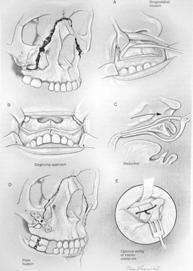
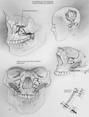
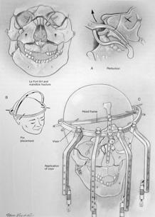
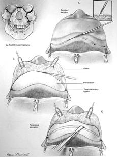
Chapter 23: Maxillofacial Trauma. Robert B. Stanley, Jr.
 Chapter 23: Maxillofacial Trauma Robert B. Stanley, Jr. Traditionally, fractures of the facial skeleton have been evaluated and treated in a segmentalized fashion, even if complex injuries were obvious
SKULL BASE AND. Bert De Foer A. Bernaerts, E. Loney +, J. Van Dinther, Th. Somers, E. Offeciers, A. Zarowski, J.W.Casselman / /* FACIAL TRAUMA
 MANDIBULO- Bert De Foer A. Bernaerts, E. Loney +, J. Van Dinther, Th. Somers, E. Offeciers, A. Zarowski, J.W.Casselman / /* DEPARTMENT OF RADIOLOGY, GZA HOSPITALS SINT-AUGUSTINUS, ANTWERP, BELGIUM DEPARTMENT
Introduction to Local Anesthesia and Review of Anatomy
 5-Sep Introduction and Anatomy Review 12-Sep Neurophysiology and Pain 19-Sep Physiology and Pharmacology part 1 26-Sep Physiology and Pharmacology part 2 Introduction to Local Anesthesia and Review of
Facial Trauma Emergencies in Sports: Recognition and Management
 Facial Trauma Emergencies in Sports: Recognition and Management CENTRAL CONNECTICUT STATE UNIVERSITY SPORTS MEDICINE SYMPOSIUM MARCH 5, 2019 MARK C. FLETCHER, DMD, MD, FACS CLINICAL ASSISTANT PROFESSOR;
Skeletal System -Axial System. Chapter 7 Part A
 Skeletal System -Axial System Chapter 7 Part A Skeleton Learn: Names of the s. Identify specific landmarks that allow: Bones to fit into each other, Organs to fit into the cavities, Muscles to attach,
Mandibular Fractures June 2000
 TITLE: Mandibular Fractures DATE: June 14, 2000 RESIDENT PHYSICIAN: Karen L. Stierman, M.D. FACULTY ADVISOR: Byron J. Bailey, M.D., FACS SERIES EDITOR: Francis B. Quinn, Jr., M.D. FACS ARCHIVIST: Melinda
Clues of a Ruptured Globe
 Definition any eye that has sustained a full thickness traumatic disruption of the cornea or sclera Overwhelmingly, rupture accidents occur in young men, small children and the elderly Corneal laceration
Trigeminal Nerve Worksheets, Distributions Page 1
 Trigeminal Nerve Worksheet #1 Distribution by Nerve Dr. Darren Hoffmann Dental Gross Anatomy, Spring 2013 We have drawn out each of the branches of CN V in lecture and you have an idea now for their basic
Cranium Facial bones. Sternum Rib
 Figure 7.1 The human skeleton. Skull Thoracic cage (ribs and sternum) Cranium Facial bones Sternum Rib Bones of pectoral girdle Vertebral column Sacrum Vertebra Bones of pelvic girdle (a) Anterior view
Introduction. patterns of injury. The injury pattern produced vanes with. j the object striking the face.
 Dolan et al. Facial fractures I Introduction Facial injury constitutes a frequent finding among emergency room patients. Schultz and Oldham estimate that 54% of such patients will have significant trauma.
OPEN ACCESS ATLAS OF OTOLARYNGOLOGY, HEAD & NECK OPERATIVE SURGERY
 OPEN ACCESS ATLAS OF OTOLARYNGOLOGY, HEAD & NECK OPERATIVE SURGERY INFERIOR MAXILLECTOMY Tumours of the hard palate and superior alveolus may be resected by inferior maxillectomy (Figure 1). A Le Fort
The diagnostic value of Computed Tomography in evaluation of maxillofacial Trauma
 The diagnostic value of Computed Tomography in evaluation of maxillofacial Trauma Qais H. Muassa FICMS College of Dentistry, Babylon University Ibrahim S. Gataa, BDS, FICMS College of Dentistry, Sulaimania
The orbit-1. Dr. Heba Kalbouneh Assistant Professor of Anatomy and Histology
 The orbit-1 Dr. Heba Kalbouneh Assistant Professor of Anatomy and Histology Orbital plate of frontal bone Orbital plate of ethmoid bone Lesser wing of sphenoid Greater wing of sphenoid Lacrimal bone Orbital
Oral Surgery Dr. Labeed Sami جامعة تكريت كلية طب االسنان املرحلة اخلامسة م.د. لبيد سامي حسن
 جامعة تكريت كلية طب االسنان جراحة الفم مادة املرحلة اخلامسة م.د. لبيد سامي حسن 6102-6102 1 5 th stage Fracture zygomatic complex As the zygomatic bone is closely associated with the maxilla, frontal and
By JOHN MARQUIS CONVERSE, M.D., and DAUBERT TELSEY, D.D.S.
 THE TRIPARTITE OSTEOTOMY OF THE MID-FACE FOR ORBITAL EXPANSION AND CORRECTION OF THE DEFORMITY IN CRANIOSTENOSIS By JOHN MARQUIS CONVERSE, M.D., and DAUBERT TELSEY, D.D.S. Center for Craniofacial Anomalies
Nasal Orbital Ethmoid (NOE) Fractures November, 2014
 Fractures November, 2014") TITLE: Nasal Orbital Ethmoid (NOE) Fractures SOURCE: Grand Rounds Presentation, The University of Texas Medical Branch at Galveston, Department of Otolaryngology DATE: November 19, 2014 RESIDENT PHYSICIAN:
TITLE: Nasal Orbital Ethmoid (NOE) Fractures SOURCE: Grand Rounds Presentation, The University of Texas Medical Branch at Galveston, Department of Otolaryngology DATE: November 19, 2014 RESIDENT PHYSICIAN:
MRI masterfile Part 5 WM Heme Strokes.ppt 1
 Ocular and Orbital Trauma Eye Trauma: Incidence 1.3 million eye injuries in the US per year. 40,000 of these injuries lead to blindness in the US. Patrick Sibony, MD March 23, 2013 Ophthalmic Emergencies
Dr.Sepideh Falah-kooshki
 Dr.Sepideh Falah-kooshki MAXILLA Premaxillary/median palatal suture (radiolucent). Incisive fossa and foramen (radiolucent). Nasal passages (radiolucent). Nasal septum (radiopaque). Anterior nasal spine
Among the myriad injuries seen in the emergency department,
 Maxillofacial Trauma: Challenges In ED Diagnosis And Management 2:20 a.m. Saturday morning: You just finished with a nine-year-old, newonset diabetic in ketoacidosis and a 37-year-old with an inferior
Extraoral radiography Introduction: Extraoral radiographs (outside the mouth) are taken when large areas of the skull or jaw must be examined or when
 are taken when large areas of the skull or jaw must be examined or when") Extraoral radiography Introduction: Extraoral radiographs (outside the mouth) are taken when large areas of the skull or jaw must be examined or when patients are unable to open their mouths for film placement.
Extraoral radiography Introduction: Extraoral radiographs (outside the mouth) are taken when large areas of the skull or jaw must be examined or when patients are unable to open their mouths for film placement.
MRI masterfile Part 5 WM Heme Strokes.ppt 2
 Imaging of Orbital Trauma Corneal Abrasion CT scan is preferable to MRI Bone, Rapid, Easy to monitor patient Foreign bodies, air, hemorrhage Fractures Cost Needed for an MRI MRI Globe and intraocular injuries
Skull basic structures. Neurocranium
 Assoc. Prof. Květuše Lovásová, M.V.D., PhD. Skull basic structures Skull consists of two groups of bones: neurocranium (bones forming the brain box) splanchnocranium (bones forming the facial skeleton)
Management Strategies for Communited Fractures of Frontal Skull Base: An Institutional Experience
 80 Original Article THIEME Management Strategies for Communited Fractures of Frontal Skull Base: An Institutional Experience V. Velho 1 Hrushikesh U. Kharosekar 1 Jasmeet S. Thukral 1 Shonali Valsangkar
Complications of Midface Fractures
 557 Kirkland Lozada, MD 1 Sameep Kadakia, MD 1 Manoj T. Abraham, MD 2 Yadranko Ducic, MD, FRCS(C), FACS 3 1 Department of Otolaryngology, New York Eye and Ear Infirmary of MountSinai,NewYork,NewYork 2
SYLLABUS OF ORAL AND MAXILLOFACIAL SURGERY
 MEDICAL UNIVERSITY OF VARNA FACULTY OF DENTAL MEDICINE DEPARTMENT OF ORAL AND MAXILLOFACIAL SURGERY AND SID SYLLABUS OF ORAL AND MAXILLOFACIAL SURGERY (State examination) ACADEMIC YEAR 2015 2016 1. Asepsis
Eyes, ears, teeth and everything in between
 Eyes, ears, teeth and everything in between E M E R G E N C Y D E P A R T M E N T J U N I O R T E A C H created 14/11/10 by S.R. Bruijns, version 1.0 Objectives Eyes Ears Teeth Maxilla- facial EYES Approaching
3. The Jaw and Related Structures
 Overview and objectives of this dissection 3. The Jaw and Related Structures The goal of this dissection is to observe the muscles of jaw raising. You will also have the opportunity to observe several
Technique Guide. Titanium Wire with Barb and Needle. Surgical Technique Guide for Canthal Tendon Prodecures.
 Technique Guide Titanium Wire with Barb and Needle. Surgical Technique Guide for Canthal Tendon Prodecures. Indications/Features Indications The Synthes Titanium Wire with Barb and straight Needle is
Oral Surgery Dr. Labeed Sami جامعة تكريت كلية طب االسنان مادة جراحة الفم املرحلة اخلامسة م.د. لبيد سامي حسن
 جامعة تكريت كلية طب االسنان مادة جراحة الفم املرحلة اخلامسة م.د. لبيد سامي حسن 6102-6102 Mandibular fractures And Dentoalveolar fractures MANDIBULAR ANATOMY The mandible is a U- or V-shaped structure consisting
The sebaceous glands (glands of Zeis) open directly into the eyelash follicles, ciliary glands (glands of Moll) are modified sweat glands that open
 open directly into the eyelash follicles, ciliary glands (glands of Moll) are modified sweat glands that open") The Orbital Region The orbits are a pair of bony cavities that contain the eyeballs; their associated muscles, nerves, vessels, and fat; and most of the lacrimal apparatus upper eyelid is larger and more
The Orbital Region The orbits are a pair of bony cavities that contain the eyeballs; their associated muscles, nerves, vessels, and fat; and most of the lacrimal apparatus upper eyelid is larger and more
Infratemporal fossa: Tikrit University college of Dentistry Dr.Ban I.S. head & neck Anatomy 2 nd y.
 Infratemporal fossa: This is a space lying beneath the base of the skull between the lateral wall of the pharynx and the ramus of the mandible. It is also referred to as the parapharyngeal or lateral pharyngeal
Chapter 7: Head & Neck
 Chapter 7: Head & Neck Osteology I. Overview A. Skull The cranium is composed of irregularly shaped bones that are fused together at unique joints called sutures The skull provides durable protection from
The Skull and Temporomandibular joint II Prof. Abdulameer Al-Nuaimi. E. mail:
 The Skull and Temporomandibular joint II Prof. Abdulameer Al-Nuaimi E-mail: a.al-nuaimi@sheffield.ac.uk E. mail: abdulameerh@yahoo.com Temporal fossa The temporal fossa is a depression on the temporal
MEDICAL CODING FOR FACIAL INJURIES & RECONSTRUCTION
 MEDICAL CODING FOR FACIAL INJURIES & RECONSTRUCTION Tirbod Fattahi, MD, DDS, FACS Chief & Associate Professor Division of Oral & Maxillofacial Surgery University of Florida Health Science Center, Jacksonville
Trigeminal Nerve (V)
") Trigeminal Nerve (V) Lecture Objectives Discuss briefly how the face is developed. Follow up the course of trigeminal nerve from its point of central connections, exit and down to its target areas. Describe
Trigeminal Nerve (V) Lecture Objectives Discuss briefly how the face is developed. Follow up the course of trigeminal nerve from its point of central connections, exit and down to its target areas. Describe
Titanium Wire with Barb and Needle. Surgical Technique Guide for Canthal Tendon Procedures.
 Titanium Wire with Barb and Needle. Surgical Technique Guide for Canthal Tendon Procedures. Technique Guide This publication is not intended for distribution in the USA. Instruments and implants approved
Human Anatomy and Physiology - Problem Drill 07: The Skeletal System Axial Skeleton
 Human Anatomy and Physiology - Problem Drill 07: The Skeletal System Axial Skeleton Question No. 1 of 10 Which of the following statements about the axial skeleton is correct? Question #01 A. The axial
Australian Dental Journal
 Australian Dental Journal The official journal of the Australian Dental Association Australian Dental Journal 2018; 63:(1 Suppl): S35 S47 doi: 10.1111/adj.12589 Current and evolving trends in the management
Anatomic Relations Summary. Done by: Sohayyla Yasin Dababseh
 Anatomic Relations Summary Done by: Sohayyla Yasin Dababseh Anatomic Relations Lecture 1 Part-1 - The medial wall of the nose is the septum. - The vestibule lies directly inside the nostrils (Nares). -
Multidetector computed tomographic evaluation of maxillofacial trauma
 ORIGINAL ARTICLE ASIAN JOURNAL OF MEDICAL SCIENCES Multidetector computed tomographic evaluation of maxillofacial trauma Kaleem Ahmad 1, R. K. Rauniyar 2, Mukesh Kumar Gupta 3, Sajid Ansari 4, Ashok Raj
Facial Trauma 76. John H. Burton and Karen Nolan Kuehl PERSPECTIVE APPROACH TO MULTITRAUMA PATIENTS WITH FACIAL INJURIES GENERAL ANATOMY
 Facial Trauma 76 John H. Burton and Karen Nolan Kuehl Treatment of all facial injuries should initially be directed toward maintaining the airway and stabilizing lifethreatening injuries. Facial computed
3-Deep fascia: is absent (except over the parotid gland & buccopharngeal fascia covering the buccinator muscle)
") The Face 1-Skin of the Face The skin of the face is: Elastic Vascular (bleed profusely however heal rapidly) Rich in sweat and sebaceous glands (can cause acne in adults) It is connected to the underlying
The Face 1-Skin of the Face The skin of the face is: Elastic Vascular (bleed profusely however heal rapidly) Rich in sweat and sebaceous glands (can cause acne in adults) It is connected to the underlying
Structure Location Function
 Frontal Bone Cranium forms the forehead and roof of the orbits Occipital Bone Cranium forms posterior and inferior portions of the cranium Temporal Bone Cranium inferior to the parietal bone forms the
Prophylactic Midface Lift in Midfacial Trauma
 Rapid Communication 347 Ryan Brown, MD 1 Kirk Lozada, MD 2 Sameep Kadakia, MD 2 Eli Gordin, MD 3 Yadranko Ducic, MD 4 1 Department of Otolaryngology, Kaiser Permanente, Denver, Colorado 2 Department of
Two Hundred Ninety-Four Consecutive Facial Fractures in an Urban Trauma Center: Lessons Learned
 CME Two Hundred Ninety-Four Consecutive Facial Fractures in an Urban Trauma Center: Lessons Learned Patrick Kelley, M.D., Marcus Crawford, M.D., Stephen Higuera, M.D., and Larry H. Hollier, M.D. Houston,
AXIAL SKELETON SKULL
 AXIAL SKELETON SKULL CRANIAL BONES (8 total flat bones w/ 2 paired) 1. Frontal forms forehead & upper portion of eyesocket (orbital) 2. Parietal paired bones; form superior & lateral walls of cranium 3.
Trigeminal Nerve Anatomy. Dr. Mohamed Rahil Ali
 Trigeminal Nerve Anatomy Dr. Mohamed Rahil Ali Trigeminal nerve Largest cranial nerve Mixed nerve Small motor root and large sensory root Motor root Nucleus of motor root present in the pons and medulla
Correction of Dentofacial Deformities (Orthognathic Surgery)
") Correction of Dentofacial Deformities (Orthognathic Surgery) BDS, MSc, German board of Oral and Maxillofacial Surgery ( Berlin-Germany), Doctoral degree by LBMS Definition Orthognathic surgery is a combination
Correction of Dentofacial Deformities (Orthognathic Surgery) BDS, MSc, German board of Oral and Maxillofacial Surgery ( Berlin-Germany), Doctoral degree by LBMS Definition Orthognathic surgery is a combination
LESSON ASSIGNMENT. Positioning for Exams of the Cranium, Sinuses, and Mandible. After completing this lesson, you should be able to:
 LESSON ASSIGNMENT LESSON 5 Positioning for Exams of the Cranium, Sinuses, and Mandible. LESSON ASSIGNMENT Paragraphs 5-1 through 5-9. LESSON OBJECTIVES After completing this lesson, you should be able
Bisection of Head & Nasal Cavity 頭部對切以及鼻腔. 解剖學科馮琮涵副教授 分機
 Bisection of Head & Nasal Cavity 頭部對切以及鼻腔 解剖學科馮琮涵副教授 分機 3250 E-mail: thfong@tmu.edu.tw Outline: The structure of nose The concha and meatus in nasal cavity The openings of paranasal sinuses Canals, foramens
Chapter 7. Skeletal System
 Chapter 7 Skeletal System 1 Skull A. The skull is made up of 22 bones: 8 cranial bones, 13 facial bones, and the mandible. B. The Cranium encloses and protects the brain, provides attachments for muscles,
Maxilla, ORBIT and infratemporal fossa. Neophytos C Demetriades MD, DDS, MSc Associate professor European University of Cyprus School of Medicine
 Maxilla, ORBIT and infratemporal fossa Neophytos C Demetriades MD, DDS, MSc Associate professor European University of Cyprus School of Medicine MAXILLA Superior, middle, and inferior meatus Frontal sinus
University of Palestine. Midterm Exam 2013/2014 Total Grade:
 Course No: DNTS2208 Course Title: Head and Neck Anatomy Date: 09/11/2013 No. of Questions: (50) Time: 1hour Using Calculator (No) University of Palestine Midterm Exam 2013/2014 Total Grade: Instructor
Parameters of Care: Clinical Practice Guidelines for Oral and Maxillofacial Surgery (AAOMS ParCare 2017) TRAUMA SURGERY
 TRAUMA SURGERY") 1 2 3 4 5 6 7 8 9 10 11 12 13 14 15 16 17 18 19 20 21 22 23 24 25 26 27 28 29 30 31 32 33 34 35 36 37 38 39 40 41 42 43 44 45 46 47 48 49 50 51 52 53 54 Parameters of Care: Clinical Practice Guidelines
1 2 3 4 5 6 7 8 9 10 11 12 13 14 15 16 17 18 19 20 21 22 23 24 25 26 27 28 29 30 31 32 33 34 35 36 37 38 39 40 41 42 43 44 45 46 47 48 49 50 51 52 53 54 Parameters of Care: Clinical Practice Guidelines
Dentistry and OMFS. Dalhousie Mini-Medical School 2018 Dr. Trish Brady BSc, DDS Dr. James Brady BSc, DDS, MD, MSc, FRCDC
 Dentistry and OMFS Dalhousie Mini-Medical School 2018 Dr. Trish Brady BSc, DDS Dr. James Brady BSc, DDS, MD, MSc, FRCDC Introduction Dr. Trish Brady, BSc, DDS Grew up in Halifax Bachelor of Science degree