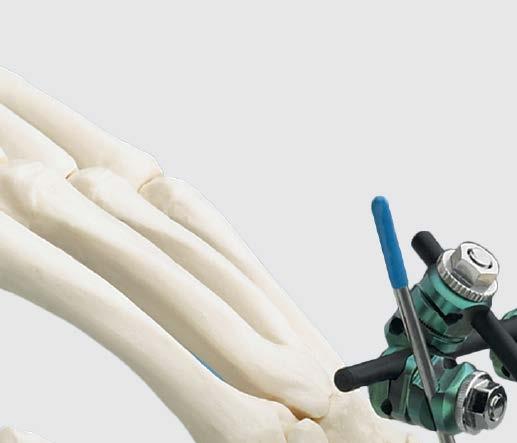
For the Treatment of Wrist Fractures. Small External Fixator Nonspanning Wrist Frame
|
|
|
- Milo Merritt
- 5 years ago
- Views:
Transcription


1 For the Treatment of Wrist Fractures Small External Fixator Nonspanning Wrist Frame Surgical Technique
2 Small External Fixator Nonspanning Wrist Frame DePuy Synthes Small External Fixation devices are labeled MR Conditional according to the terminology specified in ASTM F , Standard Practice for Marking Medical Devices and Other Items for Safety in the Magnetic Resonance Environment. Nonclinical testing demonstrated that, when used in the specific configurations stated in DePuy Synthes labeling, DePuy Synthes Small External Fixation devices are MR Conditional. Representative DePuy Synthes Small External Fixation devices used in a typical construct include clamps, rods and various attachments. A patient with a DePuy Synthes Small External Fixation frame may be scanned safely after placement of the frame under the following conditions. Static magnetic field of 1.5-Tesla when the fixator frame is positioned outside the MRI bore at Normal Operator or in First Level Control Mode. Static magnetic field when the fixator frame is positioned outside the MRI bore at Normal Operator or in First Level Control Mode. Highest spatial gradient magnetic field of 720-Gauss/cm or less. Maximum MR system reported whole body averaged specific absorption rate (SAR) of 2 W/kg for the Normal Operating Mode and 4 W/kg for the First Level Controlled Mode for 15 minutes of scanning. Use only whole body RF transmit coil, no other transmit coils are allowed, local receive only coils are allowed. Specialty Coils, such as knee or head coils, should not be used as they have not been evaluated for RF heating and may result in higher localized heating. Note: In nonclinical testing, the DePuy Synthes External Fixation Devices were tested in several different configurations. This testing was conducted with the construct positioned at the edge of the MRI bore, with the entire construct outside the MRI bore. The results showed a maximum observed heating for a wrist fixator frame of less than 4 C for 1.5T and less than 2 C for 3.0T with a machine reported whole body averaged SAR of 2 W/kg. The results showed a maximum observed heating for a pelvic frame less than 1 C for 1.5 and 3.0T with a machine reported whole body averaged SAR of 2 W/kg. Patients may be safely scanned in the MRI chamber at the above conditions. Under such conditions, the maximal expected temperature rise is less than 6ºC. Because higher in vivo heating cannot be excluded, close patient monitoring and communication with the patient during the scan is required. Immediately abort the scan if the patient reports burning sensation or pain. To minimize heating, the scan time should be as short as possible, the SAR as low as possible, and the device should be as far as possible from the edge of the bore. Temperature rise values obtained were based upon a scan time of 15 minutes. The above field conditions should be compared with those of the user s MR system, to determine if the item can safely be brought into the user s MR environment. If placed in the bore of the MR scanner during scanning, DePuy Synthes MR Conditional external fixation devices may have the potential to cause artifact in the diagnostic imaging. All components of DePuy Synthes external fixation frames must be identified as MR Conditional prior to being placed in or near an MR environment. Artifact information MR image quality may be compromised if the area of interest is in the same area or relatively close to the position of the DePuy Synthes Small External Fixation construct, and it may be necessary to optimize MR imaging parameters, to compensate for the presence of the fixation frame. Representative devices used to assemble a typical DePuy Synthes Small External Fixation frame have been evaluated in the MRI chamber and worst-case artifact information is provided below. Overall, artifacts created by DePuy Synthes Small External Fixation devices may present issues if the MR imaging area of interest is in or near the area where the fixation frame is located. Small External Fixator Nonspanning Wrist Frame Surgical Technique DePuy Synthes 1
3 Indications and MRI Information For FFE sequence: Scan duration: 3 min, TR 100 ms, TE 15 ms, flip angle 15º and SE sequence: Scan duration: 4 min, TR 500 ms, TE 20 ms, flip angle 70º radio echo sequence, worst-case artifact will extend approximately 5 cm from the device. Warning: Do not place any radio frequency (RF) transmit coils over the external fixation frame. Indications for Synthes Small External Fixation System The Synthes Small External Fixation System is intended to stabilize and provide treatment for fractures of the small bones, such as the hand, wrist, forearm, foot, and ankle. Specifically, the components can be used for: Preliminary fixation before ORIF Unstable fractures of the distal radius (both intra- and extra-articular) Open and/or comminuted bilateral fractures Fractures in combination with extensive soft tissue injury, bone loss, and vascular and/or neural involvement Fracture dislocations Failed closed reduction with casting resulting in secondary deformity (radial shortening and angulations) Pediatric open fractures with bone loss and osteotomies 2 DePuy Synthes Small External Fixator Nonspanning Wrist Frame Surgical Technique
4 DePuy Synthes Small External Fixation Devices Warning: DePuy Synthes self-drilling and self-tapping Schanz screws are not approved for screw attachment or fixation to the posterior elements (pedicles) of the cervical, thoracic, or lumbar spine. Precautions: Select the appropriate Schanz screw (self-tapping, self-drilling) for the patient s bony anatomy. Instruments and screws may have sharp edges or moving joints that may pinch or tear user s glove or skin. Handle devices with care and dispose of worn bone cutting instruments in an approved sharps container. The self-drilling Schanz screw has been developed to minimize heat development. Nevertheless, slow insertion and additional cooling (for example with a Ringer solution) are recommended. The tip of the self-drilling Schanz screw should be embedded in the far cortex to effectively resist cantilever forces and to provide sufficient stability. Only when bones are osteoporotic does the self-drilling Schanz screw have to be screwed a bit further into the distant cortical bone, and it may even slightly penetrate through it since this can increase anchoring stability. The tip of the self-tapping Schanz screw should be embedded in the far cortex to effectively resist cantilever forces and to provide sufficient stability. Implant sites should be meticulously cared for to avoid pin-tract infection. Schanz screws may be surrounded with antiseptic-coated foam sponges in an effort to avoid infection. An implant-site care procedure should be reviewed with the patient. To help minimize the risk of pin-tract infection the following points should be observed: a. Placement of Schanz screws, taking anatomy into consideration (ligaments, nerves, arteries). b. Slow insertion and/or cooling, particularly in dense, hard bone to avoid heat necrosis. c. Release of skin tension at soft tissue entry point of implant. Small External Fixator Nonspanning Wrist Frame Surgical Technique DePuy Synthes 3
5 Small External Fixator Nonspanning Wrist Frame Basic principles and application technique Although the exact frame construct chosen is dictated by the soft tissue injury and fracture pattern, basic technique principles apply to all small external fixator frames. Fracture reduction can be accomplished manually with longitudinal traction, percutaneous fixation or open reduction and internal fixation prior to the application of an external fixator. Fracture reduction is also possible after the frame has been assembled using the modular technique, which allows complete freedom of pin placement. When using the modular technique, the small external fixator frame can be applied as a wrist spanning frame or a nonspanning frame. The fracture location and complexity determine which frame application is appropriate. 4 DePuy Synthes Small External Fixator Nonspanning Wrist Frame Surgical Technique


6 Small External Fixator Nonspanning Wrist Frame continued Relevant anatomy for Schanz screw or Kirschner wire placement Target area for proximal pins Compartment 4 Extensor digitorum and extensor indicis Compartment 5 Extensor digiti minimi Extensor retinaculum Compartment 6 Extensor carpi ulnaris Compartment 3 Extensor pollicis longus Compartment 2 Extensor carpi radialis brevis Extensor carpi radialis longus Compartment 1 Extensor pollicis brevis Abductor pollicis longus Retinaculum Superficial branch of the radial nerve Radial artery Dorsal view Compartment 5 Extensor digiti minimi Compartment 6 Extensor carpi ulnaris Compartment 4 Extensor digitorum and extensor indicis Compartment 3 Extensor pollicis longus Compartment 2 Extensor carpi radialis brevis Extensor carpi radialis longus Compartment 1 Extensor pollicis brevis Abductor pollicis longus Dorsal view References 1. Jacob, Roland P. and Ralph Hertel. The Small AO External Fixator A Versatile Device. Injury 1994, Vol. 25, Suppl. 4: S-D28 S-D Jenkins, N. H., D. G. Jones, S. R. Johnson, W. J. Mintowt-Czyz. External Fixation of Colles Fractures. The Journal of Bone and Joint Surgery. Vol. 69-B, No. 2. March, 1987: Jupiter, Jesse B., and Diego L. Fernandez. Complications Following Distal Radius Fractures. The Journal of Bone and Joint Surgery (American) 83: McQueen, Margaret M. and P.J. Mackenney. Bridging and Non-bridging External Fixation of Distal Radius Fractures. Orthopaedics Today. January/February 1999:8. Small External Fixator Nonspanning Wrist Frame Surgical Technique DePuy Synthes 5
7 Technique Overview When constructing a small external fixator frame, choices for pins include: 2.5 mm Kirschner wires with thread 4.0 mm/ 2.5 mm self-drilling Schanz screws 4.0 mm/ 3.0 mm self-drilling Schanz screws 4.0 mm self-drilling Schanz screws Schanz screws provide more frame stiffness than 2.5 mm Kirschner wires. If using 2.5 mm Kirschner wires in the distal and proximal radius fragments, K-wires should converge. This improves fixation by lengthening the bone-pin interface. If using Schanz screws in the proximal radius fragment, they should be perpendicular to the long axis of the radius. The small adjustable clamp accepts only Schanz screws with 4.0 mm shafts. 6 DePuy Synthes Small External Fixator Nonspanning Wrist Frame Surgical Technique


8 Technique Overview continued 1 Insert pins in the distal radius fragment Make a small incision at the site of planned pin insertion. Make a longitudinal incision through the extensor retinaculum. Separate the appropriate compartments. Bluntly dissect the soft tissues and push the drill sleeve to the bone. Insert the pins through the drill sleeve, parallel to the articular surface. Avoid penetrating the far cortex with the pin. When using Schanz screws, insert by hand to avoid damage to soft tissues. Whenever possible, insert the pins at an angle to the sagittal plane to achieve more bony contact and increase the stability of the frame. Verify position of the pins with lateral and AP images. They should be parallel to the articular surface. 2 Build first module Connect one small combination clamp to each pin in the distal radius fragment. Connect the clamps with a 4.0 mm carbon fiber rod (straight, curved or angled T-bar). Small External Fixator Nonspanning Wrist Frame Surgical Technique DePuy Synthes 7


9 Technique Overview continued 3 Insert pins in the proximal radius Make a small incision at the site of planned pin insertion. Bluntly dissect the soft tissues and push the drill sleeve to the bone. Insert the pins through the drill sleeve. Optional technique Instrument mm Parallel Drill Guide If using the small adjustable clamp in the proximal segment, insert Schanz screws using the 4.0 mm parallel drill guide. Notes Take care to avoid the superficial branch of the radial nerve. For accuracy and ease of insertion in hard bone, consider predrilling for the Schanz screws with a 2.0 mm drill bit. For best stability, pin spacing should be maximized, and the distal pin should be as close to the fracture as possible. 4 Build second module Connect one small combination clamp to each pin in the proximal radius. Connect the clamps with a 4.0 mm carbon fiber rod. Reduce the fracture using partial frames as handles to aid in reduction. Optional technique If using the small adjustable clamp, place the clamp over parallel Schanz screws and tighten bolts onto Schanz screws. 8 DePuy Synthes Small External Fixator Nonspanning Wrist Frame Surgical Technique


10 Technique Overview continued 5 Connect modules Connect the modules with a third rod and two small combination clamps. Perform final fracture reduction, if required, and tighten the nuts securely. Optional frame construct Build the second module with a 4.0 mm carbon fiber rod which is long enough to connect to the first carbon fiber rod. Ensure that all clamps are tight. See Optional Frame Configurations page for sample frames. 6 Increase stiffness Use Schanz screws in place of 2.5 mm K-wires and/or add a bar to increase stiffness and to prevent rotation. This bar will also prevent shortening of the distal radius fragment. Connect the fourth bar to the pin in the distal radius fragment and to either the pin or the bar in the proximal radius. Small External Fixator Nonspanning Wrist Frame Surgical Technique DePuy Synthes 9
11 Recommended Components mm Kirschner Wire with Thread mm/ 2.5 mm Self-Drilling Schanz Screw mm/ 3.0 mm Self-Drilling Schanz Screw Small Combination Clamp, MR Conditional 395.6x 4.0 mm Carbon Fiber Rod, MR Conditional Protective Caps, for 2.5 mm Kirschner wires Protective Caps, for 4.0 mm Fixation Pins 11 DePuy Synthes Small External Fixator Nonspanning Wrist Frame Surgical Technique
12 Small External Fixator Nonspanning Wrist Frame Nonspanning frames Do not span the joint. When to use Simple intra-articular distal radius fractures Extra-articular distal radius fractures with a relatively large distal fragment Corrective distal radius osteotomies due to malunion Spanning frames Bridge or span of the wrist joint is the most common application for wrist fixators. Commonly used for Intra-articular distal radius fractures Extra-articular distal radius fractures Adjunct to percutaneous Kirschner wires Small External Fixator Nonspanning Wrist Frame Surgical Technique DePuy Synthes 11


13 Small External Fixator Nonspanning Wrist Frame AO Fracture Classifications 23-A Extra-articular fracture Ulna, radius intact Radius, simple and impacted Radius, multifragmentary 23-B Partial articular fracture Radius, sagittal Radius, frontal, dorsal rim Radius, frontal, volar rim 23-C Complete articular fracture of radius Articular simple, metaphyseal simple Articular simple, metaphyseal multifragmentary Articular multifragmentary Reproduced from Müller, AO Classification of Fractures/Long Bones Edition 2001, 01/1, ISBN , with permission of AO Publishing, Copyright 2001 AO Publishing, Switzerland. 11 DePuy Synthes Small External Fixator Nonspanning Wrist Frame Surgical Technique


14 Optional Frame Configurations Modular frame with angled T-bar Modular frame with curved bar Modular straight T frame Modular straight T frame with delta bar Modular box frame Small External Fixator Nonspanning Wrist Frame Surgical Technique DePuy Synthes 11

15 Small External Fixator Set with Self-Drilling Schanz Screws Stainless Steel ( ) or Titanium ( ) Graphic Case Small External Fixator Graphic Case Implants in Set , MR Conditional mm Kirschner Wire, 150 mm, trocar point, 1 pkg. of mm Kirschner Wire, 150 mm, trocar point, 1 pkg. of mm Kirschner Wire with Thread, 150 mm, trocar point, 15 mm thread length, 1 pkg. of 10 Self-Drilling Schanz Screws mm/2.5 mm, 20 mm thread length, 80 mm, 2 ea mm/3.0 mm, 20 mm thread length, 80 mm, 4 ea mm/3.0 mm, 18 mm thread length, 65 mm, 4 ea mm, 20 mm thread length, 80 mm, 2 ea mm, 30 mm thread length, 100 mm, 2 ea. Implants in Set , MR Conditional mm Kirschner Wire, 150 mm, trocar point, 1 pkg. of mm Kirschner Wire, 150 mm, trocar point, 1 pkg. of mm Kirschner Wire with Thread, 150 mm, trocar point, 15 mm thread length, 1 pkg. of 10 Graphic Case shown with additionally available Mini External Fixator and K-Wire modules Titanium Self-Drilling Schanz Screws mm/2.5 mm, 20 mm thread length, 80 mm, 2 ea mm/3.0 mm, 20 mm thread length, 80 mm, 4 ea mm, 20 mm thread length, 80 mm, 2 ea mm, 30 mm thread length, 100 mm, 2 ea. For detailed cleaning and sterilization instructions, please refer to or sterilization instructions, if provided. 11 DePuy Synthes Small External Fixator Nonspanning Wrist Frame Surgical Technique
16 Small External Fixator Set with Self-Drilling Schanz Screws Stainless Steel ( ) or Titanium ( ) continued Instruments (for both sets) Wire Cutters mm Parallel Drill Guide mm/ 2.5 mm Drill Sleeve Drive Adaptor with quick coupling, for 4.0 mm Schanz Screws, 2 ea mm Trocar Combination Wrench, 7 mm width across flats Socket Wrench, 7 mm width across flats Simple T-Handle Drill Sleeve Handle mm Threaded Drill Sleeve Fixation Material (for both sets), MR Conditional Small Combination Clamp, 12 ea * Protective Caps, for 2.5 mm Kirschner Wires (light blue), 1 pkg. of * Protective Caps, for 4.0 mm Fixation Pins (yellow), 1 pkg. of mm Carbon Fiber Rods mm, 2 ea mm, 4 ea, mm, 4 ea mm, 4 ea mm, 4 ea mm, 2 ea mm, 2 ea mm, 2 ea. Also Available Sets Kirschner Wire Implant Module Set Mini External Fixator Instrument and Implant Set Also Available Implants, MR Conditional mm/3.0 mm Schanz Screw, 20 mm thread, 80 mm length mm/2.5 mm Schanz Screw, trocar point, 18 mm thread, 65 mm length mm/2.5 mm Schanz Screw, trocar point, 20 mm thread, 80 mm length mm Self-Drilling Schanz Screw, 40 mm thread Length, 125 mm mm Titanium Self-Drilling Schanz Screw, 40 mm thread Length, 125 mm Also Available Instruments mm Drill Bit, quick coupling, 100 mm mm Drill Bit, Jacobs chuck, 85 mm Wire-Bending Pliers, 160 mm Bending Iron mm Parallel Drill Guide mm Drill Sleeve, 78 mm (long) mm Drill Sleeve, 42 mm (short) Small Distractor Mini Lengthening Apparatus, 110 mm Mini Lengthening Apparatus, 160 mm mm Trocar, 88 mm (long) mm Trocar, 49 mm (short) Open Compressor mm Threaded Drill Sleeve, 69 mm (long) mm Threaded Drill Sleeve, 33 mm (short) Also Available Fixation Material * Protective Caps, for 4.0 mm Connecting Bars and Carbon Fiber Rods (black), (10/ pkg.) *This item has not been tested for safety in the MR environment. Small External Fixator Nonspanning Wrist Frame Surgical Technique DePuy Synthes 15
17 Limited Warranty and Disclaimer: DePuy Synthes products are sold with a limited warranty to the original purchaser against defects in workmanship and materials. Any other express or implied warranties, including warranties of merchantability or fitness, are hereby disclaimed. Please also refer to the package insert(s) or other labeling associated with the devices identified in this surgical technique for additional information. CAUTION: Federal Law restricts these devices to sale by or on the order of a physician. Some devices listed in this surgical technique may not have been licensed in accordance with Canadian law and may not be for sale in Canada. Please contact your sales consultant for items approved for sale in Canada. Not all products may currently be available in all markets. Manufactured or distributed by: Synthes USA Products, LLC 1302 Wrights Lane East West Chester, PA Synthes USA, LLC 1101 Synthes Avenue Monument, CO To order (USA): To order (Canada): Note: For recognized manufacturer, refer to the product label. DePuy Synthes All rights reserved. DSUS/TRM/1016/1184 3/17 DV
Small External Fixator Nonspanning Wrist Frame. For the treatment of wrist fractures.
 Small External Fixator Nonspanning Wrist Frame. For the treatment of wrist fractures. Technique Guide Part of the Small External Fixation System Small External Fixator Nonspanning Wrist Frame When to use
Small External Fixator Wrist Spanning Frame. For the treatment of wrist fractures.
 Small External Fixator Wrist Spanning Frame. For the treatment of wrist fractures. Technique Guide Part of the Small External Fixation System Small External Fixator Wrist Spanning Frame When to use The
For Small-Statured Adults and Pediatric Patients. Medium External Fixator Basic Modular Frame
 For Small-Statured Adults and Pediatric Patients Medium External Fixator Basic Modular Frame Surgical Technique MRI Information DePuy Synthes Medium External Fixation devices are labeled MR Conditional
Large External Fixator Modular Knee Bridge
 Using Multi-pin Clamps Large External Fixator Modular Knee Bridge Surgical Technique Large External Fixator Modular Knee Bridge DePuy Synthes Large External Fixation devices are labeled MR Conditional
Mini External Fixator
 Stabilize the Phalanges and Metacarpals Mini External Fixator Surgical Technique Table of Contents Introduction Mini External Fixator 2 Indications 4 Surgical Technique Technique Overview 5 Product Information
Medium External Fixator Pediatric Femoral Shaft Frame. Using medium multi-pin clamps.
 Medium External Fixator Pediatric Femoral Shaft Frame. Using medium multi-pin clamps. Technique Guide Part of the Medium External Fixation System MRI Information Synthes Medium External Fixation devices
Medium External Fixator Humeral Shaft Frame. Modular frame for upper extremity use.
 Medium External Fixator Humeral Shaft Frame. Modular frame for upper extremity use. Technique Guide Part of the Medium External Fixation System MRI Information Synthes Medium External Fixation devices
Medium External Fixator Delta Frame Ankle Bridge
 For Small-Statured Adults Medium External Fixator Delta Frame Ankle Bridge Surgical Technique MRI Information DePuy Synthes Medium External Fixation devices are labeled MR Conditional according to the
Low-Profile Wrist Fixator. For stabilization of fractures of the distal radius.
 Low-Profile Wrist Fixator. For stabilization of fractures of the distal radius. Technique Guide Part of the External Fixation System Low-Profile Wrist Fixator Indications Intended for stabilization of
Large External Fixator Traveling Traction
 Used to Correct Angular Deformity Large External Fixator Traveling Traction Surgical Technique Large External Fixator Traveling Traction DePuy Synthes Large External Fixation devices are labeled MR Conditional
Distal Radius Plate Instrument and Implant Set. Discontinued December 2017 DSUS/TRM/0916/1063(1)
") Distal Radius Plate Instrument and Implant Set Surgical Technique Discontinued December 2017 DSUS/TRM/0916/1063(1) The Distal Radius Plates Indications For fixation of fractures and osteotomies, including
Distal Radius Plate Instrument and Implant Set Surgical Technique Discontinued December 2017 DSUS/TRM/0916/1063(1) The Distal Radius Plates Indications For fixation of fractures and osteotomies, including
External Distal Radius Fixator. Supplement to the 8 mm rod fixator system
 External Distal Radius Fixator. Supplement to the 8 mm rod fixator system Surgical technique This publication is not intended for distribution in the USA. Instruments and implants approved by the AO Foundation
Large External Fixator Pelvic Frame
 For Treatment of Unstable Pelvic Ring Injuries Large External Fixator Pelvic Frame Surgical Technique LARGE EXTERNAL FIXATOR PELVIC FRAME DePuy Synthes Companies of Johnson & Johnson Large External Fixation
Large Distractor Femur
 Fracture Reduction and Provisional Stabilization Large Distractor Femur Surgical Technique Table of Contents Introduction Standard Femoral Distraction 2 Large Distractor System 4 Surgical Technique Prepare
Large External Fixator Delta Frame Ankle Bridge. Using pin clamps with outrigger posts.
 Large External Fixator Delta Frame Ankle Bridge. Using pin clamps with outrigger posts. Technique Guide Part of the Large External Fixation System Large External Fixator Delta Frame Ankle Bridge Technique
Large External Fixator Modular Knee Bridge. Using multi-pin clamps.
 Large External Fixator Modular Knee Bridge. Using multi-pin clamps. Technique Guide Part of the Large External Fixation System Large External Fixator Modular Knee Bridge Technique Overview Insert Schanz
3.5 mm Locking Attachment Plate
 For Treatment of Periprosthetic Fractures 3.5 mm Locking Attachment Plate Surgical Technique Table of Contents Introduction 3.5 mm Locking Attachment Plate 2 Indications 4 Surgical Technique Preparation
Large External Fixator Delta Frame Ankle Bridge. For staged fixation of the distal tibia.
 Large External Fixator Delta Frame Ankle Bridge. For staged fixation of the distal tibia. Technique Guide Part of the Large External Fixation System Large External Fixator Delta Frame Ankle Bridge Technique
Mandible External Fixator II
 Provides Treatment for Fractures of the Mandible Mandible External Fixator II Surgical Technique Table of Contents Introduction Mandible External Fixator II 2 AO Principles 4 Indications 5 MRI Information
Femur Frame. The Distraction Osteogenesis Ring System
 Femur Frame The Distraction Osteogenesis Ring System Surgical Technique Table of Contents Introduction The Distraction Osteogenesis Ring System 2 MRI Information 4 AO Principles 5 Indications 6 Surgical
Distal Radius Plate 2.4/2.7 dorsal and volar
 Distal Radius Plate 2.4/2.7 dorsal and volar Surgical Technique This publication is not intended for distribution in the USA. Instruments and implants approved by the AO Foundation. Distal Radius Plate
For Distal Femur Fractures. 95º Condylar Plate. Quick Reference Chart
 For Distal Femur Fractures 95º Condylar Plate Quick Reference Chart 95 Condylar Plate. Quick reference chart for distal femur fractures. Insert guide wires Fix condylar fragments with 6.5 mm cancellous
Long Volar Plates for Diaphyseal-Metaphyseal Radius Fractures LCP. Dia-Meta Volar Distal Radius Plates. Surgical Technique
 Long Volar Plates for Diaphyseal-Metaphyseal Radius Fractures LCP Dia-Meta Volar Distal Radius Plates Surgical Technique Table of Contents Introduction LCP Dia-Meta Volar Distal Radius Plates 2 AO Principles
3.5 mm LCP Extra-articular Distal Humerus Plate
 Part of the DePuy Synthes Locking Compression Plate (LCP ) System 3.5 mm LCP Extra-articular Distal Humerus Plate Surgical Technique Table of Contents Introduction 3.5 mm LCP Extra-articular Distal Humerus
Lag Screw Device Intended for symphyseal fracture fixation of the mandible
 Lag Screw Device Intended for symphyseal fracture fixation of the mandible SUrgicaL TecHNiqUe Lag Screw Device Intended for symphyseal fracture fixation of the mandible Simplifies the lag screw fixation
3.5 mm Clavicle Hook Plates
 A Single Solution for Lateral Clavicle Fractures and Acromioclavicular Joint Dislocations 3.5 mm Clavicle Hook Plates Surgical Technique Discontinued December 2017 DSUS/TRM/1016/1126(1) Table of Contents
Low Profile Neuro Plating System. Surgical Technique
 Low Profile Neuro Plating System Surgical Technique TABLE OF CONTENTS INTRODUCTION Low Profile Neuro Plating System 2 SURGICAL TECHNIQUE Technique 5 PRODUCT INFORMATION Low Profile Neuro Plates 10 Low
3.5 mm LCP Olecranon Plates
 Part of the DePuy Synthes Locking Compression Plate (LCP ) System 3.5 mm LCP Olecranon Plates Surgical Technique Table of Contents Introduction 3.5 mm LCP Olecranon Plates 2 AO Principles 3 Indications
The Distraction Osteogenesis Ring System
 Nonarticular Tibia Frame The Distraction Osteogenesis Ring System Surgical Technique Table of Contents Introduction The Distraction Osteogenesis Ring System 2 MRI Information 4 AO Principles 5 Indications
Sterile-Packaged Large External Fixator Kits. For treatment of long bone and pelvic fractures that require external fixation.
 Sterile-Packaged Large External Fixator Kits. For treatment of long bone and pelvic fractures that require external fixation. Large External Fixator Ankle Frame Kit Large External Fixator Trauma Kit Large
The Distraction Osteogenesis Ring System
 Intra-articular Distal Tibia Frame The Distraction Osteogenesis Ring System Surgical Technique Table of Contents Introduction The Distraction Osteogenesis Ring System 2 MRI Information 4 AO Principles
Small External Fixator
 Surgical Technique This publication is not intended for distribution in the USA. Instruments and implants approved by the AO Foundation Table of Contents System Description 3 Indications/ Contraindications
Mini External Fixator.
 Mini External Fixator. Assembly and Surgical Technique This publication is not intended for distribution in the USA. Instruments and implants approved by the AO Foundation. Image intensifier control Warning
3.0/3.5/4.0/4.5/6.5/7.0/7.3. Cannulated Screws. Surgical Technique
 3.0/3.5/4.0/4.5/6.5/7.0/7.3 Cannulated Screws Surgical Technique Image intensifier control This description alone does not provide sufficient background for direct use of DePuy Synthes products. Instruction
Mandible External Fixator II. Provides treatment for fractures of the maxillofacial area.
 Mandible External Fixator II. Provides treatment for fractures of the maxillofacial area. Technique Guide This publication is not intended for distribution in the USA. Instruments and implants approved
Technique Guide. The Distraction Osteogenesis Ring System. Intra-articular distal tibia frame.
 Technique Guide The Distraction Osteogenesis Ring System. Intra-articular distal tibia frame. Table of Contents Introduction The Distraction Osteogenesis Ring System 2 MRI Information 4 AO Principles 5
The Locking Calcaneal Plate Instrument and Implant Sets
 Part of the DePuy Synthes Locking Compression Plate (LCP ) System The Locking Calcaneal Plate Instrument and Implant Sets Surgical Technique Table of Contents Introduction Locking Calcaneal Plate 2 AO
3.5 mm LCP Clavicle Hook Plates
 Part of the Synthes Locking Compression Plate (LCP ) System 3.5 mm LCP Clavicle Hook Plates Surgical Technique Table of Contents Introduction 3.5 mm LCP Clavicle Hook Plates 2 AO Principles 4 Indications
Low Bend Distal Tibia Plates
 Part of the DePuy Synthes Locking Compression Plate (LCP ) System 3.5 mm LCP Low Bend Medial Distal Tibia Plates Surgical Technique Table of Contents Introduction 3.5 mm LCP Low Bend Medial Distal Tibia
Small External Fixator, Radiolucent, Sterile. Specific design for distal radius fractures.
 Small External Fixator, Radiolucent, Sterile. Specific design for distal radius fractures. Surgical technique This publication is not intended for distribution in the USA. Instruments and implants approved
Trochanter Stabilization Plate for DHS Implants
 Extends DHS Plate Construct to Help Stabilize Greater Trochanter Trochanter Stabilization Plate for DHS Implants Surgical Technique Table of Contents Introduction Trochanter Stabilization Plate for DHS
Distal Radius Sterile Kit. Optimization And Efficiency You Can Rely On
 Distal Radius Sterile Kit Optimization And Efficiency You Can Rely On Introducing The Distal Radius Sterile Kit Improved OR Workflow The Distal Radius Sterile Kit provides high-quality single-use implants
LCP Proximal Radius Plates 2.4. Plates for radial head rim and for radial head neck address individual fracture patterns of the proximal radius.
 LCP Proximal Radius Plates 2.4. Plates for radial head rim and for radial head neck address individual fracture patterns of the proximal radius. Surgical Technique This publication is not intended for
Part of the DePuy Synthes Locking Compression Plate (LCP ) System. 3.5 mm LCP Medial Proximal Tibia Plates
 System. 3.5 mm LCP Medial Proximal Tibia Plates") Part of the DePuy Synthes Locking Compression Plate (LCP ) System 3.5 mm LCP Medial Proximal Tibia Plates Surgical Technique Table of Contents Introduction 3.5 mm LCP Medial Proximal Tibia Plates 2 AO
Part of the DePuy Synthes Locking Compression Plate (LCP ) System 3.5 mm LCP Medial Proximal Tibia Plates Surgical Technique Table of Contents Introduction 3.5 mm LCP Medial Proximal Tibia Plates 2 AO
2.4 mm Variable Angle LCP Volar Extra-Articular Distal Radius System. For fragment-specific fracture fixation with variable angle locking technology.
 2.4 mm Variable Angle LCP Volar Extra-Articular Distal Radius System. For fragment-specific fracture fixation with variable angle locking technology. Surgical Technique This publication is not intended
low ProfIle neuro PlaTIng system
 low ProfIle neuro PlaTIng system surgical TeChnIque Table of Contents Introduction Low Profile Neuro Cranial Plating System 2 Surgical Technique Technique 5 Product Information Low Profile Neuro Plates
2.7 mm/3.5 mm LCP Distal Fibula Plate
 Part of the DePuy Synthes Locking Compression Plate (LCP ) System 2.7 mm/3.5 mm LCP Distal Fibula Plate Surgical Technique Table of Contents Introduction 2.7 mm/3.5 mm LCP Distal Fibula Plates 2 AO Principles
The Calcaneal Plate. The Synthes non-locking solution for the Calcaneus.
 The Calcaneal Plate. The Synthes non-locking solution for the Calcaneus. Surgical Technique This publication is not intended for distribution in the USA. Instruments and implants approved by the AO Foundation.
Mandible External Fixator II. Provides treatment for fractures of the maxillofacial area.
 Mandible External Fixator II. Provides treatment for fractures of the maxillofacial area. Lightweight single-phase system Adjustable throughout application Rigid construct using three basic components
MEFiSTO. Monolateral External Fixation System for Trauma and Orthopaedics.
 MEFiSTO. Monolateral External Fixation System for Trauma and Orthopaedics. Surgical Technique This publication is not intended for distribution in the USA. Instruments and implants approved by the AO Foundation.
MEFiSTO. Monolateral External Fixation System for Trauma and Orthopaedics.
 MEFiSTO. Monolateral External Fixation System for Trauma and Orthopaedics. Surgical Technique This publication is not intended for distribution in the USA. Instruments and implants approved by the AO Foundation.
Technique Guide. Adjustable Distal Radius Fixator. Part of the Synthes External Fixation Systems.
 Technique Guide Adjustable Distal Radius Fixator. Part of the Synthes External Fixation Systems. Table of Contents Introduction Adjustable Distal Radius Fixator 2 Indications 3 Surgical Technique Configure
The Distraction Osteogenesis Ring System
 Nonarticular tibia frame The Distraction Osteogenesis Ring System Surgical Technique Image intensifier control This description alone does not provide sufficient background for direct use of DePuy Synthes
Variable Angle LCP Locking Technology
 BUILDING ON SUCCESS Variable Angle LCP Locking Technology The Evolution of Plating Technology Round Screw Hole Dynamic Compression Plate Limited-Contact Dynamic Compression Less Invasive Stabilization
Mandible External Fixator II
 Provides treatment for fractures of the mandible Mandible External Fixator II Surgical Technique Image intensifier control This description alone does not provide sufficient background for direct use of
2.0 mm Mandible Locking Plate System
 Advanced Plating System for Trauma, Microvascular Reconstruction, and Orthognathic Surgery 2.0 mm Mandible Locking Plate System Surgical Technique TABLE OF CONTENTS INTRODUCTION 2.0 mm Mandible Locking
3.5 mm LCP Hook Plate
 Part of the DePuy Synthes Locking Compression Plate (LCP ) System 3.5 mm LCP Hook Plate Surgical Technique Table of Contents Introduction 3.5 mm LCP Hook Plate 2 AO Principles 4 Indications 5 Clinical
3.5 mm LCP Low Bend Medial Distal Tibia Plate Aiming Instruments
 Part of the 3.5 mm LCP 3.5 mm LCP Low Bend Medial Distal Tibia Plate Aiming Instruments Surgical Technique TABLE OF CONTENTS INTRODUCTION 3.5 mm LCP Low Bend Medial Distal Tibia Plate 2 Aiming Instruments
Surgical Technique. Distal Radius and Foot
 Surgical Technique Distal Radius and Foot JET-X BAR Unilateral Fixator Distal Radius and Foot Surgical Technique Contents Design Features...2 Distal Radius Surgical Technique Indications...10 Surgical
2.4 mm LCP Radial Head Plates. Part of the Synthes LCP Distal Radius Plate System.
 2.4 mm LCP Radial Head Plates. Part of the Synthes LCP Distal Radius Plate System. Technique Guide Instruments and Implants approved by the AO Foundation Table of Contents Introduction 2.4 mm LCP Radial
3.5 mm LCP Distal Humerus Plates
 Part of the DePuy Synthes Locking Compression Plate (LCP ) System 3.5 mm LCP Distal Humerus Plates Surgical Technique Table of Contents Introduction 3.5 mm LCP Distal Humerus Plates 2 AO Principles 4 Indications
2.4 mm Variable Angle LCP Volar Rim Distal Radius Plates
 For Fragment-Specific Fracture Fixation With Variable Angle Locking Technology 2.4 mm Variable Angle LCP Volar Rim Distal Radius Plates Surgical Technique Table of Contents Introduction 2.4 mm Variable
Hoffmann II Compact External Fixation System. Modular System for
 Hoffmann II Compact External Fixation System Modular System for Introduction In 1938, Raoul Hoffmann, a surgeon from Geneva, Switzerland, designed a revolutionary External Fixation System. The basic features
LCP Metaphyseal Plates. For extra-articular fractures.
 LCP Metaphyseal Plates. For extra-articular fractures. Surgical Technique This publication is not intended for distribution in the USA. Instruments and implants approved by the AO Foundation. Image intensifier
OBSOLETED. LCP Medial Distal Tibia Plate, without Tab. The Low Profile Anatomic Fixation System with Angular Stability and Optimal Screw Orientation.
 LCP Medial Distal Tibia Plate, without Tab. The Low Profile Anatomic Fixation System with Angular Stability and Optimal Screw Orientation. Surgical Technique LCP Small Fragment System This publication
LCP Proximal Radius Plates 2.4. Plates for radial head rim and for radial head neck address individual fracture patterns of the proximal radius.
 Technique Guide LCP Proximal Radius Plates 2.4. Plates for radial head rim and for radial head neck address individual fracture patterns of the proximal radius. Table of Contents Introduction LCP Proximal
2.4 mm Variable Angle LCP Dorsal Distal Radius Plate
 For Fragment-Specific Fracture Fixation With Variable Angle (VA) Locking Technology 2.4 mm Variable Angle LCP Dorsal Distal Radius Plate Surgical Technique Table of Contents Introduction 2.4 mm VA LCP
Cannulated Pediatric Osteotomy System (CAPOS)
") A Single System of Osteotomy Blade Plates and Cannulated Instrumentation Cannulated Pediatric Osteotomy System (CAPOS) Surgical Technique Table of Contents Introduction Cannulated Pediatric Osteotomy System
A Single System of Osteotomy Blade Plates and Cannulated Instrumentation Cannulated Pediatric Osteotomy System (CAPOS) Surgical Technique Table of Contents Introduction Cannulated Pediatric Osteotomy System
Small External Fixator
 Surgical Technique Original Instruments and Implants of the Association for the Study of Internal Fixation AO/ASIF Table of Contents Description of System 2 Indications/ Contraindications 3 Bridging Surgical
Elbow Hinge Fixator. Guided Flexion/Extension for Unstable Elbow Fractures.
 Elbow Hinge Fixator. Guided Flexion/Extension for Unstable Elbow Fractures. Surgical Technique MR Safe Radiolucent Table of Contents System Description 3 Indications and Contraindications 4 Fixation Components
LCP Proximal Radius Plates 2.4. Plates for radial head rim and for radial head neck address individual fracture patterns of the proximal radius.
 LCP Proximal Radius Plates 2.4. Plates for radial head rim and for radial head neck address individual fracture patterns of the proximal radius. Surgical Technique This publication is not intended for
4.5 mm LCP Medial Proximal Tibia Plates
 Part of the DePuy Synthes LCP Periarticular Plating System 4.5 mm LCP Medial Proximal Tibia Plates Surgical Technique Table of Contents Introduction 4.5 mm LCP Medial Proximal Tibia Plates 2 AO Principles
WINSTA-R. Distal Radius System
 Distal Radius System Table of Contents Introduction WINSTA-R System 2 Indication 2 Surgical Technique Palmar Access for Radius Plate 3 Dorsal Access for Radius Plate 3 Positioning of the Radius Plate
Technique Guide. The Distraction Osteogenesis Ring System. Nonarticular tibia frame.
 Technique Guide The Distraction Osteogenesis Ring System. Nonarticular tibia frame. Table of Contents Introduction The Distraction Osteogenesis Ring System 2 AO Principles 4 Indications 5 Surgical Technique
Hoffmann II Micro External Fixation System
 Trauma Hoffmann II Micro External Fixation System Indications for the Hand Components 9 3 4 5 1 2 7 8 6 1 Hoffmann II Micro Multi-Pin Clamp 2 Hoffmann II Micro 90 Multi-Pin Clamp 3 Hoffmann II Micro Rod
3.5 mm LCP Anterolateral Distal Tibia Plates
 Part of the DePuy Synthes Locking Compression Plate (LCP ) System 3.5 mm LCP Anterolateral Distal Tibia Plates Surgical Technique Table of Contents Introduction 3.5 mm LCP Anterolateral Distal Tibia Plates
Technique Guide. Self-Drilling Schanz Screws. Designed to optimize the bone/pin interface.
 Technique Guide Self-Drilling Schanz Screws. Designed to optimize the bone/pin interface. Self-Drilling Schanz Screws The self-drilling Schanz screw has been specifically designed to optimize the bone/pin
3.5 mm LCP Distal Tibia T-Plates
 Part of the DePuy Synthes Locking Compression Plate (LCP ) System 3.5 mm LCP Distal Tibia T-Plates Surgical Technique Table of Contents Introduction 3.5 mm LCP Distal Tibia T-Plates 2 AO Principles 4 Indications
2.4 mm Variable Angle LCP Volar Extra-Articular Distal Radius System. For fragment-specific fracture fixation with variable angle locking technology.
 Technique Guide 2.4 mm Variable Angle LCP Volar Extra-Articular Distal Radius System. For fragment-specific fracture fixation with variable angle locking technology. Table of Contents Introduction 2.4
VA LOCKING CALCANEAL PLATES 2.7
 VA LOCKING CALCANEAL PLATES 2.7 Instruments and Implants approved by the AO Foundation. This publication is not intended for distribution in the USA. SURGICAL TECHNIQUE Image intensifier control Warning
Titanium Wire With Barb and Needle
 For Canthal Tendon Procedures Titanium Wire With Barb and Needle Surgical Technique Table of Contents Introduction Titanium Wire With Barb and Needle 2 Indications 2 Surgical Technique Preoperative Planning
Technique Guide. 2.4 mm Variable Angle LCP Distal Radius System. For fragment-specific fracture fixation with variable angle locking technology.
 Technique Guide 2.4 mm Variable Angle LCP Distal Radius System. For fragment-specific fracture fixation with variable angle locking technology. Table of Contents Introduction 2.4 mm Variable Angle LCP
orthodontic Bone anchor (oba) SYStem
 SYStem") orthodontic Bone anchor (oba) SYStem Skeletal implants for the orthodontic movement of teeth SurgIcal technique Table of Contents Introduction Orthodontic Bone Anchor (OBA) System 2 Indications and Contraindications
orthodontic Bone anchor (oba) SYStem Skeletal implants for the orthodontic movement of teeth SurgIcal technique Table of Contents Introduction Orthodontic Bone Anchor (OBA) System 2 Indications and Contraindications
Knee spanning solutions
 Knee spanning solutions System features Indications Intended to be used on adults or pediatric patients as required for fracture fixation (open or closed); post-traumatic joint contracture which has resulted
Hybrid Fixator Proximal Tibia Frame. Using rings with clamps.
 Hybrid Fixator Proximal Tibia Frame. Using rings with clamps. Technique Guide Part of the External Fixation System Hybrid Fixator Proximal Tibia Frame Technique Overview 1 Insert wires 2 Attach wires to
TITANIUM TIBIAL NAIL SySTEM
 TITANIUM TIBIAL NAIL SySTEM Solid and Cannulated Nails SURGICAL TEChNIqUE Table of contents Introduction Indications 2 Preoperative Implant Selection 6 Surgical Technique Instruments for Opening the Tibia
Technique Guide. 3.5 mm LCP Low Bend Medial Distal Tibia Plates. Part of the Synthes locking compression plate (LCP) system.
 system.") Technique Guide 3.5 mm LCP Low Bend Medial Distal Tibia Plates. Part of the Synthes locking compression plate (LCP) system. Table of Contents Introduction 3.5 mm LCP Low Bend Medial Distal Tibia Plates
Technique Guide 3.5 mm LCP Low Bend Medial Distal Tibia Plates. Part of the Synthes locking compression plate (LCP) system. Table of Contents Introduction 3.5 mm LCP Low Bend Medial Distal Tibia Plates
Technique Guide. 3.5 mm LCP Olecranon Plates. Part of the Synthes locking compression plate (LCP) system.
 system.") Technique Guide 3.5 mm LCP Olecranon Plates. Part of the Synthes locking compression plate (LCP) system. Table of Contents Introduction 3.5 mm LCP Olecranon Plates 2 AO Principles 3 Indications 3 Clinical
Technique Guide 3.5 mm LCP Olecranon Plates. Part of the Synthes locking compression plate (LCP) system. Table of Contents Introduction 3.5 mm LCP Olecranon Plates 2 AO Principles 3 Indications 3 Clinical
PediLoc 3.5mm and 4.5mm Contour Femur Plate Surgical Technique
 PediLoc 3.5mm and 4.5mm Contour Femur Plate Surgical Technique Surgical Technique Contour Femur Plate The technique description herein is made available to the healthcare professional to illustrate the
Compact Distal Radius System. Consolidated solution for Distal Radius Plating Systems
 Compact Distal Radius System. Consolidated solution for Distal Radius Plating Systems Designed to allow customized system configurations Variable Angle (VA) and Locking Compression Plate (LCP ) options
Orthopaedic Cable System
 Cerclage Solutions for General Surgery Orthopaedic Cable System Surgical Technique Table of Contents Introduction Orthopaedic Cable System 2 Indications 4 Contraindications 4 Surgical Technique Cerclage
For the Attention of the Operating Surgeon: IMPORTANT INFORMATION ON THE MATRIXRIB FIXATION SYSTEM
 For the Attention of the Operating Surgeon: IMPORTANT INFORMATION ON THE MATRIXRIB FIXATION SYSTEM 10/16 GP2685-E-CAN DESCRIPTION The MatrixRIB Fixation System consists of locking plates, locking screws,
LCP DISTAL TIBIA PLATE
 LCP DISTAL TIBIA PLATE Instruments and implants approved by the AO Foundation. This publication is not intended for distribution in the USA. SURGICAL TECHNIQUE Image intensifier control This description
3.5 MM VA-LCP PROXIMAL TIBIA PLATE SYSTEM
 3.5 MM VA-LCP PROXIMAL TIBIA PLATE SYSTEM Part of the DePuy Synthes Variable Angle Periarticular Plating System SURGICAL TECHNIQUE TABLE OF CONTENTS INTRODUCTION 3.5 mm VA-LCP Proximal Tibial Plate 2 AO
DISTRACTION PRODUCT OVERVIEW. For a wide variety of facial applications
 DISTRACTION PRODUCT OVERVIEW For a wide variety of facial applications DISTRACTION PRODUCT OVERVIEW. STRONG, MODULAR, VERSATILE CRANIOFACIAL DISTRACTION External Midface Distractor Distraction of the maxilla,
Variable Angle LCP Volar Rim Distal Radius Plate 2.4. For fragment-specific fracture fixation with variable angle locking technology.
 Technique Guide Variable Angle LCP Volar Rim Distal Radius Plate 2.4. For fragment-specific fracture fixation with variable angle locking technology. Image intensifier control Warning This description
MINI TIBIAL PLATEAU LEVELING OSTEOTOMY (TPLO) SYSTEM
 SYSTEM") MINI TIBIAL PLATEAU LEVELING OSTEOTOMY (TPLO) SYSTEM For stabilizing osteotomies of the canine and feline proximal tibia SURGICAL TECHNIQUE TABLE OF CONTENTS INTRODUCTION Mini Tibial Plateau Leveling
MINI TIBIAL PLATEAU LEVELING OSTEOTOMY (TPLO) SYSTEM For stabilizing osteotomies of the canine and feline proximal tibia SURGICAL TECHNIQUE TABLE OF CONTENTS INTRODUCTION Mini Tibial Plateau Leveling
LOW PROFILE NEURO. This publication is not intended for distribution in the USA. SURGICAL TECHNIQUE
 LOW PROFILE NEURO This publication is not intended for distribution in the USA. SURGICAL TECHNIQUE TABLE OF CONTENTS INTRODUCTION Low Profile Neuro Plating System 2 Intended Use, Indications, Contraindications
FastFrame External Fixation System. Damage Control Surgical Technique
 FastFrame External Fixation System Damage Control Surgical Technique 1 FastFrame External Fixation System Damage Control Surgical Technique Table of Contents Introduction... 2 Indications and Contradictions...
2.4 mm Variable Angle Locking Intercarpal Fusion System
 For Partial Wrist Arthrodesis With Variable Angle Locking Technology 2.4 mm Variable Angle Locking Intercarpal Fusion System Surgical Technique Table of Contents Introduction 2.4 mm Variable Angle Locking