Large External Fixator Pelvic Frame
|
|
|
- Colin Patterson
- 5 years ago
- Views:
Transcription


1 For Treatment of Unstable Pelvic Ring Injuries Large External Fixator Pelvic Frame Surgical Technique
2 LARGE EXTERNAL FIXATOR PELVIC FRAME DePuy Synthes Companies of Johnson & Johnson Large External Fixation devices are labeled MR Conditional according to the terminology specified in ASTM F , Standard Practice for Marking Medical Devices and Other Items for Safety in the Magnetic Resonance Environment. Nonclinical testing demonstrated that, when used in the specific configurations stated in DePuy Synthes Companies labeling, DePuy Synthes Companies Large External Fixation devices are MR Conditional. Representative DePuy Synthes Companies Large External Fixation devices used in a typical construct include clamps, rods and various attachments. A patient with a DePuy Synthes Companies Large External Fixation frame may be scanned safely after placement of the frame under the following conditions. Static magnetic field of 1.5-Tesla when the fixator frame is positioned outside the MRI bore at Normal Operator or in First Level Control Mode. Static magnetic field of 3.0-Tesla when the fixator frame is positioned outside the MRI bore at Normal Operator or in First Level Control Mode. Highest spatial gradient magnetic field of 720-Gauss/cm or less. Maximum MR system reported whole body averaged specific absorption rate (SAR) of 2 W/kg for the Normal Operating Mode and 4 W/kg for the First Level Controlled Mode for 15 minutes of scanning. Use only whole body RF transmit coil, no other transmit coils are allowed, local receive only coils are allowed. Specialty Coils, such as knee or head coils, should not be used as they have not been evaluated for RF heating and may result in higher localized heating. Note: In nonclinical testing, the DePuy Synthes Companies External Fixation Devices were tested in several different configurations. This testing was conducted with the construct positioned at the edge of the MRI bore, with the entire construct outside the MRI bore. The results showed a maximum observed heating for a wrist fixator frame of less than 4 C for 1.5T and less than 2 C for 3.0T with a machine reported whole body averaged SAR of 2 W/kg The results showed a maximum observed heating for a pelvic frame less than 1 C for 1.5 and 3.0T with a machine reported whole body averaged SAR of 2 W/kg Patients may be safely scanned in the MRI chamber at the above conditions. Under such conditions, the maximal expected temperature rise is less than 6ºC. Because higher in vivo heating cannot be excluded, close patient monitoring and communication with the patient during the scan is required. Immediately abort the scan if the patient reports burning sensation or pain. To minimize heating, the scan time should be as short as possible, the SAR as low as possible, and the device should be as far as possible from the edge of the bore. Temperature rise values obtained were based upon a scan time of 15 minutes. The above field conditions should be compared with those of the user s MR system, to determine if the item can safely be brought into the user s MR environment. If placed in the bore of the MR scanner during scanning, DePuy Synthes Companies MR Conditional external fixation devices may have the potential to cause artifact in the diagnostic imaging. All components of DePuy Synthes Companies external fixation frames must be identified as MR Conditional prior to being placed in or near an MR environment. Artifact information MR image quality may be compromised if the area of interest is in the same area or relatively close to the position of the DePuy Synthes Companies Large External Fixation construct, and it may be necessary to optimize MR imaging parameters, to compensate for the presence of the fixation frame. Representative devices used to assemble a typical DePuy Synthes Companies Large External Fixation frame have been evaluated in the MRI chamber and worst-case artifact information is provided below. Overall, artifacts created by DePuy Synthes Companies Large External Fixation devices may present issues if the MR imaging area of interest is in or near the area where the fixation frame is located. Large External Fixator Pelvic Frame Surgical Technique DePuy Synthes 1
3 INDICATIONS AND MRI INFORMATION For FFE sequence: Scan duration: 3 min, TR 100 ms, TE 15 ms, flip angle 15º and SE sequence: Scan duration: 4 min, TR 500 ms, TE 20 ms, flip angle 70º radio echo sequence, worst-case artifact will extend approximately 5 cm from the device. Warning: Do not place any radio frequency (RF) transmit coils over the external fixation frame. INDICATIONS The Synthes Large External Fixation Systems is intended to provide treatment for long bone and pelvic fractures that require external fixation. Specifically, the components can be used for: Stabilization of soft tissues and fractures Polytrauma/multiple orthopaedic trauma Vertically stable pelvic fractures, or as a treatment adjunct for vertically unstable pelvic fractures Arthrodesis and osteotomies with soft tissue problems; failures of total joints Neutralization of fractures stabilized with limited internal fixation Non-unions/septic non-unions Intraoperative reductions/stabilization tool to assist with indirect reduction Unilateral rectilinear bone segment transport or leg lengthening 2 DePuy Synthes Large External Fixator Pelvic Frame Surgical Technique
4 DEPUY SYNTHES LARGE EXTERNAL FIXATION SYSTEM Warning: DePuy Synthes self-drilling, self-tapping Schanz screws and Steinmann pins are not approved for screw attachment or fixation to the posterior elements (pedicles) of the cervical, thoracic, or lumbar spine. Precautions: To keep from damaging the femoral cutaneous nerve, avoid pin insertion up to 15 mm in a dorsal direction from the superior anterior iliac spine. When dealing with the humerus, primary consideration should be given to the radial and axillary nerves. Distally, a dorsal approach to the humerus is appropriate. Proximally, it is recommended to introduce the Schanz screws from a ventrolateral direction, caudal to the path of the axillary nerve. Select the appropriate Schanz screw (self-tapping, self-drilling), or Steinmann pin for the patient s bony anatomy. Instruments and screws may have sharp edges or moving joints that may pinch or tear user s glove or skin. Handle devices with care and dispose of worn bone cutting instruments in an approved sharps container. The self-drilling Schanz screw has been developed to minimize heat development. Nevertheless, slow insertion and additional cooling (for example with a Ringer solution) are recommended. The tip of the self-drilling Schanz screw should be embedded in the far cortex to effectively resist cantilever forces and to provide sufficient stability. Only when bones are osteoporotic does the self-drilling Schanz screw have to be screwed a bit further into the distant cortical bone, and it may even slightly penetrate through it since this can increase anchoring stability. The tip of the self-tapping Schanz screw should be embedded in the far cortex to effectively resist cantilever forces and to provide sufficient stability. Implant sites should be meticulously cared for to avoid pin-tract infection. Schanz screws and Steinmann pins may be surrounded with antiseptic-coated foam sponges in an effort to avoid infection. An implant-site care procedure should be reviewed with the patient. To help minimize the risk of pin-tract infection the following points should be observed: a. Placement of Schanz screws and Steinmann pins, taking anatomy into consideration (ligaments, nerves, arteries). b. Slow insertion and/or cooling, particularly in dense, hard bone to avoid heat necrosis. c. Release of skin tension at soft tissue entry point of implant. Large External Fixator Pelvic Frame Surgical Technique DePuy Synthes 3


.")


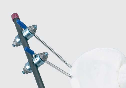
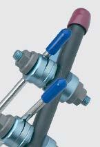
5 Large External Fixator Pelvic Frame TECHNIQUE OVERVIEW Manual reduction should be attempted prior to making skin incisions. The patient may be held in temporary reduction with a circumferential sheet, as long as the iliac crest area remains exposed. 1 Prepare for Schanz screw insertion on one side of pelvis Palpate bony landmarks and make a transverse incision 2 cm posterior to the anterior superior iliac spine (ASIS) , Predrill for the first Schanz screw Place the first Schanz screw Insert Schanz screw using the universal chuck with T-handle 8 4 Repeat for second Schanz screw Place the second Schanz screw approximately 2 cm 3 cm posterior to the first, in the middle of the gluteus medius pillar. 5 Repeat Steps 1 4 for Schanz screw insertion in the opposite hemipelvis 6 Attach one short carbon fiber rod to each set of Schanz screws Connect one open adjustable clamp to each Schanz screw. Connect with a short 11.0 mm carbon fiber rod. 7 Connect modules Use two long rods connected to the modules and to each other with combination clamps. 8 Reduce fracture Use the rods of the first and second modules as handles for reduction. Tighten the nuts. 1. Kellam JF. The role of external fixation in pelvic disruptions. Clin Orthop Relat Res. 1989;(241): Note: By adjusting the clamps, the center rods can be maneuvered to facilitate access to the abdominal region. 4 DePuy Synthes Large External Fixator Pelvic Frame Surgical Technique
6 Large External Fixator Pelvic Frame RECOMMENDED COMPONENTS FOR BASIC FRAME Product Item Quantity Number Needed 294.5x 5.0 mm Schanz Screw, 4 blunted trocar point Large Combination Clamp, 3 MR Conditional Large Open Adjustable Clamp, 4 MR Conditional 394.8x 11.0 mm Carbon Fiber Rod, MR Conditional Short ( mm) 2 Long ( 250 mm) Protective Cap, 8 for 11.0 mm Rods Protective Cap, for 5.0 mm 4 Fixation Pins Large External Fixator Pelvic Frame Surgical Technique DePuy Synthes 5
7 Large External Fixator Pelvic Frame When to use External fixation of the pelvis can be utilized for resuscitation of the hemo dynamically unstable patient with an open book fracture or as an adjunct treatment for completely unstable pelvic ring injuries. Pelvic external fixation is frequently provisional; more definitive methods of achieving pelvic stability are carried out once the patient has been stabilized. Pin placement Standard technique is to place two Schanz screws in each hemipelvis. A third Schanz screw can be added for increased stability. Schanz screws can be placed in the iliac crest where they should be inserted anterior to the gluteal tubercle. As a resuscitation frame, a single Schanz screw can be placed in each iliac crest. 2 Another option is to place one Schanz screw (see illustration) in the supra-acetabular area, slightly superior and lateral to the anterior inferior iliac spine (AIIS). This Schanz screw is oriented posteriorly, lateral to the sacroiliac joint. Iliac crest pin placement offers easier entry points but good pin purchase can be more easily achieved with supra-acetabular pin placement. Insertion of pins is facilitated by the use of image intensification, especially in the interspinous region and the AIIS. Schanz screw insertion technique Schanz screws are usually inserted through small (1 cm 2 cm) incisions which are made over the intended postreduction position of the pins. If made perpendicular to the iliac wing, 3 the incisions allow the surgeon to find the appropriate orientation of the iliac wing and can be extended to release skin tension when the frame is used for definitive treatment. 4, 5 x For placement of Schanz screws in the iliac crest, it may be useful in some patients to place two K-wires along the inner and outer tables of the ilium as guides when drilling. x Predrill only the outer cortex and insert Schanz screws by hand, using the Universal Chuck with T-Handle (393.10), allowing the Schanz screw to find its path in the ilium. Iliac crest Schanz screw insertion technique Supra-acetabular Note: When self-drilling Schanz screws are used, they must be inserted by hand. Drill sleeves must be used to protect soft tissue. 2. Tucker MC, Nork SE, Simonian PT, Routt ML Jr. Simple anterior pelvic external fixation. J Trauma. 2000;49(6): Kellam, op. cit. 4. Ibid. 5. Yang AP, Iannacone WM. External fixation for pelvic ring disruptions. Orthop Clin North Am. 1997;28(3): DePuy Synthes Large External Fixator Pelvic Frame Surgical Technique


:581 584. 2. Pohleman T. Pelvic Ring Injuries: Assessment and Concepts of Surgical Management.")
8 Large External Fixator Pelvic Frame OPTIONAL FRAME CONFIGURATIONS Supra-acetabular frames One Schanz screw can be placed in each AIIS and joined either by one curved carbon fiber rod or two straight carbon fiber rods. For greater stability, an additional Schanz screw can be placed above the first on each hemipelvis, allowing creation of a second frame that can be connected to the first. Resuscitation frame One Schanz screw can be placed in each hemipelvis and joined either by one or two carbon fiber rods, depending on patient anatomy. Iliac crest frame The use of three carbon fiber rods allows the frame to be constructed to fit larger patients. Additional reading 1. Noordeen MHH, Taylor BA, Briggs TWR, Lavy CBD. Pin placement in pelvic external fixation. Injury. 1993;24(9): Pohleman T. Pelvic Ring Injuries: Assessment and Concepts of Surgical Management. AO Principles of Fracture Management. Thomas P. Rüedi and William M. Murphy, ed. Dübendorf, Switzerland; AO Publishing Large External Fixator Pelvic Frame Surgical Technique DePuy Synthes 7

9 LARGE EXTERNAL FIXATOR SET WITH SELF-DRILLING SCHANZ SCREWS Stainless Steel ( ) or Titanium ( ) Graphic Case Large External Fixator Graphic Case Implants in Set , MR Conditional mm Steinmann Pin with Central Thread, 200 mm, 4 ea mm Schanz Screw, blunted trocar point, 200 mm, 8 ea 5.0 mm Self-Drilling Schanz Screws mm thread/150 mm, 4 ea mm thread/175 mm, 8 ea mm thread/200 mm, 8 ea mm Transfixation Pin, 225 mm, 4 ea Implants in Set , MR Conditional mm Steinmann Pin with Central Thread, 200 mm, 4 ea mm Schanz Screw, blunted trocar point, 200 mm, 8 ea mm Transfixation Pin, 225 mm, 4 ea 5.0 mm Titanium Self-Drilling Schanz Screws mm thread/150 mm, 4 ea mm thread/175 mm, 8 ea mm thread/200 mm, 8 ea Instruments (for both sets) mm Drill Bit, quick coupling, 195 mm, 2 ea mm Drill Bit, quick coupling, 195 mm, 2 ea Ratchet Wrench, 11 mm width across flats, 2 ea Cannulated Socket Wrench mm/6.0 mm Threaded Drill Sleeve, short mm/6.0 mm Threaded Drill Sleeve, long Position Drill Guide Handle Universal Chuck with T-Handle Drive Adaptor with quick coupling, for 5.0 mm Schanz Screws Drive Adaptor with quick coupling, for 6.0 mm Schanz Screws For detailed cleaning and sterilization instructions, please refer to or sterilization instructions, if provided. 8 DePuy Synthes Large External Fixator Pelvic Frame Surgical Technique
10 Large External Fixator Set with Self-Drilling Schanz Screws Stainless Steel ( ) or Titanium ( ) Split Tissue Protection Sleeve, 5.0 mm Open Compressor, 2 ea mm Trocar, short mm Trocar, long Drill Sleeve Handle mm/ 3.5 mm Drill Sleeve, short mm/ 3.5 mm Drill Sleeve, long mm/5.0 mm Threaded Drill Sleeve, short mm/5.0 mm Threaded Drill Sleeve, long Fixation Material (for both sets), MR Conditional Large Multi-Pin Clamp, 6 position, 4 ea Rod Attachment, for Large Multi-Pin Clamp, 6 ea Large Multi-Pin Clamp, 4 position, 2 ea Large Combination Clamp, 12 ea Dynamization Clip, for Large Combination Clamp, 4 ea Tube-to-Tube Clamp, 2 ea Large Open Adjustable Clamp, 8 ea * Transverse Clamp, 2 ea 11.0 mm Carbon Fiber Rods, 4 ea mm mm mm mm mm mm mm Protective Caps * For 11.0 mm Rods, 1 pkg of * For 5.0 mm Fixation Pins, 1 pkg of * For 6.0 mm Fixation Pins, 1 pkg of 10 *This item has not been tested for safety in the MR environment. Large External Fixator Pelvic Frame Surgical Technique DePuy Synthes 9


11 ALSO AVAILABLE Implants, MR Conditional Schanz Screws mm, spade point, 60 mm 150 mm mm, blunted trocar point, 100 mm 250 mm mm, blunted trocar point, 80 mm 200 mm Self-Drilling Schanz Screws mm, 60 mm 175 mm mm, 100 mm 250 mm mm, 100 mm 250 mm Titanium Self-Drilling Schanz Screws mm, 60 mm 175 mm mm, 100 mm 250 mm mm, 100 mm 250 mm S Fixation Material, MR Conditional * Spring-Loaded Nut * Adjustable Clamp * Open Clamp * Universal Joint for Two Tubes * Universal Clamp 11.0 mm Carbon Fiber Bridging Rods mm, short mm, long mm, short mm, long Protective Caps * For 4.0 mm Fixation Pins (10/pkg) * For 4.5 mm Fixation Pins (10/pkg) S terile-packaged Large External Fixator Kits S Large External Fixator Ankle Frame Kit, sterile S Large External Fixator Trauma Kit, sterile S Large External Fixator Pelvic Frame Kit, sterile *This item has not been tested for safety in the MR environment. 10 DePuy Synthes Large External Fixator Pelvic Frame Surgical Technique
12 Also Available Sets Power Drive Set ComPact Air Drive II Set Accessories for Graphic Case Label Sheet Pack, for Large External Fixator Clamps Label Sheet Pack, for Schanz Screws Replacement Brackets (3 sizes) Replacement Screws (10/pkg) Label Sheet, for Large External Fixator MR Conditional clamps Large External Fixator Pelvic Frame Surgical Technique DePuy Synthes 11
13 Limited Warranty and Disclaimer: DePuy Synthes products are sold with a limited warranty to the original purchaser against defects in workmanship and materials. Any other express or implied warranties, including warranties of merchantability or fitness, are hereby disclaimed. Please also refer to the package insert(s) or other labeling associated with the devices identified in this surgical technique for additional information. CAUTION: Federal Law restricts these devices to sale by or on the order of a physician. Some devices listed in this surgical technique may not have been licensed in accordance with Canadian law and may not be for sale in Canada. Please contact your sales consultant for items approved for sale in Canada. Not all products may currently be available in all markets. Manufactured or distributed by: Synthes USA Products, LLC 1302 Wrights Lane East West Chester, PA Synthes USA, LLC 1101 Synthes Avenue Monument, CO To order (USA): To order (Canada): Note: For recognized manufacturer, refer to the product label. DePuy Synthes All rights reserved. DSUS/TRM/0814/0215(1) 3/17 DV
Large External Fixator Modular Knee Bridge
 Using Multi-pin Clamps Large External Fixator Modular Knee Bridge Surgical Technique Large External Fixator Modular Knee Bridge DePuy Synthes Large External Fixation devices are labeled MR Conditional
For Small-Statured Adults and Pediatric Patients. Medium External Fixator Basic Modular Frame
 For Small-Statured Adults and Pediatric Patients Medium External Fixator Basic Modular Frame Surgical Technique MRI Information DePuy Synthes Medium External Fixation devices are labeled MR Conditional
Medium External Fixator Delta Frame Ankle Bridge
 For Small-Statured Adults Medium External Fixator Delta Frame Ankle Bridge Surgical Technique MRI Information DePuy Synthes Medium External Fixation devices are labeled MR Conditional according to the
Large External Fixator Traveling Traction
 Used to Correct Angular Deformity Large External Fixator Traveling Traction Surgical Technique Large External Fixator Traveling Traction DePuy Synthes Large External Fixation devices are labeled MR Conditional
Medium External Fixator Humeral Shaft Frame. Modular frame for upper extremity use.
 Medium External Fixator Humeral Shaft Frame. Modular frame for upper extremity use. Technique Guide Part of the Medium External Fixation System MRI Information Synthes Medium External Fixation devices
Medium External Fixator Pediatric Femoral Shaft Frame. Using medium multi-pin clamps.
 Medium External Fixator Pediatric Femoral Shaft Frame. Using medium multi-pin clamps. Technique Guide Part of the Medium External Fixation System MRI Information Synthes Medium External Fixation devices
Mini External Fixator
 Stabilize the Phalanges and Metacarpals Mini External Fixator Surgical Technique Table of Contents Introduction Mini External Fixator 2 Indications 4 Surgical Technique Technique Overview 5 Product Information
Large External Fixator Modular Knee Bridge. Using multi-pin clamps.
 Large External Fixator Modular Knee Bridge. Using multi-pin clamps. Technique Guide Part of the Large External Fixation System Large External Fixator Modular Knee Bridge Technique Overview Insert Schanz
For the Treatment of Wrist Fractures. Small External Fixator Nonspanning Wrist Frame
 For the Treatment of Wrist Fractures Small External Fixator Nonspanning Wrist Frame Surgical Technique Small External Fixator Nonspanning Wrist Frame DePuy Synthes Small External Fixation devices are labeled
Low-Profile Wrist Fixator. For stabilization of fractures of the distal radius.
 Low-Profile Wrist Fixator. For stabilization of fractures of the distal radius. Technique Guide Part of the External Fixation System Low-Profile Wrist Fixator Indications Intended for stabilization of
Large External Fixator Delta Frame Ankle Bridge. For staged fixation of the distal tibia.
 Large External Fixator Delta Frame Ankle Bridge. For staged fixation of the distal tibia. Technique Guide Part of the Large External Fixation System Large External Fixator Delta Frame Ankle Bridge Technique
Large External Fixator Delta Frame Ankle Bridge. Using pin clamps with outrigger posts.
 Large External Fixator Delta Frame Ankle Bridge. Using pin clamps with outrigger posts. Technique Guide Part of the Large External Fixation System Large External Fixator Delta Frame Ankle Bridge Technique
Large Distractor Femur
 Fracture Reduction and Provisional Stabilization Large Distractor Femur Surgical Technique Table of Contents Introduction Standard Femoral Distraction 2 Large Distractor System 4 Surgical Technique Prepare
Sterile-Packaged Large External Fixator Kits. For treatment of long bone and pelvic fractures that require external fixation.
 Sterile-Packaged Large External Fixator Kits. For treatment of long bone and pelvic fractures that require external fixation. Large External Fixator Ankle Frame Kit Large External Fixator Trauma Kit Large
Femur Frame. The Distraction Osteogenesis Ring System
 Femur Frame The Distraction Osteogenesis Ring System Surgical Technique Table of Contents Introduction The Distraction Osteogenesis Ring System 2 MRI Information 4 AO Principles 5 Indications 6 Surgical
3.5 mm Locking Attachment Plate
 For Treatment of Periprosthetic Fractures 3.5 mm Locking Attachment Plate Surgical Technique Table of Contents Introduction 3.5 mm Locking Attachment Plate 2 Indications 4 Surgical Technique Preparation
External Distal Radius Fixator. Supplement to the 8 mm rod fixator system
 External Distal Radius Fixator. Supplement to the 8 mm rod fixator system Surgical technique This publication is not intended for distribution in the USA. Instruments and implants approved by the AO Foundation
The Distraction Osteogenesis Ring System
 Nonarticular Tibia Frame The Distraction Osteogenesis Ring System Surgical Technique Table of Contents Introduction The Distraction Osteogenesis Ring System 2 MRI Information 4 AO Principles 5 Indications
Mandible External Fixator II
 Provides Treatment for Fractures of the Mandible Mandible External Fixator II Surgical Technique Table of Contents Introduction Mandible External Fixator II 2 AO Principles 4 Indications 5 MRI Information
The Distraction Osteogenesis Ring System
 Intra-articular Distal Tibia Frame The Distraction Osteogenesis Ring System Surgical Technique Table of Contents Introduction The Distraction Osteogenesis Ring System 2 MRI Information 4 AO Principles
3.5 mm LCP Extra-articular Distal Humerus Plate
 Part of the DePuy Synthes Locking Compression Plate (LCP ) System 3.5 mm LCP Extra-articular Distal Humerus Plate Surgical Technique Table of Contents Introduction 3.5 mm LCP Extra-articular Distal Humerus
Technique Guide. The Distraction Osteogenesis Ring System. Intra-articular distal tibia frame.
 Technique Guide The Distraction Osteogenesis Ring System. Intra-articular distal tibia frame. Table of Contents Introduction The Distraction Osteogenesis Ring System 2 MRI Information 4 AO Principles 5
For Distal Femur Fractures. 95º Condylar Plate. Quick Reference Chart
 For Distal Femur Fractures 95º Condylar Plate Quick Reference Chart 95 Condylar Plate. Quick reference chart for distal femur fractures. Insert guide wires Fix condylar fragments with 6.5 mm cancellous
Distal Radius Plate Instrument and Implant Set. Discontinued December 2017 DSUS/TRM/0916/1063(1)
") Distal Radius Plate Instrument and Implant Set Surgical Technique Discontinued December 2017 DSUS/TRM/0916/1063(1) The Distal Radius Plates Indications For fixation of fractures and osteotomies, including
Distal Radius Plate Instrument and Implant Set Surgical Technique Discontinued December 2017 DSUS/TRM/0916/1063(1) The Distal Radius Plates Indications For fixation of fractures and osteotomies, including
Lag Screw Device Intended for symphyseal fracture fixation of the mandible
 Lag Screw Device Intended for symphyseal fracture fixation of the mandible SUrgicaL TecHNiqUe Lag Screw Device Intended for symphyseal fracture fixation of the mandible Simplifies the lag screw fixation
3.5 mm LCP Olecranon Plates
 Part of the DePuy Synthes Locking Compression Plate (LCP ) System 3.5 mm LCP Olecranon Plates Surgical Technique Table of Contents Introduction 3.5 mm LCP Olecranon Plates 2 AO Principles 3 Indications
3.5 mm Clavicle Hook Plates
 A Single Solution for Lateral Clavicle Fractures and Acromioclavicular Joint Dislocations 3.5 mm Clavicle Hook Plates Surgical Technique Discontinued December 2017 DSUS/TRM/1016/1126(1) Table of Contents
Trochanter Stabilization Plate for DHS Implants
 Extends DHS Plate Construct to Help Stabilize Greater Trochanter Trochanter Stabilization Plate for DHS Implants Surgical Technique Table of Contents Introduction Trochanter Stabilization Plate for DHS
Small External Fixator Nonspanning Wrist Frame. For the treatment of wrist fractures.
 Small External Fixator Nonspanning Wrist Frame. For the treatment of wrist fractures. Technique Guide Part of the Small External Fixation System Small External Fixator Nonspanning Wrist Frame When to use
Low Profile Neuro Plating System. Surgical Technique
 Low Profile Neuro Plating System Surgical Technique TABLE OF CONTENTS INTRODUCTION Low Profile Neuro Plating System 2 SURGICAL TECHNIQUE Technique 5 PRODUCT INFORMATION Low Profile Neuro Plates 10 Low
The Distraction Osteogenesis Ring System
 Nonarticular tibia frame The Distraction Osteogenesis Ring System Surgical Technique Image intensifier control This description alone does not provide sufficient background for direct use of DePuy Synthes
Part of the DePuy Synthes Locking Compression Plate (LCP ) System. 3.5 mm LCP Medial Proximal Tibia Plates
 System. 3.5 mm LCP Medial Proximal Tibia Plates") Part of the DePuy Synthes Locking Compression Plate (LCP ) System 3.5 mm LCP Medial Proximal Tibia Plates Surgical Technique Table of Contents Introduction 3.5 mm LCP Medial Proximal Tibia Plates 2 AO
Part of the DePuy Synthes Locking Compression Plate (LCP ) System 3.5 mm LCP Medial Proximal Tibia Plates Surgical Technique Table of Contents Introduction 3.5 mm LCP Medial Proximal Tibia Plates 2 AO
3.0/3.5/4.0/4.5/6.5/7.0/7.3. Cannulated Screws. Surgical Technique
 3.0/3.5/4.0/4.5/6.5/7.0/7.3 Cannulated Screws Surgical Technique Image intensifier control This description alone does not provide sufficient background for direct use of DePuy Synthes products. Instruction
Hybrid Fixator Proximal Tibia Frame. Using rings with clamps.
 Hybrid Fixator Proximal Tibia Frame. Using rings with clamps. Technique Guide Part of the External Fixation System Hybrid Fixator Proximal Tibia Frame Technique Overview 1 Insert wires 2 Attach wires to
MEFiSTO. Monolateral External Fixation System for Trauma and Orthopaedics.
 MEFiSTO. Monolateral External Fixation System for Trauma and Orthopaedics. Surgical Technique This publication is not intended for distribution in the USA. Instruments and implants approved by the AO Foundation.
3.5 mm LCP Clavicle Hook Plates
 Part of the Synthes Locking Compression Plate (LCP ) System 3.5 mm LCP Clavicle Hook Plates Surgical Technique Table of Contents Introduction 3.5 mm LCP Clavicle Hook Plates 2 AO Principles 4 Indications
MEFiSTO. Monolateral External Fixation System for Trauma and Orthopaedics.
 MEFiSTO. Monolateral External Fixation System for Trauma and Orthopaedics. Surgical Technique This publication is not intended for distribution in the USA. Instruments and implants approved by the AO Foundation.
2.7 mm/3.5 mm LCP Distal Fibula Plate
 Part of the DePuy Synthes Locking Compression Plate (LCP ) System 2.7 mm/3.5 mm LCP Distal Fibula Plate Surgical Technique Table of Contents Introduction 2.7 mm/3.5 mm LCP Distal Fibula Plates 2 AO Principles
Small External Fixator Wrist Spanning Frame. For the treatment of wrist fractures.
 Small External Fixator Wrist Spanning Frame. For the treatment of wrist fractures. Technique Guide Part of the Small External Fixation System Small External Fixator Wrist Spanning Frame When to use The
Mandible External Fixator II. Provides treatment for fractures of the maxillofacial area.
 Mandible External Fixator II. Provides treatment for fractures of the maxillofacial area. Lightweight single-phase system Adjustable throughout application Rigid construct using three basic components
Low Bend Distal Tibia Plates
 Part of the DePuy Synthes Locking Compression Plate (LCP ) System 3.5 mm LCP Low Bend Medial Distal Tibia Plates Surgical Technique Table of Contents Introduction 3.5 mm LCP Low Bend Medial Distal Tibia
The Calcaneal Plate. The Synthes non-locking solution for the Calcaneus.
 The Calcaneal Plate. The Synthes non-locking solution for the Calcaneus. Surgical Technique This publication is not intended for distribution in the USA. Instruments and implants approved by the AO Foundation.
low ProfIle neuro PlaTIng system
 low ProfIle neuro PlaTIng system surgical TeChnIque Table of Contents Introduction Low Profile Neuro Cranial Plating System 2 Surgical Technique Technique 5 Product Information Low Profile Neuro Plates
Hoffmann 3 Modular External Fixation. Operative technique
 Hoffmann 3 Modular External Fixation Operative technique Hoffmann 3 External Fixation Contents Indications and contraindications............... 4 Technical details & MRI Information.... 5 Components...
Mandible External Fixator II. Provides treatment for fractures of the maxillofacial area.
 Mandible External Fixator II. Provides treatment for fractures of the maxillofacial area. Technique Guide This publication is not intended for distribution in the USA. Instruments and implants approved
Cannulated Pediatric Osteotomy System (CAPOS)
") A Single System of Osteotomy Blade Plates and Cannulated Instrumentation Cannulated Pediatric Osteotomy System (CAPOS) Surgical Technique Table of Contents Introduction Cannulated Pediatric Osteotomy System
A Single System of Osteotomy Blade Plates and Cannulated Instrumentation Cannulated Pediatric Osteotomy System (CAPOS) Surgical Technique Table of Contents Introduction Cannulated Pediatric Osteotomy System
3.5 mm LCP Low Bend Medial Distal Tibia Plate Aiming Instruments
 Part of the 3.5 mm LCP 3.5 mm LCP Low Bend Medial Distal Tibia Plate Aiming Instruments Surgical Technique TABLE OF CONTENTS INTRODUCTION 3.5 mm LCP Low Bend Medial Distal Tibia Plate 2 Aiming Instruments
Distal Radius Sterile Kit. Optimization And Efficiency You Can Rely On
 Distal Radius Sterile Kit Optimization And Efficiency You Can Rely On Introducing The Distal Radius Sterile Kit Improved OR Workflow The Distal Radius Sterile Kit provides high-quality single-use implants
FastFrame External Fixation System. Damage Control Surgical Technique
 FastFrame External Fixation System Damage Control Surgical Technique 1 FastFrame External Fixation System Damage Control Surgical Technique Table of Contents Introduction... 2 Indications and Contradictions...
Distal Radius Plate 2.4/2.7 dorsal and volar
 Distal Radius Plate 2.4/2.7 dorsal and volar Surgical Technique This publication is not intended for distribution in the USA. Instruments and implants approved by the AO Foundation. Distal Radius Plate
4.5 mm LCP Medial Proximal Tibia Plates
 Part of the DePuy Synthes LCP Periarticular Plating System 4.5 mm LCP Medial Proximal Tibia Plates Surgical Technique Table of Contents Introduction 4.5 mm LCP Medial Proximal Tibia Plates 2 AO Principles
LCP Metaphyseal Plates. For extra-articular fractures.
 LCP Metaphyseal Plates. For extra-articular fractures. Surgical Technique This publication is not intended for distribution in the USA. Instruments and implants approved by the AO Foundation. Image intensifier
Variable Angle LCP Locking Technology
 BUILDING ON SUCCESS Variable Angle LCP Locking Technology The Evolution of Plating Technology Round Screw Hole Dynamic Compression Plate Limited-Contact Dynamic Compression Less Invasive Stabilization
Mandible External Fixator II
 Provides treatment for fractures of the mandible Mandible External Fixator II Surgical Technique Image intensifier control This description alone does not provide sufficient background for direct use of
3.5 mm LCP Hook Plate
 Part of the DePuy Synthes Locking Compression Plate (LCP ) System 3.5 mm LCP Hook Plate Surgical Technique Table of Contents Introduction 3.5 mm LCP Hook Plate 2 AO Principles 4 Indications 5 Clinical
Elbow Hinge Fixator. Guided Flexion/Extension for Unstable Elbow Fractures.
 Elbow Hinge Fixator. Guided Flexion/Extension for Unstable Elbow Fractures. Surgical Technique MR Safe Radiolucent Table of Contents System Description 3 Indications and Contraindications 4 Fixation Components
Large and Medium-Size External Fixators. Modular rod systems.
 Large and Medium-Size External Fixators. Modular rod systems. Surgical technique This publication is not intended for distribution in the USA. Instruments and implants approved by the AO Foundation Table
3.5 mm LCP Distal Humerus Plates
 Part of the DePuy Synthes Locking Compression Plate (LCP ) System 3.5 mm LCP Distal Humerus Plates Surgical Technique Table of Contents Introduction 3.5 mm LCP Distal Humerus Plates 2 AO Principles 4 Indications
Mini External Fixator.
 Mini External Fixator. Assembly and Surgical Technique This publication is not intended for distribution in the USA. Instruments and implants approved by the AO Foundation. Image intensifier control Warning
Small External Fixator
 Surgical Technique This publication is not intended for distribution in the USA. Instruments and implants approved by the AO Foundation Table of Contents System Description 3 Indications/ Contraindications
The Locking Calcaneal Plate Instrument and Implant Sets
 Part of the DePuy Synthes Locking Compression Plate (LCP ) System The Locking Calcaneal Plate Instrument and Implant Sets Surgical Technique Table of Contents Introduction Locking Calcaneal Plate 2 AO
Long Volar Plates for Diaphyseal-Metaphyseal Radius Fractures LCP. Dia-Meta Volar Distal Radius Plates. Surgical Technique
 Long Volar Plates for Diaphyseal-Metaphyseal Radius Fractures LCP Dia-Meta Volar Distal Radius Plates Surgical Technique Table of Contents Introduction LCP Dia-Meta Volar Distal Radius Plates 2 AO Principles
2.4 mm Variable Angle LCP Volar Extra-Articular Distal Radius System. For fragment-specific fracture fixation with variable angle locking technology.
 2.4 mm Variable Angle LCP Volar Extra-Articular Distal Radius System. For fragment-specific fracture fixation with variable angle locking technology. Surgical Technique This publication is not intended
orthodontic Bone anchor (oba) SYStem
 SYStem") orthodontic Bone anchor (oba) SYStem Skeletal implants for the orthodontic movement of teeth SurgIcal technique Table of Contents Introduction Orthodontic Bone Anchor (OBA) System 2 Indications and Contraindications
orthodontic Bone anchor (oba) SYStem Skeletal implants for the orthodontic movement of teeth SurgIcal technique Table of Contents Introduction Orthodontic Bone Anchor (OBA) System 2 Indications and Contraindications
OBSOLETED. LCP Medial Distal Tibia Plate, without Tab. The Low Profile Anatomic Fixation System with Angular Stability and Optimal Screw Orientation.
 LCP Medial Distal Tibia Plate, without Tab. The Low Profile Anatomic Fixation System with Angular Stability and Optimal Screw Orientation. Surgical Technique LCP Small Fragment System This publication
Titanium Wire With Barb and Needle
 For Canthal Tendon Procedures Titanium Wire With Barb and Needle Surgical Technique Table of Contents Introduction Titanium Wire With Barb and Needle 2 Indications 2 Surgical Technique Preoperative Planning
RAPIDSORB RAPID ReSORBABLe CRANIAL CLAMP
 RAPIDSORB RAPID ReSORBABLe CRANIAL CLAMP Resorbable fixation of cranial bone flaps SURGICAL TeCHNIQUe RAPIDSORB Rapid Resorbable CRANIAL CLAMP Indication DePuy Synthes Companies of Johnson & Johnson RAPIDSORB
2.0 mm Mandible Locking Plate System
 Advanced Plating System for Trauma, Microvascular Reconstruction, and Orthognathic Surgery 2.0 mm Mandible Locking Plate System Surgical Technique TABLE OF CONTENTS INTRODUCTION 2.0 mm Mandible Locking
For the Attention of the Operating Surgeon: IMPORTANT INFORMATION ON THE MATRIXRIB FIXATION SYSTEM
 For the Attention of the Operating Surgeon: IMPORTANT INFORMATION ON THE MATRIXRIB FIXATION SYSTEM 10/16 GP2685-E-CAN DESCRIPTION The MatrixRIB Fixation System consists of locking plates, locking screws,
PROXIMAL FEMORAL NAIL REMOVAL SET
 PROXIMAL FEMORAL NAIL REMOVAL SET for PFN, TFN and PFNA/PFNA-II Instruments and Implants approved by the AO Foundation. This publication is not intended for distribution in the USA. SURGICAL TECHNIQUE
Surgical Technique. Distal Radius and Foot
 Surgical Technique Distal Radius and Foot JET-X BAR Unilateral Fixator Distal Radius and Foot Surgical Technique Contents Design Features...2 Distal Radius Surgical Technique Indications...10 Surgical
Access Pelvic Fixator
 Access Pelvic Fixator Attila Poka, MD Director, Orthopedic Trauma Service Grant Medical Center Columbus, OH Patents Pending CONTENTS 1 Introduction...Page 2 Equipment Required...Page 3 Design Rationale...Page
Button Plate. Reinforcement for transosseous fixations.
 Button Plate. Reinforcement for transosseous fixations. Product Information This publication is not intended for distribution in the USA. Instruments and implants approved by the AO Foundation. Image intensifier
Cannulated Pediatric Osteotomy System (CAPOS). A single system of osteotomy blade plates and cannulated instrumentation.
. A single system of osteotomy blade plates and cannulated instrumentation.") Cannulated Pediatric Osteotomy System (CAPOS). A single system of osteotomy blade plates and cannulated instrumentation. Surgical Technique This publication is not intended for distribution in the USA.
Cannulated Pediatric Osteotomy System (CAPOS). A single system of osteotomy blade plates and cannulated instrumentation. Surgical Technique This publication is not intended for distribution in the USA.
TOMOFIX Medial Distal Femur Plate
 For Closed-Wedge Varus Femoral Osteotomies TOMOFIX Medial Distal Femur Plate Surgical Techniquee TABLE OF CONTENTS INTRODUCTION TOMOFIX Medial Distal Femur Plate 2 AO Principles 4 Indications 5 SURGICAL
Orthopaedic Cable System
 Cerclage Solutions for General Surgery Orthopaedic Cable System Surgical Technique Table of Contents Introduction Orthopaedic Cable System 2 Indications 4 Contraindications 4 Surgical Technique Cerclage
Compact Distal Radius System. Consolidated solution for Distal Radius Plating Systems
 Compact Distal Radius System. Consolidated solution for Distal Radius Plating Systems Designed to allow customized system configurations Variable Angle (VA) and Locking Compression Plate (LCP ) options
TITANIUM TIBIAL NAIL SySTEM
 TITANIUM TIBIAL NAIL SySTEM Solid and Cannulated Nails SURGICAL TEChNIqUE Table of contents Introduction Indications 2 Preoperative Implant Selection 6 Surgical Technique Instruments for Opening the Tibia
2.4 mm Variable Angle Locking Intercarpal Fusion System
 For Partial Wrist Arthrodesis With Variable Angle Locking Technology 2.4 mm Variable Angle Locking Intercarpal Fusion System Surgical Technique Table of Contents Introduction 2.4 mm Variable Angle Locking
Technique Guide. The Distraction Osteogenesis Ring System. Nonarticular tibia frame.
 Technique Guide The Distraction Osteogenesis Ring System. Nonarticular tibia frame. Table of Contents Introduction The Distraction Osteogenesis Ring System 2 AO Principles 4 Indications 5 Surgical Technique
Small External Fixator, Radiolucent, Sterile. Specific design for distal radius fractures.
 Small External Fixator, Radiolucent, Sterile. Specific design for distal radius fractures. Surgical technique This publication is not intended for distribution in the USA. Instruments and implants approved
Surgical Technique Guide
 Sacroiliac Joint Fusion System Surgical Technique Guide Moving Life Forward Table of Contents SiCure Implant Overview...2 SiCure System Information...3 X-ray Basics...4 Patient Positioning....5 Surgical
matrixwave tm mmf expand your possibilities A novel system that expands and compresses to achieve maxillomandibular fixation
 matrixwave tm mmf expand your possibilities A novel system that expands and compresses to achieve maxillomandibular fixation Designed for adaptability, versatility, and patient comfort MatrixWAVE TM MMF
QUICK REFERENCE GUIDE. The PreFix Fixator (92000 Series) ALWAYS INNOVATING
 ALWAYS INNOVATING") 21 The PreFix Fixator (92000 Series) ALWAYS INNOVATING INTRODUCTION The PreFix fixator is designed to provide temporary external fixation. This may be needed when local facilities or the condition of the
21 The PreFix Fixator (92000 Series) ALWAYS INNOVATING INTRODUCTION The PreFix fixator is designed to provide temporary external fixation. This may be needed when local facilities or the condition of the
Surgical Technique. 3.5mm and 4.5mm Lateral Proximal Tibia Locking Plates
 Surgical Technique 3.5mm and 4.5mm Lateral Proximal Tibia Locking Plates PERI-LOC Periarticular Locked Plating System 3.5mm and 4.5mm Lateral Proximal Tibia Locking Plate Surgical Technique Contents Product
Technique Guide. Adjustable Distal Radius Fixator. Part of the Synthes External Fixation Systems.
 Technique Guide Adjustable Distal Radius Fixator. Part of the Synthes External Fixation Systems. Table of Contents Introduction Adjustable Distal Radius Fixator 2 Indications 3 Surgical Technique Configure
LCP Proximal Radius Plates 2.4. Plates for radial head rim and for radial head neck address individual fracture patterns of the proximal radius.
 LCP Proximal Radius Plates 2.4. Plates for radial head rim and for radial head neck address individual fracture patterns of the proximal radius. Surgical Technique This publication is not intended for
3.5 mm LCP Distal Tibia T-Plates
 Part of the DePuy Synthes Locking Compression Plate (LCP ) System 3.5 mm LCP Distal Tibia T-Plates Surgical Technique Table of Contents Introduction 3.5 mm LCP Distal Tibia T-Plates 2 AO Principles 4 Indications
3.5 mm LCP Anterolateral Distal Tibia Plates
 Part of the DePuy Synthes Locking Compression Plate (LCP ) System 3.5 mm LCP Anterolateral Distal Tibia Plates Surgical Technique Table of Contents Introduction 3.5 mm LCP Anterolateral Distal Tibia Plates
Monolateral External Fixation System for Trauma and Orthopaedics
 MEFiSTO Monolateral External Fixation System for Trauma and Orthopaedics Surgical Technique Original Instruments and Implants of the Association for the Study of Internal Fixation AO/ASIF MEFiSTO Table
Technique Guide. Self-Drilling Schanz Screws. Designed to optimize the bone/pin interface.
 Technique Guide Self-Drilling Schanz Screws. Designed to optimize the bone/pin interface. Self-Drilling Schanz Screws The self-drilling Schanz screw has been specifically designed to optimize the bone/pin
designed to advance the treatment of hip fractures.
 designed to advance the treatment of hip fractures. introducing the tfn-advanced proximal femoral nailing system (tfna). the tfna system is a new system designed to solve a wide range of unmet needs for
Titanium Distal Femoral Nail System
 For Retrograde Insertion Titanium Distal Femoral Nail System Surgical Technique Table of Contents Introduction Titanium Distal Femoral Nail System 2 AO Principles 4 Indications 5 Clinical Cases 6 Surgical
Tissue Sparing. DEsign Rationale
 Tissue Sparing Solutions DEsign Rationale TISSUE SPARING SOLUTIONS Instruments designed to accommodate your surgical approach preference and implant choice Enabling efficient, repeatable, tissue-sparing
Pediatric LCP Plate System. For osteotomies and fracture fixation of the proximal and distal femur.
 Pediatric LCP Plate System. For osteotomies and fracture fixation of the proximal and distal femur. Angular stability Intraoperative correction and flexibility Universal design Indications The Pediatric
TABLE OF CONTENTS. Limited intercarpal joint arthrodesis (including isolated capitolunate, two-column and four-corner fusions)
") PROCEDURE MANUAL TABLE OF CONTENTS 3 4 5 6 7 8 9 10 11 12 13 Limited intercarpal joint arthrodesis (including isolated capitolunate, two-column and four-corner fusions) Thumb carpometacarpal (CMC) joint
PROCEDURE MANUAL TABLE OF CONTENTS 3 4 5 6 7 8 9 10 11 12 13 Limited intercarpal joint arthrodesis (including isolated capitolunate, two-column and four-corner fusions) Thumb carpometacarpal (CMC) joint
For Fast and Stable Fixation of the Sternum. Sternal ZIPFIX. System. Surgical Technique
 For Fast and Stable Fixation of the Sternum Sternal ZIPFIX System Surgical Technique TABLE OF CONTENTS INTRODUCTION Sternal ZIPFIX System 2 AO Principles 7 Indications and Contraindications 8 SURGICAL
Wrist Fusion Instrument and Implant Set.
 Wrist Fusion Instrument and Implant Set. Surgical Technique Discontinued December 2016 DSEM/TRM/0815/0479(2) This publication is not intended for distribution in the USA. Instruments and implants approved
PHILOS and PHILOS Long. The anatomic fixation system for the proximal humerus.
 PHILOS and PHILOS Long. The anatomic fixation system for the proximal humerus. Surgical Technique This publication is not intended for distribution in the USA. Instruments and implants approved by the
Cannulated Pediatric Osteotomy System (CAPOS). A single system of osteotomy blade plates and cannulated instrumentation.
. A single system of osteotomy blade plates and cannulated instrumentation.") Cannulated Pediatric Osteotomy System (CAPOS). A single system of osteotomy blade plates and cannulated instrumentation. Surgical Technique This publication is not intended for distribution in the USA.
Cannulated Pediatric Osteotomy System (CAPOS). A single system of osteotomy blade plates and cannulated instrumentation. Surgical Technique This publication is not intended for distribution in the USA.