Bone and Joint Part 2. Leslie G Dodd, MD
|
|
|
- Amberlynn Ball
- 5 years ago
- Views:
Transcription
1 Bone and Joint Part 2 Leslie G Dodd, MD
2 Relative rates of cancer Sarcomas are relatively uncommon tumors New cancer cases 2007 All sites 1.4 million prostate 218,890 lung 213,380 breast 180,510 Soft tissue 9,220 Bone 2,370 Jenal A, Siegel R, Ward E et al. Cancer Statistics, CA Cancer J Clin 2007:57:43-66
3
4


5 Bone tumor classification Osseous lesions benign malignant Cartilage forming lesions benign malignant Other Fibrous Cyst Giant cell tumor Round cell Metastases P jpg
6 Osteoid Osteoma Clinical: Pain of increasing severity; worse at night and relieved by aspirin referred pain to joints; scoliosis, muscle atrophy and neurological disorder Diaphysis/metaphysis of long bones and appendicular skeleton

7 Osteoid Osteoma Radiology: small lucency, surrounded by sclerosis and cortical reaction At center of lucency is a nidus---small area of ossification lesion is easily missed on conventional exam; may require tomo or CT Ddx: Brodie s abscess, stress fracture or osteoblastoma


8 Osteoid osteoma Peripheral bone sclerotic Inner nidus

9 Osteoblastoma Radiology: may simulate osteoid osteoma but can be highly variable in appearance Can be destructive and confused for malignant process Central ossification is common

10 Osteoblastoma Histopathology: Similar to osteoid osteoma although not as well organized Tends to be highly vascular in central portion
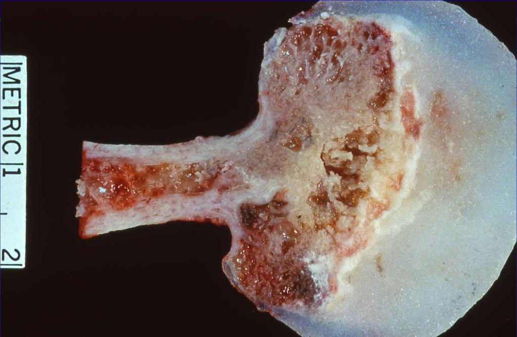
11 Osteosarcoma Clinical: Pain and tender mass of relatively short duration Knee is most common site **Aggressive treatment approach with neoadjuvant chemotherapy and limb salvage Early development of pulmonary mets Predisposing conditions: Pagets and Radiation
12 Large peak in children/young adults. Small peak in later years. Most frequently affected site is in the metaphysial area in long bones, but can happen elsewhere.
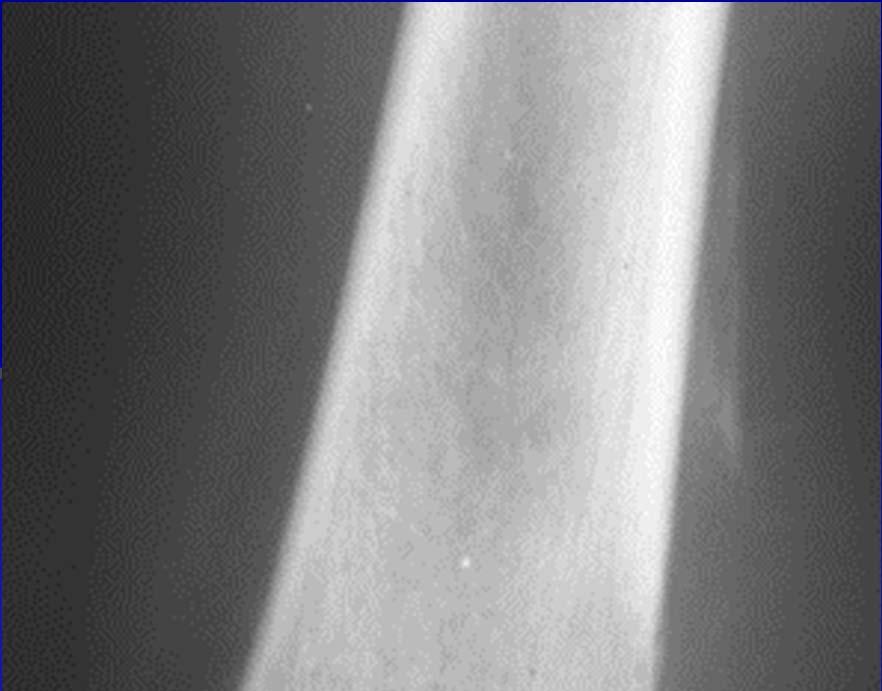
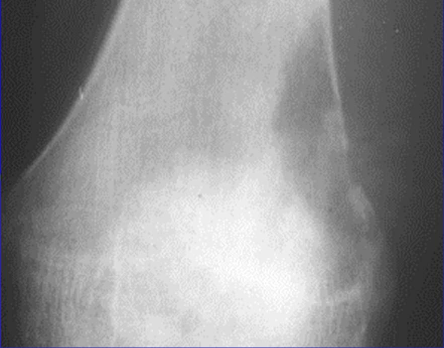
13 Osteosarcoma Radiology: metaphyseal lesion which can be mixed lytic and blastic Poorly delineated; cortical destruction with soft tissue extension Spiculation and Codman s triangle MRI/CT to define extent of intramedullary spread
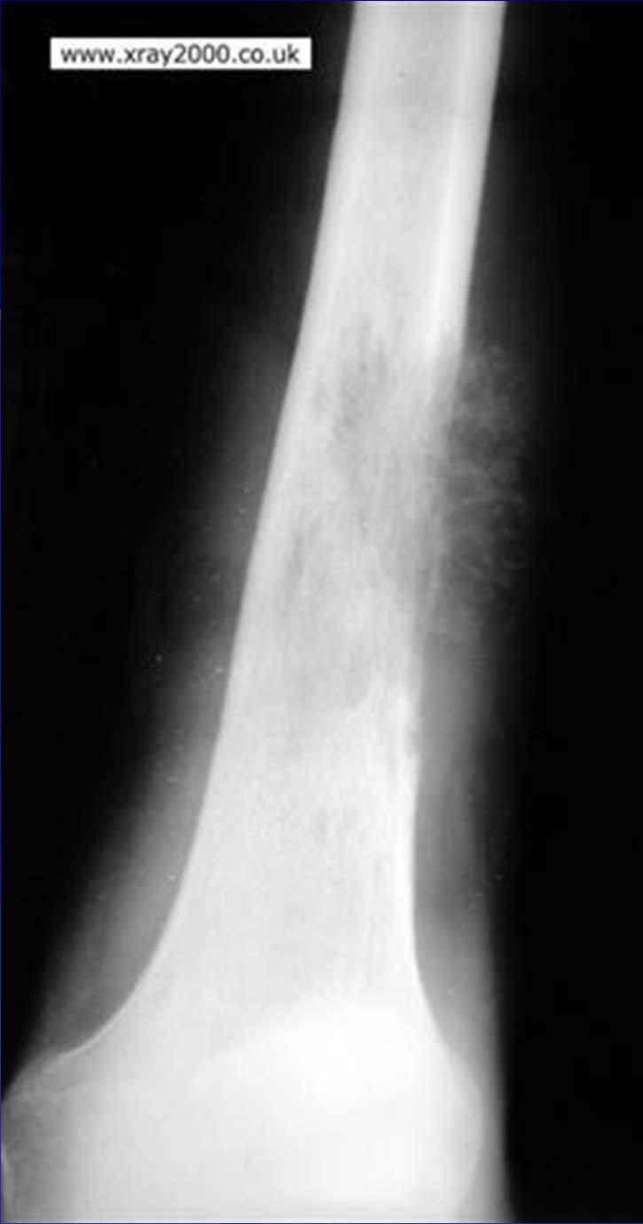
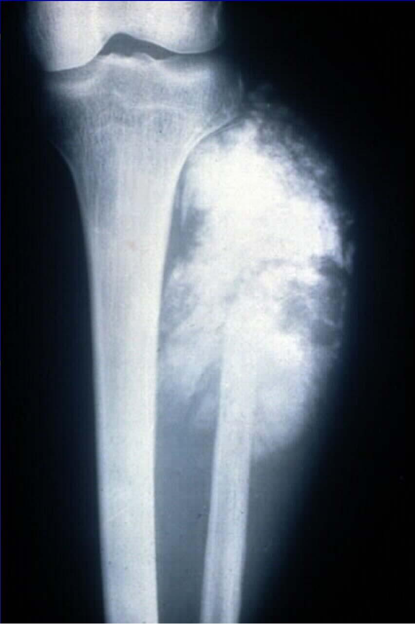
14 Osteosarcoma
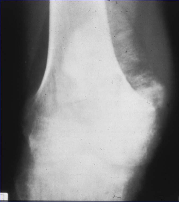
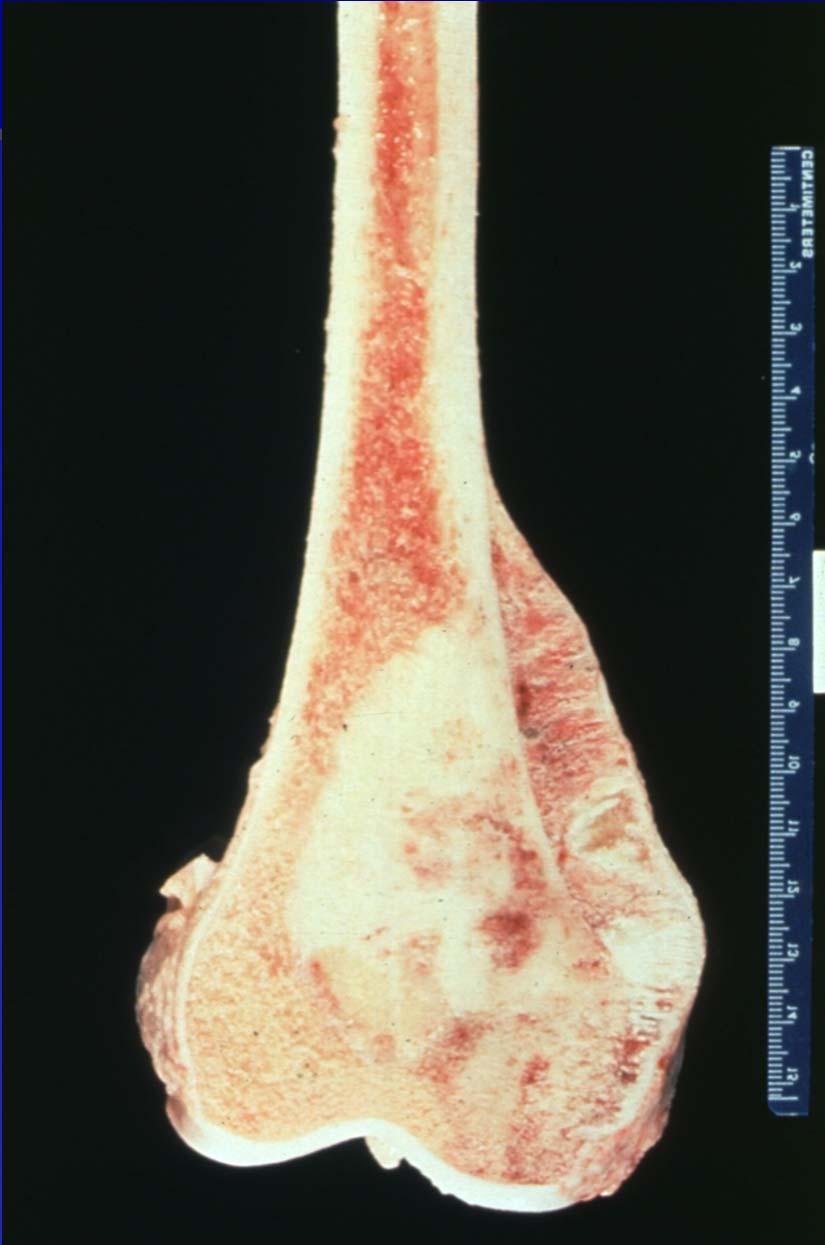
15 Osteosarcoma
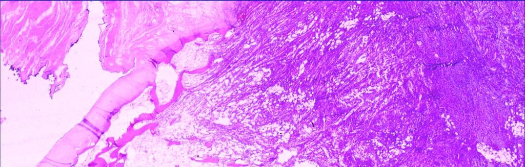
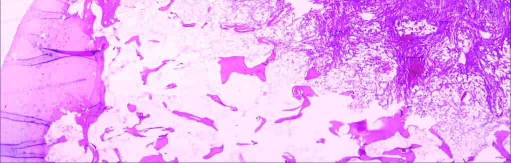
16 Osteosarcoma Histopathology: Highly variable with osteoid, chondroid or fibrous matrix predominant All have in common the production of tumor osteoid and malignant osteoblasts Spindled sarcomatous stromal element with anaplasia and mitotic figures Foci of degeneration, infarction or heavy osteoid formation with sclerosis Lace-like osteoid pattern is common

17
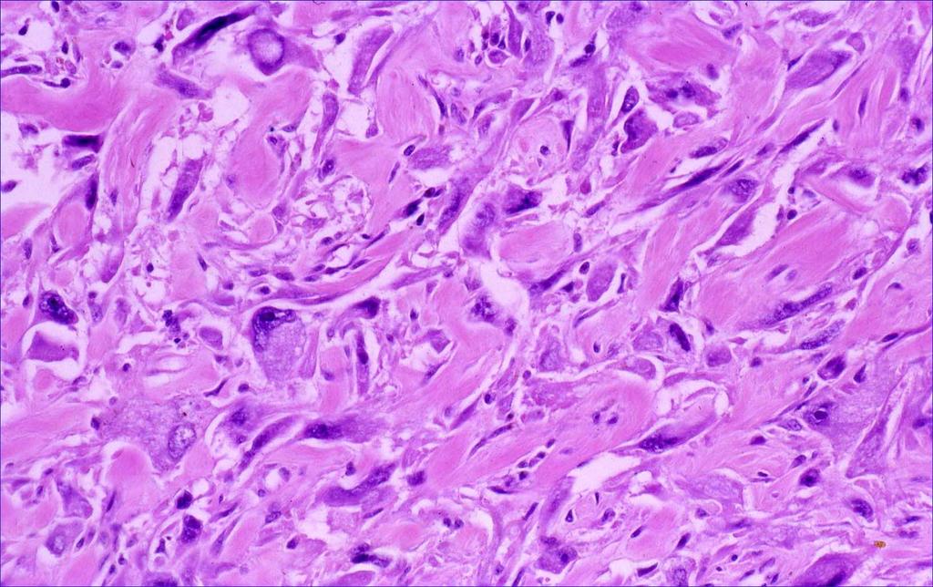
18 Osteosarcoma--- conventional
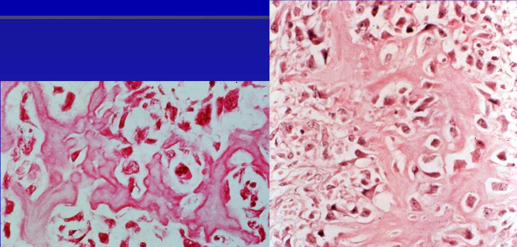
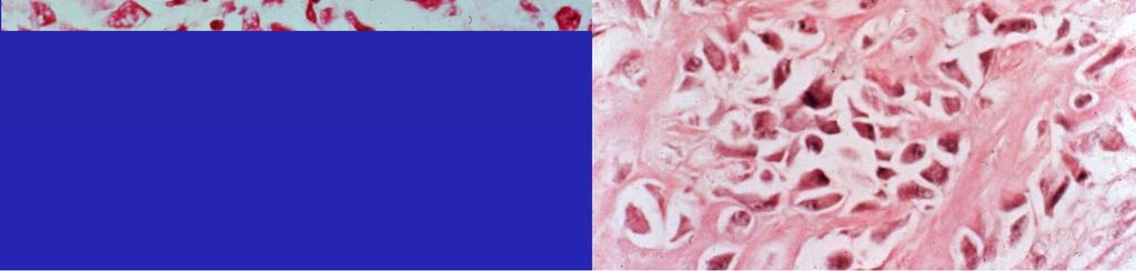
19 Osteoid


20 Treatment Amputation produced 20% 5 yr DFS Neo-adjuvant chemotherapy plus surgery results in 60-65% 5 yr DFS Limb salvage surgery appropriate in 80% of cases


21 Chondroid tumors Osteochondroma Enchondroma Chondroblastoma Chondromyxoid fibroma Chondrosarcoma


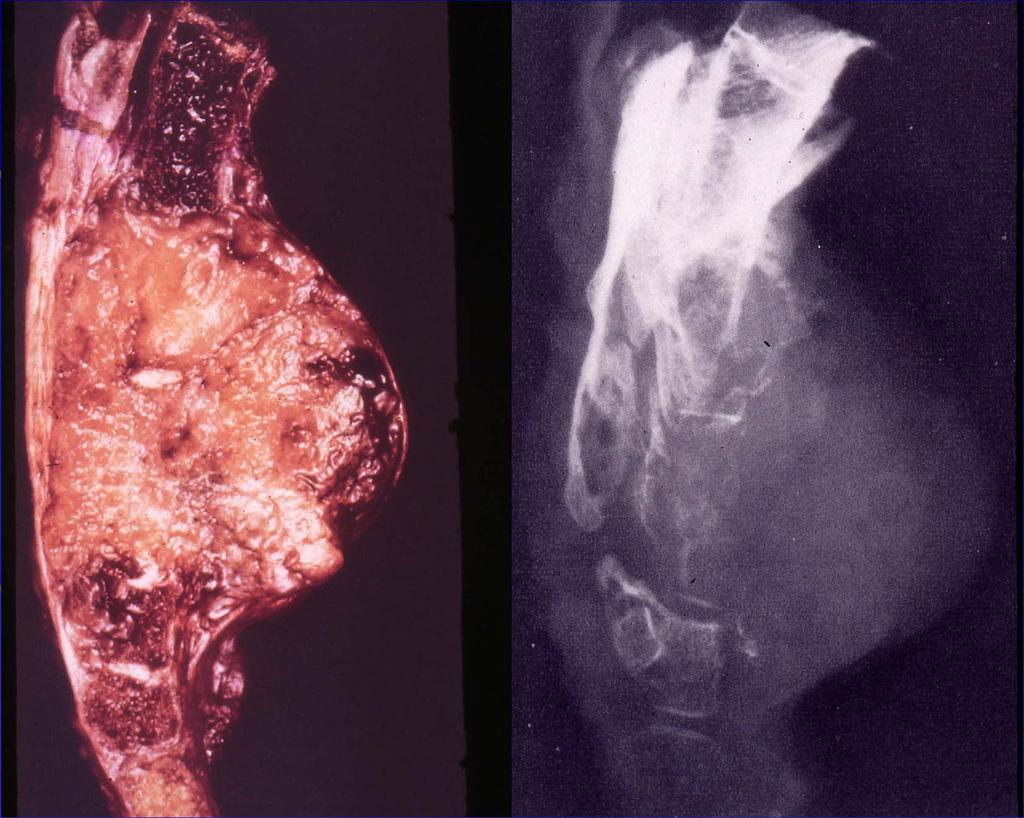
22 Osteochondroma Clinical: Obvious mass lesion; often of long duration Not painful unless impinges on a structure (bursa) Risk of chondrosarcomatous degeneration Hereditary multiple exostoses syndrome
23 Favored site for osteochondromas
24
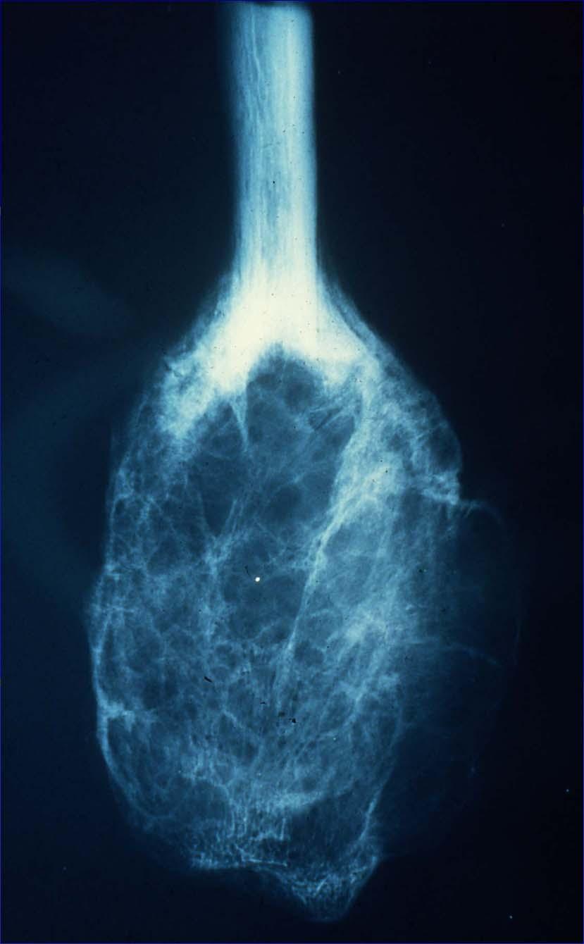
25 Enchondroma Clinical: Common benign lesion; asymptomatic, painless and incidentally discovered Grow up to 3 cm; mass lesion in hands and feet Peripheral skeleton is common (Vs. central for chondrosarcoma) Up to 10% of all bone tumors Multiple enchondromas associated with Ollier s and Maffuci s syndromes

26 Enchondroma-Radiology Variable intralesional calcification Rings and arcs pattern in long bone have metaphyseal location and can simulate a bone infarct

27 Enchondroma Histopathology: Circumscribed, lobulated lesion comprised of lobules of cartilage separated by thin septae micro appearance of benign cartilage
; may have bi-nucleate chondrocytes Focal calcification and enchondral")
28 Enchondroma Peripheral concentration of chondrocytes Minimal cytologic atypia (digits); may have bi-nucleate chondrocytes Focal calcification and enchondral ossification


29 Multiple enchondromas- Ollier s and Maffucci s Ollier s disease Maffuci s disease hemangiomas

30 Chondroblastoma Clinical: Less than 1% of all bone tumors In young individuals with open epiphyses Pain in affected region May have corresponding joint effusion Most common site is distal femur/proximal tibia

 May have fine to course intralesional")
31 Chondroblastoma Radiologic: Located in epiphyses of long bones in skeletally immature individuals Sharply demarcated oval/round lesion surrounded by sclerotic bone Typically does not alter bone contour unless accompanied by ABC (20%) May have fine to course intralesional calcification


32 Chondroblastoma Histopathology: Chondroblasts, characterized by small cells with high N/C ratio and clefted nuclei Variable mature or immature cartilaginous matrix Chicken-wire calcification pattern associated with blastic foci Few multinucleate giant cells

33 Chondromyxoid fibroma Extremely rare lesion (< 1%) with wide age range Most common location is knee; proximal tibia accounts for 25% of all may present with tenderness or swelling but is often an incidental finding


34 Chondromyxoid fibroma Radiologic: Eccentric, metaphyseal lytic defect Long axis of lesion parallels long axis of affected bone Sharp, sclerotic and scalloped margins; may be lobulated or septated Typically lacks intralesional calcification
35
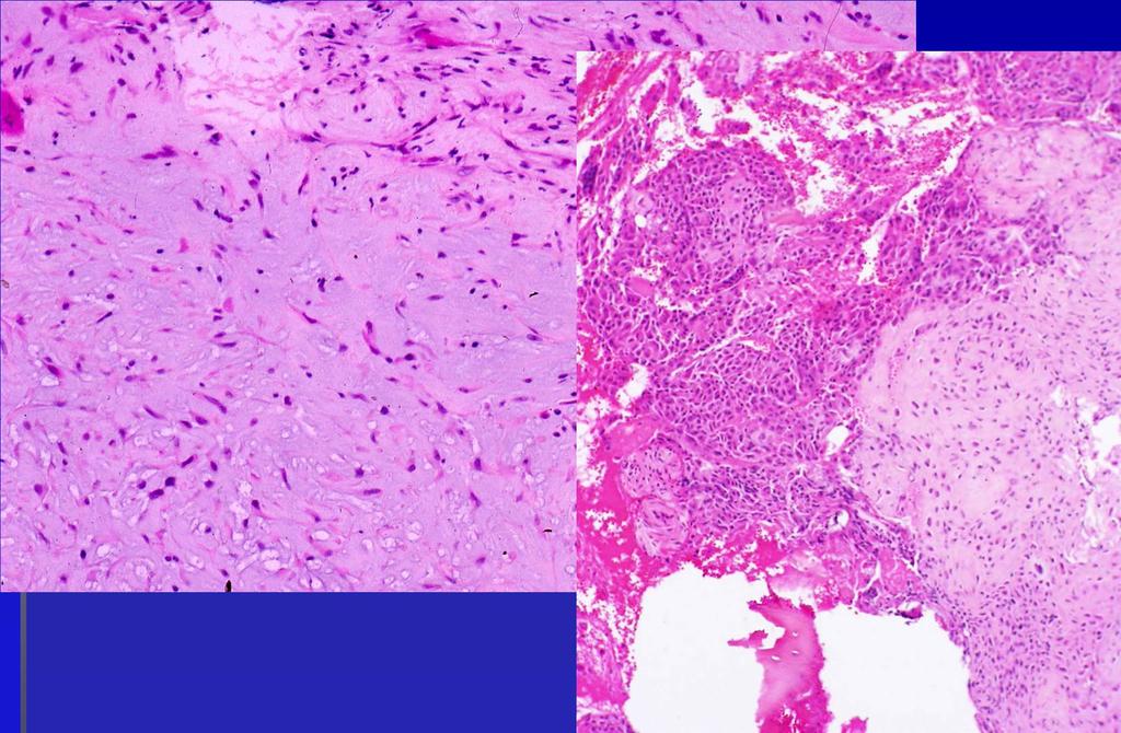
36 Chondrosarcoma Clinical: Wide age range; tumor in adults is most common; predisposing factors Appendicular skeleton and proximal extremities are most common sites presents with pain and local tenderness; often of long duration Palpable firm mass
37 Chondrosarcoma Radiographic: Predilection for central skeleton; acetabulum or metaphysis/diaphysis of long bones Intralesional calcification classically in form or rings and arcs (65%) Cortical destruction; soft tissue extension with large lesions
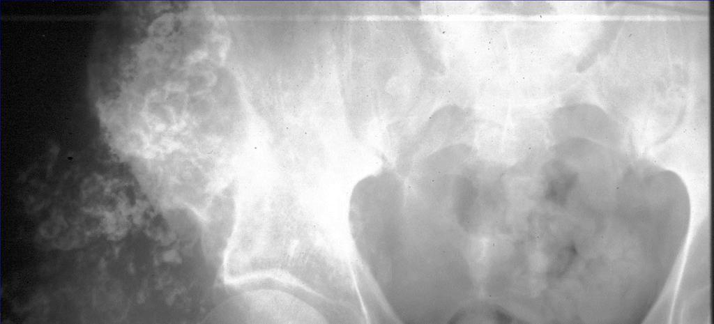
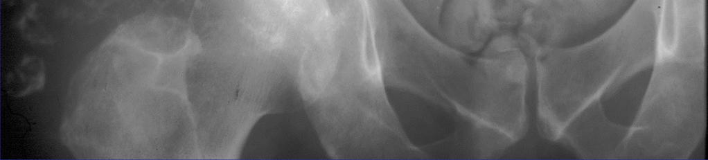
38 Chondrosarcoma
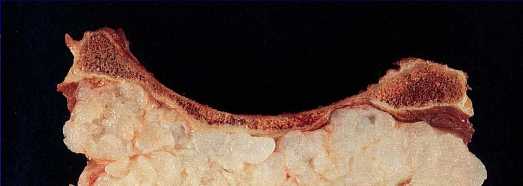

39 Chondrosarcoma


40 Chondrosarcoma Binucleation and myxoid change of cartilage are helpful but not absolute indicators of malignancy Lack of osteoid (conventional)


 or multiple")
41 Fibrous dysplasia Clinical: Often asymptomatic; incidental finding or bone growth deformity Solitary (monostotic) or multiple bones involved (polyostotic)
 Cystic degeneration is")
42 Fibrous dysplasia Radiologic: Metaphyseal or diaphyseal based lesion Lytic or ground glass appearance Bowing and pathologic fracture (shepherd's crook deformity) Cystic degeneration is common

43 Fibrous dysplasia Can be uni- or multifocal (polyostotic) Multifocal FD seen in Albright-McCune and Mazabraud s syndromes
44 Fibrous dysplasia Histopathology: Fibrous stroma of variable density; small bland spindled cells Osteoid trabeculae of unusual shapes; alphabet soup or psammoma bodies Lack of osteoid rimming Foci of cartilaginous metaplasia, lipid laden macrophages and cystic degeneration Rare multinucleated giant cells


45 This resembles a membranous growth process without fibrous rimming. Metaplastic process where osteoid forms right out of fibrous matrix. Simulates bone tumor clinically.


46 Fibroma Also known as fibrous metaphyseal defect, non-ossifying fibroma Peak incidence in adolescents and young adults May be discovered incidentally or present with pathologic fracture Metaphyseally centered lesion, lower extremity is most common location

47 Fibroma/NOF Rad: sharply demarcated, expansile lesion of the cortex; metaphyseal Lesion is parallel to long axis of affected bone


48 Fibroma/ NOF Because of pathognomonic appearance and tendency for spontaneous resolution, seldom encountered in SP

49 Bone cysts
50 Giant cell tumor Most often in skeletally mature individuals (third decade) Most common location = knee, distal radius, sacrum Pain, swelling and pathologic fracture Classically epiphyseal


51 Giant cell tumor Radiology: Epiphyseal centered; extends to articular surface lytic lesion lacking significant sclerosis or periosteal reaction Confined to bone or break through cortex into soft tissue Radiologically simulates malignancy: osteosarcoma

52 Giant cell tumor Histopathology: May have necrosis or secondary cyst formation Uniform pattern of growth with numerous giant cells Background of round /oval mononuclear cells- -same as multinucleate cells; no cytologic atypia
; related cytogenetically to PNET and DSRCT,")
53 Ewings Sarcoma Clinical: Lesion of adolescents and young adults Presents with local pain and swelling; may have fever and simulate infection **Rx d with radiation and adjuvant chemotherapy t (11;22); related cytogenetically to PNET and DSRCT, Askins

54 Ewing Sarcoma Radiologic: Extensive, poorly marginated diaphyseal lesion Periosteal new bone formation and soft tissue mass May be lytic and /or sclerotic Ddx: lymphoma and osteomyelitis


55 Ewing Sarcoma Histopathology: Small round blue cell tumor Pas +/diastaseintracytoplasmic glycogen may show extensive necrosis and crush artifact
56 Chordoma Clinical: Associated with wide age range majority of lesions of sacrum or clivus pain but also nerve/spinal cord compression, change in bowel, cranial nerve dysfunction or nasopharyngeal mass

57

58 Pigmented villonodular synovitis PVNS Common proliferative/neoplastic lesions of joints; progressive discomfort nodular, villous thickening of the synovium with hemosiderin deposition
 can be present Hemosiderin")
59 PVNS Periarticular soft tissue swelling and mass effect Extensive bone destruction (simulating aggressive disease) can be present Hemosiderin identified on MRI


60 PVNS Large villi filled with dense infiltrate of fibrohistiocytic mononuclear cells, hemosiderin laden macrophages and giant cells. Numerous mitoses may be identified; NOT an indicator of aggressive potential
61 Soft tissue tumor/ Sarcoma Lesions of adipose tissue: Lipoma/Liposarcoma Lesions of skeletal muscle: Rhabdomysarcoma Lesions of primitive/progenitor/multipotential cell: Malignant fibrous histiocytoma (MFH) Lesion of? Tissue/cell: Synovial sarcoma
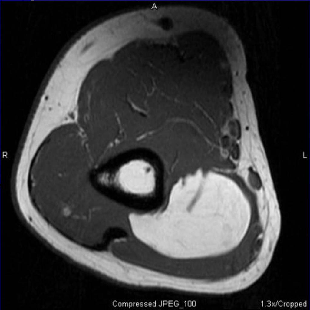
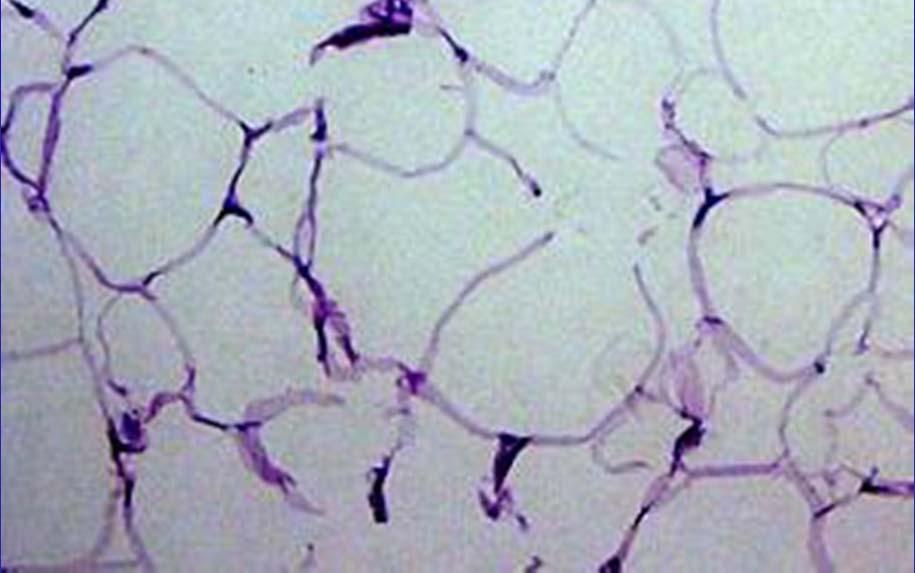
62 Lipoma

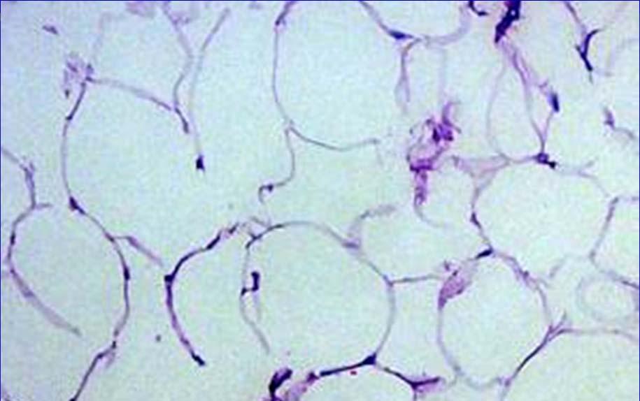
63 Liposarcoma One of the more common sarcomas Develop in the deep soft tissue; thigh and retroperitoneum Treatment is surgical; prognosis is dependent on grade, size, location


64 Liposarcoma Different variants with prognostic significance: well differentiated, myxoid/round cell and pleomorphic Histology = Lipoblasts


65 Rhabdomyosarcoma One of the more common tumors of children Occurs in unusual sites: head and neck, orbit, urogenital region
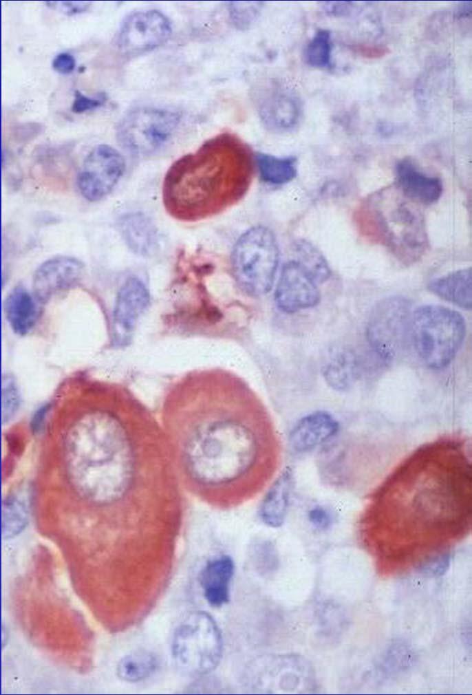
66 Rhabdomyosarcoma Key to Dx is recognition of rhabdomyoblast


67 Synovial sarcoma Commonly arises near joints/extremities Histologically biphasic t(x;18)

68 Chromosomal abnormalities in Soft tissue sarcoma

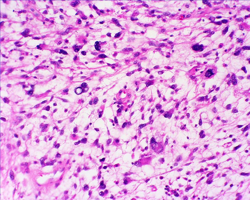
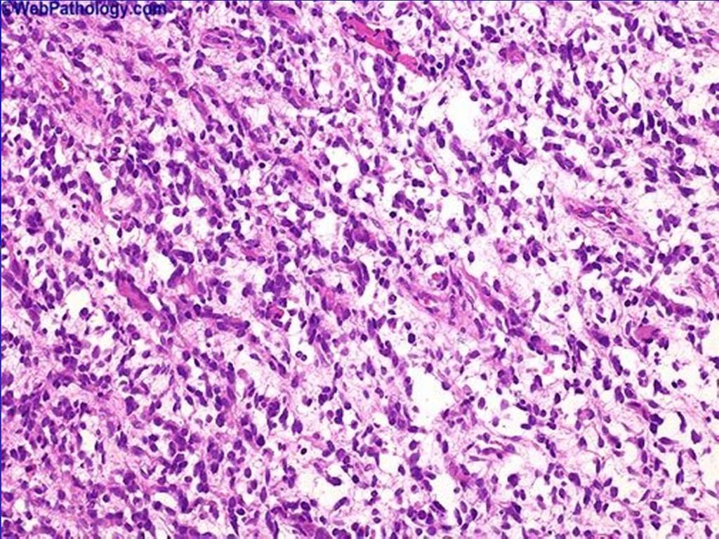
69 Malignant fibrous histiocytoma
Primary bone tumors > metastases from other sites Primary bone tumors widely range -from benign to malignant. Classified according to the normal cell
 Primary bone tumors > metastases from other sites Primary bone tumors widely range -from benign to malignant. Classified according to the normal cell counterpart and line of differentiation. Among the
Bone Tumors Clues and Cues
 William Herring, M.D. 2002 Bone Tumors Clues and Cues In Slide Show mode, advance the slides by pressing the spacebar All Photos Retain the Copyright of their Authors Clues by Appearance of Lesion Patterns
The Radiology Assistant : Bone tumor - well-defined osteolytic tumors and tumor-like lesions
 Bone tumor - well-defined osteolytic tumors and tumor-like lesions Henk Jan van der Woude and Robin Smithuis Radiology department of the Onze Lieve Vrouwe Gasthuis, Amsterdam and the Rijnland hospital,
Bone Tumours - a synopsis. Dr Zena Slim SpR in Histopathology QAH 2009
 Bone Tumours - a synopsis Dr Zena Slim SpR in Histopathology QAH 2009 Aims General approach to diagnosis Common entities.and not so common ones. Mini quiz Challenge of bone tumour diagnosis Bone tumours
Introduction to Musculoskeletal Tumors. James C. Wittig, MD Orthopedic Oncologist Sarcoma Surgeon
 Introduction to Musculoskeletal Tumors James C. Wittig, MD Orthopedic Oncologist Sarcoma Surgeon www.tumorsurgery.org Definitions Primary Bone / Soft tissue tumors Mesenchymally derived tumors (Mesodermal)
MARK D. MURPHEY MD, FACR. Physician-in-Chief, AIRP. Chief, Musculoskeletal Imaging
 ALPHABET SOUP AND CYSTIC LESIONS OF THE BONE MARK D. MURPHEY MD, FACR Physician-in-Chief, AIRP Chief, Musculoskeletal Imaging ALPHABET SOUP AND CYSTIC LESIONS OF THE BONE Giant cell tumor (GCT) Unicameral
Bread and Butter Bone Pathology
 Bread and Butter Bone Pathology NICOLE D. RIDDLE, MD RUFFOLO, HOOPER, AND ASSOC. / UNIVERSITY OF SOUTH FLORIDA Goals: Fundamentals of neoplastic bone pathology Bone Producing Cartilage Producing Miscellaneous
APMA 2018 Radiology Track Bone Tumors When to say Gulp!
 APMA 2018 Radiology Track Bone Tumors When to say Gulp! DANIEL P. EVANS, DPM, FACFAOM Professor, Department of Podiatric Medicine and Radiology Dr. Wm. Scholl College of Podiatric Medicine Conflict of
Bone tumors. RMG: jan
 Bone tumors RMG: jan 217. @Kijohs KIZZA JOHN KIJOHS Diseases arising in bone Lipoma Fibrous cortical defects Non-ossifying fibroma Bone island Benign simple cysts Enchondroma Osteochondroma Osteoid osteoma
USCAP 2014 Common problems in bone and soft tissue pathology: Cartilage tumors
 USCAP 2014 Common problems in bone and soft tissue pathology: Cartilage tumors Andrew Horvai MD PhD Clinical Professor, Pathology UCSF, San Francisco, CA Outline Common intramedullary tumors Enchondroma
Typical skeletal location and differential diagnosis of bone tumors.
 Typical skeletal location and differential diagnosis of bone tumors. Poster No.: C-2418 Congress: ECR 2015 Type: Educational Exhibit Authors: M. Barros, L. A. Ferreira, Y. Costa, P. J. V. Coelho, F. Caseiro
Multifocal fibrous Dysplasia with enchondroma-like areas: Fibrocartilaginous Dysplasia
 ISPUB.COM The Internet Journal of Pathology Volume 7 Number 2 Multifocal fibrous Dysplasia with enchondroma-like areas: Fibrocartilaginous Dysplasia V Monappa, R Kudva Citation V Monappa, R Kudva. Multifocal
Grading of Bone Tumors
 Grading of Bone Tumors Joon Hyuk Choi, M.D. Department of Pathology College of Medicine, Yeungnam University Introduction to grading system of bone tumor used at Mayo Clinic WHO Histologic Classification
COPYRIGHT 2004 BY THE JOURNAL OF BONE AND JOINT SURGERY, INCORPORATED
 84 COPYRIGHT 2004 BY THE JOURNAL BONE AND JOINT SURGERY, INCORPORATED Radiographic Evaluation of Pathological Bone Lesions: Current Spectrum of Disease and Approach to Diagnosis BY BENJAMIN G. DOMB, MD,
Fluid-fluid levels in bone tumors: A pictorial review
 Fluid-fluid levels in bone tumors: A pictorial review Poster No.: C-578 Congress: ECR 2009 Type: Educational Exhibit Topic: Musculoskeletal Authors: L. Figueroa Nasra, C. Martín Hervás, M. Tapia-Viñé,
Disclosures. Giant Cell Rich Tumors of Bone. Outline. The osteoclast. Giant cell rich tumors 5/21/11
 Disclosures Giant Cell Rich Tumors of Bone Andrew Horvai, MD, PhD Associate Clinical Professor, Pathology This lecture discusses "off label" uses of a number of pharmaceutical agents. The speaker is describing
Essential Dermatopathology. Jinah Kim, MD, PhD Department of Pathology and Dermatology Stanford University Medical Center
 Essential Dermatopathology Jinah Kim, MD, PhD Department of Pathology and Dermatology Stanford University Medical Center OBJECTIVES Review clinical, pathologic and molecular aspects of bone and fat tumors
Musculoskeletal Sarcomas
 Musculoskeletal Sarcomas Robert C. Orth, M.D., Ph.D. Edward B. Singleton Department of Pediatric Radiology Texas Children s Hospital Page 0 xxx00.#####.ppt 9/23/2012 9:01:18 AM No disclosures Page 1 xxx00.#####.ppt
Imaging Findings Of Bone Tumors: A Pictorial Review
 Imaging Findings Of Bone Tumors: A Pictorial Review Poster No.: C-2511 Congress: ECR 2015 Type: Educational Exhibit Authors: M. Limeme, N. Benzina, A. BelKhiria, H. Zaghouani, S. Majdoub, N. Mallat, H.
The Radiology Assistant : Bone tumor - ill defined osteolytic tumors and tumor-like lesions
 Bone tumor - ill defined osteolytic tumors and tumor-like lesions Henk Jan van der Woude and Robin Smithuis Radiology department of the Onze Lieve Vrouwe Gasthuis, Amsterdam and the Rijnland hospital,
Malignant bone tumors. Incidence Myeloma 45% Osteosarcoma 24% Chondrosarcoma 12% Lyphoma 8% Ewing s Sarcoma 7%
 Malignant bone tumors Incidence Myeloma 45% Osteosarcoma 24% Chondrosarcoma 12% Lyphoma 8% Ewing s Sarcoma 7% Commonest primary bone sarcoma is osteosarcoma X ray Questions to ask 1. Solitary or Multiple
Bubbly Lesions of Bone
 Residents Section Pattern of the Month w79 08.18.09 Eisenberg Residents Section Pattern of the Month Residents inradiology Ronald L. Eisenberg 1 Eisenberg RL Keywords: bubbly lesions, fegnomashic, skeletal
Fluid fluid levels in bone tumors and tumoral lesions - Pictorial essay
 Review Fluid fluid levels in bone tumors and tumoral lesions - Pictorial essay Subbarao Kakarla 1,* 1 KIMS Foundation and Research Centre, Minister Road, Secunderabad - 500003, Telangana, India Abstract
Primary bone tumors according to the WHO classification: a review of 13 years with illustrative examples
 Primary bone tumors according to the WHO classification: a review of 13 years with illustrative examples Poster No.: C-1741 Congress: ECR 2015 Type: Educational Exhibit Authors: J. Silva, M. A. Ramírez
Case 8 Soft tissue swelling
 Case 8 Soft tissue swelling 26-year-old female presented with a swelling on the back of the left knee joint since the last 6 months and chronic pain in the calf and foot since the last 2 months. Pain in
Fibrocartilaginous Dysplasia of the Bone: A Rare Variant of Fibrous Dysplasia
 Open Access Case Report DOI: 10.7759/cureus.448 Fibrocartilaginous Dysplasia of the Bone: A Rare Variant of Fibrous Dysplasia Raju Vaishya 1, Amit Kumar Agarwal 1, Nishint Gupta 2, Vipul Vijay 1 1. Department
A review of Tumoral lesions of the shoulder
 A review of Tumoral lesions of the shoulder Poster No.: P-0109 Congress: ESSR 2013 Type: Scientific Exhibit Authors: M. M. Milán Rodríguez, Á. E. Moreno Puertas, J. M. Giménez, 1 1 1 1 2 1 A. Rubio Fernández,
Recognizing Cartilaginous Tumors: Spectrum of Imaging Characteristics with Radiologic-Pathologic correlation.
 Recognizing Cartilaginous Tumors: Spectrum of Imaging Characteristics with Radiologic-Pathologic correlation. Poster No.: C-1451 Congress: ECR 2012 Type: Educational Exhibit Authors: E. Barcina García,
ORTHOPAEDIC ONCOLOGY OITE REVIEW COURSE
 ORTHOPAEDIC ONCOLOGY OITE REVIEW COURSE Richard D. Lackman, MD FACS Director, Orthopaedic Oncology Center Cancer Institute Introduction In the evaluation of a patient with a bone tumor, there are several
GIANT CELL-RICH OSTEOSARCOMA: A CASE REPORT
 Nagoya J. Med. Sci. 59. 151-157, 1996 CASE REPORTS GIANT CELL-RICH OSTEOSARCOMA: A CASE REPORT KEIJI SATO!, SHIGEKI YAMAMURA!, HISASHI IWATA!, HIDESHI SUGIURA 2, NOBUO NAKASHIMA 3 and TETSURO NAGASAKA
04/27/2017. The Spectrum of Cartilaginous Tumors
 The Spectrum of Cartilaginous Tumors L I S A E R C O L A N O, M D M U S C U L O S K E L E T A L O N C O L O G Y D E P A R T M E N T O F O R T H O P A E D I C S A L L E G H E N Y H E A L T H N E T W O R
History. 33 y/o F with hx of palpable anterior tibial mass x 2 years, only painful with palpation
 History 33 y/o F with hx of palpable anterior tibial mass x 2 years, only painful with palpation Imaging Photo Album Patient also had a smaller lesion 1 cm proximal to this lesion, not seen radiographically.
Immunohistochemistry in Bone and Soft Tissue Tumors. Sahar Rassi Zankoul, MD
 Immunohistochemistry in Bone and Soft Tissue Tumors Sahar Rassi Zankoul, MD Introduction Bone tumors represent a wide variety of tumors of various origins and malignant potentials. These different tumor
* I have no disclosures or any
 Howard Rosenthal, M.D. Associate Professor of Orthopedic Surgery University of Kansas Sarcoma Center I have no disclosures or any conflicts related to the content of this presentation. Objectives 1. Describe
Case Report Intramedullary Chondrosarcoma of Proximal Humerus
 Hindawi Publishing Corporation Case Reports in Radiology Volume 2012, Article ID 642062, 7 pages doi:10.1155/2012/642062 Case Report Intramedullary Chondrosarcoma of Proximal Humerus Pratiksha Yadav, Dolly
Bone/Osteoid Producing Lesions
 Chapter 2 Bone/Osteoid Producing Lesions Introduction There are many lesions that are associated with reactive new bone formation; this chapter predominantly covers those in which deposition of osteoid/bone
5/10. Pathology Soft tissue tumors. Farah Bhani. Mohammed Alorjani
 5/10 Pathology Soft tissue tumors Mohammed Alorjani Farah Bhani Slides are included in this sheet. Objectives: Soft tissue tumors 1. Describe soft tissue tumors. 2. Understand the classification of soft
FEGNOMASHIC: from x-ray to MRI
 FEGNOMASHIC: from x-ray to MRI Poster No.: C-2441 Congress: ECR 2015 Type: Educational Exhibit Authors: S. Fouassier, A. L. C. Duarte, C. Ruivo, J. Velez ; Évora/PT, 1 2 1 2 3 1 3 Coimbra/PT, PT Keywords:
Malignant Bone Tumors - Part I: a brief revision of diagnostic aspects with conventional radiology
 Malignant Bone Tumors - Part I: a brief revision of diagnostic aspects with conventional radiology Poster No.: C-2473 Congress: ECR 2013 Type: Educational Exhibit Authors: I. Candelaria, L. B. Barbosa,
GIANT CELL TUMOR OF BONE
 GIANT CELL TUMOR OF BONE Definition. First described by Jaffe et al. 1, giant cell tumor of bone is a locally aggressive primary neoplasm of bone that is composed of proliferation of bland looking oval
Incidental bone tumors are asymptomatic lesions that are. Incidental Bone Lesions. When to Refer to the Tumor Specialist
 Bulletin of the NYU Hospital for Joint Diseases 2012;70(4):235-40 235 Incidental Bone Lesions When to Refer to the Tumor Specialist LT Suezie Kim, M.D., M.C., U.S.N., Catherine N. Laible, M.D., Leon D.
The Radiology Assistant : Bone tumor A-G
 Bone tumor A-G Bone tumors and tumor-like lesions in alphabethic order Henk Jan van de Woude and Robin Smithuis Radiology department of the Onze Lieve Vrouwe Gasthuis, Amsterdam and the Rijnland hospital,
Section II Musculoskeletal Radiology
 Section II Musculoskeletal Radiology Figure 1 25. You are shown a noncontrast CT (Figure 1) of the thigh. What is the MOST LIKELY diagnosis? A. Synovial sarcoma B. Hemangioma C. Organizing hematoma D.
Review Course «Musculoskeletal Oncology» October 6, 2011 UNIKLINIK BALGRIST. Imaging of Bone and Soft Tissue. Tumors
 Imaging of Bone and Soft Tissue Tumors Approach from a radiologist s point of view Florian Buck Radiology Radio- Radio- Oncologist Oncologist Orthopedist Orthopedist Patient Management Oncologist Oncologist
Malignant Peripheral Nerve Sheath Tumor
 C H A P T E R 120 Malignant Peripheral Nerve Sheath Tumor Currently, malignant peripheral nerve sheath tumor (MPNST) is the most commonly used generic name for the neoplasms known in the past as neurosarcoma,
Giant cell tumour of the sternum-two cases
 Giant cell tumour of the sternum-two cases Nishaa.P 1, Raghuram.P 2, Navin patil 3, Jaipal B.R 4 Akkamahadevi patel 5 Assistant Professor ESIC medical college and PGIMSR 1 Professor and HOD, 2 Professor
Malignant Bone Tumours. PathoBasic, Daniel Baumhoer
 Malignant Bone Tumours PathoBasic, 20.03.18 Daniel Baumhoer FNCLCC Grading The differentiation score is defined as the extent to which a tumor resembles adult mesenchymal tissue (score 1), the extent to
Mousa Al-Abadi. Abd. Kharabsheh. Rand Abu Anzeh
 7 Mousa Al-Abadi Abd. Kharabsheh Rand Abu Anzeh 1 Recap The histological appearance of Giant cell tumor of bone shows only multi-nucleated giant cells. The histological appearance of Aneurysmal bone cyst
Tumors of Adipose Tissue Tumors Epidemiology Clinical Features. Morphology. Mature Adipocytes Separated by delicate fibrous septa
 Tumors of Adipose Tissue Lipoma Liposarcoma Most commonly happens in female The most common soft tissue tumor o Originates from matured Adipocytes Most commonly happes at the 4 th and 5 th decade of life
Common Primary Tumors of Bone
 Special Report Common Primary Tumors of Bone Primary bone tumors are a relatively rare occurrence, however, they can have serious deleterious consequences. Many possess the ability to degenerate into malignant
Radiologic approach to pediatric lytic bone lesions
 Radiologic approach to pediatric lytic bone lesions Poster No.: C-1177 Congress: ECR 2016 Type: Educational Exhibit Authors: J. L. LERMA GALLARDO, I. de la Pedraja, A. Lancharro 1 1 1 2 1 1 Zapata, J.
Benign Tumors of Bone
 REVIEW ARTICLE Benign Tumors of Bone Subbarao K Padmshri Prof. Dr. Kakarla Subbara, Hyderabad, India. Benign tumors of bone are common while malignant tumors are rare. Benign tumors constitute about 75%
SMALL ROUND BLUE CELL LESION OF BONE
 DISCLOSURE SMALL ROUND BLUE CELL LESION OF BONE Dr. Alistair Jordan University of South Alabama No financial support or endorsement OBJECTIVES Describe the more common small round cell lesions of bone
LAC + USC.
 Jeff McDavit,, M.D. LAC + USC mcdavit@usc.edu Clinical History 55 year old male with large, deep, non- tender left thigh mass. Seen at LAC+USC Med Ctr FNA clinic No h/o trauma or radiation Vimentin
Department of Radiology, University of Szeged. Imaging of the skeleton
 Imaging of the skeleton Methods of examination: plain x-ray (radiography, densitometry) x-ray with contrast material (fistulography, angiography) ultrasound (b-mode, Doppler, color, duplex) computed tomography
Mark D. Murphey, MD, FACR
 Fundamental Concepts of Musculoskeletal Neoplasm: CT and MRI Mark D. Murphey, MD, FACR Important Features in Evaluation of Musculoskeletal Masses Differential diagnosis Preoperative assessment and staging
Radiography in the Initial Diagnosis of Primary Bone Tumors
 Residents Section Structured Review Costelloe and Madewell Radiography of Primary Bone Tumors Residents Section Structured Review Colleen M. Costelloe 1 John E. Madewell Costelloe CM, Madewell JE Keywords:
ORTHOPAEDIC TUMOURS DJM FRANTZEN 2012 AUGUST TUMOURS 1
 ORTHOPAEDIC TUMOURS DJM FRANTZEN 2012 AUGUST TUMOURS 1 PRINCIPLES STAGING WORKUP RADIOLOGY BIOPSY PROCEDURES CHEMOTHERAPY RADIOTHERAPY 2012 AUGUST TUMOURS 2 STAGING ENNEKING'S SURGICAL STAGES (ENNEKING)
Fun with Fat. General Rules. Case
 Fun with Fat General Rules Imaging: location (deep vs. superficial) Superficial lesions are seldom liposarcomas Deep lesions may be benign or malignant Myxoid stroma is common in benign and malignant lesions
What Do You Need to Know About Bone Pathology? Benjamin L. Hoch M.D. Associate Professor Department of Pathology University of Washington
 What Do You Need to Know About Bone Pathology? Benjamin L. Hoch M.D. Associate Professor Department of Pathology University of Washington What s Do You Need To Know About Bone Pathology? Reactive/pseudosarcomatous
PATHOLOGY OF THE MUSCULOSKELETAL SYSTEM
 PATHOLOGY OF THE MUSCULOSKELETAL SYSTEM BONES & JOINTS CONGENITAL DISEASES OF BONE Osteogenesis Imperfecta (OI) (Brittle bone diseases) is a group of hereditary disorders caused by gene mutations that
INDEX. in this web service Cambridge University Press
 actin 14 adamantinoma 202, 290 292, 297 adenocarcinoma 136 adipocytes in hibernoma 149, 150 in lipoblastoma 148 in lipoma 141, 142, 145 in liposarcoma 152 in myelolipoma 151 adrenal gland tumors see myelolipoma
Index. Note: Page numbers of article titles are in boldface type.
 Magn Reson Imaging Clin N Am 12 (2004) 185 189 Index Note: Page numbers of article titles are in boldface type. A Acromioclavicular joint, MR imaging findings concerning, 161 Acromion, types of, 77 79
Bone Tumors: In 1 Simple Chart
 Bone Tumors with PowerPoint Interactivity Download this entire slideshow from When running this on your own computer you can jump from slide to slide using these buttons at bottom of each slide: Last slide
A 24 year old male patient presented with a swelling on the dorsal aspect of left foot since 3 years. He was operated thrice before, outside, for
 A 24 year old male patient presented with a swelling on the dorsal aspect of left foot since 3 years. He was operated thrice before, outside, for same. Came to us with recurrence since last one year with
UCLA UCLA Previously Published Works
 UCLA UCLA Previously Published Works Title Benign bone tumors Permalink https://escholarship.org/uc/item/7h86k14t Journal Radiologic Clinics of North America, 49(6) ISSN 0033-8389 Authors Motamedi, K Seeger,
University Journal of Surgery and Surgical Specialities
 University Journal of Surgery and Surgical Specialities Volume 1 Issue 1 2015 EXTRA SKELETAL MESENCHYMAL CHONDROSARCOMA :A CASE REPORT Rajaraman R Subbiah S Navin Naushad Kilpaulk Medical College Abstract:
2003 PIP-A A Cases. Paul K. Shitabata, M.D. APMG May 21, 2003
 2003 PIP-A A Cases Paul K. Shitabata, M.D. APMG May 21, 2003 Case 1 47F pelvic mass involving the right fallopian tube 5.5 cm intraluminal mass Fallopian Tube Adenocarcinoma Risk factors Breast, endometrial,
Imaging in breast cancer. Mammography and Ultrasound Donya Farrokh.MD Radiologist Mashhad University of Medical Since
 Imaging in breast cancer Mammography and Ultrasound Donya Farrokh.MD Radiologist Mashhad University of Medical Since A mammogram report is a key component of the breast cancer diagnostic process. A mammogram
warwick.ac.uk/lib-publications
 A Thesis Submitted for the Degree of PhD at the University of Warwick Permanent WRAP URL: http://wrap.warwick.ac.uk/102063/ Copyright and reuse: This thesis is made available online and is protected by
Medical Student Rotation Guide Tumor Service
 Medical Student Rotation Guide Tumor Service Overview Welcome to the medical student rotation on the Tumor service in the Department of Orthopaedic Surgery at Rush University Medical Center! We are excited
Medical Student Rotation Guide Tumor Service
 Medical Student Rotation Guide Tumor Service Overview Welcome to the medical student rotation on the Tumor service in the Department of Orthopaedic Surgery at Rush University Medical Center! We are excited
Bone Tumors: Epidemiology, Classification, Pathology
 Bone Tumors: Epidemiology, Classification, Pathology 1 Lars Gunnar Kindblom C O N T E N T S 1.1 Introduction 2 1.2 Epidemiology 2 1.3 Morphologic Diagnosis of Bone Tumors 5 1.4 Types of Bone Tumor Specimens
Bizarre parosteal osteochondromatous proliferation
 * * Bizarre Parosteal Osteochondromatous Proliferation A Case Report with Literature Review Chi-Fu Kao Yang-Chih Lin Yu-Hung Wu Be-Fong Chen* We report the case of a 12-year-old female with a slowly erythematous
Intraosseous lipoma of the talus
 Intraosseous lipoma of the talus Shigeki Maruyama, M.D. 1, Tetsuji Yamamoto, M.D (!). 2, Masataka Hashimura, M.D. 1, and Nobuhiro Ohsaki, M.D. 1, Toshihiro Akisue, M.D. 2, Shinichi Yoshiya, M.D 2 1 Department
Radiology-Pathology Conference
 July 31, 2009 Radiology-Pathology Conference Daniel T Ginat, M.D., M.S. Sharlin Johnykutty,, M.D. Presentation material is for education purposes only. All rights reserved. 2009 URMC Radiology Page 1 of
3/27/2017. Disclosure of Relevant Financial Relationships
 Ophthalmic Pathology Evening Specialty Conference USCAP 2017 5 th March, 2017 Mukul K. Divatia, MD Assistant Professor Department of Pathology & Genomic Medicine Weill Cornell Medical College Houston Methodist
MRI XR, CT, NM. Principal Modality (2): Case Report # 2. Date accepted: 15 March 2013
: Case Report # 2. Date accepted: 15 March 2013") Radiological Category: Musculoskeletal Principal Modality (1): Principal Modality (2): MRI XR, CT, NM Case Report # 2 Submitted by: Hannah Safia Elamir, D.O. Faculty reviewer: Naga R. Chinapuvvula, M.D.
Radiological Category: Musculoskeletal Principal Modality (1): Principal Modality (2): MRI XR, CT, NM Case Report # 2 Submitted by: Hannah Safia Elamir, D.O. Faculty reviewer: Naga R. Chinapuvvula, M.D.
International Journal of Health Sciences and Research ISSN:
 International Journal of Health Sciences and Research www.ijhsr.org ISSN: 2249-9571 Case Report Maffucci Syndrome with Composite Hemangioendothelioma: Two Rare Entities Ghanshyam Verma 1, Kirti Makhija
JMSCR Vol 3 Issue 11 Page November 2015
 www.jmscr.igmpublication.org Impact Factor 3.79 Index Copernicus Value: 5.88 ISSN (e)-2347-176x ISSN (p) 2455-0450 DOI: http://dx.doi.org/10.18535/jmscr/v3i11.36 Diagnostic Dilemmas in Cytodiagnosis of
Albert Leung, HMS III Gillian Lieberman, M.D. BIDMC Radiology Clerkship February 22, 2010
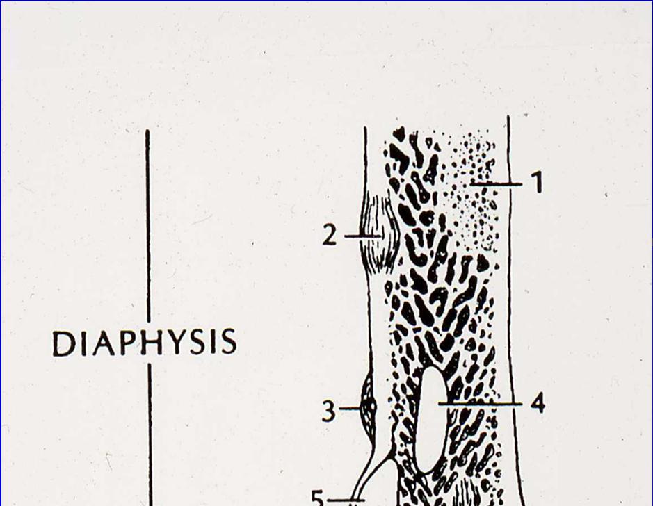
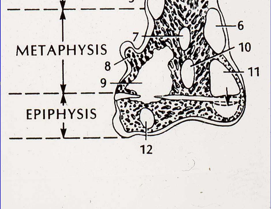
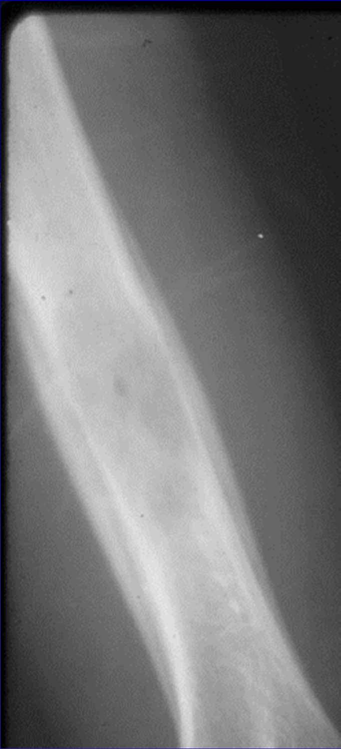
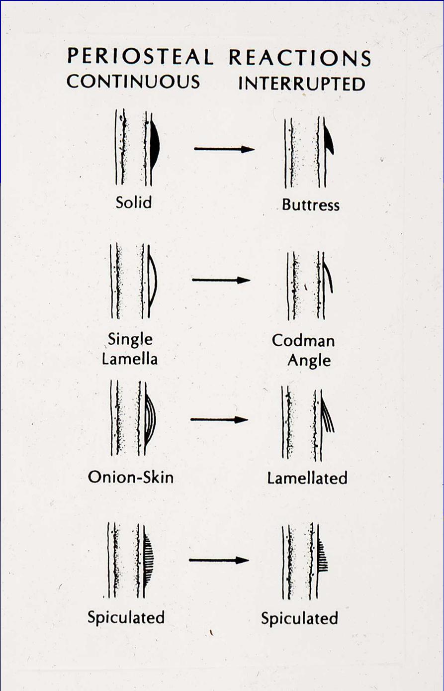
 Albert Leung, HMS III Gillian Lieberman, M.D. BIDMC Radiology Clerkship February 22, 2010 Overview Index Patient Periosteal Reactions Differential Diagnosis Principles of Osteoid Osteomas Bone Anatomy
Cytology of Neoplasms that Occur on the Limbs Rick Alleman, DVM, PhD, DABVP, DACVP
 Cytology of Neoplasms that Occur on the Limbs Rick Alleman, DVM, PhD, DABVP, DACVP I. Introduction The purpose of this material is to provide information that may be useful in the identification of tumors
GIANT CELL TUMOR OF LOWER END OF FEMUR IN A SKELETALLY IMMATURE-A RARE CASE
 GIANT CELL TUMOR OF LOWER END OF FEMUR IN A SKELETALLY IMMATURE-A RARE CASE *Surojit Mondal 1, Aniket Chowdhury 2 and Goutam Bandyopadhyay 3 1 Department of Orthopaedics, B.S.Medical College, Bankura,
RESEARCH INFORMATION AWARENESS SUPPORT PRIMARY BONE CANCER CHONDROSARCOMA. Visit bcrt.org.uk for more information
 RESEARCH INFORMATION AWARENESS SUPPORT PRIMARY BONE CANCER CHONDROSARCOMA Visit bcrt.org.uk for more information CONTENTS What is it? Who does it affect? Symptoms Types of Chondrosarcoma Cause and Risk
MRI of Pediatric Ankle and Foot. Mahesh Thapa, MD Associate Professor Seattle Children s University of Washington School of Medicine
 MRI of Pediatric Ankle and Foot Mahesh Thapa, MD Associate Professor Seattle Children s University of Washington School of Medicine Disclosures Under contract with Lippincott Williams and Wilkins (LWW)
Contents Part I Introduction 1 General Description 2 Natural History: Importance of Size, Site, Histopathology
 Contents Part I Introduction 1 General Description... 3 1.1 Introduction... 3 1.2 Incidence and Prevalence... 5 1.3 Predisposing and Genetic Factors... 8 References... 16 2 Natural History: Importance
VALORACIÒN RADIOLÓGICA DE LA LESIÒN ÒSEA SOLITARIA IMAGENOLOGIA MEDICA UNIVERSIDAD HISPANOAMERICANA
 VALORACIÒN RADIOLÓGICA DE LA LESIÒN ÒSEA SOLITARIA IMAGENOLOGIA MEDICA UNIVERSIDAD HISPANOAMERICANA TUMORES ÓSEOS SE PRESENTAN POR RANGOS DE EDAD, PRINCIPALMENTE: MENORES DE 20 AÑOS 20 A 40 AÑOS MAYORES
Case Report Giant Cell Tumor of Bone: Documented Progression over 4 Years from Its Origin at the Metaphysis to the Articular Surface
 Volume 2016, Article ID 9786925, 5 pages http://dx.doi.org/10.1155/2016/9786925 Case Report Giant Cell Tumor of Bone: Documented Progression over 4 Years from Its Origin at the Metaphysis to the Articular
A peculiar location of a rare bone tumor: sternal lipoma
 A peculiar location of a rare bone tumor: sternal lipoma Poster No.: P-0033 Congress: ESSR 2016 Type: Authors: Keywords: DOI: Scientific Poster Z. Akkaya, C. Uzun, S. Enon, G. Kocaman, G. Sahin; Ankara/TR
ISSN: DISTRIBUTION OF BONE AND CARTILAGINOUS TUMORS IN PEDIATRIC AGE GROUP IN WESTERN UTTAR-PRADESH: AN EVALUATIVE STUDY
 : 289-295 ISSN: 2277 4998 DISTRIBUTION OF BONE AND CARTILAGINOUS TUMORS IN PEDIATRIC AGE GROUP IN WESTERN UTTAR-PRADESH: AN EVALUATIVE STUDY QADRI S, HASAN M, AKHTAR K * AND SHERWANI RK The Departments
Heterogeneous osteoblastic activity in the right ischium of unclear etiology seen on NaF18-PET/CT
 CASE REPORT Heterogeneous osteoblastic activity in the right ischium of unclear etiology seen on NaF18-PET/CT Aung Zaw Win, Carina Mari Aparici Dept. Radiology, Nuclear Medicine section, San Francisco
Update On Lipomatous Tumors: Old Standbys and New Concepts
 Update On Lipomatous Tumors: Old Standbys and New Concepts John R. Goldblum, M.D. Chairman, Department of Anatomic Pathology Cleveland Clinic Professor of Pathology Cleveland Clinic Lerner College of Medicine
A Modified Lodwick-Madewell Grading System for the Evaluation of Lytic Bone Lesions
 Musculoskeletal Imaging Original Research Caracciolo et al. Evaluation of Lytic one Lesions Musculoskeletal Imaging Original Research Jamie T. Caracciolo 1 H. Thomas Temple 2 G. Douglas Letson 3 Mark J.
Key points in the evaluation of focal bone lesions: from plain film to multidetector CT
 Key points in the evaluation of focal bone lesions: from plain film to multidetector CT Poster No.: C-2060 Congress: ECR 2011 Type: Educational Exhibit Authors: I. Rubio Marco, M. Arraiza Sarasa, H. Gómez
General Approach to Lytic Bone Lesions D. Lee Bennett, MD, MA, Georges Y. El Khoury, MD Appl Radiol. 2004;33(5)
") General Approach to Lytic Bone Lesions D. Lee Bennett, MD, MA, Georges Y. El Khoury, MD Appl Radiol. 2004;33(5) www.medscape.com Abstract and Introduction Abstract When interpreting musculoskeletal radiographs,
General Approach to Lytic Bone Lesions D. Lee Bennett, MD, MA, Georges Y. El Khoury, MD Appl Radiol. 2004;33(5) www.medscape.com Abstract and Introduction Abstract When interpreting musculoskeletal radiographs,
FRACTURE CALLUS ASSOCIATED WITH BENIGN AND MALIGNANT BONE LESIONS AND MIMICKING OSTEOSARCOMA
 THE AMERICAN JOURNAL OF CLINICAL PATHOLOGY Vol. 52, No. 1 Copyright 1969 by The Williams & Wilkins Co. Printed in U.S.A. FRACTURE CALLUS ASSOCIATED WITH BENIGN AND MALIGNANT BONE LESIONS AND MIMICKING
Osteosarcoma Anatomic and Histologic Variants
 natomic Pathology / OSTEOSRCOM Osteosarcoma natomic and Histologic Variants Michael J. Klein, MD, and Gene P. Siegal, MD, PhD Key Words: Osteosarcoma; Malignant bone tumors; ones; Imaging DOI: 10.1309/UC6KQHLD9LV2KENN
Update on Sarcomas of the Head and Neck. Kevin Harrington
 Update on Sarcomas of the Head and Neck Kevin Harrington Overview Classification and incidence of sarcomas Clinical presentation Challenges to treatment Management approaches Prognostic factors Radiation-induced
MANAGEMENT OF OSTEOFIBROUS DYSPLASIA OF THE ULNAAFTER RESECTION WITH ELASTIC INTRAMEDULLARY NAIL AND NON VASCULAR FIBULAR GRAFT: A CASE REPORT
 MANAGEMENT OF OSTEOFIBROUS DYSPLASIA OF THE ULNAAFTER RESECTION WITH ELASTIC INTRAMEDULLARY NAIL AND NON VASCULAR FIBULAR GRAFT: A CASE REPORT *Ujwal Ramteke and Hitesh Mangukiya Department of Orthopaedics,