The Urinary System. Lecture Presentation by Lori Garrett Pearson Education, Inc.
|
|
|
- Scarlett Jackson
- 5 years ago
- Views:
Transcription

1 24 The Urinary System Lecture Presentation by Lori Garrett
2 Section 1: Anatomy of the Urinary System Learning Outcomes 24.1 Identify the organs of the urinary system, and cite a primary function of each Describe the location and structural features of the kidneys Describe the gross structural features of the kidney, and distinguish between cortical and juxtamedullary nephrons.
3 Section 1: Anatomy of the Urinary System Learning Outcomes (continued) 24.4 Describe the segments of the nephron and collecting system, including their general functions and histological appearance Trace the pathway of blood flow through a kidney, and compare the pattern of blood flow in cortical and juxtamedullary nephrons.
4 Module 24.1: The urinary system organs are the kidneys, ureters, urinary bladder, and urethra Urinary system Two kidneys Receive 25 percent of the cardiac output Major excretory organs of the urinary system Produce urine (fluid containing water, ions, and small soluble substances)

5 Module 24.1: Urinary system organs Urinary tract Ureters receive urine from the kidneys Conduct urine to the urinary bladder by gravity and peristalsis Urinary bladder receives and stores urine Contraction of muscle in walls drives urination Urethra conducts urine from the bladder to outside the body
6
7 Module 24.1: Review A. Name the major excretory organs of the urinary system. B. Describe the functions of the urinary system. Learning Outcome: Identify the organs of the urinary system, and cite a primary function of each.
8 Module 24.2: The kidneys are paired retroperitoneal organs Kidney structure Reddish brown Dimensions ~10 cm (4 in.) long; ~5.5 cm (2.2 in.) wide; ~3 cm (1.2 in.) thick Weight: ~150 g (5.25 oz) Hilum Medial indentation Point of entry/exit for the renal artery, renal nerves, renal vein, and the ureter

9 Module 24.2: Kidney location and structure Located in a retroperitoneal position Between the muscles of the posterior body wall and the parietal peritoneum Connected to the urinary bladder by the ureters Empty into the posterior, inferior surface of the urinary bladder
")
10 Module 24.2: Kidney location and structure Kidney location Either side of vertebral column Protected by: Visceral organs (anteriorly) Body wall musculature and the 11th and 12th ribs (posteriorly and laterally) Left kidney slightly superior to the right
11 Module 24.2: Kidney location and structure Kidney location (continued) Position maintained by: 1. Overlying peritoneum 2. Adjacent visceral organs 3. Supporting connective tissues
12 Module 24.2: Kidney location and structure Connective tissues supporting kidney Fibrous capsule (layer of collagen fibers) Covers the outer surface of the kidney Projects collagen fibers through the perinephric fat to the renal fascia
 Perinephric fat (perinephric fat capsule)")
13 Module 24.2: Kidney location and structure Connective tissues supporting kidney (continued) Perinephric fat (perinephric fat capsule) Thick layer of adipose tissue Renal fascia (dense, fibrous outer layer) Anchors the kidney to surrounding structures
14 Module 24.2: Review A. What structures enter and exit the kidney at the hilum? B. Describe the concentric layers of connective tissue that protect and anchor the kidney. C. What would happen to a kidney s position if the perinephric fat layer were depleted and the collagen fibers of the fibrous capsule were to become detached? Learning Outcome: Describe the location and structural features of the kidneys.
15 Module 24.3: The kidneys are complex at the gross and microscopic levels Major structural landmarks Fibrous capsule Lines the renal sinus (internal cavity within the kidney) Renal cortex (superficial region of the kidney)
16 Module 24.3: Gross and microscopic anatomy of the kidney Major structural landmarks (continued) Renal medulla (inner region of the kidney) Renal pyramid (conical structure in the medulla) Renal papilla (tip of the pyramid) Renal column (separates adjacent pyramids)
 Kidney lobe")
17 Module 24.3: Gross and microscopic anatomy of the kidney Major structural landmarks (continued) Kidney lobe (pyramid, the overlying cortex, and adjacent renal columns) Each kidney contains 6 18 lobes
18 Module 24.3: Gross and microscopic anatomy of the kidney Other features of the kidney Hilum Medial indentation in the kidney Minor calyx Collects urine from a single kidney lobe Major calyx Forms from the fusion of 4 5 minor calyces
 Renal")
19 Module 24.3: Gross and microscopic anatomy of the kidney Other features of the kidney (continued) Renal pelvis Funnel-shaped structure that collects urine from major calyces Continuous with the ureter
20 Module 24.3: Gross and microscopic anatomy of the kidney Two types of nephrons Microscopic functional units of the kidney 1. Cortical nephrons 85 percent of all nephrons Located primarily in the cortex Responsible for most regulatory functions

21 Module 24.3: Gross and microscopic anatomy of the kidney Two types of nephrons (continued) 2. Juxtamedullary nephrons 15 percent of all nephrons Long nephron loop extending deep into medulla Essential to producing concentrated urine
22 Module 24.3: Review A. Which structure is a conical mass within the renal medulla that ends at the papilla? B. Compare the minor and major calyces. C. Which type of nephron is essential for water conservation and concentrated urine production? Learning Outcome: Describe the gross structural features of the kidney, and distinguish between cortical and juxtamedullary nephrons.
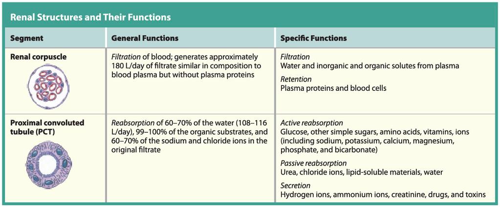
23 Module 24.4: A nephron is divided into segments; each segment has specific functions Two components 1. Renal corpuscle Blood pressure forces water and solutes out of the glomerular capillaries in a process called filtration Produces filtrate (protein-free solution, similar to blood plasma) Collected in the surrounding capsular space 2. Renal tubule Tubular passageway up to 50 mm long Receives filtrate and modifies it to create urine

24 Module 24.4: Segments of a nephron Renal corpuscle Glomerular capsule (Bowman s capsule) Cup-shaped chamber Capillary network (glomerulus)
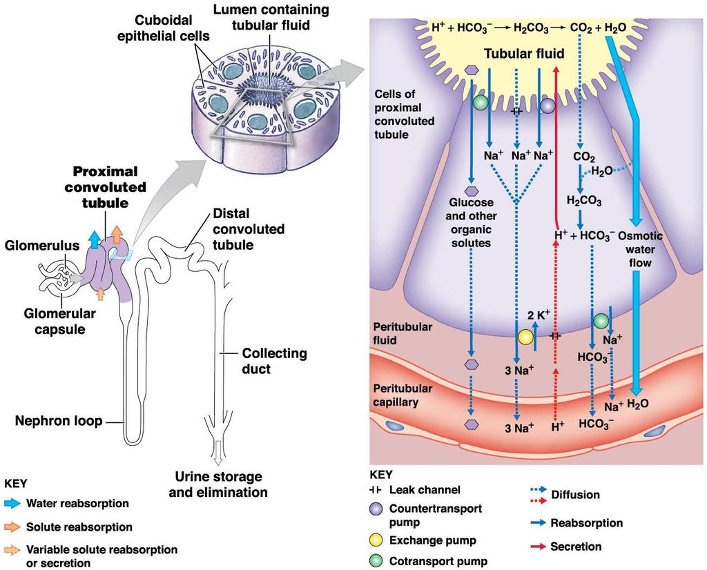
25 Module 24.4: Segments of a nephron Proximal convoluted tubule (PCT) Reabsorbs nutrients from the filtrate (now called tubular fluid)


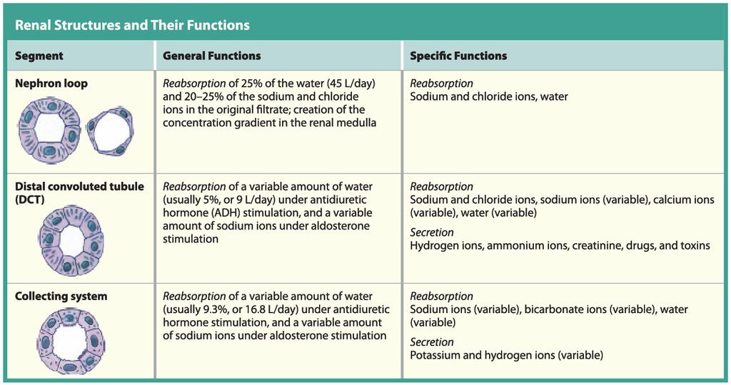
26 Module 24.4: Segments of a nephron Nephron loop Establishes osmotic gradient for water reabsorption Each limb contains a thin segment and a thick segment
 Adjusts")
27 Module 24.4: Segments of a nephron Distal convoluted tubule (DCT) Adjusts tubular fluid composition by reabsorption and secretion

28 Segments of a nephron
29 Module 24.4: Segments of a nephron Collecting system Series of tubes carrying tubular fluid away from the nephron
30 Module 24.4: Segments of a nephron Collecting system (continued) Collecting duct Collects fluid from many nephrons Carries fluid through the renal medulla
 Collecting duct (continued) Lined with two main types of cells: Intercalated cells")
31 Module 24.4: Segments of a nephron Collecting system (continued) Collecting duct (continued) Lined with two main types of cells: Intercalated cells (play a role in secreting and reabsorbing hydrogen and bicarbonate ions) Principal cells (reabsorb water and secrete potassium)
 Papillary duct")
32 Module 24.4: Segments of a nephron Collecting system (continued) Papillary duct Collects fluid from multiple collecting ducts Delivers fluid to minor calyx

33 The Nephron and collecting system
34 Module 24.4: Review A. Describe filtrate. B. Identify the structures of the renal corpuscle. C. Describe the structures of the collecting system. Learning Outcome: Describe the segments of the nephron and collecting system, including their general functions and histological appearance.
35 Module 24.5: The kidneys are highly vascular, and the circulation patterns are complex Arterial system Renal artery delivers blood to kidney, branching into: Segmental arteries in the renal sinus
36 Module 24.5: Circulation patterns in the kidney Arterial system (continued) Renal artery Segmental arteries, which branch into: Interlobar arteries running within renal columns
37 Module 24.5: Circulation patterns in the kidney Arterial system (continued) Renal artery Segmental arteries Interlobar arteries, which branch into: Arcuate arteries, which arch along the boundary between the renal cortex and renal medulla
38 Module 24.5: Circulation patterns in the kidney Arterial system (continued) Renal artery Segmental arteries Interlobar arteries Arcuate arteries, which branch into: Cortical radiate arteries, which branch into:
 Renal artery Segmental arteries Interlobar arteries Arcuate arteries Cortical")
39 Module 24.5: Circulation patterns in the kidney Arterial system (continued) Renal artery Segmental arteries Interlobar arteries Arcuate arteries Cortical radiate arteries, which branch into: Afferent arterioles, which supply each nephron, specifically a capillary knot known as a glomerulus

40 Module 24.5: Circulation patterns in the kidney Venous system Cortical radiate veins collect blood from the capillaries of the nephron and drain into: Arcuate veins, then to: Interlobar veins, which drain into the: o Renal vein, which drains into the inferior vena cava
41 Module 24.5: Circulation patterns in the kidney Blood flow around a cortical nephron Afferent arteriole Supplies blood to each individual nephron Glomerulus Efferent arteriole Carries blood from the glomerulus to the peritubular capillaries
 Peritubular capillaries Surround the entire renal tubule Surrounded by")
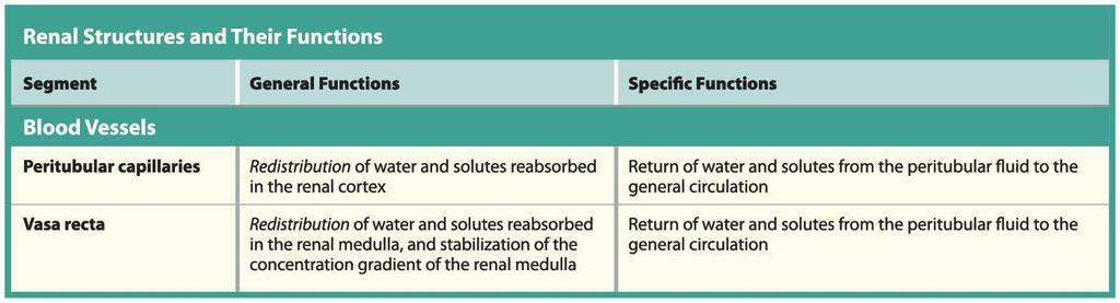
42 Module 24.5: Circulation patterns in the kidney Blood flow around a cortical nephron (continued) Peritubular capillaries Surround the entire renal tubule Surrounded by peritubular fluid Collect water and solutes absorbed by the nephron Deliver other solutes to the nephron for secretion Drain into cortical radiate veins
 Connected to")
43 Module 24.5: Circulation patterns in the kidney Blood flow around a juxtamedullary nephron Same blood flow until after the peritubular capillaries Vasa recta (vasa, vessels + recta, straight) Connected to the distal end of the peritubular capillaries Long, straight capillaries that parallel the nephron loop Transport water and solutes within the renal medulla Drain into cortical radiate veins
44 Module 24.5: Circulation patterns in the kidney Nephron innervation Each kidney has ~1.25 million nephrons Both cortical and juxtamedullary nephrons are innervated by renal nerves Enter at the hilum and follow the branches of the renal artery Most of the nerve fibers are sympathetic postganglionic fibers from the celiac plexus and inferior splanchnic nerves Sympathetic stimuli adjust blood flow and blood pressure at the glomeruli Also stimulate the release of renin
45 Module 24.5: Review A. Trace the pathway of blood from the renal artery to the renal vein. B. Describe how blood enters and leaves the glomerulus. C. Define the vasa recta. Learning Outcome: Trace the pathway of blood flow through a kidney, and compare the pattern of blood flow in cortical and juxtamedullary nephrons.
46 Section 2: Overview of Renal Physiology Learning Outcomes 24.6 Briefly describe how the kidneys maintain homeostasis and produce urine Describe filtration, reabsorption, and secretion along each segment of the nephron and collecting system Describe the structural features of a renal corpuscle, and explain the functions of the filtration membrane components.
47 Section 2: Overview of Renal Physiology Learning Outcomes (continued) 24.9 Describe the factors that influence filtration pressure and the glomerular filtration rate Identify the types of transport mechanisms along the proximal and distal convoluted tubules of the nephron Explain the role of countercurrent multiplication in the formation of a concentration gradient in the renal medulla.
48 Section 2: Overview of Renal Physiology Learning Outcomes (continued) Describe how antidiuretic hormone influences the volume and concentration of urine Summarize the major steps involved in \water reabsorption and urine production Clinical Module: Compare and contrast chronic and acute renal failure, and explain the process of hemodialysis.
49 Module 24.6: The kidneys maintain homeostasis by removing wastes and producing urine Renal physiology Urinary system maintains homeostasis by regulating the volume and composition of blood Concentrates urine to mosm/l Excretes solutes, especially metabolic wastes

50 Module 24.6: Renal physiology Examples of metabolic wastes Urea Most abundant organic waste By-product of amino acid breakdown Creatinine By-product of creatine phosphate breakdown in muscles Uric acid Formed during recycling of nitrogenous bases of RNA

51 Module 24.6: Renal physiology Three processes in urine formation 1. Filtration Blood pressure forces water and solutes across the membranes of the glomerular capillaries into the capsular space
52 Module 24.6: Renal physiology Three processes in urine formation (continued) 2. Reabsorption Transport of water and solutes from the tubular fluid across tubular epithelium into the peritubular fluid

53 Module 24.6: Renal physiology Three processes in urine formation (continued) 3. Secretion Transport of solutes from the peritubular fluid across tubular epithelium into the tubular fluid
54 Module 24.6: Review A. In which direction do fluids and solutes move in each of the three kidney processes? Learning Outcome: Briefly describe how the kidneys maintain homeostasis and produce urine.
55 Module 24.7: Filtration, reabsorption, and secretion occur in specific segments of the nephron and collecting system Functions of each nephron segment Three processes in urine formation: filtration, reabsorption, and secretion Filtration occurs only in the renal corpuscle Balance between reabsorption and secretion varies in remaining nephron segments Regulation of final volume and solute concentration is from the interaction between the collecting system and the nephron loops

56 Filtration, reabsorption, and secretion
57
58
59
60 Module 24.7: Review A. Identify the three distinct processes of urine formation in the kidney. B. Where does filtration exclusively occur in the kidney? C. Which segment of the nephron is solely involved in the reabsorption of water and sodium and chloride ions? Learning Outcome: Describe filtration, reabsorption, and secretion along each segment of the nephron and collecting system.
61 Module 24.8: Filtration occurs at the renal corpuscle Afferent arteriole (delivers blood to the corpuscle) Glomerulus (capillary knot) Filtration occurs here
62 Module 24.8: Filtration Efferent arteriole (delivers blood to peritubular capillaries) Smaller diameter than the afferent arteriole Increases the blood pressure in the glomerulus, aiding filtration

63 Module 24.8: Filtration Capsular space Between layers of the glomerular capsule Juxtaglomerular complex Secretes renin when glomerular blood pressure decreases

64 Module 24.8: Filtration Intraglomerular mesangial cells Supporting cells Lie between adjacent glomerular capillaries Control capillary diameter and rate of blood flow
65 Module 24.8: Filtration Glomerular capsule Forms the outer wall of the renal corpuscle and covers the glomerular capillaries Parietal layer (forms outer capsule)
 Visceral layer (covers the capillaries) Composed of podocytes (podos, foot + -cyte, cell) Large")
66 Module 24.8: Filtration Glomerular capsule (continued) Visceral layer (covers the capillaries) Composed of podocytes (podos, foot + -cyte, cell) Large cells with foot processes (pedicels) that wrap around the glomerular capillaries Gaps between adjacent pedicels are filtration slits

67 Module 24.8: Filtration Filtration membrane 1. Fenestrated glomerular capillaries Contain large diameter pores 2. Dense layer Specialized basement membrane 3. Filtration slits from podocytes Combination of these layers prevents most plasma proteins from entering the capsular space
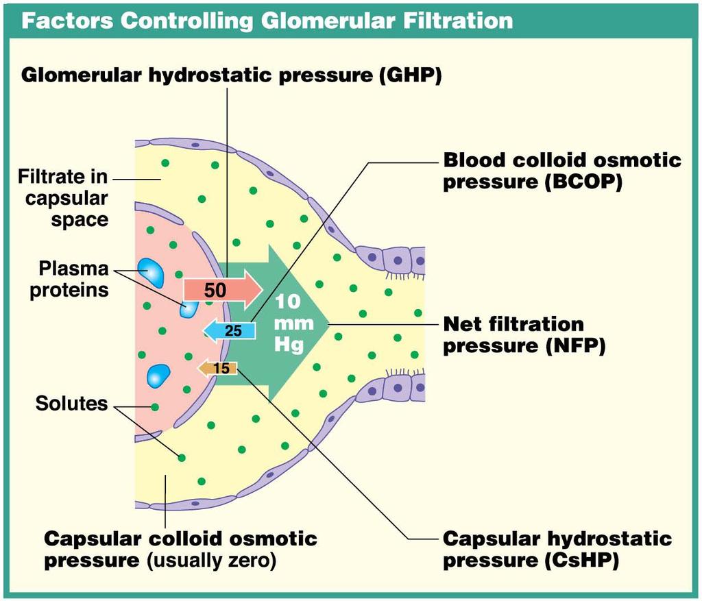
68 Module 24.8: Filtration Factors controlling glomerular filtration Glomerular hydrostatic pressure (GHP) Blood pressure in the glomerular capillaries Tends to push water and solutes out of plasma and into the filtrate Capsular colloid osmotic pressure Few, if any, plasma proteins enter the capsular space Blood colloid osmotic pressure (BCOP) Pressure due to materials in solution Tends to draw water out of the filtrate and into the plasma (opposes filtration)
69 Module 24.8: Filtration Factors controlling glomerular filtration (continued) Capsular hydrostatic pressure (CsHP) Opposes GHP Tends to push water and solutes out of the filtrate and into the plasma Results from resistance of filtrate already in the nephron
70 Module 24.8: Filtration Factors controlling glomerular filtration (continued) Net filtration pressure (NFP) Pressure acting across the glomerular capillaries Represents the sum of the hydrostatic pressures and colloid osmotic pressures Average pressure forcing water and dissolved substances out of the glomerular capillaries and into the capsular space
71 Filtration
72 Module 24.8: Review A. What three elements form the filtration membrane? B. Explain why blood pressure is higher in glomerular capillaries than in other systemic capillaries. C. Blood colloid osmotic pressure tends to draw water out of the filtrate and into the plasma. Why does this occur? Learning Outcome: Describe the structural features of a renal corpuscle, and explain the functions of the filtration membrane components.
73 Module 24.9: The glomerular filtration rate is the amount of filtrate produced each minute Two interacting levels of control help stabilize Glomerular filtration rate (GFR) 1. Autoregulation at the local level 2. Central regulation Endocrine component o Initiated by the kidneys Neural component o Involves the sympathetic division of the ANS

74 Autoregulation maintains adequate GFR

75 Module 24.9: Glomerular filtration If autoregulation is ineffective, central regulation is involved Juxtaglomerular complex

76 Central regulation
77 Module 24.9: Review A. Describe autoregulation at the kidneys. B. What does the juxtaglomerular complex do in response to decreased filtration pressure? C. Angiotensin II has what effect on the CNS? Learning Outcome: Describe the factors that influence filtration pressure and the glomerular filtration rate.
78 Module 24.10: Reabsorption predominates along the proximal convoluted tubule Reabsorption in the PCT includes: >99 percent of glucose, amino acids, and other organic nutrients Sodium, potassium, bicarbonate, magnesium, phosphate, sulfate ions Water (about 108 liters each day) Solute concentration of tubular fluid decreases Water moves into the peritubular fluid
79 Reabsorption
80 Module 24.10: whereas reabsorption and secretion are often linked along the distal convoluted tubule Movement of water and solutes out of peritubular fluid into the tubular fluid Only percent of the initial filtrate volume reaches the distal convoluted tubule (DCT) Combination of reabsorption and secretion in the DCT alters solute composition in the tubular fluid Sodium ions are reabsorbed in exchange for potassium ions (pumps are stimulated by aldosterone) Hydrogen ions are secreted in exchange for sodium ions (to increase ph of body fluids) Carrier proteins also secrete toxins or drugs

81 Secretion and reabsorption
82 Module 24.10: Review A. Identify the segment of the nephron that makes final adjustments to the composition of tubular fluid. B. What effect would increased amounts of aldosterone have on the K + concentration in urine? C. What effect would a decrease in the Na + concentration of filtrate have on the ph of tubular fluid? Learning Outcome: Identify the types of transport mechanisms along the proximal and distal convoluted tubules of the nephron.
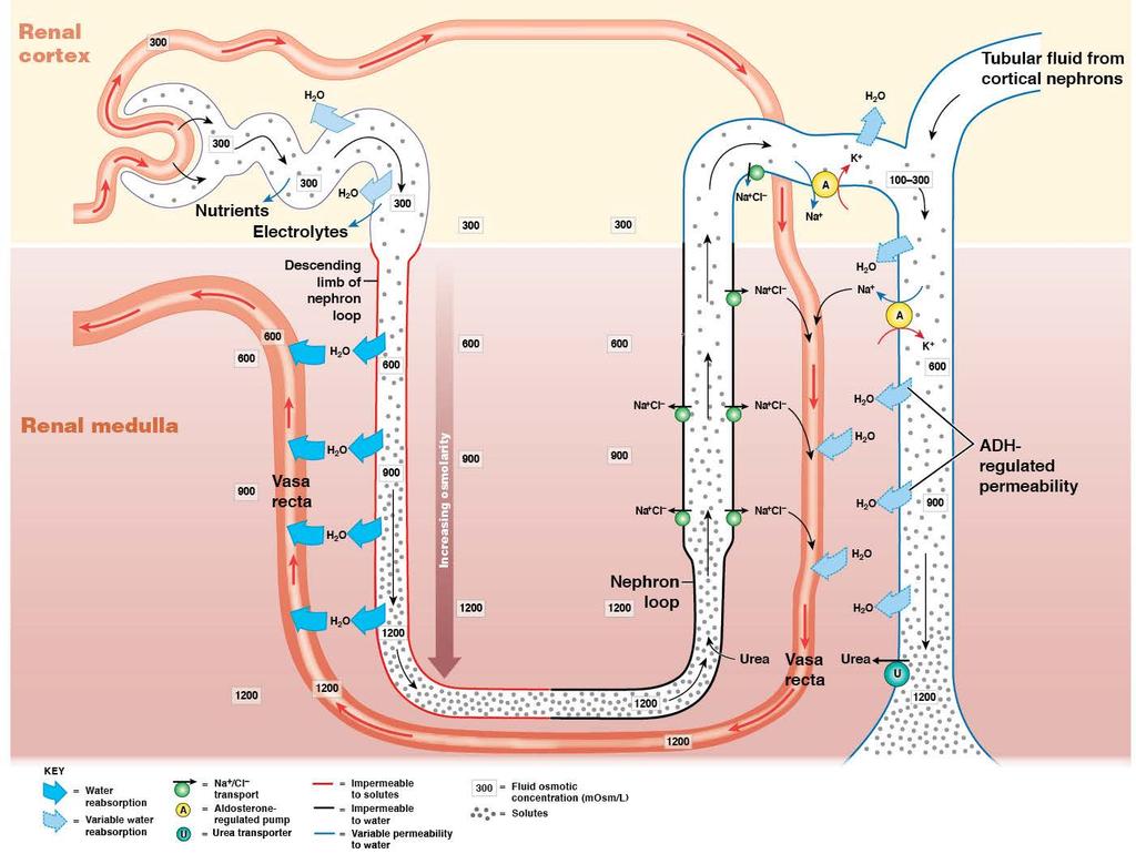
83 Module 24.11: Exchange between the limbs of the nephron create an osmotic concentration gradient in the renal medulla Countercurrent multiplication Thin descending limb and thick ascending limb Located very close to each other Separated by peritubular fluid Exchange between these adjacent limbs is called countercurrent multiplication Countercurrent fluids moving in opposite directions Multiplication effect increases with fluid movement Responsible for creating concentration gradient in the renal medulla Enables production of highly concentrated urine
84 Nephron loop

85 Module 24.11: Nephron loop Thick ascending limb Actively transports Na + and Cl out of the tubular fluid Impermeable to water Tubular fluid solute concentration decreases Peritubular fluid solute concentration increases

86 Module 24.11: Nephron loop Osmotic concentration of peritubular fluid is increased from activity of the thick ascending limb Thin descending limb Permeable to water Impermeable to solutes Water moves from tubular fluid into the peritubular fluid by osmosis Tubular fluid solute concentration increases

87 Module 24.11: Nephron loop Concentration of urine Water is reabsorbed along the DCT and collecting duct Increases concentration of solutes within the tubular fluid, particularly urea Tubular fluid reaching the papillary duct has a typical urea concentration of ~450 mosm/l
88 Module 24.11: Review A. Define countercurrent multiplication as it occurs in the kidneys. B. The thick ascending limb of the nephron loop actively pumps what substances into the peritubular fluid? C. An increase in sodium and chloride ions in the peritubular fluid affects the descending thin limb in what way? Learning Outcome: Explain the role of countercurrent multiplication in the formation of a concentration gradient in the renal medulla.
89 Module 24.12: Urine volume and concentration are hormonally regulated Obligatory water reabsorption Occurs in locations where water movements cannot be prevented PCT and descending limb of nephron loop Rate cannot be adjusted Recovers 85 percent of filtrate

90 Module 24.12: Urine volume and concentration Facultative water reabsorption Occurs in the DCT and collecting tubule Allows precise control of water reabsorption Adjusts urine volume by reabsorbing a portion (or all) of the remaining 15 percent of filtrate volume
 No")
91 Module 24.12: Urine volume and concentration Urine volume without ADH (antidiuretic hormone) No water is reabsorbed in DCT and collecting tubule No facultative water reabsorption

92 Module 24.12: Urine volume and concentration Urine volume with ADH ADH allows water channels to form Aquaporins appear in the apical plasma membranes of the DCT and collecting tubule cells Water permeability of the last tubular segments increases, increasing water reabsorption

93 Module 24.12: Urine volume and concentration Normal urine Normal volume is about 1200 ml/day with an osmotic concentration of 1000 mosm/l Values differ from person to person and from day to day Kidneys alter their function to maintain homeostasis
94 Module 24.12: Review A. Can the water permeability of the PCT or DCT ever change? Explain. B. Compare obligatory water reabsorption with facultative water reabsorption. C. What effect does an increase in ADH levels have on the DCT? D. When ADH levels in the DCT decrease, what happens to the urine osmotic concentration? E. What effect does ADH have on the apical plasma membranes lining the DCT and collecting ducts? Learning Outcome: Describe how antidiuretic hormone influences the volume and concentration of urine.
95 Module 24.13: Renal function is an integrative process involving filtration, reabsorption, and secretion Renal corpuscle Filtrate has the same osmotic composition as plasma (~300 mosm/l) Same composition as plasma except for plasma proteins
96 Module 24.13: Renal function as an integrative process Proximal convoluted tubules Ions and organic nutrients removed from tubular fluid Water follows by osmosis Reduces tubular fluid volume but keeps tubular fluid and peritubular fluid isotonic PCT and descending limb of nephron loop Obligatory water reabsorption concentrates the tubular fluid
97 Filtration, reabsorption, and secretion
98 Module 24.13: Renal function as an integrative process Ascending limb of nephron loop Actively transports Na + and Cl out of the tubule (impermeable to water) Lowers the osmotic concentration of tubular fluid
99 Module 24.13: Renal function as an integrative process DCT and collecting system make adjustments Reabsorption and secretion of solutes Hormonally controlled water reabsorption

100 Module 24.13: Renal function as an integrative process Vasa recta Absorbs solutes and water from the tubules into the systemic circuit Maintains concentration gradient of medulla
101 Production of urine
102 Module 24.13: Review A. In the PCT, ions and organic substrates are actively removed, thus causing what to occur? Learning Outcome: Summarize the major steps involved in water reabsorption and urine production.
103 Module 24.14: Clinical Module: Renal failure is a life-threatening condition Occurs when the kidneys cannot filter wastes from blood and can no longer maintain homeostasis Impairs all systems in the body, resulting in: Decrease in urine production Rise in blood pressure Anemia from decline in erythropoietin production Central nervous system problems (sleeplessness, seizures, delirium, and coma)
104 Module 24.14: Renal failure Chronic renal failure Kidney function deteriorates gradually Associated problems accumulate over time Progression can be slowed, but the condition is not reversible Management involves restricted water, salt, and protein intake Reduces strain on urinary system by minimizing: Volume of urine produced Amount of nitrogenous waste generated Acidosis (a common problem with renal failure) can be countered by ingesting bicarbonate ions
105 Module 24.14: Renal failure Acute renal failure Kidney function deteriorates rapidly in just a few days May be impaired for weeks Sudden slowing or stopping of filtration caused by: Exposure to toxic drugs, renal ischemia, urinary obstruction, or trauma Allergic response to antibiotics or anesthetics in sensitized individuals Recovery of partial or complete function is possible if patients survive the initial incident Survival rate ~50 percent with supportive treatment
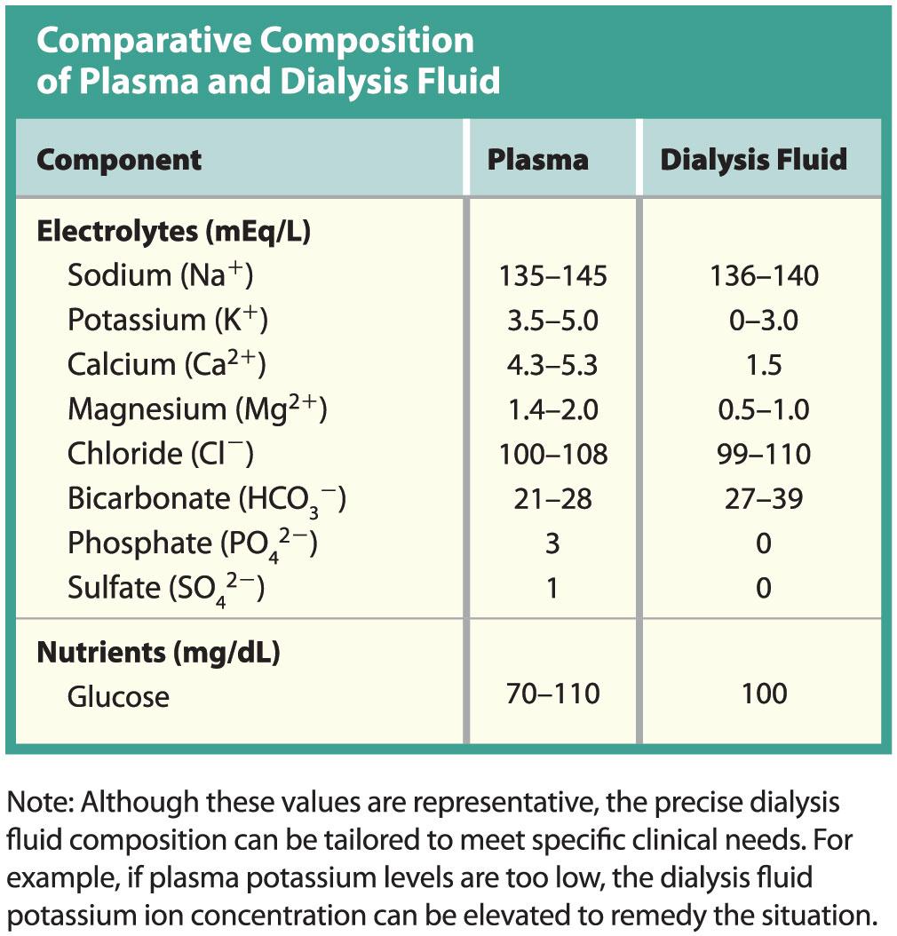
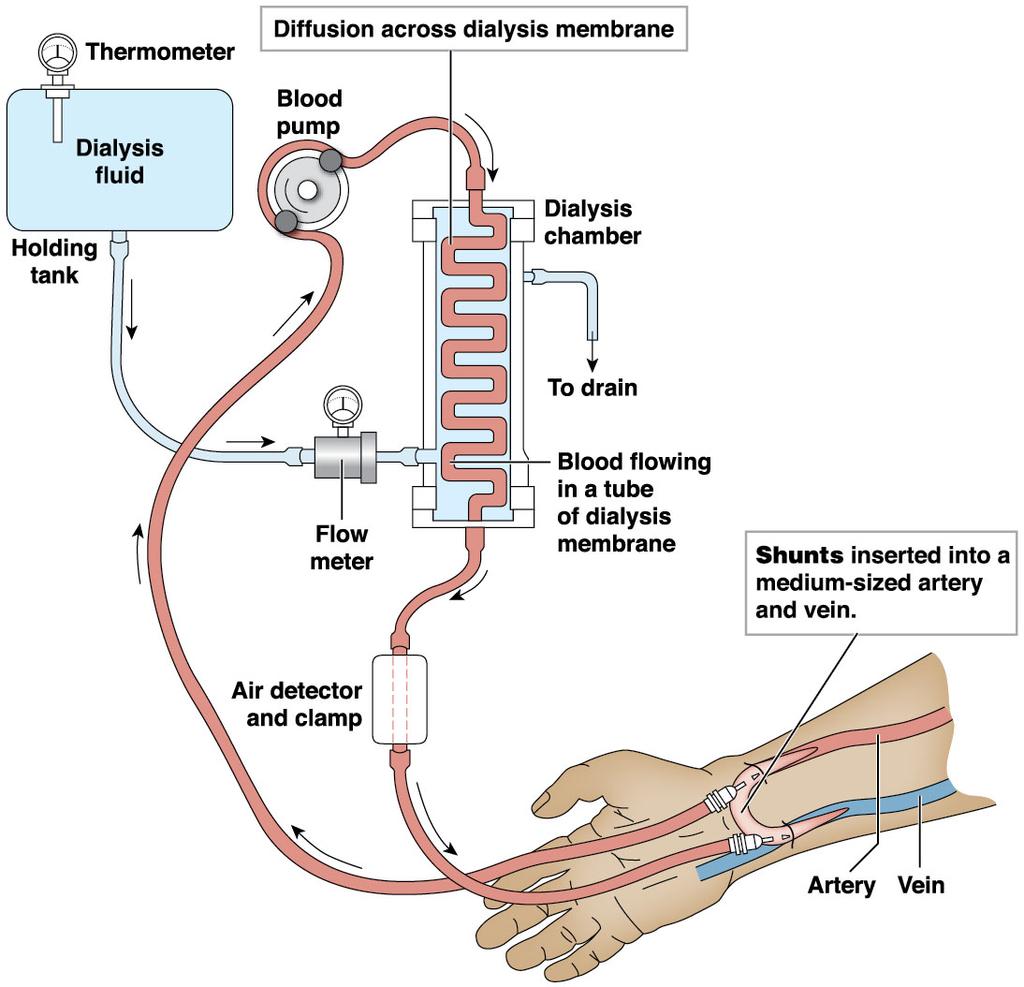
106 Module 24.14: Renal failure Dialysis Process of passive diffusion across a selectively permeable membrane Hemodialysis Uses an artificial membrane as an alternative to the kidney s normal membrane around the glomerulus Regulates the composition of blood using a dialysis machine Membrane pores allow diffusion of ions, nutrients, and organic wastes, but not plasma proteins
 Hemodialysis (continued) Dialysis fluid containing specific concentrations of solutes")
107 Module 24.14: Renal failure Dialysis (continued) Hemodialysis (continued) Dialysis fluid containing specific concentrations of solutes is run on the other side of the membrane Shunts (silicone rubber tubes) connect blood vessels with the dialysis machine
108

109 Renal failure
110 Module 24.14: Renal failure Renal failure treatment Dialysis relieves renal failure symptoms, but is not a cure Kidney transplant is the only real cure for severe renal failure Patient survival is more than 90 percent at 2 years after the transplant Close relative donor increases success rate Immunosuppressive drugs are necessary to reduce rejection of transplant
111 Module 24.14: Review A. Briefly explain the difference between chronic and acute renal failure. B. Define dialysis. C. Explain why patients on dialysis often receive Epogen or Procrit, synthetic forms of erythropoietin. Learning Outcome: Compare and contrast chronic and acute renal failure, and explain the process of hemodialysis.
112 Section 3: Urine Storage and Elimination Learning Outcomes Name the organs responsible for the transport, storage, and elimination of urine Describe the structures and functions of the ureters, urinary bladder, and urethra Describe the urine storage and urine voiding reflexes Clinical Module: Describe common urinary disorders related to output and frequency.
113 Module 24.15: The urinary tract transports, stores, and eliminates urine Urinary tract Transports, stores, and eliminates urine Includes the ureters, urinary bladder, and urethra Can be visualized using a pyelogram X-ray image of the urinary tract taken after a radiopaque dye is administered intravenously
 Retroperitoneal")
114 Module 24.15: The urinary tract Ureters Paired muscular tubes extending from the kidney to the urinary bladder (about 30 cm) Retroperitoneal and attached to the posterior abdominal wall Urinary bladder Hollow, muscular organ holding up to a liter of urine Urethra

115 Module 24.15: The urinary tract Urethra Extends from the neck of the urinary bladder to the exterior of the body Different lengths and functions in males versus females Male urethra is longer and transports semen as well as urine
116 Module 24.15: Review A. When does urine production end? B. What is a pyelogram? C. How does the urethra differ between males and females? Learning Outcome: Name the organs responsible for the transport, storage, and elimination of urine
117 Module 24.16: The ureters, urinary bladder, and urethra are specialized to conduct urine Urinary bladder Filled by the ureters and drained by the urethra Dimensions vary with state of distension Posterior, inferior, and anterior surfaces outside the peritoneal cavity Anchored to the pelvic and pubic bones by supporting ligaments Lateral umbilical ligaments Vestiges of the umbilical arteries Middle umbilical ligament
118 Module 24.16: Conduction and storage of urine Urinary bladder (continued) Rugae Folds in the bladder lining that disappear with expansion as the bladder fills Ureteric orifices Slitlike shape helps prevent backflow of urine into ureters with bladder contraction
119 Module 24.16: Conduction and storage of urine Urinary bladder (continued) Ureters penetrate posterior bladder wall at an oblique angle Trigone Triangular area bounded by the ureteral openings and the entrance to the urethra
120 Module 24.16: Conduction and storage of urine Urinary bladder (continued) Neck of the urinary bladder Surrounds the urethral opening Contains a muscular internal urethral sphincter (involuntary smooth muscle)
 External urethral sphincter Located where the urethra")
121 Module 24.16: Conduction and storage of urine Urinary bladder (continued) External urethral sphincter Located where the urethra passes through the urogenital diaphragm Under voluntary control Must be voluntarily relaxed to permit urination
122 Module 24.16: Conduction and storage of urine Ureters conduct urine to the bladder 1. Inner mucosa Transitional epithelium and surrounding lamina propria

123 Module 24.16: Conduction and storage of urine Ureters (continued) 2. Middle muscular layer Bands of smooth muscle that create peristaltic waves to move urine to the bladder 3. Outer connective tissue layer Continuous with the fibrous capsule and peritoneum

124 Module 24.16: Conduction and storage of urine Wall of the urinary bladder Contains mucosa, submucosa, and muscularis layers
 Muscularis layer has three layers Inner")
125 Module 24.16: Conduction and storage of urine Wall of the urinary bladder (continued) Muscularis layer has three layers Inner longitudinal layer Circular layer Outer longitudinal layer Collectively, the layers form the detrusor muscle
126 Module 24.16: Conduction and storage of urine Wall of the urethra Lined with stratified epithelium that varies by location Transitional at the neck Stratified columnar at midpoint Stratified squamous near the external urethral orifice
 Thick, elastic lamina propria")
127 Module 24.16: Conduction and storage of urine Wall of the urethra (continued) Thick, elastic lamina propria Longitudinal folds in the mucous membrane Mucin-secreting cells in the epithelial pockets
128 Module 24.16: Review A. Urine is transported by the, stored within the, and eliminated through the. B. What has to happen to the external urethral sphincter to allow urination? C. Name the specialized smooth muscle of the urinary bladder, and describe its function. Learning Outcome: Describe the structures and functions of the ureters, urinary bladder, and urethra.
129 Module 24.17: Urinary reflexes coordinate urine storage and voiding Micturation reflex Coordinates the process of urination Involves both: Local reflex pathway Central pathway through the cerebral cortex
130 Module 24.17: Urination Micturation reflex (continued) Urine storage reflex Stretch receptors of urinary bladder wall distort as it fills Afferent impulses stimulate sympathetic stimulation to detrusor and stimulate contraction of internal urethral sphincter Pontine storage center decreases parasympathetic activity and increases somatic motor nerve activity of external urethral sphincter
 Urine voiding reflex through pontine micturition center Interneuron relays sensation of bladder fullness to the thalamus Projection fibers relay the")
131 Module 24.17: Urination Micturation reflex (continued) Urine voiding reflex through pontine micturition center Interneuron relays sensation of bladder fullness to the thalamus Projection fibers relay the information to the cerebral cortex Voluntary relaxation of the external urethral sphincter Causes relaxation of the internal urethral sphincter Since pressure is already increased, relaxing the sphincters leads to urination
132 Module 24.17: Review A. What are the two reflexes that control urination? B. The ability to consciously control urination depends on your ability to control which muscle? Learning Outcome: Describe the urine storage and urine voiding reflexes.
133 Module 24.18: Clinical Module: Urinary disorders can often be detected by physical examinations and laboratory tests Primary signs of urinary disorders Change in volume and appearance of urine Polyuria Excessive urine production Results from hormonal or metabolic problems Oliguria o Possibly diabetes or glomerulonephritis Reduced urine production ( ml/day) Anuria Severely reduced urine production (0 50 ml/day) Oliguria and anuria indicate serious kidney problems and potential renal failure
 Change in frequency Increased urgency or frequency Can be from irritation of the lining of the ureters or urinary bladder")
134 Module 24.18: Urinary disorders Primary signs of urinary disorders (continued) Change in frequency Increased urgency or frequency Can be from irritation of the lining of the ureters or urinary bladder Incontinence Inability to control urination voluntarily May involve periodic involuntary leakage (stress incontinence), inability to delay urination (urge incontinence), or continual trickle of urine from full bladder (overflow incontinence)
135 Module 24.18: Urinary disorders Primary signs of urinary disorders (continued) Change in frequency (continued) Urinary retention Initially normal renal function Urination does not occur In males, commonly results from enlarged prostate gland and compression of prostatic urethra
136 Module 24.18: Urinary disorders Primary signs of urinary disorders (continued) Pain Pain in the superior pubic region Associated with urinary bladder disorders Pain in the superior lumbar region or in the flank that radiates to the right or left upper quadrants Associated with kidney infections (pyelonephritis) Also associated with kidney stones (renal calculi) Dysuria Painful or difficult urination Can occur with cystitis or urethritis or urinary obstructions (possibly enlarged prostate in males)

137 Urinary disorders
138 Module 24.18: Urinary disorders Clinical signs of urinary system disorders Edema (swelling) Occurs when renal disorders lead to proteinuria (protein in the urine) Facial swelling, especially around the eyes, is common Fever Commonly develops when urinary system is infected with pathogens Cystitis (bladder infection) usually low-grade fever Pyelonephritis (kidney infection) can produce very high fevers
139 Module 24.18: Review A. What is the term for painful or difficult urination? B. Why is urinary obstruction at the urethra more dangerous than at the ureter? Learning Outcome: Describe common urinary disorders related to output and frequency.
Figure 26.1 An Introduction to the Urinary System
 Chapter 26 Figure 26.1 An Introduction to the Urinary System Components of the Urinary System Kidney Produces urine Ureter Transports urine toward the urinary bladder Urinary Bladder Temporarily stores
Urinary System Organization. Urinary System Organization. The Kidneys. The Components of the Urinary System
 Urinary System Organization The Golden Rule: The Job of The Urinary System is to Maintain the Composition and Volume of ECF remember this & all else will fall in place! Functions of the Urinary System
describe the location of the kidneys relative to the vertebral column:
 Basic A & P II Dr. L. Bacha Chapter Outline (Martini & Nath 2010) list the three major functions of the urinary system: by examining Fig. 24-1, list the organs of the urinary system: describe the location
The Urinary System Pearson Education, Inc.
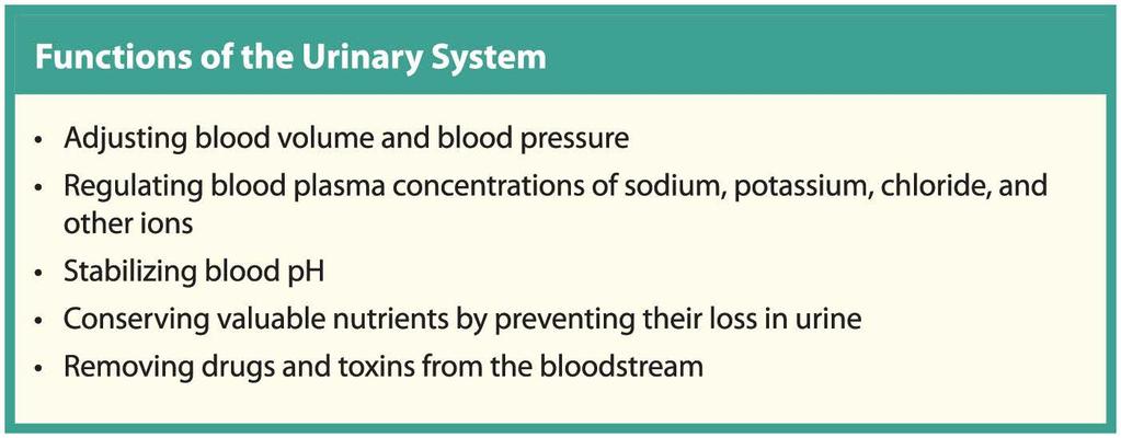
 26 The Urinary System Introduction The urinary system does more than just get rid of liquid waste. It also: Regulates plasma ion concentrations Regulates blood volume and blood pressure Stabilizes blood
Chapter 25: Urinary System
 Chapter 25: Urinary System I. Kidney anatomy: retroperitoneal from 12 th thoracic to 3 rd lumbar area A. External anatomy: hilus is the indentation 1. Adrenal gland: in the fat at the superior end of each
Kidney Functions Removal of toxins, metabolic wastes, and excess ions from the blood Regulation of blood volume, chemical composition, and ph
 The Urinary System Urinary System Organs Kidneys are major excretory organs Urinary bladder is the temporary storage reservoir for urine Ureters transport urine from the kidneys to the bladder Urethra
CHAPTER 25 URINARY. Urinary system. Kidneys 2 Ureters 2 Urinary Bladder 1 Urethra 1. functions
 CHAPTER 25 URINARY Kidneys 2 Ureters 2 Urinary Bladder 1 Urethra 1 fluid waste elimination secretion of wastes control blood volume and BP control blood ph electrolyte levels RBC levels hormone production
Human Urogenital System 26-1
 Human Urogenital System 26-1 Urogenital System Functions Filtering of blood, Removal of wastes and metabolites Regulation of blood volume and composition concentration of blood solutes ph of extracellular
General Anatomy of Urinary System
 General Anatomy of Urinary System URINARY SYSTEM ORGANS Kidneys (2) Ureters (2) Urinary bladder Urethra KIDNEY FUNCTIONS Control blood volume and composition KIDNEY FUNCTIONS Filter blood plasma, eliminate
Urinary System. Chapter 17 7/19/11. Introduction
 7/19/11 Chapter 17 Urinary System Introduction A. The urinary system consists of two kidneys that filter the blood, two ureters, a urinary bladder, and a urethra to convey waste substances to the outside.
Lab Activity 31. Anatomy of the Urinary System. Portland Community College BI 233
 Lab Activity 31 Anatomy of the Urinary System Portland Community College BI 233 Urinary System Organs Kidneys Urinary bladder: provides a temporary storage reservoir for urine Paired ureters: transport
Chapter 26. The Urinary System. Lecture Presentation by Steven Bassett Southeast Community College Pearson Education, Inc.
 Chapter 26 The Urinary System Lecture Presentation by Steven Bassett Southeast Community College Introduction The urinary system does more than just get rid of liquid waste. It also: Regulates plasma ion
Chapter 26: Urinary System By: Eddie Tribiana and Piers Frieden
 Chapter 26: Urinary System By: Eddie Tribiana and Piers Frieden The urinary system is important because it performs vital excretory functions Takes blood from renal arteries into the kidney to filtrate
Urinary System. consists of the kidneys, ureters, urinary bladder and urethra
 Urinary System 1 Urinary System consists of the kidneys, ureters, urinary bladder and urethra 2 Location of Kidneys The kidneys which are positioned retroperitoneally lie on either side of the vertebral
1. The Fibrous Capsule covers the outside of the kidney. It is made of fat and fibers.
 Slide 2 The kidney has a number of functions. First is the excretion of toxic metabolic waste through urine production. The kidneys filter blood plasma and as a result of filtering blood, the kidneys help
2) This is a Point and Click question. You must click on the required structure.
 This is a Point and Click question. You must click on the required structure.") Class: A&P2-1 Description: Test: Excretory Test Points: 144 Test Number: 28379 Printed: 31-March-10 12:03 1) This is a Point and Click question. You must click on the required structure. Click on the Bowman's
Class: A&P2-1 Description: Test: Excretory Test Points: 144 Test Number: 28379 Printed: 31-March-10 12:03 1) This is a Point and Click question. You must click on the required structure. Click on the Bowman's
Urinary System BIO 250. Waste Products of Metabolism Urea Carbon dioxide Inorganic salts Water Heat. Routes of Waste Elimination
 Urinary System BIO 250 Waste Products of Metabolism Urea Carbon dioxide Inorganic salts Water Heat Routes of Waste Elimination Skin: Variable amounts of heat, salts, and water; small amounts of urea and
BIOL2030 Human A & P II -- Exam 6
 BIOL2030 Human A & P II -- Exam 6 Name: 1. The kidney functions in A. preventing blood loss. C. synthesis of vitamin E. E. making ADH. B. white blood cell production. D. excretion of metabolic wastes.
Chapter 17: Urinary System
 Introduction Chapter 17: Urinary System Organs of the Urinary System REFERENCE FIGURE 17.1 2 kidneys filters the blood 2 ureters transport urine from the kidneys to the urinary bladder Urinary bladder
28/04/2013 LEARNING OUTCOME C13 URINARY SYSTEM STUDENT ACHIEVEMENT INDICATORS STUDENT ACHIEVEMENT INDICATORS URINARY SYSTEM & EXCRETION
 LEARNING OUTCOME C13 Analyse the functional interrelationships of the structures of the urinary system Learning Outcome C13 URINARY SYSTEM STUDENT ACHIEVEMENT INDICATORS Students who have fully met this
URINARY SYSTEM CHAPTER 28 I ANATOMY OF THE URINARY SYSTEM. Student Name
 Student Name CHAPTER 28 URINARY SYSTEM L iving produces wastes. Wherever people live or work or play, wastes accumulate. To keep these areas healthy, there must be a method of disposing of these wastes
Chapter 23. The Nephron. (functional unit of the kidney
 Chapter 23 The Nephron (functional unit of the kidney Renal capsule The Nephron Renal cortex Nephron Collecting duct Efferent arteriole Afferent arteriole (a) Renal corpuscle: Glomerular capsule Glomerulus
Chapter 16 Lecture Outline
 Chapter 16 Lecture Outline See separate PowerPoint slides for all figures and tables preinserted into PowerPoint without notes. Copyright The McGraw-Hill Companies, Inc. Permission required for reproduction
URINARY SYSTEM ANATOMY
 URINARY SYSTEM ANATOMY Adapted from Human Anatomy & Physiology Marieb and Hoehn (9 th ed.) OVERVIEW Metabolism of nutrients by the body produces wastes that must be removed from the body. Although excretory
Human Anatomy Unit 3 URINARY SYSTEM
 Human Anatomy Unit 3 URINARY SYSTEM In Anatomy Today Components Kidneys Ureters Urinary bladder Urethra Functions Storage of urine Bladder stores up to 1 L of urine Excretion of urine Transport of urine
Collin College. BIOL Anatomy & Physiology WEEK 12. Urinary System INTRODUCTION. Main functions of the kidneys are
 Collin College BIOL. 2402 Anatomy & Physiology WEEK 12 Urinary System 1 INTRODUCTION Main functions of the kidneys are regulate blood volume, water content regulate blood composition e..g. Na, Cl, K, ph
Urinary system. Urinary system
 INTRODUCTION. Several organs system Produce urine and excrete it from the body Maintenance of homeostasis. Components. two kidneys, produce urine; two ureters, carry urine to single urinary bladder for
Urinary Anatomy. Lab 40. Kidneys. Nephrons. Renal Corpuscle
 Urinary Anatomy Lab 40. Urinary Anatomy and Kidney Dissection Kidneys: filters blood, produces urine Ureters: convey urine to bladder Bladder: holding tank Urethra: carries urine to the outside for elimination
A. Incorrect! The urinary system is involved in the regulation of blood ph. B. Correct! The urinary system is involved in the synthesis of vitamin D.
 Human Anatomy - Problem Drill 22: The Urinary System Question No. 1 of 10 1. Which of the following statements about the functions of the urinary system is not correct? Question #01 (A) The urinary system
SHORT ANSWER. Write the word or phrase that best completes each statement or answers the question.
 Exam Name SHORT ANSWER. Write the word or phrase that best completes each statement or answers the question. Figure 25.1 Using Figure 25.1, match the following: 1) Glomerulus. 2) Afferent arteriole. 3)
Histology Urinary system
 Histology Urinary system Urinary system Composed of two kidneys, two ureters, the urinary bladder, and the urethra, the urinary system plays a critical role in: 1- Blood filtration,(filtration of cellular
Waste. Urinary System Anatomy Urinary Section pages 5-8. Urinary System. Urinary System. Nitrogenous Wastes. Nitrogenous Wastes 4/22/2016
 Waste Urinary System Anatomy Urinary Section pages 5-8 Metabolism produces waste products What is the primary waste product of cellular respiration? How does the body dispose of it? Urinary System Urinary
Copyright 2003 Pearson Education, Inc. publishing as Benjamin Cummings. Dr. Nabil Khouri
 Dr. Nabil Khouri Objectives: General objectives: - to identify the kidney s structures, function and location - to analyze the relationship between microscopic structure and function Specific objectives:
1. Urinary System, General
 S T U D Y G U I D E 16 1. Urinary System, General a. Label the figure by placing the numbers of the structures in the spaces by the correct labels. 7 Aorta 6 Kidney 8 Ureter 2 Inferior vena cava 4 Renal
The Urinary System. Medical Assisting Third Edition. Booth, Whicker, Wyman, Pugh, Thompson The McGraw-Hill Companies, Inc. All rights reserved
 The Urinary System PowerPoint presentation to accompany: Medical Assisting Third Edition Booth, Whicker, Wyman, Pugh, Thompson 30-2 Learning Outcomes 30.1 Describe the structure, location, and functions
URINARY SYSTEM. These organs lie posterior or inferior to the. (membrane).
.") URINARY SYSTEM I. INTRODUCTION Each kidney is made up of about a million tiny tubules called nephrons. Each nephron individually filters the blood and makes urine and it does the job completely, from start
URINARY SYSTEM I. INTRODUCTION Each kidney is made up of about a million tiny tubules called nephrons. Each nephron individually filters the blood and makes urine and it does the job completely, from start
I. Anatomy of the Urinary System A. Kidneys 1. Right lower than Left* 2. Retroperitoneal 3. Layers that secure kidneys in the abdominal cavity a.
 I. Anatomy of the Urinary System A. Kidneys 1. Right lower than Left* 2. Retroperitoneal 3. Layers that secure kidneys in the abdominal cavity a. Renal fascia b. Perinephric fat (Adipose) capsule c. Fibrous
Lesson 14.1: Learning the Key Terms
 209 Lesson 14.1: Learning the Key Terms Directions: Place the letter of the best definition next to each key term. 1. collecting duct 2. distal convoluted tubule 3. glomerulus 4. nephron 5. nephron loop
Urinary System URINARY SYSTEM
 URINARY SYSTEM The urinary system consists of two kidneys, two ureters, the urinary bladder, and the urethra. The formation of urine is the function of the kidneys, and the rest of the system is responsible
BIOH122 Human Biological Science 2
 BIOH122 Human Biological Science 2 Session 16 Urinary System 1 The Kidneys Bioscience Department Endeavour College of Natural Health endeavour.edu.au Session Plan o Functions of Urinary system o The Kidneys:
Histology / First stage The Urinary System: Introduction. Kidneys
 The Urinary System: Introduction The urinary system consists of the paired kidneys and ureters, the bladder, and the urethra. This system helps maintain homeostasis by a complex combination of processes
Nephrology - the study of the kidney. Urology - branch of medicine dealing with the male and female urinary systems and the male reproductive system
 Urinary System Nephrology - the study of the kidney Urology - branch of medicine dealing with the male and female urinary systems and the male reproductive system Functions of the Urinary System 1. Regulation
Urinary System Laboratory
 Urinary System Laboratory 1 Adrenal gland Organs of The Urinary System Renal artery and vein Kidney Ureter Urinary bladder Figure 26.1 2 Urethra Functions of the urinary system organs: Urethra expels urine
URINARY SYSTEM. Urinary System
 URINARY SYSTEM Urinary System Kidney Functions Excretion Regulation of blood volume and pressure Regulation of electrolyte and ph levels Kidney Structure Gross Anatomy Fibrous Capsule Renal Cortex Renal
Outline Urinary System. Urinary System and Excretion. Urine. Urinary System. I. Function II. Organs of the urinary system
 Outline Urinary System Urinary System and Excretion Bio105 Chapter 16 Renal will be on the Final only. I. Function II. Organs of the urinary system A. Kidneys 1. Function 2. Structure III. Disorders of
AP2, Lab 7 - THE URINARY SYSTEM
 AP2, Lab 7 - THE URINARY SYSTEM I. SYSTEM COMPONENTS (Figs. 25.1 25.4) KIDNEYS Each kidney contains approx. 1,000,000 tubular NEPHRONS which produce FILTRATE from the plasma and then add to or take from
Chapter 24: The Urinary System
 Chapter 24: The Urinary System Overview of kidney functions n Regulation of blood ionic composition n Regulation of blood ph n Regulation of blood volume n Regulation of blood pressure n Maintenance of
organs of the urinary system
 organs of the urinary system Kidneys (2) bean-shaped, fist-sized organ where urine is formed. Lie on either sides of the vertebral column, in a depression beneath peritoneum and protected by lower ribs
Urinary System. BSC 2086 A & P 2 Professor Tcherina Duncombe Palm Beach State College
 Urinary System BSC 2086 A & P 2 Professor Tcherina Duncombe Palm Beach State College Filter plasma, separate and eliminate wastes Functions Regulate blood volume and pressure Regulate osmolarity of body
Urinary bladder provides a temporary storage reservoir for urine
 Urinary System Organs Kidney Filters blood, allowing toxins, metabolic wastes, and excess ions to leave the body in urine Urinary bladder provides a temporary storage reservoir for urine Paired ureters
Chapter 11 Lecture Outline
 Chapter 11 Lecture Outline See separate PowerPoint slides for all figures and tables preinserted into PowerPoint without notes. Copyright 2016 McGraw-Hill Education. Permission required for reproduction
The Urinary System. Copyright 2003 Pearson Education, Inc. publishing as Benjamin Cummings
 The Urinary System Functions of the Urinary System Elimination of waste products Nitrogenous wastes Toxins Drugs Functions of the Urinary System Regulate aspects of homeostasis Water balance Electrolytes
Functions of the Urinary System
 The Urinary System Functions of the Urinary System Elimination of waste products Nitrogenous wastes Toxins Drugs Regulate aspects of homeostasis Water balance Electrolytes Acid-base balance in the blood
BCH 450 Biochemistry of Specialized Tissues
 BCH 450 Biochemistry of Specialized Tissues VII. Renal Structure, Function & Regulation Kidney Function 1. Regulate Extracellular fluid (ECF) (plasma and interstitial fluid) through formation of urine.
The Urinary System 15PART A. PowerPoint Lecture Slide Presentation by Patty Bostwick-Taylor, Florence-Darlington Technical College
 PowerPoint Lecture Slide Presentation by Patty Bostwick-Taylor, Florence-Darlington Technical College The Urinary System 15PART A Functions of the Urinary System Elimination of waste products Nitrogenous
Urinary System and Fluid Balance. Urine Production
 Urinary System and Fluid Balance Name Pd Date Urine Production The three processes critical to the formation of urine are filtration, reabsorption, and secretion. Match these terms with the correct statement
Vertebrates possess kidneys: internal organs which are vital to ion and water balance and excretion.
 The Kidney Vertebrates possess kidneys: internal organs which are vital to ion and water balance and excretion. The kidney has 6 roles in the maintenance of homeostasis. 6 Main Functions 1. Ion Balance
A&P 2 CANALE T H E U R I N A R Y S Y S T E M
 A&P 2 CANALE T H E U R I N A R Y S Y S T E M URINARY SYSTEM CONTRIBUTION TO HOMEOSTASIS Regulates body water levels Excess water taken in is excreted Output varies from 2-1/2 liter/day to 1 liter/hour
The functions of the kidney:
 The functions of the kidney: After reading this lecture you should be able to.. 1. List the main functions of the kidney. 2. Know the basic physiological anatomy of the kidney and the nephron 3. Describe
Urinary System and Excretion. Bio105 Lecture 20 Chapter 16
 Urinary System and Excretion Bio105 Lecture 20 Chapter 16 1 Outline Urinary System I. Function II. Organs of the urinary system A. Kidneys 1. Function 2. Structure III. Disorders of the urinary system
19. RENAL PHYSIOLOGY ROLE OF THE URINARY SYSTEM THE URINARY SYSTEM. Components and function. V BS 122 Physiology II 151 Class of 2011
 19. RENAL PHYSIOLOGY THE URINARY SYSTEM Components and function The urinary system is composed of two kidneys, the functionally filtering apparatus, which connect through two tubular structures called
Other Factors Affecting GFR. Chapter 25. After Filtration. Reabsorption and Secretion. 5 Functions of the PCT
 Other Factors Affecting GFR Chapter 25 Part 2. Renal Physiology Nitric oxide vasodilator produced by the vascular endothelium Adenosine vasoconstrictor of renal vasculature Endothelin a powerful vasoconstrictor
H I S T O L O G Y O F T H E U R I N A R Y S Y S T E M
 SCPA 602- Anatomical Basis For Pathological Study H I S T O L O G Y O F T H E U R I N A R Y S Y S T E M S O M P H O N G N A R K P I N I T, M. D. D E P A R T M E N T O F P A T H O B I O L O G Y F A C U
RNPDC CCNP Anatomy and Physiology: Renal System Pre-Quiz 2015
 RNPDC CCNP Anatomy and Physiology: Renal System Pre-Quiz 2015 1. In which abdominal cavity do the kidneys lie? a) Peritoneum. b) Anteperitoneal. c) Retroperitoneal. d) Parietal peritoneal 2. What is the
First is Urine Production. We ll discuss the specifics of this process momentarily.
 1 2 The kidney has a number of functions. First is Urine Production. We ll discuss the specifics of this process momentarily. Next, the kidneys filter blood. As a result of filtering blood, the kidneys
Unit 15: The Urinary System
 Unit 15: The Urinary System I. Functions of the Urinary System A. Elimination of waste products 1. Nitrogenous wastes 2. Toxins 3. Drugs B. Regulate aspects of homeostasis 1. Water balance 2. Electrolytes
Unit 15 - The Urinary System 1
 Unit 15 - The Urinary System 1 I. Unit 15: The Urinary System A. Functions of the Urinary System 1. Elimination of waste products a) Nitrogenous wastes b) Toxins c) Drugs 2. Regulate aspects of homeostasis
Urinary System Review Questions:
 Urinary System Review Questions: 1. This system would be lined with what type of membrane? 2. What type of epithelial tissue would line the opening of the urethra (the exit of the tract)? 3. What type
Urinary System kidneys, ureters, bladder & urethra
 Urinary System kidneys, ureters, bladder & urethra Kidney Function Filters blood removes waste products conserves salts, glucose, proteins, nutrients and water Produces urine Endocrine functions regulates
URINARY SYSTEM ANATOMY PART
 URINARY SYSTEM ANATOMY PART 1 DANIL HAMMOUDI.MD Urinary System Composed of kidneys, ureters, urinary bladder, and urethra Eliminates nitrogenous wastes from the body Regulates water, electrolyte, and ph
Human Anatomy and Physiology - Problem Drill 23: The Urinary System, Fluid, Electrolyte and Acid-Base Balance
 Human Anatomy and Physiology - Problem Drill 23: The Urinary System, Fluid, Electrolyte and Acid-Base Balance Question No. 1 of 10 Which of the following statements about the functions of the urinary system
Urinary System kidneys, ureters, bladder & urethra
 Urinary System kidneys, ureters, bladder & urethra Filters blood removes waste products conserves salts, glucose, proteins, nutrients and water Produces urine Kidney Function Endocrine functions regulates
HISTOLOGY OF THE URINARY SYSTEM
 HISTOLOGY OF THE URINARY SYSTEM The Urinary System Kidneys, ureters, urinary bladder & urethra Urine flows from each kidney, down its ureter to the bladder and to the outside via the urethra Filter the
Renal System and Excretion
 Renal System and Excretion Biology 105 Lecture 19 Chapter 16 Outline Renal System I. Functions II. Organs of the renal system III. Kidneys 1. Structure 2. Function IV. Nephron 1. Structure 2. Function
Urinary System. Analyze the Anatomy and Physiology of the urinary system
 Urinary System Analyze the Anatomy and Physiology of the urinary system Kidney Bean-shaped Located between peritoneum and the back muscles (retroperitoneal) Renal pelvis funnelshaped structure at the beginning
active transport of! Na. C. Tubular Reabsorption of Nutrients, Water, and Ions (p. 979; Fig )
") The Urinary System Outline 25.1 The kidneys have three distinct regions and a rich blood supply (pp. 963 965; Figs. 25.1 25.5) A. Location and External Anatomy (p. 963; Figs. 25.1 25.3) 1. The kidneys
The Urinary System Outline 25.1 The kidneys have three distinct regions and a rich blood supply (pp. 963 965; Figs. 25.1 25.5) A. Location and External Anatomy (p. 963; Figs. 25.1 25.3) 1. The kidneys
Urinary System (Chapter 26) Lecture Materials for Amy Warenda Czura, Ph.D. Suffolk County Community College Eastern Campus
 Lecture Materials for Amy Warenda Czura, Ph.D. Suffolk County Community College Eastern Campus") Urinary System (Chapter 26) Lecture Materials for Amy Warenda Czura, Ph.D. Suffolk County Community College Eastern Campus Primary Sources for figures and content: Marieb, E. N. Human Anatomy & Physiology
Urinary System (Chapter 26) Lecture Materials for Amy Warenda Czura, Ph.D. Suffolk County Community College Eastern Campus Primary Sources for figures and content: Marieb, E. N. Human Anatomy & Physiology
Chapter 26 The Urinary System
 Chapter 26 The Urinary System Kidneys, ureters, urinary bladder & urethra Urine flows from each kidney, down its ureter to the bladder and to the outside via the urethra Filter the blood and return most
Urinary Physiology. Chapter 17 Outline. Kidney Function. Chapter 17
 Urinary Physiology Chapter 17 Chapter 17 Outline Structure and Function of the Kidney Glomerular Filtration Reabsorption of Salt and Water Renal Plasma Clearance Renal Control of Electrolyte and Acid-Base
NOTES: CH 44 Regulating the Internal Environment (Homeostasis & The Urinary System)
") NOTES: CH 44 Regulating the Internal Environment (Homeostasis & The Urinary System) HOMEOSTASIS **Recall HOMEOSTASIS is the steady-state physiological condition of the body. It includes: 1) Thermoregulation:
NOTES: CH 44 Regulating the Internal Environment (Homeostasis & The Urinary System) HOMEOSTASIS **Recall HOMEOSTASIS is the steady-state physiological condition of the body. It includes: 1) Thermoregulation:
Basic Urinary Tract Anatomy and Histology
 Basic Urinary Tract Anatomy and Histology The two kidneys are located in the retroperitoneum on either side of the vertebral bladder and the contraction of the detrusor muscle. Any mechanical barrier,
Bladder Schistosomes. Normally, urine is sterile. Presence of blood may indicate an infection.
 Bladder Schistosomes Normally, urine is sterile. Presence of blood may indicate an infection. 17.1 Introduction -Cells produce waste that can become toxic if they accumulate Functions the urinary system
Copyright 2010 Pearson Education, Inc. Urinary System
 Urinary System What are the organs that comprise the urinary system? Urinary System Organs Kidneys Urinary bladder Ureters Urethra Hepatic veins (cut) Esophagus (cut) Inferior vena cava Adrenal gland Aorta
Bio 322 Human Anatomy Objectives for the laboratory exercise Urinary System Filtration Reabsorption Secretion Concentration
 Bio 322 Human Anatomy Objectives for the laboratory exercise Urinary System Required reading before beginning this lab: Saladin, KS: Human Anatomy 5 th ed (2017) Chapter 25 For this lab you will use parts
Bio 230 Lecture Notes: THE URINARY SYSTEM
 Bio 230 Lecture Notes: THE URINARY SYSTEM NOTE: You must follow along in your text book or the powerpoint supplied while reading through these lecture notes. A picture is worth a thousand words. Urinary
Urinary System Multiple Choice Practice Test. c. Kidneys have three protective layers d. The adrenal gland is located deep within the kidney
 Urinary System Multiple Choice Practice Test 1. Which of the following is a function of the urinary system? a. Regulates water b. Regulates balance of acids, bases, and electrolytes c. Filters waste from
Outline Urinary System
 Urinary System and Excretion Bio105 Lecture Packet 20 Chapter 16 Outline Urinary System I. Function II. Organs of the urinary system A. Kidneys 1. Function 2. Structure B. Urine formation 1. Hormonal regulation
Unit #4 Waste and Excretion. The Kidneys
 Unit #4 Waste and Excretion The Kidneys Renal Hilus (Hilus) the doorway of the kidney Ureter leaves this region blood and lymphatic vessels enter and exit here Renal Capsule (Capsule) smooth fibrous tissue
Chapter 14 The Urinary System. Anatomy
 Chapter 14 The Urinary System Anatomy 14.1 Overview Role in removal of wastes to maintain homeostasis Acts as filtering system of the blood Produces urine Removes wastes, maintains ph, electrolyte composition,
The Urinary System. Chapter 17
 The Urinary System Chapter 17 Quick Overview of the Urinary System Bladder Schistosomes Normally, urine is sterile. Presence of blood may indicate an infection. 17.1 Introduction -Cells produce waste that
Waste Products & Kidney Function
 Waste Products & Kidney Function urinary system principal means of metabolic waste removal urinary system is closely associated with reproductive system urogenital system share embryonic development share
Copyright 2009 Pearson Education, Inc. Copyright 2009 Pearson Education, Inc. Figure 19-1c. Efferent arteriole. Juxtaglomerular apparatus
 /6/0 About this Chapter Functions of the Kidneys Anatomy of the urinary system Overview of kidney function Secretion Micturition Regulation of extracellular fluid volume and blood pressure Regulation of
April 08, biology 2201 ch 11.3 excretion.notebook. Biology The Excretory System. Apr 13 9:14 PM EXCRETORY SYSTEM.
 Biology 2201 11.3 The Excretory System EXCRETORY SYSTEM 1 Excretory System How does the excretory system maintain homeostasis? It regulates heat, water, salt, acid base concentrations and metabolite concentrations
Urinary System Functions of the Urinary system (renal) A. B. C.
 A. B. C.") Urinary System Functions of the Urinary system (renal) A. B. C. I. Overview of fluids A. Normal volumes 1. fluid intake a. = 1500 ml b. = 750 ml c. = 250 ml (remember ETS) d. total = 2500 ml/day 2. fluid
Urinary System Functions of the Urinary system (renal) A. B. C. I. Overview of fluids A. Normal volumes 1. fluid intake a. = 1500 ml b. = 750 ml c. = 250 ml (remember ETS) d. total = 2500 ml/day 2. fluid
PARTS OF THE URINARY SYSTEM
 EXCRETORY SYSTEM Excretory System How does the excretory system maintain homeostasis? It regulates heat, water, salt, acid-base concentrations and metabolite concentrations 1 ORGANS OF EXCRETION Skin and
5.Which part of the nephron removes water, ions and nutrients from the blood?
 Uro question 1.While reading a blood test I notice a high level of creatinine, I could assume from this that A) There is a possibility of a UTI B) There is a possibility of diabetes C) There is a possibility
RENAL PHYSIOLOGY. Danil Hammoudi.MD
 RENAL PHYSIOLOGY Danil Hammoudi.MD Functions Regulating blood ionic composition Regulating blood ph Regulating blood volume Regulating blood pressure Produce calcitrol and erythropoietin Regulating blood
Urinary System. Dr. Ahmed Maher Dr. Ahmed Manhal
 Urinary System Dr. Ahmed Maher Dr. Ahmed Manhal Presentation Map Kidney (cortex & medulla). Nephron. Duct system. Juxtaglomerular apparatus. Ureter, bladder & urethra. Definition & General Structure The
Excretory Lecture Test Questions Set 1
 Excretory Lecture Test Questions Set 1 1. The separation and ejection of metabolic wastes, usually in aqueous solution, is: a. reabsorption b. secretion c. filtration d. excretion e. endocrinology 2. Besides