RENAL SCINTIGRAPHY IN THE 21 st CENTURY
|
|
|
- Arthur Evans
- 5 years ago
- Views:
Transcription

 : A Fast and Easy")

1 RENAL SCINTIGRAPHY IN THE 21 st CENTURY 99m Tc- MAG 3 with zero time injection of Furosemide (MAG 3 -F 0 ) : A Fast and Easy Protocol, One for All Indications Clinical Experience Congenital Disorders
2 PROTOCOL FOR MAG 3 - F 0 PATIENT PREPARATION Easy (only restriction, oral hydration, no bladder cath.) DYNAMIC STUDY (iv 1-10 mci MAG mg LASIX) Simultaneous injection of Furosemide: MAG 3 -F 0 Duration of the study 25 min TOMOGRAPHY-SPECT (20 mci MAG 3 ) No diuretic needed Duration of the study 4 min
 Method applied the")

3 RENAL SCINTIGRAPHY AT UM/JMMC: (MAG3-F0) Method applied the last 18 years: FOR NATIVE KIDNEY STUDIES FOR RENAL TRANSPLANT STUDIES
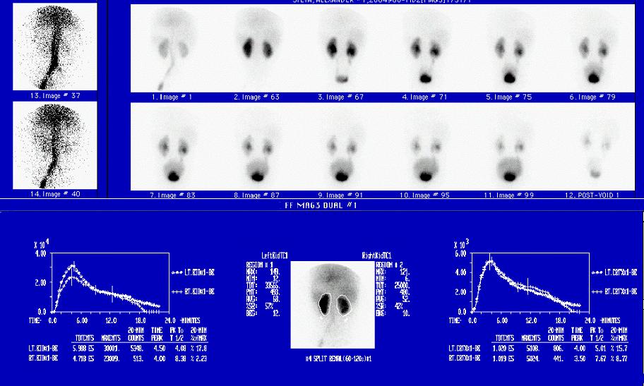
4 RENAL SCINTIGRAPHY AT UM/JMMC: (MAG3-F0) Method applied the last 18 years: A. DYNAMIC STUDY Injection iv 1-10 mci MAG mg LASIX (Furosemide) Simultaneous Injection of MAG3 and Lasix = F 0 ACQUISITION: FLOW: FUNCTION: POST VOID DELAYED 1 min ( 1 frame per 1 sec) 22 min ( 1 frame per 30 sec) 2 min static image (at min) 2 min static images (at 1 hr) GROUPING IMAGES : GRAPH GENERATION: -FLOW: in 3 sec images -FUNCTION: in 2 min images -FLOW/FUNCTION, KIDNEY/CORTEX
5 RENAL SCINTIGRAPHY AT UM/JMMC: (MAG3-F0) Method applied the last 18 years: This protocol was originally applied in the evaluation of drainage Soon it was realized that it allowed the evaluation of the parenchyma Then it was applied in all parenchymal indications (including APN) It was also utilized for the study of Renovascular Hypertension In children allowed the study of HIV and other Nephropathies In patients with renal colic unraveled the Stunned (decompressed) kidney It was finally successful in the study of complications of renal transplants
6 MAG 3 -F 0 PROTOCOL Misconception You cannot image the kidneys of a newborn You need to catheterize the urinary bladder to exclude obstruction Facts MAG 3 -F 0 works in the Newborn Infant You do not need to catheterize the urinary bladder

7 Typical NORMAL MAG 3 -F 0 in a NEWBORN Indication: Evaluate Pelviectasis found by Ultrasound Normal study; Slight Immaturity, Bladder empties
8 Typical NORMAL MAG 3 -F 0 in a NEWBORN Indication: Evaluate Pelviectasis found by Ultrasound Normal study; Slight Immaturity, Bladder does not empty
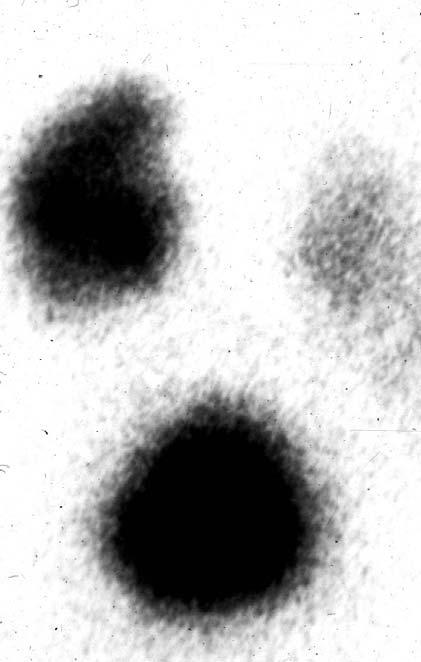
9 MAG 3 -F 0 in a 10 yo CHILD; Mature Normal Kidneys Indication: Evaluate effects of urinary infection Slight discrepancy in size and function (effect of infection on the right kidney)
10 Normal MAG 3 -F 0 in an ADULT Indication: Renal Colic
11 INDICATIONS FOR MAG 3 -F 0 STUDIES: DIAGNOSIS - PROGNOSIS - FOLLOW UP PARENCHYMAL OR DRAINAGE DISORDERS IN CONGENITAL OR ACQUIRED DISEASES FOR NATIVE OR TRANSPLANTED KIDNEYS AT ALL AGES AND FUNCTIONAL STATES
12 CONGENITAL URINARY TRACT ANOMALIES MAG 3 -F 0 Dynamic Studies Diagnosis-Prognosis-Follow up
13 MOST COMMON INDICATIONS FOR RENAL SCINTIGRAPHY NEONATE CONGENITAL RENAL INSUFFICIENCY/FAILURE PERINATAL COMPLICATIONS WORK UP OF SONOGRAPHIC FINDINGS MASSES IN THE ABDOMEN SEARCH FOR AND EVALUATION OF CONGENITAL UT ANOMALIES Diagnosis-Prognosis-Follow up
14 MAG 3 -F 0 in Congenital Renal Insufficiency or Failure Posterior Urethral Valves Bilateral Agenesis Bilateral Dysplasia
 may Prevent Loss of Function and may Promote Recovery of")
15 CONGENITAL RENAL INSUFFICIENCY/FAILURE Bilateral Obstruction Early Correction of UPJO (the first week of life) may Prevent Loss of Function and may Promote Recovery of Function
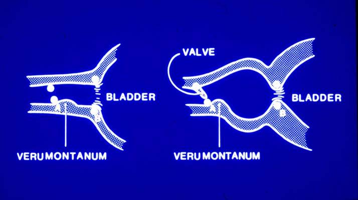
16 Congenital renal insufficiency/failure Posterior Urethral Valves in the Newborn Bilateral Obstruction
17 Congenital renal insufficiency/failure Bilateral Agenesis 2 min 20 min


18 Congenital renal insufficiency/failure Bilateral Dysplasia in the newborn 2 min


19 CONGENITAL RENAL INSUFFICIENCY/FAILURE Bilateral Dysplasias Bilateral Dysplasias or Agenesis No functioning renal parenchyma No Intervention indicated, no recovery expected
20 MOST COMMON INDICATIONS FOR RENAL SCINTIGRAPHY NEONATE CONGENITAL RENAL INSUFFICIENCY/FAILURE PERINATAL COMPLICATIONS WORK UP OF SONOGRAPHIC FINDINGS MASSES IN THE ABDOMEN SEARCH FOR AND EVALUATION OF CONGENITAL UT ANOMALIES Diagnosis-Prognosis-Follow up
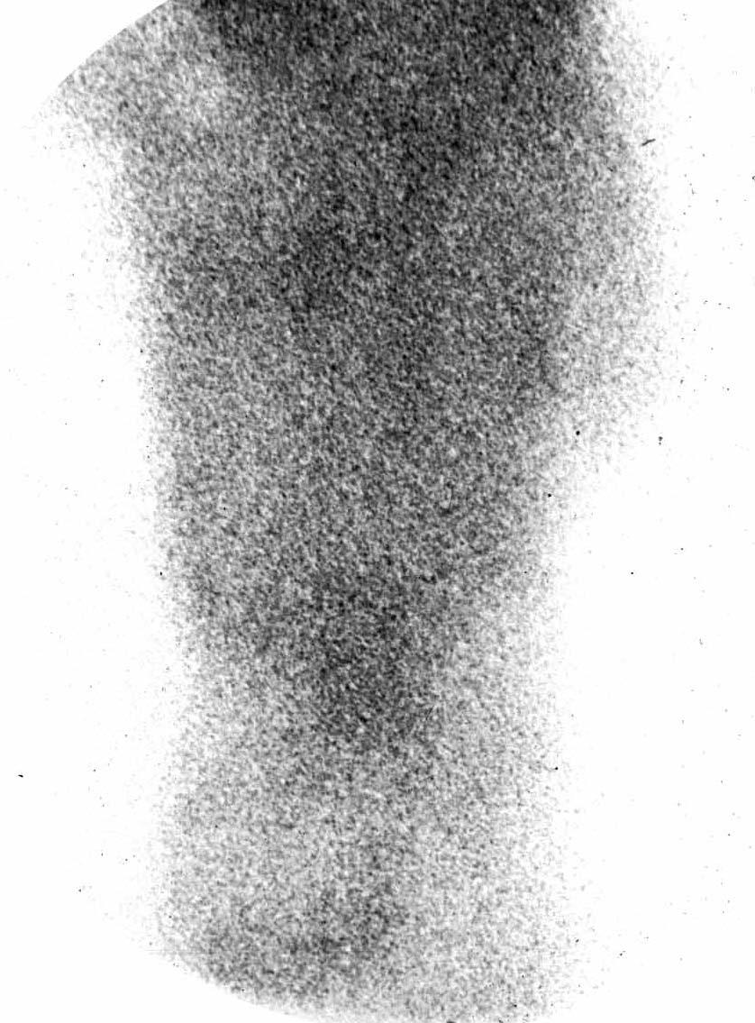
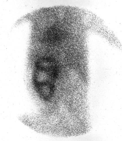
21 MAG 3 -F 0 in Perinatal Renal Disorders Acute Tubular Necrosis Renovascular Hypertension Adrenal Hemorrhage
22 Perinatal Renal Insufficiency/Failure: Newborn with Severe Oliguria Diabetic Mother and Difficult, Prolonged Delivery Normal size Solid kidneys, Preservation of Flow and Cortical Uptake; Delayed Drainage; High Residual Cortical Activity: Acute Tubular Necrosis: No intervention; Full Recovery
23 Neonates with hypertension from renal ischemia due to thrombus in the umbilical catheter should not be treated with ACE-Inhibitors

24 Neonatal Hypertension from Aortic Thrombus Around Aortic Catheter Left Infarction Right Ischemia


25 MAG 3 -F 0 in Neonatal Hypertension from renal ischemia due to thrombus around the umbilical catheter Baseline Renogram ACE Inhibition Renogram This infant should not be treated with ACE-Inhibitors

26 Perinatal Adrenal Hemorrhage
27 MOST COMMON INDICATIONS FOR RENAL SCINTIGRAPHY NEONATE INFANT OR OLDER CHILD CONGENITAL RENAL INSUFFICIENCY/FAILURE PERINATAL COMPLICATIONS WORK UP OF SONOGRAPHIC FINDINGS MASSES IN THE ABDOMEN SEARCH FOR AND EVALUATION OF CONGENITAL UT ANOMALIES Diagnosis-Prognosis-Follow up
28 MAG 3 -F 0 in Congenital Non-Obstructing Renal Diseases Unilateral Agenesis Hypoplasia Ectopia Horse-shoe kidney Multicystic Kidney Disease Polycystic Kidney Disease Megaureter
29 Agenesis

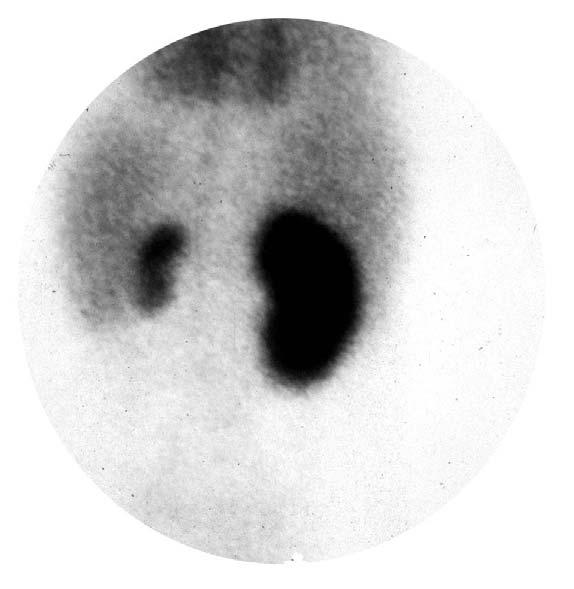

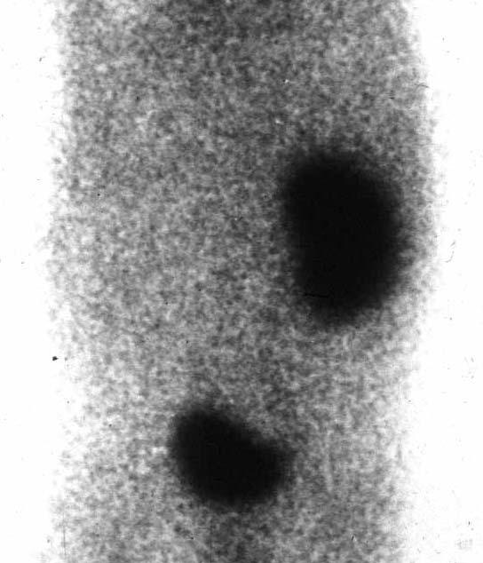
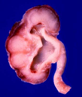
30 Hypoplasia with contralateral Compensatory Hypertrophy 2 min kc 20 min

31 Ectopic normal kidney (pelvic) Ectopic esp. normal kidneys are missed about 50% by routine Ultrasonography but they can very easily be identified by MAG 3 -F 0, within 2 min after injection
32 Horseshoe Kidney Non-obstructed
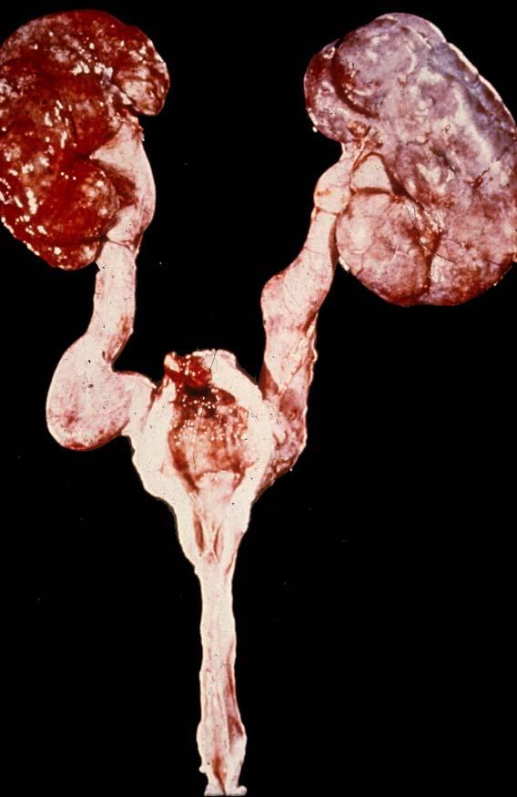
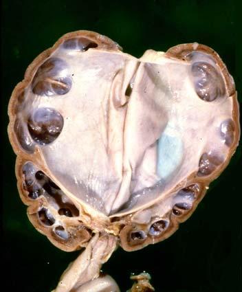
33 Multicystic Dysplastic Kidney

34 Multicystic Dysplastic Kidney
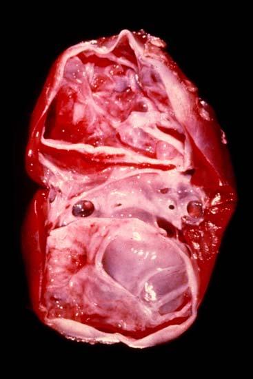
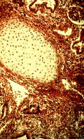
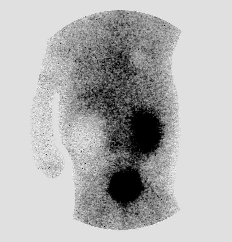
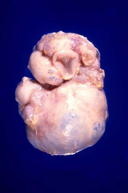
35 Multicystic Dysplastic Kidney
36 Polycystic Kidney Disease Autosomal Recessive min Mild infantile type


37 Polycystic Kidney Disease Children: Autosomal Recessive Other congenital diseases (Scan: Large kidneys with DPD) Adults: Autosomal Dominant (Scan: Large kidneys with evidence of multiple cysts)


38 Polycystic Kidney Disease Autosomal Dominant (usually in adults) Cysts Normal drainage
39 MEGAURETER without OBSTRUCTION Use of Bladder Catheterization

40 Idiopathic Megaureter NEWBORN FOLLOW UP 2 min 2 min 20 min Delayed NEWBORN FOLLOW UP
41 NEUROGENIC BLADDER
42 MAG 3 -F 0 in Congenital Renal Obstruction Anatomic Uretero-Pelvic Junction Obstruction Uretero-Vesical Junction Obstruction Posterior Urethral Valves Functional Vesico-Ureteral Reflux
43 OBSTRUCTION Can we prevent the loss? Neonate 5 year old

44 Proximal Obstruction ( Uretero-Pelvic Junction ) 2min min 18 20


45 Distal Obstruction ( Uretero-Vesical Junction ) 2min kc min
46 Pelvic Ectopic Obstructed Kidney (UPJO) 2 min 4 min 6 min Contrast enhanced images
47 Newborn with abnormal prenatal ultrasound Findings: Early Defect and Late Retention Left Upper Pole Defect Urinary Bladder Diagnosis: Duplication with Upper Moiety Obstruction, Ectopic Ureter, and Ureterocele

48 Findings: Early Defect and Late Retention Left Upper Pole Defect Urinary Bladder Diagnosis: Duplication with Upper Moiety Obstruction, Ectopic Ureter, and Ureterocele One and ½ month old with abnormal ultrasound referred from outside for evaluation

49 Duplication with Ectopic Ureter Ureterocele and Upper Moiety Obstruction
50 THE VALUE OF MAG 3 -F 0 DIURETIC RENOGRAPHY IN PREDICTING THE NEED FOR SURGERY IN THE NEONATE WITH URETEROPELVIC JUNCTION OBSTRUCTION Sfakianakis G, Vensel E, Tapia M, Policaro F, Gosalbez R, Labbie A, Zilleruelo G, Abitbol C, Montane B, Strauss J Abstract: SNM 2000


51 Right Pelvic Retention but Normal Drainage of the Cortex and Downsloping Renogram Prognosis: High probability for Spontaneous Compensation; Surgery Not Needed Newborn with pelviectasis evaluated for obstruction SRF 45/55 L/R Follow up study shows Improvement of Drainage and Preservation of Renal Function SRF 50/50 L/R

52 Newborn A down-sloping MAG 3 -F 0 renography in the neonate predicts 6 mo old Spontaneous Compensation


53 High Pelvic Retention; Abnormal Drainage of the Cortex and Upsloping Renogram: UPJO Prognosis: No probability for Spontaneous Compensation; Surgery is Needed Newborn with severe pelvicaliectasis evaluated for obstruction The infant was not operated but was followed up with scintigraphy Follow up study shows Deterioration of Drainage and Loss of Renal Function

54 Newborn An Up-sloping MAG 3 -F 0 renography in the neonate predicts 1 mo old The need of Surgical Correction
55 A Horizontal Renogram requires follow up studies
56 FOLLOW UP STUDIES

57 Follow Up Studies Horse-shoe kidney non obstructed; newborn and 6 year old

58 Follow Up Effect of Therapy At birth Posterior Urethral Valves Newborn Post Therapy S/P Therapy Posterior Urethral Valves
59 Congenital renal insufficiency/failure Posterior Urethral Valves in the Newborn and F/U post therapy
60 Recent Observations When the dilated collecting system keeps the activity but the CORTEX EMPTIES: there is no functional obstruction (reservoir effect) or there is no obstruction at all
61 MAG 3 -F 0 in Renal Obstruction: New Findings Traditional concept: To make the Diagnosis of Obstruction you need to Study the Renogram and the Collecting System of the Kidney New Horizons: To make the Diagnosis of Obstruction you better study the behavior of the Renal Parenchyma: If the Parenchyma empties, there is no obstruction! (even when the drainage system is dilated and it does not empty appropriately)
62 Clinical Experience on The Discrepancy between the Parenchyma (Empties appropriately) and the Drainage System (suggests Obstruction) Different categories of patients with Congenital or Acquired disorders Frequent finding in patients with chronic problems Data were presented at the 2003 SNM meeting Potential problems in patient care if this finding is not recognized (considering surgery etc)
63 Patients with Dilated Collecting System but Physiologic Drainage of the Parenchyma (Cortex) Extra-Renal Pelvis Post-Operatively after Pyeloplasty Congenital Pelviectasis Chronic Nephrolithiasis usually after Colics Characteristics The Parenchyma Empties in Normal Sequence but The Drainage system retains the activity (Pelvi-cali-ectasis) The Function of the kidney (SRF) does not deteriorate
64 EXTRARENAL PELVIS and OBSTRUCTION A dilated extra-renal pelvis may not be obstructed Yet there is retention of urine in the pelvis even after diuretic and an abnormal obstructive kidney renogram When the cortex empties normally there may not be obstruction and a non-obstructive cortical renogram confirms this If the patient is asymptomatic no intervention needed
65 EXTRARENAL PELVIS without OBSTRUCTION Newborn 17 month old

66 EXTRARENAL PELVIS without OBSTRUCTION
67 POST-OPERATIVE DILATED PELVIS and OBSTRUCTION After surgical correction of obstruction a dilated pelvis may persist but there may be no residual obstruction Yet there is retention of urine in the pelvis after diuretic and an abnormal obstructive kidney renogram When the cortex empties normally may not be obstruction and a non-obstructive cortical renogram confirms If the patient is asymptomatic no intervention needed
68 POST-OPERATIVE DILATED PELVIS without OBSTRUCTION A dilated pelvis after Pyeloplasty with an abnormal obstructive kidney renogram may not be obstructed when the cortex empties normally

69 DILATED PELVIS s/p PYELOPLASTY without OBSTRUCTION PRE-OPERATIVE POST-OPERATIVE
70 NON-OBSTRUCTED CORTEX in the presence of dilated, abnormal collecting system The Non-Obstructed Cortex Empties The appearance of the Renogram depends on the Appropriate Placement of the Regions Of Interest (ROI) by the Technologist
71 Obstructed v/s Non-obstructed Parenchyma in the presence of dilated, abnormal collecting system Case No 1

72 Non-obstructed Parenchyma v/s Obstructed in the presence of dilated, abnormal collecting system: non-obstructed Parenchyma Empties, obstructed does not Case No 1 In this case the left cortex empties but the right does not empty

73 IF THE CORTEX EMPTIES, THERE IS NO OBSTRUCTION In the presence of a dilated, abnormal collecting system The Non-Obstructed Cortex Empties The Obstructed Cortex does not empty Case No 1 left right left right min min Left: Non-obstructed; Right: Obstructed

74 Non-obstructed Parenchyma in the presence of dilated, abnormal collecting system Does the renogram of the Non-Obstructed Parenchyma Empty? If the ROI is placed Appropriately The Renogram is correct, that is Normal: Case No 1

75 Non-obstructed Parenchyma in the presence of dilated, abnormal collecting system Does the renogram of the Non-Obstructed Parenchyma Empty? If the ROI is placed inappropriately (too deeply) The Renogram is Wrong, that is Abnormal: Case No 1

76 Congenital Pelviectasis and Mega-ureter 11/3/2004 First Study Case No 2 11/3/2004 The cortex of the left kidney appears to empty properly
77 Congenital Pelviectasis and Mega-ureter 11/3/2004 First Study Case No 2 11/3/2004 The cortex of the left kidney appears to empty properly: Wait and See

78 Congenital Pelviectasis and Mega-ureter 9/9/2005 Follow up Study Case No 2 9/9/2005 Slight Improvement in renograms, Split Renal Function Stable
79 EXTRARENAL PELVIS without OBSTRUCTION Newborn 17 month old

80 Extra-renal Pelvis with Normally Emptying Cortex Asymptomatic 55 yo man with incidental finding of hydronephrosis on CT
81 Case No 7 Extrarenal Pelvis S/p Endo-Pyeloplasty without Obstruction
82 CORRECT WAY OF READING INVESTIGATE BOTH THE PARENCHYMA AND THE DRAINAGE SYSTEM
83 RENAL SCINTIGRAPHY IN THE 21 st CENTURY 99m Tc- MAG 3 with zero time injection of Furosemide (MAG 3 -F 0 ) : A Fast and Easy Protocol, One for All Indications Clinical Experience Congenital Disorders
Fetal Renal Malformations: The Role of Ultrasound in Diagnosis & Management
 Fetal Renal Malformations: The Role of Ultrasound in Diagnosis & Management 12 weeks Alfred Abuhamad, M.D. Eastern Virginia Medical School 13 weeks 2nd trimester Medullary pyramids Renal Sinus Cortex 2nd
Prenatal Hydronephrosis
 Prenatal Hydronephrosis What is hydronephrosis? Hydronephrosis is dilation of the kidney, specifically the renal pelvis (place where urine is stored after its production). This can be the result of an
Controversies around antenatally detected PUJ syndrom. Amy Piepsz, CHU St Pierre, Brussels, Belgium
 Controversies around antenatally detected PUJ syndrom Amy Piepsz, CHU St Pierre, Brussels, Belgium Editors : Anthony Caldamone, USA Pierre Mouriquand, France Newborn boy History of prenatally diagnosed
GU Ultrasound in First Trimester
 Fetal Renal Malformations: The Role of Ultrasound in Diagnosis & Management Outline 1. Renal Anomalies Urinary Tract Dilation Aberrant Early Development Defects Terminal Maturation Alfred Abuhamad, M.D.
Urinary Tract Abnormalities
 Urinary Tract Abnormalities Dr Hennie Lombaard Senior Specialist Maternal and Fetal Medcine Department of Obstetrics and Gynecology Level 7 Pretoria Academic Hospital Pictures from The 18 to 23 weeks scan
RENAL SCINTIGRAPHY IN THE 21 st CENTURY
 RENAL SCINTIGRAPHY IN THE 21 st CENTURY 99m Tc- MAG 3 with zero time injection of Furosemide (MAG 3 -F 0 ) : A Fast and Easy Protocol, One for All Indications Introduction George N. Sfakianakis MD Professor
Obstetrics Content Outline Obstetrics - Fetal Abnormalities
 Obstetrics Content Outline Obstetrics - Fetal Abnormalities Effective February 2007 10 16% renal agenesis complete absence of the kidneys occurs when ureteric buds fail to develop Or degenerate before
Chapter 6: Genitourinary and Gastrointestinal Systems 93
 Chapter 6: Genitourinary and Gastrointestinal Systems 93 Chapter 6 Genitourinary and Gastrointestinal Systems Embryology Three sets of excretory organs or kidneys develop in human embryos: Pronephros:
Excretory urography (EU) or IVP US CT & radionuclide imaging
 or IVP US CT & radionuclide imaging") Excretory urography (EU) or IVP US CT & radionuclide imaging MRI arteriography studies requiring catherization or direct puncture of collecting system EU & to a lesser extent CT provide both functional
Excretory urography (EU) or IVP US CT & radionuclide imaging MRI arteriography studies requiring catherization or direct puncture of collecting system EU & to a lesser extent CT provide both functional
Congenital Pediatric Anomalies: A Collection of Abdominal Scintigraphy Findings: An Imaging Atlas
 ISPUB.COM The Internet Journal of Nuclear Medicine Volume 5 Number 1 Congenital Pediatric Anomalies: A Collection of Abdominal Scintigraphy Findings: An Imaging Atlas V Vijayakumar, T Nishino Citation
Developmental Abnormalities of the Kidneys and GU System
 A5 Developmental Abnormalities of the Kidneys and GU System Erin Parilla, MD Neonatologist Pediatrix Medical Group, Tampa, FL The speaker has signed a disclosure form and indicated she has no significant
Hydronephrosis. Nephrosis. Refers to the kidney
 What is hydronephrosis? Hydro Nephrosis Refers to water or fluid Refers to the kidney A build-up of fluid (urine) in the kidney is the medical term for a build-up of urine in the kidney. As the urine builds
weighing risks against benefits ALARA principle appropriate activities (radiopharmaceutical doses)
") weighing risks against benefits ALARA principle appropriate activities (radiopharmaceutical doses) based on EANM references adequate appointment method (patient booking system) Appropriate activities (doses)
weighing risks against benefits ALARA principle appropriate activities (radiopharmaceutical doses) based on EANM references adequate appointment method (patient booking system) Appropriate activities (doses)
Nuclear medicine methods in the urogenital system
 Nuclear medicine methods in the urogenital system Anatomy of the kidneys I. Anatomy of the kidneys II. The types of examinations Static examinations (scintigraphy): 1) the radiopharmaceutical is administered
Kidney & Urinary Tract Ultrasound. Fatina Fadel Hafez Bazaraa
 Kidney & Urinary Tract Ultrasound Fatina Fadel Hafez Bazaraa Ultrasonography Ultrasound Available Rapid Inexpensive Painless & no sedation needed No adverse effects/ complications Can be repeated Useful
PROFESSIONAL SKILLS 1 3RD YEAR SEMESTER 6 RADIOGRAPHY. THE URINARY SYSTEM Uz. Fatema shmus aldeen Tel
 PROFESSIONAL SKILLS 1 3RD YEAR SEMESTER 6 RADIOGRAPHY THE URINARY SYSTEM Uz. Fatema shmus aldeen Tel. 0925111552 Professional skills-2 THE URINARY SYSTEM The urinary system (review anatomy and physiology)
Pelvi-Ureteric Junction Obstruction Revisited
 Dr. Bimalendu Mukherjee was trained in Urology in the UK between 1956 to 1961. Upon return to India, he took up a teaching position in Calcutta National Medical College and ultimately retired as Professor
Fetal Urologic Anomalies
 Fetal Urologic Anomalies Kathryn Drennan, MD Elizabeth McKinney, MD MultiCare Regional Maternal-Fetal Medicine What you should know They are common Account for 15%-20% of all congenital anomalies Associated
1. Hypogonadism is usually encountered in the following conditions, except
 1. Hypogonadism is usually encountered in the following conditions, except A. Congenital adrenal hyperplasia B. Noonan Syndrome C. Prader-Willi Syndrome D. Bardet-Biedl Syndrome 2. A 6 year old girl with
PAEDIATRIC RENAL IMAGING. Dr A Brink
 PAEDIATRIC RENAL IMAGING Dr A Brink Causes of hydronephrosis includes: Pelvi-ureteric obstruction Vesico-ureteric reflux Vesico-ureteric obstruction Posterior uretral valves Duplex kidneys Radiopharmaceutical
CYSTIC DISEASES of THE KIDNEY. Dr. Nisreen Abu Shahin
 CYSTIC DISEASES of THE KIDNEY Dr. Nisreen Abu Shahin 1 Types of cysts 1-Simple Cysts 2-Dialysis-associated acquired cysts 3-Autosomal Dominant (Adult) Polycystic Kidney Disease 4-Autosomal Recessive (Childhood)
1. Normal and pathological embryology of the urinary and genital tract 2. Nephrology 3. Infection
 1 1. Normal and pathological embryology of the urinary and genital tract 1.1. Development of the kidney and ureter 1.2. Development of the bladder and the urethra 1.3. Development of the female genital
Postnatal Imaging of Antenatal Hydronephrosis
 Review Special Issue: Pre- and Postnatal Management of Hydronephrosis TheScientificWorldJOURNAL (2009) 9, 393 399 TSW Urology ISSN 1537-744X; DOI 10.1100/tsw.2009.50 Postnatal Imaging of Antenatal Hydronephrosis
Index. Note: Page numbers of article titles are in boldface type.
 Magn Reson Imaging Clin N Am 12 (2004) 587 591 Index Note: Page numbers of article titles are in boldface type. A Adenoma(s), adrenal, gadolinium-enhanced MR imaging in, 533 534 hyperfunctioning versus
Radiological and pathologic findings of fetal renal cystic diseases and associated fetal syndromes: A pictorial review
 Radiological and pathologic findings of fetal renal cystic diseases and associated fetal syndromes: A pictorial review Poster No.: C-2835 Congress: ECR 2010 Type: Educational Exhibit Topic: Pediatric Authors:
Renal. Prof John Buscombe
 Renal Prof John Buscombe Renal nuclear Medicine Only consistent test of kidney func7on Many good tests for renal anatomy Ultrasound good looking at cysts and renal pelvis CT can look at perfusion, size
Case Report Diagnosis of Rare Association of Orthotopic Multicystic Dysplasia with Crossed Fused Renal Ectopia
 Case Reports in Urology, Article ID 140850, 4 pages http://dx.doi.org/10.1155/2014/140850 Case Report Diagnosis of Rare Association of Orthotopic Multicystic Dysplasia with Crossed Fused Renal Ectopia
Modified Differential Renal Function Measurement Revised by Renal Cross Sectional Area in Children with Ureteropelvic Junction Obstruction
 www.kjurology.org DOI:10.4111/kju.2010.51.4.271 Pediatric Urology Modified Differential Renal Function Measurement Revised by Renal Cross Sectional Area in Children with Ureteropelvic Junction Obstruction
Pelvioureteric junction obstruction of the lower collecting system associated with incomplete ureteral duplication: A case report
 Ped Urol Case Rep 2014;1(6):11-15 DOI:10.14534/PUCR.201468061 PUCR Ped Urol Case Rep PEDIATRIC UROLOGY CASE REPORTS ISSN: 2148 2969 Journal homepage: http://www.pediatricurologycasereports.com Pelvioureteric
University Clinical Centre Ljubljana, Children's hospital Ljubljana, Radiology Unit
 University Clinical Centre Ljubljana, Children's hospital Ljubljana, Radiology Unit Usporedba dinamičke scintigrafije bubrega naspram Funkcionalne MR urografije - naša iskustva Zagreb, 2014 US - ultrasound
Obstructive Uropathy. PATHOPHYSIOLOGIC CHANGES UUO vs BUO. Arry Rodjani Urology Department Ciptomangunkusumo Hospital Jakarta
 Obstructive Uropathy PATHOPHYSIOLOGIC CHANGES UUO vs BUO Arry Rodjani Urology Department Ciptomangunkusumo Hospital Jakarta INTRODUCTION Obstructive uropathy refers to the functional or anatomic obstruction
Clinical-Radiological management of congenital hydronephrosis.
 Clinical-Radiological management of congenital hydronephrosis. Poster No.: C-0983 Congress: ECR 2015 Type: Authors: Keywords: DOI: Educational Exhibit M. Vidal, D. Llanos, E. Pallares, I. de la Pedraja,
Role of imaging in evaluation of genitourinary i trauma Spectrum of GU injuries Relevance of imaging findings in determining management Focus on MDCT
 Genitourinary Tract Injuries 6 th Nordic Course Scott D. Steenburg, MD Assistant Professor University of Maryland Department of Radiology Division of Trauma and Emergency Radiology R Adams Cowley Shock
Genitourinary Radiology In-Training Test Questions for Diagnostic Radiology Residents
 Genitourinary Radiology In-Training Test Questions for Diagnostic Radiology Residents March, 2013 Sponsored by: Commission on Education Committee on Residency Training in Diagnostic Radiology 2013 by American
Multicystic dysplastic kidney: a review of eleven years ( )
") ORIGINAL ARTICLE Advance Access publication 14 February 2012 Multicystic dysplastic kidney: a review of eleven years (2000 2010) Helena Rios, Raquel Santos, Clara Gomes, António Jorge Correia Paediatric
ORIGINAL ARTICLE Advance Access publication 14 February 2012 Multicystic dysplastic kidney: a review of eleven years (2000 2010) Helena Rios, Raquel Santos, Clara Gomes, António Jorge Correia Paediatric
Indications and effectiveness of the open surgery in vesicoureteral reflux
 Indications and effectiveness of the open surgery in vesicoureteral reflux Suzi DEMIRBAG, MD Department of Pediatric Surgery, Gulhane Military Medical Academy, Ankara, TURKEY Vesicoureteral reflux (VUR)
Genetics in Nephrology. Saeid Morovvati Associate Professor of BMSU Director of Biogene Laboratory
 Genetics in Nephrology Saeid Morovvati Associate Professor of BMSU Director of Biogene Laboratory Genetics in: A. Congenital Anomalies of the Kidney and Urinary Tract B. Cystic Diseases of the Kidney C.
Division of Nuclear Medicine Procedure / Protocol University Hospital and The American Center
 KIDNEY FLOW / FUNCTION WITH DIURETIC CPT CODE: 78708 UPDATED: FEBRUARY 2017 Indications: The scan is designed to differentiate dilated renal collecting systems (calyces, pelves, or ureters) from obstructed
Acute flank pain in children: Imaging considerations
 Acute flank pain in children: Imaging considerations Carlos J. Sivit MD Rainbow Babies and Children s Hospital Case Western Reserve School of Medicine Flank pain Results from distention of ureter or renal
Dynamic Renal Scintigraphy
 Dynamic Renal Scintigraphy JP Esser, Meander Medical Centre, Amersfoort 1. Introduction Patients referred for renography either have partial kidney obstruction or no obstruction. Partial obstruction is
What s not! Imaging i.e CT scan, Sonography to localize testes. Find testes with imaging= surgery/orchiopexy
 What s Hot and What s Not in Pediatric Urology Undescended Testes Laurence Baskin, UCSF Children s Hospital Early Surgery MRI in UDT Obesity to localize Testes Bilateral Disease Non-Palpable Testes Refer
RADIO-NUCLIDE STUDIES FOR THE EVALUATION OF KIDNEY FUNCTIONS. DR RIANA NEL NUCLEAR MEDICINE DEPT 27 Sept 2010
 RADIO-NUCLIDE STUDIES FOR THE EVALUATION OF KIDNEY FUNCTIONS DR RIANA NEL NUCLEAR MEDICINE DEPT 27 Sept 2010 Alive patient RA + tracers Tc-99m(86%) + MAG 3 DMSA (DTPA) T½ = 6,5h Gamma camera ect. Radio-active
Recurrent Pediatric UTI Revisited 2013
 Recurrent Pediatric UTI Revisited 2013 PIDSP 21.2.2013 Shai Ashkenazi, MD, MSc Medicine changes constantly Some aspects of the standard practice of ~40 years are probably not valid and need to be changed
Detection of Congenital Renal Anomalies in Children being Investigated by Tc99m-DMSA Renal Scan
 Detection of Congenital Renal Anomalies in Children being Investigated by Tc99m-DMSA Renal Scan Mais Halaseh MD*, Khaled Alkhawaldeh MD*, Akram Al-Ibraheem MD*, Hamza Al Adwan MD*, Hussam Al-Kaylani MD*
Case Report Crossed Renal Ectopia without Fusion An Unusual Cause of Acute Abdominal Pain: A Case Report
 Case Reports in Urology Volume 2012, Article ID 728531, 4 pages doi:10.1155/2012/728531 Case Report Crossed Renal Ectopia without Fusion An Unusual Cause of Acute Abdominal Pain: A Case Report D. P. Ramaema,
1. Congenital Anomalies of Kidney and Ureter 1
 CONTENTS 1. Congenital Anomalies of Kidney and Ureter 1 1.1 Antenatal Pelviureteric Junction Obstruction 1 1.2 Bilateral Pelviureteric Junction Obstruction 3 1.3 Circumcaval Ureter 6 1.4 Crossed Renal
Assessment and management of newborn hydronephrosis
 World J Urol (2004) 22: 73 78 DOI 10.1007/s00345-004-0405-0 TOPIC PAPER Marcus Riccabona Assessment and management of newborn hydronephrosis Received: 15 March 2004 / Accepted: 23 March 2004 / Published
Functions of the kidney:
 Diseases of renal system : Normal anatomy of renal system : Each human adult kidney weighs about 150 gm, the ureter enters the kidney at the hilum, it dilates into a funnel-shaped cavity, the pelvis, from
Hydronephrosis. What is hydronephrosis?
 What is hydronephrosis? Hydronephrosis Hydronephrosis describes the situation where the urine collecting system of the kidney is dilated. This may be a normal variant or it may be due to an underlying
Uroradiology For Medical Students
 Uroradiology For Medical Students Lesson 4: Cystography & Urethrography - Part 2 American Urological Association Review Cystography is useful in evaluating the bladder, the urethra and the competence of
Table of Contents: OVERVIEW AND INTRODUCTION. Imaging Approaches RETROPERITONEUM. Introduction to the Retroperitoneum
 Table of ontents: OVERVIEW AND INTRODUTION Imaging Approaches RETROPERITONEUM Introduction to the Retroperitoneum Duplications and Anomalies of IV Inflammati Retroperitoneal Fibrosis Deg Pelvic Lipomatosis
Unilateral renal agenesis does it matter?
 Clinical Research Facility Central Manchester University Hospitals NHS Foundation Trust Unilateral renal agenesis does it matter? Nicholas J A Webb BMedSci DM FRCP FRCPCH Honorary Professor of Paediatric
ISUOG Basic Training. Distinguishing between Normal & Abnormal Appearances of the Urinary Tract. Seshadri Suresh, India
 ISUOG Basic Training Distinguishing between Normal & Abnormal Appearances of the Urinary Tract Seshadri Suresh, India Learning objectives 13 & 14 At the end of the lecture you will be able to: describe
Development of the urinary system
 Development of the urinary system WSO School of Biomedical Sciences, University of Hong Kong. 3 sets of kidneys developing in succession (temporally and spatially) : Pronephros ] Mesonephros ]- Intermediate
Urinary tract infections, renal malformations and scarring
 Urinary tract infections, renal malformations and scarring Yaacov Frishberg, MD Division of Pediatric Nephrology Shaare Zedek Medical Center Jerusalem, ISRAEL UTI - definitions UTI = growth of bacteria
Lower Urinary Tract Obstruction LUTO or Bladder Outlet Obstruction BOO. Miss Harriet Corbett Consultant Paediatric Urologist
 Lower Urinary Tract Obstruction LUTO or Bladder Outlet Obstruction BOO Miss Harriet Corbett Consultant Paediatric Urologist Aims To give an overview of the anomalies we encounter presentation of LUTO how
Perineal Sonography in Diagnosis of an Ectopic Ureteric Opening Into the Urethra
 Case Series Perineal Sonography in Diagnosis of an Ectopic Ureteric Opening Into the Urethra S. Boopathy Vijayaraghavan, MD, DMRD Objective. To study the role of perineal sonography in the diagnosis of
Paediatrics. John Buscombe
 Paediatrics John Buscombe Renal disease n Use of most radiopharmaceuticals limited as excreted via kidneys n However some role for infection specific studies in no-working kidneys Eg Infected polycystic
Pediatric Ure-Radiology*
 Pediatric Ure-Radiology* HERMAN GROSSMAN, M.D. Professor of Radiology and Pediatrics, Duke University Medical Center, Durham, North Carolina "Routine" radiologic studies do not, often enough, concentrate
Foreword: Our Kidneys: A Lot Is Riding on Their Shoulders. Preface: Renal and Urologic Abnormalities in the Perinatal Period
 Renal and Urologic Issues Foreword: Our Kidneys: A Lot Is Riding on Their Shoulders Lucky Jain xv Preface: Renal and Urologic Abnormalities in the Perinatal Period Michelle N. Rheault and Larry A. Greenbaum
CONGENITAL ANOMALIES UROLOGY SUB DIVISION DEPARTMENT OF SURGERY MEDICAL SCHOOL UNIVERSITY OF SUMATERA UTARA
 CONGENITAL ANOMALIES UROLOGY SUB DIVISION DEPARTMENT OF SURGERY MEDICAL SCHOOL UNIVERSITY OF SUMATERA UTARA UPPER URINARY TRACT Abnormalities of the kidney position & number 1. Simple ectopia 2. Thoracic
URINARY SYSTEM I. Kidneys II. Nephron Unit and Urine Formation
 URINARY SYSTEM I. Kidneys A. Location and Structure 1. Retroperitoneal 2. Between T12 and L3 3. Rt. kidney slightly lower 4. Two bean shaped organs 5. Adrenal gland 6. Internal construction a. Renal cortex
UNIVERSITY OF MEDICINE AND PHARMACY OF CRAIOVA IMAGING ASPECTS OF RENO-URINARY MALFORMATIONS IN CHILD ABSTRACT
 UNIVERSITY OF MEDICINE AND PHARMACY OF CRAIOVA IMAGING ASPECTS OF RENO-URINARY MALFORMATIONS IN CHILD ABSTRACT SCIENTIFIC COORDINATOR UNIV. PROF. DR. BONDARI ANDREI Ph.D. CANDIDATE DINU IRINA ELENA CRAIOVA
Find Medical Solutions to Your Problems HYDRONEPHROSIS. (Distension of Renal Calyces & Pelvis)
") HYDRONEPHROSIS (Distension of Renal Calyces & Pelvis) Hydronephrosis is the distension of the renal calyces and pelvis due to accumulation of the urine as a result of the obstruction to the outflow of
HYDRONEPHROSIS (Distension of Renal Calyces & Pelvis) Hydronephrosis is the distension of the renal calyces and pelvis due to accumulation of the urine as a result of the obstruction to the outflow of
Evaluation of dilated upper renal tracts by technetium-99m ethylenedicysteine F+0 diuresis renography in infants and children
 ORIGINAL ARTICLE Annals of Nuclear Medicine Vol. 18, No. 8, 681 687, 2004 Evaluation of dilated upper renal tracts by technetium-99m ethylenedicysteine F+0 diuresis renography in infants and children Madhavi
R adio logical investigations of urinary system
 R adio logical investigations of urinary system There are 4 main radiological Ix: 1 IVU: Intravenous urography. 2- U/S 3-CT scan 4-Radioisotope scan. Others (not frequently used): MRI, arteriography, antegrade
DISCLOSURES THE FETAL GENITOURINARY TRACT LEARNING OBJECTIVES OUTLINE THE NORMAL URINARY TRACT THE NORMAL URINARY TRACT. Normal Fetal Kidneys
 THE FETAL GENITOURINARY TRACT Beverly G., MD Emeritus Professor of Radiology Perelman School of Medicine University of Pennsylvania Director of Fetal Imaging The Children s Hospital of Philadelphia Philadelphia,
Imaging Ejaculatory Disorders and Hematospermia
 ATHENS 4-6 October 2018 European Society of Urogenital Radiology Imaging Ejaculatory Disorders and Hematospermia Parvati Ramchandani, MD Professor, Radiology and Surgery University of Pennsylvania Medical
The clinical and laboratory classification of a urinary tract
 Diuretic MAG3 Scintigraphy (F 0 ) in Acute Pyelonephritis: Regional Parenchymal Dysfunction and Comparison with DMSA George N. Sfakianakis, Felipe Cavagnaro, Gaston Zilleruelo, Carolyn Abitbol, Brenda
Urinary system Ultrasound (Renal & Urinary bladder)
") Urinary system Ultrasound (Renal & Urinary bladder) Edited & Presented by ; Hussien A.B ALI DINAR. Msc.Phd ISRRT Associate Member Lecturer (National university) Reporting Sonographer (PHC) Objective By
Urinary system Ultrasound (Renal & Urinary bladder) Edited & Presented by ; Hussien A.B ALI DINAR. Msc.Phd ISRRT Associate Member Lecturer (National university) Reporting Sonographer (PHC) Objective By
SNMMI Procedure Standard/EANM Practice Guideline for Diuretic Renal Scintigraphy in Adults with Suspected Upper Urinary Tract Obstruction 1.
 SNMMI Procedure Standard/EANM Practice Guideline for Diuretic Renal Scintigraphy in Adults with Suspected Upper Urinary Tract Obstruction 1.0 Andrew T. Taylor 1 (Chair), M. Donald Blaufox 2, David C. Brandon
Case Report Symptomatic Infundibulopelvic Dysgenesis in an Adolescent
 Case Reports in Urology Volume 2015, Article ID 307319, 4 pages http://dx.doi.org/10.1155/2015/307319 Case Report Symptomatic Infundibulopelvic Dysgenesis in an Adolescent Daniel Pitts, 1 David Chalmers,
16.1 Risk of UTI recurrence in children
 16. UTI prognosis 16.1 Risk of UTI recurrence in children Key question: What is the risk of recurrent UTI in children with no known structural or functional abnormalities of the urinary tract with a first
Proceedings of the 34th World Small Animal Veterinary Congress WSAVA 2009
 www.ivis.org Proceedings of the 34th World Small Animal Veterinary Congress WSAVA 2009 São Paulo, Brazil - 2009 Next WSAVA Congress : Reprinted in IVIS with the permission of the Congress Organizers IMAGING
Uroradiology Tutorial For Medical Students
 Uroradiology Tutorial For Medical Students Lesson 3: Cystography & Urethrography Part 1 American Urological Association Introduction Conventional radiography of the urinary tract includes several diagnostic
hoofdstuk :07 Pagina ix Introduction
 hoofdstuk 00 08-03-2001 15:07 Pagina ix Introduction Incontinence at pediatric age is a problem that can harm the psychological and physical development of children. Starting in 1986 we have searched for
Large spectrum of complete urinary collecting system duplication exemplified by cases. Pictorial essay.
 Pictorial essay Med Ultrason 2013, Vol. 15, no. 4, 315-315 DOI: 10.11152/mu.2013.2066.154.of2 Large spectrum of complete urinary collecting system duplication exemplified by cases. Pictorial essay. Otilia
Kidneycentric. Follow this and additional works at:
 Washington University School of Medicine Digital Commons@Becker All Kidneycentric 2014 Renal Dysplasia Halana V. Whitehead Washington University School of Medicine in St. Louis Follow this and additional
Topic 5: Screening of the neonate/infant with prenatal hydronephrosis
 Topic 5: Screening of the neonate/infant with prenatal hydronephrosis Contents Index patient... 2 Introduction... 2 Methodology... 2 Outcomes Analysis... 3 Summary... 11 References... 12 Copyright 2010
The relationship Between The Divided Shape of Kidney and the Duplication of Ureter
 The relationship Between The Divided Shape of Kidney and the Duplication of Ureter Poster No.: C-1373 Congress: ECR 2014 Type: Authors: Keywords: DOI: Scientific Exhibit J. Lee, B. S. Cho, S. J. Kim, K.
Urine Blockage in Newborns
 Urine Blockage in Newborns National Kidney and Urologic Diseases Information Clearinghouse What is the urinary tract? The urinary tract is the body s drainage system for removing wastes and extra fluid.
A STUDY ON LONGTERM OUTCOMES OF POSTERIOR URETHRAL VALVES
 3 Original article A STUDY ON LONGTERM OUTCOMES OF POSTERIOR URETHRAL VALVES Dr. Urvish R. Parikh [1], Dr Sudhir B. Chandana [], Dr Vinay M. Rohra [3],, Dr Jay B. Pandya [5], Dr Ankit B. Kothari [4] Assistant
Downloaded from Medico Research Chronicles Pelvi-ureteric junction obstruction - A ten year single center review in north central Nigeria.
 PELVI-URETERIC JUNCTION OBSTRUCTION - A TEN YEAR SINGLE CENTER REVIEW IN NORTH CENTRAL NIGERIA. 1,2 1,2 1 1 Atim T, Aisuodionoe-Shadrach O, Ajibola O. H, Magnus F. E Division of Urology, Departments of
THE SNMMI AND EANM PROCEDURAL GUIDELINES FOR DIURESIS RENOGRAPHY IN INFANTS AND CHILDREN.
 The Society of Nuclear Medicine and Molecular Imaging (SNMMI) is an international scientific and professional organization founded in 1954 to promote the science, technology, and practical application
Vesicoureteral Reflux in Neonates with Hydronephrosis; Role of Imaging Tools
 Original Article Iran J Pediatr Dec 2009; Vol 19 (No 4), Pp:347-353 Vesicoureteral Reflux in Neonates with Hydronephrosis; Role of Imaging Tools Hamid Mohammadjafari 1, MD; Alireza Alam* 2,MD; Mehrnoosh
Renal ultrasonography not required in babies with isolated minor ear. anomalies
 ADC-FNN Online First, published on October 13, 2005 as 10.1136/adc.2005.083329 Renal ultrasonography not required in babies with isolated minor ear Corresponding author: S A Deshpande Royal Shrewsbury
Sonographic Evaluation of Hydronephrosis in the Pediatric Population
 ORIGINAL RESEARCH Sonographic Evaluation of Hydronephrosis in the Pediatric Population Is Well-Tempered Sonography Necessary? Marc R. Walker, MD, Sarkis Babikian, MD, Alexander J. Ernest, MD, Troy S. Koch,
IMAGING PROFILE OF CHILDREN BIRTH TO 12 YEARS PRESENTING WITH FIRST URINARY TRACT INFECTION (UTI) AT A TERTIARY CARE HOSPITAL
 AT A TERTIARY CARE HOSPITAL") IMAGING PROFILE OF CHILDREN BIRTH TO 12 YEARS PRESENTING WITH FIRST URINARY TRACT INFECTION (UTI) AT A TERTIARY CARE HOSPITAL Yengkhom Rameshwor Singh 1, Okram Pusparani Devi 2, Tonjam Hemchand Singh 3
IMAGING PROFILE OF CHILDREN BIRTH TO 12 YEARS PRESENTING WITH FIRST URINARY TRACT INFECTION (UTI) AT A TERTIARY CARE HOSPITAL Yengkhom Rameshwor Singh 1, Okram Pusparani Devi 2, Tonjam Hemchand Singh 3
Medical Management of childhood UTI and VUR. Dr Patrina HY Caldwell Paediatric Continence Education, CFA 15 th November 2013
 Medical Management of childhood UTI and VUR Dr Patrina HY Caldwell Paediatric Continence Education, CFA 15 th November 2013 Terminology According to the current ICCS terminology guidelines Bladder and
Pediatric Urology. Access Center 24/7 access for referring physicians (866) 353-KIDS (5437)
 353-KIDS (5437)") Pediatric Urology The Urology practice at Valley Children s provides specialized care for infants, children and adolescents with genital and urological problems. In addition to pediatric urologists, the
Pediatric Urology The Urology practice at Valley Children s provides specialized care for infants, children and adolescents with genital and urological problems. In addition to pediatric urologists, the
Comparison of DMSA Scan 99 m and EC Scan 99 m in Diagnosis of Cortical Defect and Differential Renal Function
 Global Journal of Health Science; Vol. 6, No. 7; 2014 ISSN 1916-9736 E-ISSN 1916-9744 Published by Canadian Center of Science and Education Comparison of DMSA Scan 99 m and EC Scan 99 m in Diagnosis of
The functional anatomy of the urinary system. Human Anatomy Department Dr. Anastasia Bendelic
 The functional anatomy of the urinary system Human Anatomy Department Dr. Anastasia Bendelic Plan Development of the kidneys and their abnormalities Development of the urinary ways and their abnormalities
27-Apr-15 1 UAF ANOMALIES OF DEVELOPMENT RENAL SYSTEM - 1 DR. MUHAMMAD TARIQ JAVED UAF UAF
 RENAL SYSTEM - 1 DR. MUHAMMAD TARIQ JAVED Professor, Department of Pathology, Faculty of Veterinary Science, University of Agriculture, Faisalabad, Pakistan. Email: mtjaved@uaf.edu.pk RENAL AGENESIS Renal
The Minimally Invasive Management of Ureteropelvic Junction Obstruction in Horseshoe Kidneys
 Thomas Jefferson University Jefferson Digital Commons Department of Urology Faculty Papers Department of Urology 1-25-2011 The Minimally Invasive Management of Ureteropelvic Junction Obstruction in Horseshoe
2 Radiographic Imaging
 2 Radiographic Imaging Lane S. Palmer Abstract The evaluation of children with urologic complaints or possible congenital anomalies cannot go forward without proper imaging. The mainstay of this imaging
Article. A New Grading System for the Management of Antenatal Hydronephrosis
 CJASN epress. Published on July 31, 2015 as doi: 10.2215/CJN.12861214 Article A New Grading System for the Management of Antenatal Hydronephrosis Joana Dos Santos,* Rulan S. Parekh,* Tino D. Piscione,*
PYELONEPHRITIS. Wendy Glaberson 11/8/13
 PYELONEPHRITIS Wendy Glaberson 11/8/13 A 19mo infant girl was seen in the ED 3 days ago and diagnosed with a UTI. She was afebrile at the time and discharged on broad spectrum antibiotics. The child returns
Cystic Renal Disease, for USMLE Step One. Howard J. Sachs, MD
 Cystic Renal Disease, for USMLE Step One Howard J. Sachs, MD www.12daysinmarch.com The Major Players Medullary Sponge Kidney (MSK) Polycystic Kidney Disease (PKD) Autosomal Recessive: Childhood Autosomal
The SNMMI and EANM Procedural Guidelines for Diuresis Renography in Infants and Children
 SPECIAL CONTRIBUTION The SNMMI and EANM Procedural Guidelines for Diuresis Renography in Infants and Children Massoud Majd 1, Zvi Bar-Sever 2, Ana Isabel Santos 3, and Diego De Palma 4 1 SNMMI Pediatric