University Clinical Centre Ljubljana, Children's hospital Ljubljana, Radiology Unit
|
|
|
- Cory Fletcher
- 6 years ago
- Views:
Transcription

1 University Clinical Centre Ljubljana, Children's hospital Ljubljana, Radiology Unit Usporedba dinamičke scintigrafije bubrega naspram Funkcionalne MR urografije - naša iskustva Zagreb, 2014
2 US - ultrasound of urinary tract: first imaging method Renal scintigraphy is known to be a gold standard for renal function evaluation MRU - magnetic resonance urography: next step in evaluation in pediatric uroradiology fmru (functional MRU) could acquire both morphological and functional data of genitourinary system in one study
3 MRI was introduced in pediatric urology non contrast MRI sequence (static fluid MRU) - first method to visualize the urinary tract MRU in pediatric urogenital imaging 2000 fmru in an animal model 2002 fmru in children with congenital urinary tract dilatation Cerwinka; MRU in pediatric urology. J Ped Urol 2008;4:74-83.


4 Preparation: good hydration Catheterization of the urinary bladder if needed Injection of a radiotracer 99mTc MAG3 ~ 100 MBq Acquisition : 1 2 sec scintigraph duration up to 1 min, sec scintigraph duration up to 30 minut. If detect kidney blockages, diuretic is applied within 15 min postvoiding imaging takes few min. Acquisition is the same as at beginning Effective dose: 0,012mSv/MBq.
5 ANATOMIC-MORPHOLOGICAL GROUP static-fluid MRU excretory MRU conventional (semi-functional) MRU FUNCTIONAL GROUP functional MRU (fmru)


6 SYNONIMS: static MRU, T2-W MRU, MR hydrourography TECHNIQUE: water weighted sequences without application of CM INDICATION: visualization of large fluid-filled structures such as megaureters, large ureteroceles, hydronephrosis, dilated ectopic ureter and cystic renal disease. hydro ureter duplication of collecting system with hydronephrosis of upper pole and ectopic megaureter

7 SYNONYM: T1-weighted MRU TECHNIQUES: roughly analogous to CT urography and conventional i.v. urography, i.v. administered of paramagnetic CM INDICATION: visualization of small and subtile anomalies in the non-dilated urinary tract. calyceal diverticulum Iliacal ureter diverticula
8 SYNONYMS: classic, semi-quantitative TECHNIQUES: conventional MR imaging sequences beside static-fluid and excretory MRU INDICATION: visualization of kidney parenchyma and collecting system, good for aorta and renal vessels subjective kidney function evaluation (renal parenchyma enhancement, excretion of contrast medium)
9 TECHNIQUES: no standardization, no consensus about patient preparation: hydration (i.v., per os) catheterization of the urinary bladder when is recommended? Time of application of diuretic Time and dose of i.v. paramagnetic CM MR sequences: for morphology, for fluid sequences, for dynamic renography. INDICATION: when we need quantitative assessment of kidney function and urinary drainage besides anatomical and morphological information all in one method, Holy Grail
10 congenital anomalies of kidney and urinary tract hydronephrosis and suspected obstruction of the urinary tract renal dysplasia, chronic pyelonephritis, renal scarring renal masses pre and postoperative evaluation transplanted kidney evaluation
11 deep sedation/general anesthesia in younger children semi-invasive method still need bladder catheterization in certain cases and injection of CM time consuming method high cost low sensitivity in detecting calcifications be aware of limitations in interpretation of functional results

12 MORPHOLOGIC/ANATOMIC FUNCTIONAL size, position and morphology of the kidneys, presence or absence of cortical or medullary scars, excretion curve split renal function time intervals demonstrate vascular flow and ishemic region, kidney volume degree of hydronephrosis identification of transition in ureteric caliber, the ureter throughout its length, including insertion into the bladder, anomalies of the bladder, number and position of renal arteries
13 fmru HOW WE DO IT? preparation min. imaging post-proc 45min sedation lasix 0,5 mg/kg CM (0,05 mmol/kg) CM (0,1 mmol/kg) 30-45min 15-20min 20-30min 45 min HYDRATION saline sol ml/kg water seq morfological seq dynamic imaging angio T1 VIBE uro Postproc scout high spatial resolution imaging
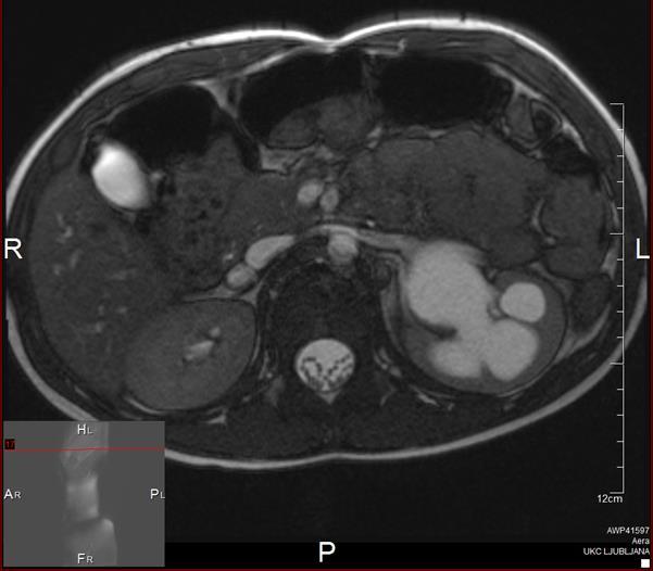
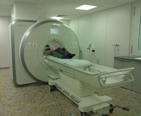
14 Parameters of standard MRU protocol in an 18-month-old child with a 1,5 T imager (Aera, Siemens, Ljubljana, Slovenia) Assessment Renal Excretory Renography Weighting parenchyma system T2 Heavily T2 Thick slab Excretory system Excretory system Bladder / ureterocele T2 T2 T2 T1 T1 Siemens name HASTE HASTE SPACE TSE HASTE Turbo FLASH VIBE Parenchyma and cavitas after injection Orientation Coronal Coronal Coronal Coronal Sagital Coronal Coronal TR (ms) ,29 TE (ms) ,26 1,57 FA ( ) TI (ms) Fat-saturation Yes Yes Yes Yes Yes No Yes Slice thickness (mm) ,3 Space between slices 0% 50% - 10% 0% 10% 20% FOV (mm) Acquisition matrix 200x x x x x x x224 Acquisition time 3,15 min 9 sec 6,10 min 4,1 min 1,39 min 10 min (150 meritev) 2,22 min Sirnik A (2014)
15 ImageJ MRU processing software Vivier PH et al Pediatr Radiol (2010) 40: CHOP-fMRU Khrichenko D, Darge K. Pediatr Radiol (2010) 40:


16 Damasio MB, MD, PhD, Radiologia IRCCS G. Gaslini, Genova Kljucevsek D, MD, PhD, Radiology Unit, Children's hospital Ljubljana,


17


18
19 double collecting systems on both sides with normal right kidney left kidney: dysplastic upper pole with very poor function, non obstructive hydronephrosis of lower pole no ectopic insertion of left ureters both ureter end in bladder normal renal vessel anatomy Volume of RF 20 ml, LK 14 ml Excretion curve: normal on both sides Delayed excretion of CM in the upper pole of the left kidney Split Renal function: LK= 26%, RK=74% Therapy : heminephrectomiam of upper moiety of LK and neoimplantation of distal LU (Politano-Leadbetter) Renal Scintigraphy: manually calculated function of kidney LK= 28% RK= 72%


20


21
22 HN gr. IV of LK Volume of RF 36 ml, LK 33 ml Excretion curve: RK in accommulation, LK- in accommulation (obstruction) Split Renal function: LK = 48%, RK = 52 % Symmetric CTT (delayed on the left) and RTT more than 8 minutes obstruction Renal Scintigraphy: manually calculated function of kidney LK= 41% RK= 59%


23

24 HN gr. IV of LK with thinned and edematous renal parenchyma, lost corticomedular differentiation left PUJ is oriented posteriorly with kinking of proximal ureter information about renal vessels, no crossing vessel volume of RF 38 ml, LK 60 ml excretion curve: RK normal, LK- in accommulation (obstruction) Split Renal function: LK = 77%, RK = 23 % asymmetric CCT (delayed on the left) and RTT more than 8 minutes obstruction Renal Scintigraphy: manually calculated function of kidney LK= 25% RK= 75%
25 US first imaging method MRU - promising tool in evaluation of urinary system fmru - all in one method - with anatomic and quantitative functional information team work: radiologist, radiographer (MR specialist), nephrologist, urologist, nuclear medicine specialist, anesthesiologist


26
RENAL SCINTIGRAPHY IN THE 21 st CENTURY
 RENAL SCINTIGRAPHY IN THE 21 st CENTURY 99m Tc- MAG 3 with zero time injection of Furosemide (MAG 3 -F 0 ) : A Fast and Easy Protocol, One for All Indications Clinical Experience Congenital Disorders PROTOCOL
Controversies around antenatally detected PUJ syndrom. Amy Piepsz, CHU St Pierre, Brussels, Belgium
 Controversies around antenatally detected PUJ syndrom Amy Piepsz, CHU St Pierre, Brussels, Belgium Editors : Anthony Caldamone, USA Pierre Mouriquand, France Newborn boy History of prenatally diagnosed
PROFESSIONAL SKILLS 1 3RD YEAR SEMESTER 6 RADIOGRAPHY. THE URINARY SYSTEM Uz. Fatema shmus aldeen Tel
 PROFESSIONAL SKILLS 1 3RD YEAR SEMESTER 6 RADIOGRAPHY THE URINARY SYSTEM Uz. Fatema shmus aldeen Tel. 0925111552 Professional skills-2 THE URINARY SYSTEM The urinary system (review anatomy and physiology)
G.Manzoni has documented that he has no relevant financial relationships to disclose or conflict of interest to resolve.
 G.Manzoni has documented that he has no relevant financial relationships to disclose or conflict of interest to resolve. ANTENATAL HYRONEPHROSIS UROLOGICAL ASPECTS Dr Gianantonio Manzoni, FEAPU FRCS U.O.
Hydronephrosis. Nephrosis. Refers to the kidney
 What is hydronephrosis? Hydro Nephrosis Refers to water or fluid Refers to the kidney A build-up of fluid (urine) in the kidney is the medical term for a build-up of urine in the kidney. As the urine builds
RENAL SCINTIGRAPHY IN THE 21 st CENTURY
 RENAL SCINTIGRAPHY IN THE 21 st CENTURY 99m Tc- MAG 3 with zero time injection of Furosemide (MAG 3 -F 0 ) : A Fast and Easy Protocol, One for All Indications Introduction George N. Sfakianakis MD Professor
Congenital Pediatric Anomalies: A Collection of Abdominal Scintigraphy Findings: An Imaging Atlas
 ISPUB.COM The Internet Journal of Nuclear Medicine Volume 5 Number 1 Congenital Pediatric Anomalies: A Collection of Abdominal Scintigraphy Findings: An Imaging Atlas V Vijayakumar, T Nishino Citation
Obstetrics Content Outline Obstetrics - Fetal Abnormalities
 Obstetrics Content Outline Obstetrics - Fetal Abnormalities Effective February 2007 10 16% renal agenesis complete absence of the kidneys occurs when ureteric buds fail to develop Or degenerate before
Radiography/Radiology
 Radiography/Radiology Activity for 2017 Activity No: A3(17) Topic MR Urography Article Pediatric MR Urography: indications, techniques and approach to review Approved for (3) Clinical Continuing Educational
Indications and effectiveness of the open surgery in vesicoureteral reflux
 Indications and effectiveness of the open surgery in vesicoureteral reflux Suzi DEMIRBAG, MD Department of Pediatric Surgery, Gulhane Military Medical Academy, Ankara, TURKEY Vesicoureteral reflux (VUR)
Index. Note: Page numbers of article titles are in boldface type.
 Magn Reson Imaging Clin N Am 12 (2004) 587 591 Index Note: Page numbers of article titles are in boldface type. A Adenoma(s), adrenal, gadolinium-enhanced MR imaging in, 533 534 hyperfunctioning versus
Acute flank pain in children: Imaging considerations
 Acute flank pain in children: Imaging considerations Carlos J. Sivit MD Rainbow Babies and Children s Hospital Case Western Reserve School of Medicine Flank pain Results from distention of ureter or renal
Prenatal Hydronephrosis
 Prenatal Hydronephrosis What is hydronephrosis? Hydronephrosis is dilation of the kidney, specifically the renal pelvis (place where urine is stored after its production). This can be the result of an
Pediatric* MR Urography
 Abdominal Imaging Clinical Pediatric* MR Urography Richard A. Jones; Stephen Little; J. Damien Grattan-Smith Children s Healthcare of Atlanta, Department of Radiology, Atlanta, GA, USA MR urography represents
Fetal Renal Malformations: The Role of Ultrasound in Diagnosis & Management
 Fetal Renal Malformations: The Role of Ultrasound in Diagnosis & Management 12 weeks Alfred Abuhamad, M.D. Eastern Virginia Medical School 13 weeks 2nd trimester Medullary pyramids Renal Sinus Cortex 2nd
Pelvi-Ureteric Junction Obstruction Revisited
 Dr. Bimalendu Mukherjee was trained in Urology in the UK between 1956 to 1961. Upon return to India, he took up a teaching position in Calcutta National Medical College and ultimately retired as Professor
Excretory urography (EU) or IVP US CT & radionuclide imaging
 or IVP US CT & radionuclide imaging") Excretory urography (EU) or IVP US CT & radionuclide imaging MRI arteriography studies requiring catherization or direct puncture of collecting system EU & to a lesser extent CT provide both functional
Excretory urography (EU) or IVP US CT & radionuclide imaging MRI arteriography studies requiring catherization or direct puncture of collecting system EU & to a lesser extent CT provide both functional
Uroradiology For Medical Students
 Uroradiology For Medical Students Lesson 4: Cystography & Urethrography - Part 2 American Urological Association Review Cystography is useful in evaluating the bladder, the urethra and the competence of
weighing risks against benefits ALARA principle appropriate activities (radiopharmaceutical doses)
") weighing risks against benefits ALARA principle appropriate activities (radiopharmaceutical doses) based on EANM references adequate appointment method (patient booking system) Appropriate activities (doses)
weighing risks against benefits ALARA principle appropriate activities (radiopharmaceutical doses) based on EANM references adequate appointment method (patient booking system) Appropriate activities (doses)
Pelvioureteric junction obstruction of the lower collecting system associated with incomplete ureteral duplication: A case report
 Ped Urol Case Rep 2014;1(6):11-15 DOI:10.14534/PUCR.201468061 PUCR Ped Urol Case Rep PEDIATRIC UROLOGY CASE REPORTS ISSN: 2148 2969 Journal homepage: http://www.pediatricurologycasereports.com Pelvioureteric
Postnatal Imaging of Antenatal Hydronephrosis
 Review Special Issue: Pre- and Postnatal Management of Hydronephrosis TheScientificWorldJOURNAL (2009) 9, 393 399 TSW Urology ISSN 1537-744X; DOI 10.1100/tsw.2009.50 Postnatal Imaging of Antenatal Hydronephrosis
PICTORIAL ESSAY. Experiences of using a single post-contrast CT scan of the urinary tract after triphasic contrast injection
 Experiences of using a single post-contrast CT scan of the urinary tract after triphasic contrast injection P C Pretorius, FCRad (Diag) SA Drs Visser, Erasmus, Vawda & Partners, Port Elizabeth Corresponding
Chapter 6: Genitourinary and Gastrointestinal Systems 93
 Chapter 6: Genitourinary and Gastrointestinal Systems 93 Chapter 6 Genitourinary and Gastrointestinal Systems Embryology Three sets of excretory organs or kidneys develop in human embryos: Pronephros:
Pediatric Ure-Radiology*
 Pediatric Ure-Radiology* HERMAN GROSSMAN, M.D. Professor of Radiology and Pediatrics, Duke University Medical Center, Durham, North Carolina "Routine" radiologic studies do not, often enough, concentrate
Urinary Tract Abnormalities
 Urinary Tract Abnormalities Dr Hennie Lombaard Senior Specialist Maternal and Fetal Medcine Department of Obstetrics and Gynecology Level 7 Pretoria Academic Hospital Pictures from The 18 to 23 weeks scan
Proceedings of the 34th World Small Animal Veterinary Congress WSAVA 2009
 www.ivis.org Proceedings of the 34th World Small Animal Veterinary Congress WSAVA 2009 São Paulo, Brazil - 2009 Next WSAVA Congress : Reprinted in IVIS with the permission of the Congress Organizers IMAGING
Case MDCT 3D reconstructed features of posterior urethral valve
 Case 12688 MDCT 3D reconstructed features of posterior urethral valve Hidayatullah Hamidi Third year Resident of Radiology French medical institute for children Radiology Department; Kabul, Afghanistan;
Case Report Congenital Midureteric Stricture: Challenges in Diagnosis and Management
 Case Reports in Urology Volume 2015, Article ID 969246, 5 pages http://dx.doi.org/10.1155/2015/969246 Case Report Congenital Midureteric Stricture: Challenges in Diagnosis and Management Raashid Hamid,
Comparison of DMSA Scan 99 m and EC Scan 99 m in Diagnosis of Cortical Defect and Differential Renal Function
 Global Journal of Health Science; Vol. 6, No. 7; 2014 ISSN 1916-9736 E-ISSN 1916-9744 Published by Canadian Center of Science and Education Comparison of DMSA Scan 99 m and EC Scan 99 m in Diagnosis of
Rare case of urinary bladder agenesis - Multislice CT abdomen imaging
 Rare case of urinary bladder agenesis - Multislice CT abdomen imaging Venkatraman Indiran 1*, Kabilan Chokkappan 2, Emmanuel Gunaseelan 2 1. Department of Radiodiagnosis, Sree Balaji Medical College and
Division of Nuclear Medicine Procedure / Protocol University Hospital and The American Center
 KIDNEY FLOW / FUNCTION WITH DIURETIC CPT CODE: 78708 UPDATED: FEBRUARY 2017 Indications: The scan is designed to differentiate dilated renal collecting systems (calyces, pelves, or ureters) from obstructed
Nephrographic and Pyelographic Analysis of CT Urography: Principles, Patterns, and Pathophysiology
 Genitourinary Imaging Review Wolin et al. CT Urography Principles, Patterns, and Genitourinary Imaging Review FOCUS ON: Ely A. Wolin 1 David S. Hartman J. Ryan Olson Wolin EA, Hartman DS, Olson JR Keywords:
Nuclear medicine methods in the urogenital system
 Nuclear medicine methods in the urogenital system Anatomy of the kidneys I. Anatomy of the kidneys II. The types of examinations Static examinations (scintigraphy): 1) the radiopharmaceutical is administered
Efficacy of Magnetic Resonance urography in detecting crossing renal vessels in children with ureteropelvic junction obstruction
 Efficacy of Magnetic Resonance urography in detecting crossing renal vessels in children with ureteropelvic junction obstruction Ann. Ital. Chir., 2015 86: 443-449 pii: S0003469X15023611 Polina K. Pavicevic*,
Current Trends in Pediatric GU Imaging European Perspective
 Current Trends in Pediatric GU Imaging European Perspective Pierre-Hugues Vivier, MD, PhD CHU C. Nicolle, Rouen, France Générale de Santé, Hôpital Privé de l Estuaire, Le Havre, France 1.6% of boys / 7.8%
The relationship Between The Divided Shape of Kidney and the Duplication of Ureter
 The relationship Between The Divided Shape of Kidney and the Duplication of Ureter Poster No.: C-1373 Congress: ECR 2014 Type: Authors: Keywords: DOI: Scientific Exhibit J. Lee, B. S. Cho, S. J. Kim, K.
MRI Abdomen Protocol Pancreas/MRCP with Contrast
 MRI Abdomen Protocol Pancreas/MRCP with Contrast Reviewed By: Brett Mollard, MD; Anna Ellermeier, MD Last Reviewed: July 2018 Contact: (866) 761-4200 Standard uses: 1. Characterization of cystic and solid
Acute renal colic Radiological investigation in patients with renal colic
 Acute renal colic Radiological investigation in patients with renal colic Mikael Hellström Professor Department of Radiology Sahlgrenska University Hospital Göteborg University 0.9-1.8/1.000 inhabitants
Clinical-Radiological management of congenital hydronephrosis.
 Clinical-Radiological management of congenital hydronephrosis. Poster No.: C-0983 Congress: ECR 2015 Type: Authors: Keywords: DOI: Educational Exhibit M. Vidal, D. Llanos, E. Pallares, I. de la Pedraja,
Abdominal Ultrasound : Aorta, Kidneys, Bladder
 Abdominal Ultrasound : Aorta, Kidneys, Bladder Nilam J. Soni, MD, MSc Associate Professor of Medicine Divisions of Hospital Medicine and Pulmonary/Critical Care Medicine Department of Medicine University
Lower-dose CT urography (CTU) with iterative reconstruction technique in children initial experience and examination protocol
 with iterative reconstruction technique in children initial experience and examination protocol") Signature: Pol J Radiol, 2014; 79: 137-144 DOI: 10.12659/PJR.890729 ORIGINAL ARTICLE Received: 2014.03.21 Accepted: 2014.04.14 Published: 2014.06.08 Authors Contribution: A Study Design B Data Collection
Signature: Pol J Radiol, 2014; 79: 137-144 DOI: 10.12659/PJR.890729 ORIGINAL ARTICLE Received: 2014.03.21 Accepted: 2014.04.14 Published: 2014.06.08 Authors Contribution: A Study Design B Data Collection
Information for Patients
 Information for Patients Congenital Malformation in the Urinary Tract: Ureteral Duplication, Ureterocele, and Ectopic Ureter English Table of contents Ureteral Duplication... 3 Symptoms and Diagnosis...
US in non-traumatic acute abdomen. Lalita, M.D. Radiologist Department of radiology Faculty of Medicine ChiangMai university
 US in non-traumatic acute abdomen Lalita, M.D. Radiologist Department of radiology Faculty of Medicine ChiangMai university Sagittal Orientation Transverse (Axial) Orientation Coronal Orientation Intercostal
Imaging Ejaculatory Disorders and Hematospermia
 ATHENS 4-6 October 2018 European Society of Urogenital Radiology Imaging Ejaculatory Disorders and Hematospermia Parvati Ramchandani, MD Professor, Radiology and Surgery University of Pennsylvania Medical
Evaluation of Imaging Abnormalities of Ureter using MDCT Urography
 DOI: 10.7860/IJARS/2017/23458:2250 Radiology Section Original Article Evaluation of Imaging Abnormalities of Ureter using MDCT Urography Vedaraju Kadaba Shamachar, Vijay Kumar Kenchanahalli Rangaswamy,
Functions of the kidney:
 Diseases of renal system : Normal anatomy of renal system : Each human adult kidney weighs about 150 gm, the ureter enters the kidney at the hilum, it dilates into a funnel-shaped cavity, the pelvis, from
2 Radiographic Imaging
 2 Radiographic Imaging Lane S. Palmer Abstract The evaluation of children with urologic complaints or possible congenital anomalies cannot go forward without proper imaging. The mainstay of this imaging
R adio logical investigations of urinary system
 R adio logical investigations of urinary system There are 4 main radiological Ix: 1 IVU: Intravenous urography. 2- U/S 3-CT scan 4-Radioisotope scan. Others (not frequently used): MRI, arteriography, antegrade
ESPR. Keywords Uroradiology. Recommendations. Procedures. IVU. MRU. Uro-CT. Child. Pediatric uroradiology. Children. Imaging.
 Pediatr Radiol (2010) 40:1315 1320 DOI 10.1007/s00247-010-1686-7 ESPR ESPR uroradiology task force and ESUR paediatric working group: imaging and procedural recommendations in paediatric uroradiology,
MR Tumor Staging for Treatment Decision in Case of Wilms Tumor
 MR Tumor Staging for Treatment Decision in Case of Wilms Tumor G. Schneider, M.D., Ph.D.; P. Fries, M.D. Dept. of Diagnostic and Interventional Radiology, Saarland University Hospital, Homburg/Saar, Germany
UNIVERSITY OF MEDICINE AND PHARMACY OF CRAIOVA IMAGING ASPECTS OF RENO-URINARY MALFORMATIONS IN CHILD ABSTRACT
 UNIVERSITY OF MEDICINE AND PHARMACY OF CRAIOVA IMAGING ASPECTS OF RENO-URINARY MALFORMATIONS IN CHILD ABSTRACT SCIENTIFIC COORDINATOR UNIV. PROF. DR. BONDARI ANDREI Ph.D. CANDIDATE DINU IRINA ELENA CRAIOVA
Recurrent Pediatric UTI Revisited 2013
 Recurrent Pediatric UTI Revisited 2013 PIDSP 21.2.2013 Shai Ashkenazi, MD, MSc Medicine changes constantly Some aspects of the standard practice of ~40 years are probably not valid and need to be changed
MRI PEDIATRIC PROTOCOLS (Updated 6/19/2018)
") MRI PEDIATRIC PROTOCOLS (Updated 6/19/2018) *Please get or let us know where radiologist can review plain films. *For Texas Orthopedics and other Docs requesting only MSK section read for their pediatric
MRI PEDIATRIC PROTOCOLS (Updated 6/19/2018) *Please get or let us know where radiologist can review plain films. *For Texas Orthopedics and other Docs requesting only MSK section read for their pediatric
Anatomical and Functional MRI of the Pancreas
 Anatomical and Functional MRI of the Pancreas MA Bali, MD, T Metens, PhD Erasme Hospital Free University of Brussels Belgium mbali@ulb.ac.be Introduction The use of MRI to investigate the pancreas has
IMAGING OF UPPER UT TCC
 IMAGING OF UPPER UT TCC IS THERE AN EVIDENCE BASED STRATEGY? S A MOUSSA FRCS Ed, FRCR WESTERN GENERAL HOSPITAL EDINBURGH UPPER TRACT TCC 0.7-4% of patients with primary bladder cancer develops UT-TCC.
GU Ultrasound in First Trimester
 Fetal Renal Malformations: The Role of Ultrasound in Diagnosis & Management Outline 1. Renal Anomalies Urinary Tract Dilation Aberrant Early Development Defects Terminal Maturation Alfred Abuhamad, M.D.
Renal. Prof John Buscombe
 Renal Prof John Buscombe Renal nuclear Medicine Only consistent test of kidney func7on Many good tests for renal anatomy Ultrasound good looking at cysts and renal pelvis CT can look at perfusion, size
Spectrum of Micturating Cystourethrogram Revisited: A Pictorial Assay
 603 International Journal of Collaborative Research on Internal Medicine & Public Health Spectrum of Micturating Cystourethrogram Revisited: A Pictorial Assay Abhinav Jain 1, Vivek Setia 1, Shweta Agnihotri
Find Medical Solutions to Your Problems HYDRONEPHROSIS. (Distension of Renal Calyces & Pelvis)
") HYDRONEPHROSIS (Distension of Renal Calyces & Pelvis) Hydronephrosis is the distension of the renal calyces and pelvis due to accumulation of the urine as a result of the obstruction to the outflow of
HYDRONEPHROSIS (Distension of Renal Calyces & Pelvis) Hydronephrosis is the distension of the renal calyces and pelvis due to accumulation of the urine as a result of the obstruction to the outflow of
URINARY TRACT IMAGING - BASIC PRINCIPLES
 URINARY TRACT IMAGING - BASIC PRINCIPLES Clinical Radiology Every physician needs a basic understanding of diagnostic imaging to understand how to order the appropriate studies and to understand the resulting
8/14/2017. Kidney location & visualization. Brief Review with tips & Case Based Illustrations. Size = x L2. Size =
 Dr. Russell Tucker, DACVR Brief Review with tips & Case Based Illustrations Kidney location & visualization K9 Kidneys: Rt @ T13-L1 Lt @ L2-L4 Kidney visualization K9 Kidneys: Rt @ T13-L1 Lt @ L2-L4 Size
Case Report Symptomatic Infundibulopelvic Dysgenesis in an Adolescent
 Case Reports in Urology Volume 2015, Article ID 307319, 4 pages http://dx.doi.org/10.1155/2015/307319 Case Report Symptomatic Infundibulopelvic Dysgenesis in an Adolescent Daniel Pitts, 1 David Chalmers,
Index. mri.theclinics.com. Note: Page numbers of article titles are in boldface type.
 Index Note: Page numbers of article titles are in boldface type. A Angiogenesis, and cancer of prostate, 689 690 Angiography, MR. See MR angiography. Apoptosis, MR imaging of, 637 Apparent diffusion coefficient,
Can SCMR CMR protocol recommendations
 Can SCMR CMR protocol recommendations V1.3 - April 2009 CanSCMR CMR Protocol and SOP Recommendation 2009 (15 minutes) 2 Planning of LV fct. real time multiple axes Realtime 3 cine long axis 6 long axes
UNILATERAL DOUBLE PELVIS BIFID URETER ASSOCIATED WITH MULTIPLE VARIATIONS OF RENAL VESSELS
 Original Research Article UNILATERAL DOUBLE PELVIS BIFID URETER ASSOCIATED WITH MULTIPLE VARIATIONS OF RENAL VESSELS Ajay Babu Kannabathula * 1, Gunjan Rai 2, K. C. Singh 3. ABSTRACT International Journal
ISUOG Basic Training. Distinguishing between Normal & Abnormal Appearances of the Urinary Tract. Seshadri Suresh, India
 ISUOG Basic Training Distinguishing between Normal & Abnormal Appearances of the Urinary Tract Seshadri Suresh, India Learning objectives 13 & 14 At the end of the lecture you will be able to: describe
Imaging the Urogenital System
 maging the Urogenital System Tony Pease, DVM, MS, DACVR Assistant Professor of Radiology North Carolina State University Reading Thrall Chapters 42-46 Prostate Gland Not visible radiographically in normal
RADIO-NUCLIDE STUDIES FOR THE EVALUATION OF KIDNEY FUNCTIONS. DR RIANA NEL NUCLEAR MEDICINE DEPT 27 Sept 2010
 RADIO-NUCLIDE STUDIES FOR THE EVALUATION OF KIDNEY FUNCTIONS DR RIANA NEL NUCLEAR MEDICINE DEPT 27 Sept 2010 Alive patient RA + tracers Tc-99m(86%) + MAG 3 DMSA (DTPA) T½ = 6,5h Gamma camera ect. Radio-active
Case report Paediatrics Today 2016;12(1):87-91 DOI /p
:87-91 DOI /p") Case report Paediatrics Today 2016;12(1):87-91 DOI 10.5457/p2005-114.140 URINARY INCONTINENCE FOLLOWING VAGINAL SEPTUM RESECTION IN HERLYN-WERNER-WUNDERLICH SYNDROME A CASE REPORT Slavica PONORAC 1*, Anamarija
Case report Paediatrics Today 2016;12(1):87-91 DOI 10.5457/p2005-114.140 URINARY INCONTINENCE FOLLOWING VAGINAL SEPTUM RESECTION IN HERLYN-WERNER-WUNDERLICH SYNDROME A CASE REPORT Slavica PONORAC 1*, Anamarija
Expert systems for diuresis renography Andrew Taylor, MD Emory University, Atlanta, GA, USA
 Expert systems for diuresis renography Andrew Taylor, MD Emory University, Atlanta, GA, USA Collaborators Ernest V. Garcia, PhD Amita Manatunga, PhD Jose Binonngo, PhD Jieqiong Bao, MS Russell Folks, CNMT
Annual Report 2016 PEDIATRIC UROLOGY. Seppo Taskinen
 Annual Report 2016 PEDIATRIC UROLOGY Seppo Taskinen Contents Introduction 2 Personnel and resources 3 Pediatric surgery appointments 3 Children s urological surgeries 4 Quality 7 Networking of urology
Dynamic Contrast-Enhanced MR Urography in the Evaluation of Pediatric Hydronephrosis: Part 1, Functional Assessment
 Jones et al. MR Urography of Pediatric Hydronephr osis Pediatric Imaging Original Research Richard A. Jones 1,2 Kirk Easley 3 Stephen B. Little 2 Hal Scherz 4,5 Andrew J. Kirsch 4,5 J. Damien Grattan-Smith
URINARY SYSTEM I. Kidneys II. Nephron Unit and Urine Formation
 URINARY SYSTEM I. Kidneys A. Location and Structure 1. Retroperitoneal 2. Between T12 and L3 3. Rt. kidney slightly lower 4. Two bean shaped organs 5. Adrenal gland 6. Internal construction a. Renal cortex
Perineal Sonography in Diagnosis of an Ectopic Ureteric Opening Into the Urethra
 Case Series Perineal Sonography in Diagnosis of an Ectopic Ureteric Opening Into the Urethra S. Boopathy Vijayaraghavan, MD, DMRD Objective. To study the role of perineal sonography in the diagnosis of
INTERNATIONAL SPINAL CORD INJURY DATA SETS URINARY TRACT IMAGING BASIC DATA SET - COMMENTS
 1 INTERNATIONAL SPINAL CORD INJURY DATA SETS URINARY TRACT IMAGING BASIC DATA SET - COMMENTS The working-group consists of: Fin Biering-Sørensen representing the Executive Committee of The International
MR Advance Techniques. Vascular Imaging. Class II
 MR Advance Techniques Vascular Imaging Class II 1 Vascular Imaging There are several methods that can be used to evaluate the cardiovascular systems with the use of MRI. MRI will aloud to evaluate morphology
Kidney Biopsy. Table of Contents. Test Overview. Why It Is Done. Test Overview. Why It Is Done. How to Prepare. How It Is Done.
 Kidney Biopsy Table of Contents Test Overview Why It Is Done How to Prepare How It Is Done Results What Affects the Test? Test Overview The two kidneys {IMAGE LINK 1} are found on either side of the spine,
Case Report Crossed Renal Ectopia without Fusion An Unusual Cause of Acute Abdominal Pain: A Case Report
 Case Reports in Urology Volume 2012, Article ID 728531, 4 pages doi:10.1155/2012/728531 Case Report Crossed Renal Ectopia without Fusion An Unusual Cause of Acute Abdominal Pain: A Case Report D. P. Ramaema,
Transverse Comparisons Between Ultrasound and Radionuclide Parameters in Children with Pelvi-Ureteric Junction Obstruction
 The Egyptian Journal of Hospital Medicine (July 2018) Vol. 72 (5), Page 4543-4550 Transverse Comparisons Between Ultrasound and Radionuclide Parameters in Children with Pelvi-Ureteric Junction Obstruction
Audit of split-bolus CT urography for the investigation of haematuria over a 12 month period at two district general hospitals
 Audit of split-bolus CT urography for the investigation of haematuria over a 12 month period at two district general hospitals Poster No.: C-1349 Congress: ECR 2010 Type: Educational Exhibit Topic: Genitourinary
Downloaded from Medico Research Chronicles Pelvi-ureteric junction obstruction - A ten year single center review in north central Nigeria.
 PELVI-URETERIC JUNCTION OBSTRUCTION - A TEN YEAR SINGLE CENTER REVIEW IN NORTH CENTRAL NIGERIA. 1,2 1,2 1 1 Atim T, Aisuodionoe-Shadrach O, Ajibola O. H, Magnus F. E Division of Urology, Departments of
1. Normal and pathological embryology of the urinary and genital tract 2. Nephrology 3. Infection
 1 1. Normal and pathological embryology of the urinary and genital tract 1.1. Development of the kidney and ureter 1.2. Development of the bladder and the urethra 1.3. Development of the female genital
Evaluation of dilated upper renal tracts by technetium-99m ethylenedicysteine F+0 diuresis renography in infants and children
 ORIGINAL ARTICLE Annals of Nuclear Medicine Vol. 18, No. 8, 681 687, 2004 Evaluation of dilated upper renal tracts by technetium-99m ethylenedicysteine F+0 diuresis renography in infants and children Madhavi
Large spectrum of complete urinary collecting system duplication exemplified by cases. Pictorial essay.
 Pictorial essay Med Ultrason 2013, Vol. 15, no. 4, 315-315 DOI: 10.11152/mu.2013.2066.154.of2 Large spectrum of complete urinary collecting system duplication exemplified by cases. Pictorial essay. Otilia
9. Renal duplication. Essentials in Pediatric Urology, 2012: ISBN: Editor: George Sakellaris
 Research Signpost 37/661 (2), Fort P.O. Trivandrum-695 023 Kerala, India Essentials in Pediatric Urology, 2012: 89-94 ISBN: 978-81-308-0511-5 Editor: George Sakellaris 9. Renal duplication Consultant Pediatric
Unilateral supernumerary kidney with contra lateral hydronephrosis a rare case report
 Unilateral supernumerary kidney with contra lateral hydronephrosis a rare case report Ezzat Khalda, Gyanendra Narain Singh, Ashok Kumar Mandal, Rajiv Kumar Department of Radiodiagnosis, Patna Medical College,
Clinics in Diagnostic Imaging (61)
") Singapore Med J 2001 Vol 42(5) : 233-237 M e d i c a l E d u c a t i o n Clinics in Diagnostic Imaging (61) K B J Sheah, S K H Yip, V T Joseph Fig. 1a Control abdominal radiograph. Fig. 1b Coned oblique
Singapore Med J 2001 Vol 42(5) : 233-237 M e d i c a l E d u c a t i o n Clinics in Diagnostic Imaging (61) K B J Sheah, S K H Yip, V T Joseph Fig. 1a Control abdominal radiograph. Fig. 1b Coned oblique
ESPR. Pediatr Radiol (2014) 44: DOI /s
 44: DOI /s") Pediatr Radiol (2014) 44:1478 1484 DOI 10.1007/s00247-014-3135-5 ESPR ESPR uroradiology task force imaging recommendations in paediatric uroradiology, part VII: standardised terminology, impact of existing
Pediatr Radiol (2014) 44:1478 1484 DOI 10.1007/s00247-014-3135-5 ESPR ESPR uroradiology task force imaging recommendations in paediatric uroradiology, part VII: standardised terminology, impact of existing
Dynamic Contrast-Enhanced MR Renography for Renal Function Evaluation in Ureteropelvic Junction Obstruction: Feasibility Study
 Genitourinary Imaging Original Research Krepkin et al. Genitourinary Imaging Original Research Konstantin Krepkin 1 Eugene Won 1 Krishna Ramaswamy 2 Michael Triolo 1 Michael Stiffelman 2 Henry Rusinek
The Evolving Role of Antibiotic Prophylaxis for Vesicoureteral Reflux. Stephen Canon, MD Children s Urology
 The Evolving Role of Antibiotic Prophylaxis for Vesicoureteral Reflux Stephen Canon, MD Children s Urology www.childrensurology.com May 17, 2008 Objectives Review literature establishing the value of antibiotic
이학종분당서울대학교병원. Ultrasound in Urinary Colic
 이학종분당서울대학교병원 Ultrasound in Urinary Colic U l t r a s o u n d i n U r i n a US: Normal Kidney r y C o l i c Contents 1. 1. Definition and clinical consideration 2. 2. Pathophysiology 3. 3. US in in obstructive
Non Contrast MRA. Mayil Krishnam. Director, Cardiovascular and Thoracic Imaging University of California, Irvine
 Non Contrast MRA Mayil Krishnam Director, Cardiovascular and Thoracic Imaging University of California, Irvine No disclosures Non contrast MRA-Why? Limitations of CTA Radiation exposure Iodinated contrast
Abdomen and Retroperitoneum Ultrasound Protocols
 Abdomen and Retroperitoneum Ultrasound Protocols Reviewed By: Anna Ellermeier, MD Last Reviewed: March 2018 Contact: (866) 761-4200, Option 1 **NOTE for all examinations: 1. If documenting possible flow
Outline. Introduction to imaging modalities of the urinary system. Case base learning of common diseases in urinary tract
 Outline Introduction to imaging modalities of the urinary system Case base learning of common diseases in urinary tract Outline Introduction to imaging modalities of the urinary system Case base learning
Outline. Introduction to imaging modalities of the urinary system. Case base learning of common diseases in urinary tract
 Outline Introduction to imaging modalities of the urinary system Case base learning of common diseases in urinary tract Diagnostic Investigations in Urinary System PLAIN KUB EXCRETORY UROGRAPHY RETROGRADE
Lower Urinary Tract Obstruction LUTO or Bladder Outlet Obstruction BOO. Miss Harriet Corbett Consultant Paediatric Urologist
 Lower Urinary Tract Obstruction LUTO or Bladder Outlet Obstruction BOO Miss Harriet Corbett Consultant Paediatric Urologist Aims To give an overview of the anomalies we encounter presentation of LUTO how
Contents. Review anatomy of the urinary tract Imaging modalities
 Contents Review anatomy of the urinary tract Imaging modalities The Urinary Tract Kidneys ตาแหน งไต (position) อย ใน retroperitoneum ระด บ T12-L3 โดยไต ขวาจะม ระด บตากว าไตซ ายเล กน อย ร ปร าง (shape)
Comparative Effectiveness of a Pilot Patient-Centered Ultrasound Report for Hydronephrosis Management
 Comparative Effectiveness of a Pilot Patient-Centered Ultrasound Report for Hydronephrosis Management Geolani W. Dy, MD Society of Women in Urology 6 th Annual Winter Meeting January 21, 2017 Cultural
Case Report Transplantation of Horseshoe Kidney from Living, Genetically Unrelated Donor
 Case Reports in Transplantation Volume 2015, Article ID 390381, 4 pages http://dx.doi.org/10.1155/2015/390381 Case Report Transplantation of Horseshoe Kidney from Living, Genetically Unrelated Donor Kazuro
Sonographic Evaluation of Hydronephrosis in the Pediatric Population
 ORIGINAL RESEARCH Sonographic Evaluation of Hydronephrosis in the Pediatric Population Is Well-Tempered Sonography Necessary? Marc R. Walker, MD, Sarkis Babikian, MD, Alexander J. Ernest, MD, Troy S. Koch,