Urinary system Ultrasound (Renal & Urinary bladder)
|
|
|
- Lee Hamilton
- 5 years ago
- Views:
Transcription
 Edited & Presented by ; Hussien A.")

1 Urinary system Ultrasound (Renal & Urinary bladder) Edited & Presented by ; Hussien A.B ALI DINAR. Msc.Phd ISRRT Associate Member Lecturer (National university) Reporting Sonographer (PHC)
2 Objective By the end of this lecture you should : Review Anatomy,physiology and pathology of Renal system Evaluate normal and abnormal finding in urinary system sonography Describe sonographic appearance of pathological finding
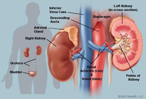
3 Anatomy The kidneys are two bean-shaped organs situated in the retroperitoneum on each side of the vertebral column. Their posterior surfaces are separated from the psoas major and the quadratus lumborum muscles by the posterior pararenal space. Each kidney consists of an upper and lower pole, anterior and posterior surfaces, a convex lateral margin and a concave medial margin. The long axis of the kidney runs obliquely and parallels the lateral border of the psoas major muscle. This means the upper poles are closer to midline than the lower poles.
4 Anatomy

5 Anatomy

6 The kidneys regulate the composition and the volume of the blood and remove wastes from the blood in the form of urine. Formation of active vitamin D. Production and secretion of erythropoietin, the hormone responsible for controlling the rate of formation of red blood cells. Production and secretion of rennin, an important enzyme in the control of blood pressure. The regulation of blood acid-base balance. (Blood PH). Functions

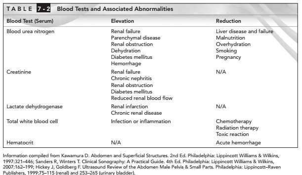
7 Urinalysis/Blood test and associations
8 Indications Indications for an ultrasound examination of the kidney and/or bladder include but are not limited to: Flank and/or back pain; Signs or symptoms that may be referred from the kidney and/or bladder regions such as hematuria; Abnormal laboratory values or abnormal findings on other imaging examinations suggestive of kidney and/or bladder pathology; Follow-up of known or suspected abnormalities in the kidney and/or bladder; Evaluation of suspected congenital abnormalities; Abdominal trauma; Pretransplantation and posttransplantation evaluation; and Planning and guidance for an invasive procedure
9 Scanning Protocol Transducer: MHz Patient generally supine. Right kidney: The right liver provides a good acoustic window for scanning the right kidney. The lower pole is occasionally obscured by the right colic flexure but is accessible to scanning from the posterior side. Left kidney: An acoustic window is not available for the left kidney. Scanning from the posterolateral side is advantageous as it avoids overlying gas in the colon and gastric fornix.
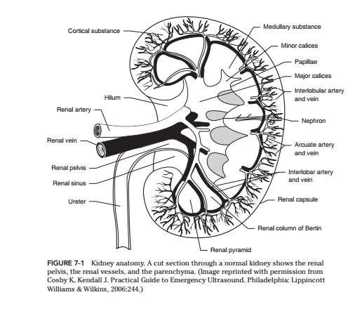
10 Renal sonographic Anatomy They typically measure approximately 8 13 cm in length, 2 3 cm in the anteroposterior dimension, and 4 5 cm in width. The renal sinus is central in the kidney and has an echogenic appearance. The renal cortex appears as medium-to-low level echoes surrounding the central sinus. The normal cortex should be more hypoechoic than, or isoechoic to, the liver or spleen.


11 Renal sonographic Anatomy Adult kidney Neonatal kidney


12 Renal Variants in Appearance and Location Congenital variations pelvic kidney - fails to migrate from pelvic area during embryology Dromedary hump agenesis Ectopic kidney horseshoe - isthmus of tissue that connects both kidneys Duplex kidney Hypoplastic kidney Pelvic kidney

 Empty right")


13 Renal Variants in Appearance and Location Dromedary hump(upper) and agenesis kidneys(lower) Empty right renal fossa, caused by a partial horseshoe kidney on the left side (K). AO = aorta, V = compressed vena cava, M = lumbar muscle, L = liver

.")
.")

14 Renal Variants in Appearance and Location Renal hypoplasia. The absent left kidney is probably a tiny hypoplastic kidney (cursors). b Malrotated kidney at a slightly ectopic location (cursors). The renal hilum is directed anteriorly Large kidneys. a Duplex kidney (K, cursors 132.5mm) with parenchymal band, b Acromegaly (cursors 138.1mm)

15
16 SONOGRAPHY FINDING Overview and Classification of Findings : size changes: Acute diffuse diseases are generally associated with renal enlargement due to inflammatory edematous swelling, whereas chronic diseases are marked by a decrease in renal size caused by loss of parenchyma. In chronic glomerulonephritis and diabetic nephropathy, the kidneys do not shrink in size until the disease has progressed to the dialysis stage. Echogenicity changes: Increased or decreased echogenicity reflects tissue changes at the histologic level.

17 Kidney pathology finding RENAL CYSTIC DISEASE Simple Renal Cysts Sonographic criteria: Anechoic Strong back walldistal acoustic enhancement No measurable wall thickness

18 Kidney pathology finding RENAL CYSTIC DISEASE Atypical Renal Cysts Sonographic criteria: cysts may have thick walls. May contain septations. generate low levels of echogenicity from particulate matter associated with hemorrhage or infection The echogenic cyst contents may be diffuse or show dependent layering. The cyst wall may be calcified and is considered a benign finding if all other US criteria for simple cyst are met Complicated cyst with internal hemorrhage

19 Kidney pathology finding RENAL CYSTIC DISEASE Multicystic Dysplastic Kidney MCDK Sonographic criteria: kidney is large and filled with cysts of various sizes The cysts do not communicate and appear benign There is no evidence of renal parenchyma, pelvis or ureter Polycystic kidney: The kidney is enlarged and poorly defined with little evidence of residual parenchyma. A central echo complex is not visualized
20 Kidney pathology finding Hydronephrosis Hydronephrosis refers to dilatation of the renal collecting system most frequently caused by incomplete or complete obstruction. Hydroureter is dilatation of the ureter also caused by complete or incomplete obstruction Causes - In infants and children ureteropelvic junction obstruction, posterior urethralvalves in males and Prune Belly Syndrome are the most common causes of obstruction. Calculi is the most common cause in adults followed by tumors of the kidney, ureter and bladder. Less common causes are inflammatory ureteral strictures, neurogenic bladder and bladder outlet obstruction. There are three grade of hydronephrosis Grade I - renal pelvis dilated Grade II - renal pelvis & major calyces dilated Grade III - renal pelvis, major & minor calyces dilated
21 Grade 2 hydronephrosis Grade 1 hydronephrosis Grade 3 hydronephrosis
 and renal colic are most often associated with ureteric calculi.")
22 Kidney pathology finding Renal Calculus Disease Urolithiasis is most prevalent in males aged years.1 Calculi can form in any part of the urinary tract but most form in the kidneys. They may be clinically silent or associated with flank pain. Hematuria (gross or microscopic) and renal colic are most often associated with ureteric calculi. Stones can occur within any part of the kidneys - the renal cortex, medulla, vessels, calyces or renal pelvis.

23 Stone overview Stones in renal sinus may not be visualized due to echogenicity Stones in ureter may not be visualized due to overlying bowel gas Acoustic shadow may not be present if stone is small Stones may be visualized at the UVJ with a full bladder Renal stone Acoustic Shadow
24

25 Kidney pathology finding Cortical Nephrocalcinosis Sonographic criteria: increased corticalechogenicity which may be associated with acoustic shadowing Cortical nephrocalcinosis.
 projected over the medullary")
26 Medullary Nephrocalcinosis Sonographic criteria: The renal pyramids appear more echogenic than the adjacent renal cortex Pronounced medullary nephrocalcinosis: small atrophic kidney with a band of residual parenchyma and hyperechoic areas, some with acoustic shadows (S) projected over the medullary pyramids (arrows)

27 Kidney pathology finding NEOPLASMS BENIGN LESIONS Angiomyolipoma (AML) AML is a benign solid tumor containing variable amounts of blood vessels (angio), smooth muscle (myo) and fat (lipoma). Sonographic criteria: AMLs are extremely hyperechoic indicating the predominance of fat however, if muscle or vascular components predominate the lesion may be hypoechoic. Angiomyolipoma: echogenic tumor with smooth margins (arrow) causing an almost imperceptible bulge in the renal contour

28 BENIGN LESIONS Renal adenoma Sonographic criteria: Round, hypoechoic mass with smooth margins Complex internal echo pattern due to regressive changes Occasionally hyperechoic Renal adenoma

29 MALIGNANT LESIONS MALIGNANT LESIONS Renal Cell Carcinoma (RCC) is a primary tumor of the renal parenchyma thought to originate from the renal tubular epithelium. It is also called a hypernephroma or a renal adenocarcinoma Sonographic criteria: a spherical, solitary, unilateral tumor of variable size and Echogenicity renal cell carcinoma (T)

30 MALIGNANT LESIONS Nephroblastoma nephroblastoma is a rapidly growing malignant tumor of the kidneys, consisting of embryonal elements. It is also known as Wilm's tumors, Wilm's embryoma or embryonal Carcinoma Sonographic criteria: large, intrarenal, solid mass with a well-defined margin or pseudocapsule of fibrous tissue and compressed renal parenchyma. The tumor may be homogeneous or heterogeneous, if necrosis or hemorrhage has occurred. Nephroblastoma with cystic mass

31 Kidney pathology finding Renal Infections Most renal infections occur via the ascending route. They are usually caused by contaminants from the intestinal tract. Instrumentation, stasis, calculi, and vesicoureteral reflux are predisposing factors

32 Renal Infections Acute Pyelonephritis (Acute Bacterial Nephritis) Sonographic criteria: diffuse renal enlargement. decreased parenchymal echogenicity and loss of corticomedullary differentiation. The walls of the renal pelvis or major calyces may bethickened Acute pyelonephritis: Large, hypoechoic kidney with an obliterated sinus echo and a rim of fluid in the renal pelvis

33 Renal Infections Renal Abscess Sonographic criteria: renal abscess is typically a round thick walled hypoechoic complex mass with internal debris and acoustic enhancement Abscesses: anechoic cystic masses in the central echocomplex
: Foci of parenchymal thinning due to scarring, producing a wavy surface contour.")
34 Renal Infections Chronic Pyelonephritis Sonographic criteria: The affected kidney is small lobulated due to parenchymal thinning decreased renal size in pyelonephritis (83.9mm, cursors): Foci of parenchymal thinning due to scarring, producing a wavy surface contour. C = flat cyst.

35 Urinary Bladder Ultrasound The urinary bladder is a retroperitoneal organ that functions as a reservoir for urine. It is located in the pelvis, posterior to the symphysis pubis.

36 Sonographic anatomy of U.B Sonographically, the normal distended bladder appears as a smoothwalled, anechoic structure within the pelvis. In the transverse plane, it appears as a squareshaped organ; whereas in sagittal, the urinary bladder appears more elliptical.

37 Urinary Bladder pathology finding inflammation of the urinary bladder is referred to as cystitis. Inflammation of the urinary bladder will present itself sonographically as bladder wall thickening. When the bladder wall is thickened, its diameter will exceed 4 mm in a distended state

38 Urinary Bladder pathology finding A bladder diverticulum is an outpouching in the bladder wall. A diverticulum of the bladder may be associated with a urethral obstruction or it may be congenital

39 Urinary Bladder pathology finding Bladder Stones and Other Intraluminal Objects Urolithiasis may be created or become trapped within the urinary bladder. They appear as echogenic, mobile structures that produce posterior acoustic shadowing. Blood clots may also be noted within the urinary bladder. A blood clot within the bladder will appear as an echogenic, nonshadowing mass that may be mobile or adhered to the bladder wal

40 The most common malignant tumor of the bladder is TCC.Patients typically present with gross hematuria and may pass some blood clots.the sonographic appearance of TCC within the urinary bladder is a smooth or papillary hypoechoic mass that projects into the lumen of the bladder
41 Any Quiz Reference ; Thieme Clinical Companions Ultrasound EXAMINATION REVIEW FOR ULTRASOUND ABDOMEN & OBSTETRICS AND GYNECOLOGY Manual Ultrasound

42
US in non-traumatic acute abdomen. Lalita, M.D. Radiologist Department of radiology Faculty of Medicine ChiangMai university
 US in non-traumatic acute abdomen Lalita, M.D. Radiologist Department of radiology Faculty of Medicine ChiangMai university Sagittal Orientation Transverse (Axial) Orientation Coronal Orientation Intercostal
Chapter 6: Genitourinary and Gastrointestinal Systems 93
 Chapter 6: Genitourinary and Gastrointestinal Systems 93 Chapter 6 Genitourinary and Gastrointestinal Systems Embryology Three sets of excretory organs or kidneys develop in human embryos: Pronephros:
Proceedings of the 34th World Small Animal Veterinary Congress WSAVA 2009
 www.ivis.org Proceedings of the 34th World Small Animal Veterinary Congress WSAVA 2009 São Paulo, Brazil - 2009 Next WSAVA Congress : Reprinted in IVIS with the permission of the Congress Organizers IMAGING
Excretory urography (EU) or IVP US CT & radionuclide imaging
 or IVP US CT & radionuclide imaging") Excretory urography (EU) or IVP US CT & radionuclide imaging MRI arteriography studies requiring catherization or direct puncture of collecting system EU & to a lesser extent CT provide both functional
Excretory urography (EU) or IVP US CT & radionuclide imaging MRI arteriography studies requiring catherization or direct puncture of collecting system EU & to a lesser extent CT provide both functional
My Patient Has Abdominal Pain PoCUS of the Biliary Tract and the Urinary Tract
 My Patient Has Abdominal Pain PoCUS of the Biliary Tract and the Urinary Tract Objectives PoCUS for Biliary Disease PoCUS for Renal Colic PoCUS for Urinary Retention Biliary Disease A patient presents
Kidney & Urinary Tract Ultrasound. Fatina Fadel Hafez Bazaraa
 Kidney & Urinary Tract Ultrasound Fatina Fadel Hafez Bazaraa Ultrasonography Ultrasound Available Rapid Inexpensive Painless & no sedation needed No adverse effects/ complications Can be repeated Useful
Abdominal ultrasound:
 Abdominal ultrasound: Non-traumatic acute abdomen Wittanee Na-ChiangMai, MD Department of Radiology ChiangMai University 26/04/2017 Contents Technique of examination Normal anatomy Emergency conditions
Kidneys and Urinary Tract Content Outline. Anatomy Coverings. Location. (Effective February 2007) (16%-24%)
 (16%-24%)") Kidneys and Urinary Tract Content Outline (Effective February 2007) (16%-24%) Anatomy Coverings true capsule perirenal fat surrounds capsule Gerota s fascia separates perirenal from extraperitoneal fat
Kidneys and Urinary Tract Content Outline (Effective February 2007) (16%-24%) Anatomy Coverings true capsule perirenal fat surrounds capsule Gerota s fascia separates perirenal from extraperitoneal fat
Obstetrics Content Outline Obstetrics - Fetal Abnormalities
 Obstetrics Content Outline Obstetrics - Fetal Abnormalities Effective February 2007 10 16% renal agenesis complete absence of the kidneys occurs when ureteric buds fail to develop Or degenerate before
Acute flank pain in children: Imaging considerations
 Acute flank pain in children: Imaging considerations Carlos J. Sivit MD Rainbow Babies and Children s Hospital Case Western Reserve School of Medicine Flank pain Results from distention of ureter or renal
Normal Sonographic Anatomy
 hapter 2:The Liver DUNSTAN ABRAHAM Normal Sonographic Anatomy Homogeneous, echogenic texture (Figure 2-1) Measures approximately 15 cm in length and 10 12.5 cm anterior to posterior; measurement taken
Abdominal Ultrasound : Aorta, Kidneys, Bladder
 Abdominal Ultrasound : Aorta, Kidneys, Bladder Nilam J. Soni, MD, MSc Associate Professor of Medicine Divisions of Hospital Medicine and Pulmonary/Critical Care Medicine Department of Medicine University
Role of imaging in RCC. Ultrasonography. Solid lesion. Cystic RCC. Solid RCC 31/08/60. From Diagnosis to Treatment: the Radiologist Perspective
 Role of imaging in RCC From Diagnosis to Treatment: the Radiologist Perspective Diagnosis Staging Follow up Imaging modalities Limitations and pitfalls Duangkamon Prapruttam, MD Department of Therapeutic
Fetal Renal Malformations: The Role of Ultrasound in Diagnosis & Management
 Fetal Renal Malformations: The Role of Ultrasound in Diagnosis & Management 12 weeks Alfred Abuhamad, M.D. Eastern Virginia Medical School 13 weeks 2nd trimester Medullary pyramids Renal Sinus Cortex 2nd
PROFESSIONAL SKILLS 1 3RD YEAR SEMESTER 6 RADIOGRAPHY. THE URINARY SYSTEM Uz. Fatema shmus aldeen Tel
 PROFESSIONAL SKILLS 1 3RD YEAR SEMESTER 6 RADIOGRAPHY THE URINARY SYSTEM Uz. Fatema shmus aldeen Tel. 0925111552 Professional skills-2 THE URINARY SYSTEM The urinary system (review anatomy and physiology)
Basic of Ultrasound Physics E FAST & Renal Examination. Dr Muhammad Umer Ihsan MBBS,MD, DCH CCPU,DDU1,FACEM
 Basic of Ultrasound Physics E FAST & Renal Examination Dr Muhammad Umer Ihsan MBBS,MD, DCH CCPU,DDU1,FACEM What is Sound? Sound is Mechanical pressure waves What is Ultrasound? Ultrasounds are sound waves
GU Ultrasound in First Trimester
 Fetal Renal Malformations: The Role of Ultrasound in Diagnosis & Management Outline 1. Renal Anomalies Urinary Tract Dilation Aberrant Early Development Defects Terminal Maturation Alfred Abuhamad, M.D.
Outline. Introduction to imaging modalities of the urinary system. Case base learning of common diseases in urinary tract
 Outline Introduction to imaging modalities of the urinary system Case base learning of common diseases in urinary tract Diagnostic Investigations in Urinary System PLAIN KUB EXCRETORY UROGRAPHY RETROGRADE
Outline. Introduction to imaging modalities of the urinary system. Case base learning of common diseases in urinary tract
 Outline Introduction to imaging modalities of the urinary system Case base learning of common diseases in urinary tract Outline Introduction to imaging modalities of the urinary system Case base learning
The functional anatomy of the urinary system. Human Anatomy Department Dr. Anastasia Bendelic
 The functional anatomy of the urinary system Human Anatomy Department Dr. Anastasia Bendelic Plan Development of the kidneys and their abnormalities Development of the urinary ways and their abnormalities
Abdomen and Retroperitoneum Ultrasound Protocols
 Abdomen and Retroperitoneum Ultrasound Protocols Reviewed By: Anna Ellermeier, MD Last Reviewed: March 2018 Contact: (866) 761-4200, Option 1 **NOTE for all examinations: 1. If documenting possible flow
Abdominal Ultrasound. Diane Hallinen, MD. Bloodroot
 Abdominal Ultrasound Diane Hallinen, MD Bloodroot Abdominal Ultrasound Vasculature Hepatobiliary Spleen Kidney Bladder Bowel Where to put the probe? Vasculature We are going to talk about Celiac Trunk
Guidelines, Policies and Statements D5 Statement on Abdominal Scanning
 Guidelines, Policies and Statements D5 Statement on Abdominal Scanning Disclaimer and Copyright The ASUM Standards of Practice Board have made every effort to ensure that this Guideline/Policy/Statement
Contents. Review anatomy of the urinary tract Imaging modalities
 Contents Review anatomy of the urinary tract Imaging modalities The Urinary Tract Kidneys ตาแหน งไต (position) อย ใน retroperitoneum ระด บ T12-L3 โดยไต ขวาจะม ระด บตากว าไตซ ายเล กน อย ร ปร าง (shape)
URINARY SYSTEM ANATOMY PART
 URINARY SYSTEM ANATOMY PART 1 DANIL HAMMOUDI.MD Urinary System Composed of kidneys, ureters, urinary bladder, and urethra Eliminates nitrogenous wastes from the body Regulates water, electrolyte, and ph
ISUOG Basic Training. Distinguishing between Normal & Abnormal Appearances of the Urinary Tract. Seshadri Suresh, India
 ISUOG Basic Training Distinguishing between Normal & Abnormal Appearances of the Urinary Tract Seshadri Suresh, India Learning objectives 13 & 14 At the end of the lecture you will be able to: describe
URINARY SYSTEM I. Kidneys II. Nephron Unit and Urine Formation
 URINARY SYSTEM I. Kidneys A. Location and Structure 1. Retroperitoneal 2. Between T12 and L3 3. Rt. kidney slightly lower 4. Two bean shaped organs 5. Adrenal gland 6. Internal construction a. Renal cortex
CYSTIC DISEASES of THE KIDNEY. Dr. Nisreen Abu Shahin
 CYSTIC DISEASES of THE KIDNEY Dr. Nisreen Abu Shahin 1 Types of cysts 1-Simple Cysts 2-Dialysis-associated acquired cysts 3-Autosomal Dominant (Adult) Polycystic Kidney Disease 4-Autosomal Recessive (Childhood)
Congenital Pediatric Anomalies: A Collection of Abdominal Scintigraphy Findings: An Imaging Atlas
 ISPUB.COM The Internet Journal of Nuclear Medicine Volume 5 Number 1 Congenital Pediatric Anomalies: A Collection of Abdominal Scintigraphy Findings: An Imaging Atlas V Vijayakumar, T Nishino Citation
Functions of the kidney:
 Diseases of renal system : Normal anatomy of renal system : Each human adult kidney weighs about 150 gm, the ureter enters the kidney at the hilum, it dilates into a funnel-shaped cavity, the pelvis, from
Focused Assessment Sonography of Trauma (FAST) Scanning Protocol
 Scanning Protocol") Focused Assessment Sonography of Trauma (FAST) Scanning Protocol Romolo Gaspari CHAPTER 3 GOAL OF THE FAST EXAM Demonstrate free fluid in abdomen, pleural space, or pericardial space. EMERGENCY ULTRASOUND
Focused Assessment Sonography of Trauma (FAST) Scanning Protocol Romolo Gaspari CHAPTER 3 GOAL OF THE FAST EXAM Demonstrate free fluid in abdomen, pleural space, or pericardial space. EMERGENCY ULTRASOUND
Kidney Case 1 SURGICAL PATHOLOGY REPORT
 Kidney Case 1 Surgical Pathology Report February 9, 2007 Clinical History: This 45 year old woman was found to have a left renal mass. CT urography with reconstruction revealed a 2 cm medial mass which
Caveat sonologist Mistakes to avoid in Kidney Ultrasound
 Caveat sonologist Mistakes to avoid in Kidney Ultrasound Simon Freeman Derriford Hospital, Plymouth simonfreeman@nhs.net Bear trap 1 Report: There is a 4cm solid mass arising from the left kidney likely
Genitourinary Radiology In-Training Test Questions for Diagnostic Radiology Residents
 Genitourinary Radiology In-Training Test Questions for Diagnostic Radiology Residents March, 2013 Sponsored by: Commission on Education Committee on Residency Training in Diagnostic Radiology 2013 by American
RENAL SCINTIGRAPHY IN THE 21 st CENTURY
 RENAL SCINTIGRAPHY IN THE 21 st CENTURY 99m Tc- MAG 3 with zero time injection of Furosemide (MAG 3 -F 0 ) : A Fast and Easy Protocol, One for All Indications Clinical Experience Congenital Disorders PROTOCOL
Imaging Ejaculatory Disorders and Hematospermia
 ATHENS 4-6 October 2018 European Society of Urogenital Radiology Imaging Ejaculatory Disorders and Hematospermia Parvati Ramchandani, MD Professor, Radiology and Surgery University of Pennsylvania Medical
Hydronephrosis. What is hydronephrosis?
 What is hydronephrosis? Hydronephrosis Hydronephrosis describes the situation where the urine collecting system of the kidney is dilated. This may be a normal variant or it may be due to an underlying
ASSESSING THE PLAIN ABDOMINAL RADIOGRAPH M A A M E F O S U A A M P O F O
 ASSESSING THE PLAIN ABDOMINAL RADIOGRAPH M A A M E F O S U A A M P O F O Introduction The abdomen (less formally called the belly, stomach, is that part of the body between the thorax (chest) and pelvis,
Anatomy of the renal system. Professor Nawfal K. Al-Hadithi
 Anatomy of the renal system Professor Nawfal K. Al-Hadithi Objectives To describe the posterior abdominal wall To identify the main anatomical landmarks of the kidneys & ureters To describe the suprarenal
Pediatric Retroperitoneal Masses Radiologic-Pathologic Correlation
 Acta Radiológica Portuguesa, Vol.XVIII, nº 70, pág. 61-70, Abr.-Jun., 2006 Pediatric Retroperitoneal Masses Radiologic-Pathologic Correlation Marilyn J. Siegel Mallinckrodt Institute of Radiology, Washington
Basic Abdominal Sonography
 24S Basic Abdominal Sonography Procedural Overview JOHN FATCHETT II, RDMS is provided. Patient preparation (i.e., fasting) scanning techniques, spleen, transducer. evaluation of abdominal anatomy in the
General Anatomy of Urinary System
 General Anatomy of Urinary System URINARY SYSTEM ORGANS Kidneys (2) Ureters (2) Urinary bladder Urethra KIDNEY FUNCTIONS Control blood volume and composition KIDNEY FUNCTIONS Filter blood plasma, eliminate
RADIOLOGY OF THE URINARY TRACT CHAPTER 9 239
 RADIOLOGY OF THE URINARY TRACT CHAPTER 9 239 in length. They lie cephalad to the kidneys, with the right just posterior to the inferior vena cava (IVC) and the left anteromedial to the upper pole of the
ANATOMY OF PELVICAYCEAL SYSTEM -DR. RAHUL BEVARA
 1 ANATOMY OF PELVICAYCEAL SYSTEM -DR. RAHUL BEVARA 2 KIDNEY:ANATOMY OVERVIEW Kidneys are retroperitoneal, in posterior abdominal region, extending from T12 L3 Bean-shaped Right kidney is lower than left
Fetal Urologic Anomalies
 Fetal Urologic Anomalies Kathryn Drennan, MD Elizabeth McKinney, MD MultiCare Regional Maternal-Fetal Medicine What you should know They are common Account for 15%-20% of all congenital anomalies Associated
Category Term Definition Comments 1 Major Categories 1a
 Working Lexicon Categories, Terms & Definitions Category Term Definition Comments 1 Major Categories 1a Physiologic Category (consistent with normal ovarian physiology) Follicle Simple 3 cm in premenopausal
Obstructive Uropathy. PATHOPHYSIOLOGIC CHANGES UUO vs BUO. Arry Rodjani Urology Department Ciptomangunkusumo Hospital Jakarta
 Obstructive Uropathy PATHOPHYSIOLOGIC CHANGES UUO vs BUO Arry Rodjani Urology Department Ciptomangunkusumo Hospital Jakarta INTRODUCTION Obstructive uropathy refers to the functional or anatomic obstruction
Renal tumors of adults
 Renal tumors of adults Urinary Tract Tumors 2%-3% of all cancers in adults. The most common malignant tumor of the kidney is renal cell carcinoma. Tumors of the lower urinary tract are twice as common
the urinary system pathology Dr. Fairoz A Eltorgman
 the urinary system pathology Dr. Fairoz A Eltorgman Tumors of the renal pelvis & kidney Benign tumors of the renal pelvis: Hemangioma Leiomyoma Malignant tumors: Transitional cell carcinoma Squamous cell
Gross Anatomy of the Urinary System
 Gross Anatomy of the Urinary System Lecture Objectives Overview of the urinary system. Describe the external and internal anatomical structure of the kidney. Describe the anatomical structure of the ureter
Lecture 56 Kidney and Urinary System
 Lecture 56 Kidney and Urinary System The adrenal glands are located on the superomedial aspect of the kidney The right diagram shows a picture of the kidney with the abdominal walls and organs removed
Urinary Tract Abnormalities
 Urinary Tract Abnormalities Dr Hennie Lombaard Senior Specialist Maternal and Fetal Medcine Department of Obstetrics and Gynecology Level 7 Pretoria Academic Hospital Pictures from The 18 to 23 weeks scan
Uroradiology For Medical Students
 Uroradiology For Medical Students Lesson 4: Cystography & Urethrography - Part 2 American Urological Association Review Cystography is useful in evaluating the bladder, the urethra and the competence of
XANTHOGRANULOMATOUS PYELONEPHRITIS: radiologic review.
 XANTHOGRANULOMATOUS PYELONEPHRITIS: radiologic review. Poster No.: C-0557 Congress: ECR 2014 Type: Educational Exhibit Authors: M. Barral, J. M. Sánchez Crespo, J. C. Pérez Herrera, J. L. 1 2 3 1 1 1 Ortega
What s Your Diagnosis??? Renée Fahrenholz, Class of 2012
 Renée Fahrenholz, Class of 2012 What s Your Diagnosis??? Signalment Emma, a 9 year old, Female, Spayed, Domestic Short Haired Feline Presenting Complaint Weight loss, vomited the morning of her visit,
FHS Appendicitis US Protocol
 FHS Appendicitis US Protocol Reviewed By: Shireen Khan, MD; Sarah Farley, MD; Anna Ellermeier, MD Last Reviewed: May 2018 Contact: (866) 761-4200 **NOTE for all examinations: 1. If documenting possible
The Adnexal Mass. Handout NCUS 3/18/2017 Suzanne Dixon, MD
 The Adnexal Mass Handout NCUS 3/18/2017 Suzanne Dixon, MD Objectives: Pelvic mass differential Characteristics of the normal ovary Standard terminology for ovarian masses Benign vs. malignant features
Contents. Basic Ultrasound Principles and Terminology. Ultrasound Nodule Characteristics
 Contents Basic Ultrasound Principles and Terminology Basic Ultrasound Principles... 1 Ultrasound System... 2 Linear Transducer for Superficial Images and Ultrasound-Guided FNA... 3 Scanning Planes... 4
Kristina M. Nowitzki, M.D., Ph.D. and Hao S. Lo, M.D. University of Massachusetts Medical School, Worcester, MA
 Kristina M. Nowitzki, M.D., Ph.D. and Hao S. Lo, M.D. University of Massachusetts Medical School, Worcester, MA Outline I. Introduction highlighting normal renal enhancement physiology including normal
Policies, Standards, and Guidelines. Guidelines for Abdominal Ultrasound Examination
 Policies, Standards, and Guidelines Guidelines for Abdominal Ultrasound Examination Approved by Council Feb 2018 Disclaimer and Copyright The ASUM Standards of Practice Board have made every effort to
Intraperitoneal cysts in infancy and childhood An overview and sonographic differentiation
 Intraperitoneal cysts in infancy and childhood An overview and sonographic differentiation M. Mearadji International Foundation for Pediatric Imaging Aid Rotterdam, The Netherlands Intraperitoneal cysts
Sex: 女 Age: 51 Occupation: 無 Admission date:92/07/22
 Sex: 女 Age: 51 Occupation: 無 Admission date:92/07/22 Chief complaint Unknown fever for one month Hand tremor and left huge renal tumor was noted Present illness Suffered from fever for one month, hand
Basic Urinary Tract Anatomy and Histology
 Basic Urinary Tract Anatomy and Histology The two kidneys are located in the retroperitoneum on either side of the vertebral bladder and the contraction of the detrusor muscle. Any mechanical barrier,
Imaging the Urinary Tract
 Imaging the Urinary Tract Laura Armbrust, DVM, DACVR Gregory F. Grauer, DVM, MS, DACVIM Kansas State University Radiographic and ultrasound imaging in addition to history, physical examination, and clinicopathologic
The Urinary System Pearson Education, Inc.
 26 The Urinary System Introduction The urinary system does more than just get rid of liquid waste. It also: Regulates plasma ion concentrations Regulates blood volume and blood pressure Stabilizes blood
R adio logical investigations of urinary system
 R adio logical investigations of urinary system There are 4 main radiological Ix: 1 IVU: Intravenous urography. 2- U/S 3-CT scan 4-Radioisotope scan. Others (not frequently used): MRI, arteriography, antegrade
Figure 26.1 An Introduction to the Urinary System
 Chapter 26 Figure 26.1 An Introduction to the Urinary System Components of the Urinary System Kidney Produces urine Ureter Transports urine toward the urinary bladder Urinary Bladder Temporarily stores
Urinary system. Urinary system
 INTRODUCTION. Several organs system Produce urine and excrete it from the body Maintenance of homeostasis. Components. two kidneys, produce urine; two ureters, carry urine to single urinary bladder for
of Thyroid Lesions Comet Tail Crystals
 2 Ultrasound Features of Thyroid Lesions There are many different features indicating a certain benign or malignant tumor type, but many of these are overlapping signs. Combining several features is considered
Urinary tract obstruction
 Urinary tract obstruction Common causes : stone, blood clot Radiographic findings depend on I. Level of obstruction II. Severity of obstruction : partial or complete III. Timing of obstruction Pathophysiology
Anatomy Jessica Ferguson Ashley Dobos May 31, 2006 LIVER
 Anatomy Jessica Ferguson Ashley Dobos May 31, 2006 LIVER 1) Other Names: Reidel s Lobe normal anatomic variant; projection of the right lobe that can extend as far as the iliac crest (Tempkin, p.54, Anatomy).
Perineal Sonography in Diagnosis of an Ectopic Ureteric Opening Into the Urethra
 Case Series Perineal Sonography in Diagnosis of an Ectopic Ureteric Opening Into the Urethra S. Boopathy Vijayaraghavan, MD, DMRD Objective. To study the role of perineal sonography in the diagnosis of
Q129. Which of the following is NOT true about lymph node?
 Q129. Which of the following is NOT true about lymph node? (1). Normal lymph node is not seen in the ultrasound image (2). It is general that high frequency probe is used due to normal lymph node is located
Renal and Urographic CT Imaging
 Renal and Urographic CT Imaging Robert Chatelain MRT(R) CTIC The Ottawa Hospital May 28 30, 2015, Montréal, Québec Disclosure Statement: With a Conflict of Interest I have/had an affiliation, financial
Abdominal Ultrasonography
 Abdominal Ultrasonography David A. Masneri, DO, FACEP, FAAEM Assistant Professor of Emergency Medicine Assistant Director, Emergency Medicine Residency Medical Director, Operational Medicine Division Center
IMAGING OF THE UROGENITAL TRACT
 IMAGING OF THE UROGENITAL TRACT 1 A) URINARY TRACT There are many methods of imaging the urinary tract but plain abdominal X-ray and ultrasound scan are usually done first in most cases, especially in
Sonographic Features of Necrosed Renal Papillae Causing Hydronephrosis
 Case Series Sonographic Features of Necrosed Renal Papillae Causing Hydronephrosis S. Boopathy Vijayaraghavan, MD, DMRD, Sangampalayam Vedhanayagam Kandasamy, MS, MCh, Mylsamy Arul, MS, DNB (Uro), Muniappan
The Kidneys. (L., ren; Gk, nephros; hence the adjectives renal and nephric) & Suprarenal (Adrenal) Glands. Dr Maan Al-Abbasi PhD, MBChB
 & Suprarenal (Adrenal) Glands. Dr Maan Al-Abbasi PhD, MBChB") The Kidneys (L., ren; Gk, nephros; hence the adjectives renal and nephric) & Suprarenal (Adrenal) Glands Dr Maan Al-Abbasi PhD, MBChB Functions of Urinary System Regulate electrolytes (K+, Na+, etc) Regulate
The Kidneys (L., ren; Gk, nephros; hence the adjectives renal and nephric) & Suprarenal (Adrenal) Glands Dr Maan Al-Abbasi PhD, MBChB Functions of Urinary System Regulate electrolytes (K+, Na+, etc) Regulate
Uroradiology Tutorial For Medical Students
 Uroradiology Tutorial For Medical Students Lesson 3: Cystography & Urethrography Part 1 American Urological Association Introduction Conventional radiography of the urinary tract includes several diagnostic
Table of Contents: OVERVIEW AND INTRODUCTION. Imaging Approaches RETROPERITONEUM. Introduction to the Retroperitoneum
 Table of ontents: OVERVIEW AND INTRODUTION Imaging Approaches RETROPERITONEUM Introduction to the Retroperitoneum Duplications and Anomalies of IV Inflammati Retroperitoneal Fibrosis Deg Pelvic Lipomatosis
THE ABDOMEN SUPRARENAL GLANDS KIDNEY URETERS URINARY BLADDER
 THE ABDOMEN SUPRARENAL GLANDS KIDNEY URETERS URINARY BLADDER THE SUPRARENAL GLANDS The suprarenal (adrenal) glands lie immediately superior and slightly anterior to the upper pole of either kidney. Golden
Urinary 1 Checklist Gross Anatomy of the Urinary System
 Urinary 1 Checklist Gross Anatomy of the Urinary System Urinary system Kidneys Parietal peritoneum Retroperitoneal Renal fascia The urinary system consists of two kidneys, two ureters, the urinary bladder,
Chapter 3. Sonographic Image Interpretation
 Chapter 3 Sonographic Image Interpretation Sonograms are two-dimensional gray-scale images that allow assessment and diagnosis of many anatomic and pathologic changes that can occur in the human body.
Copyright 2003 Pearson Education, Inc. publishing as Benjamin Cummings. Dr. Nabil Khouri
 Dr. Nabil Khouri Objectives: General objectives: - to identify the kidney s structures, function and location - to analyze the relationship between microscopic structure and function Specific objectives:
Scrotum Kacey Morrison Amanda Baxter Sabrina Tucker July 18, 2006 SCROTUM
 Scrotum Kacey Morrison Amanda Baxter Sabrina Tucker July 18, 2006 SCROTUM 1) Other Names: Scrotum None Testicles Testes (Curry Tempkin, p. 236, 2/3/2) Ductus deferens spermatic cord (Tempkin, p. 279, Anatomy
DISEASES AFFECTING TUBULES AND INTERSTITIUM
 DISEASES AFFECTING TUBULES AND INTERSTITIUM Acute tubular injury (ATI) Pyelonephritis Drug-induced tubulointerstitial nephritis (TIN) Myeloma cast NP Renal stones Urinary outflow obstruction: hydronephrosis
Developmental Abnormalities of the Kidneys and GU System
 A5 Developmental Abnormalities of the Kidneys and GU System Erin Parilla, MD Neonatologist Pediatrix Medical Group, Tampa, FL The speaker has signed a disclosure form and indicated she has no significant
CYSTIC TUMORS OF THE KIDNEY JOHN N. EBLE, M.D. CYSTIC NEPHROMA
 Page 1 CYSTIC TUMORS OF THE KIDNEY JOHN N. EBLE, M.D. Department of Pathology & Laboratory Medicine Phone (317) 274-4806 Medical Science A-128 FAX: (317) 278-2018 635 Barnhill Drive jeble @iupui.edu Indianapolis,
Guide to Small Animal Reproductive Imaging using the Vevo 770
 Guide to Small Animal Reproductive Imaging using the Vevo 770 Course Objectives: After completion of this module, the participant will be able to accomplish the following: Recognize reproductive female
Renal Cystic Disease. Dr H Bierman
 Renal Cystic Disease Dr H Bierman Objectives Be able to diagnose renal cystic disease Genetic / non-genetic Be able to describe patterns of various renal cystic disease on routine imaging studies Be able
URINARY SYSTEM ANATOMY
 URINARY SYSTEM ANATOMY Adapted from Human Anatomy & Physiology Marieb and Hoehn (9 th ed.) OVERVIEW Metabolism of nutrients by the body produces wastes that must be removed from the body. Although excretory
Case-based discussion:
 Case-based discussion: Pailin Kongmebhol, M.D. Department of Radiology Faculty of Medicine Chiang Mai University There are many guidelines for managing thyroid nodules Two important guidelines: 2015 American
GASTRIC ULTRASOUND. A Point-of-care tool for aspiration risk assessment.
 GASTRIC ULTRASOUND A Point-of-care tool for aspiration risk assessment edu@gastricultrasound.org Indications Any clinical situation where aspiration risk is uncertain. For example: Lack of adherence to
Autosomal Dominant Polycystic Kidney Disease
 Case Studies [1] July 01, 2014 By Amar Udare, MBBS [2] Case History: 45-year-old female with vague pain in the abdomen. Case History: A 45-year-old female presented with vague pain in the abdomen. A USG
URINARY SYSTEM CHAPTER 28 I ANATOMY OF THE URINARY SYSTEM. Student Name
 Student Name CHAPTER 28 URINARY SYSTEM L iving produces wastes. Wherever people live or work or play, wastes accumulate. To keep these areas healthy, there must be a method of disposing of these wastes
Find Medical Solutions to Your Problems HYDRONEPHROSIS. (Distension of Renal Calyces & Pelvis)
") HYDRONEPHROSIS (Distension of Renal Calyces & Pelvis) Hydronephrosis is the distension of the renal calyces and pelvis due to accumulation of the urine as a result of the obstruction to the outflow of
HYDRONEPHROSIS (Distension of Renal Calyces & Pelvis) Hydronephrosis is the distension of the renal calyces and pelvis due to accumulation of the urine as a result of the obstruction to the outflow of
Pediatric Ure-Radiology*
 Pediatric Ure-Radiology* HERMAN GROSSMAN, M.D. Professor of Radiology and Pediatrics, Duke University Medical Center, Durham, North Carolina "Routine" radiologic studies do not, often enough, concentrate
Urinary System. Unit 6.12 (6 th Edition) Chapter 7.12 (7 th Edition)
 Chapter 7.12 (7 th Edition)") Urinary System Unit 6.12 (6 th Edition) Chapter 7.12 (7 th Edition) 1 Learning Objectives Identify the major organs of the urinary system and their functions. Explain the major functions of the kidneys.
Urinary System Unit 6.12 (6 th Edition) Chapter 7.12 (7 th Edition) 1 Learning Objectives Identify the major organs of the urinary system and their functions. Explain the major functions of the kidneys.
1. Congenital Anomalies of Kidney and Ureter 1
 CONTENTS 1. Congenital Anomalies of Kidney and Ureter 1 1.1 Antenatal Pelviureteric Junction Obstruction 1 1.2 Bilateral Pelviureteric Junction Obstruction 3 1.3 Circumcaval Ureter 6 1.4 Crossed Renal
Renal masses - the role of diagnostic imaging
 Renal masses - the role of diagnostic imaging Poster No.: C-2471 Congress: ECR 2015 Type: Educational Exhibit Authors: V. Rai#; Bjelovar/HR Keywords: Cysts, Cancer, Structured reporting, Ultrasound, MR,