Kidney & Urinary Tract Ultrasound. Fatina Fadel Hafez Bazaraa
|
|
|
- Junior McCormick
- 5 years ago
- Views:
Transcription


1 Kidney & Urinary Tract Ultrasound Fatina Fadel Hafez Bazaraa
2 Ultrasonography
3
4
5 Ultrasound Available Rapid Inexpensive Painless & no sedation needed No adverse effects/ complications Can be repeated Useful for screening
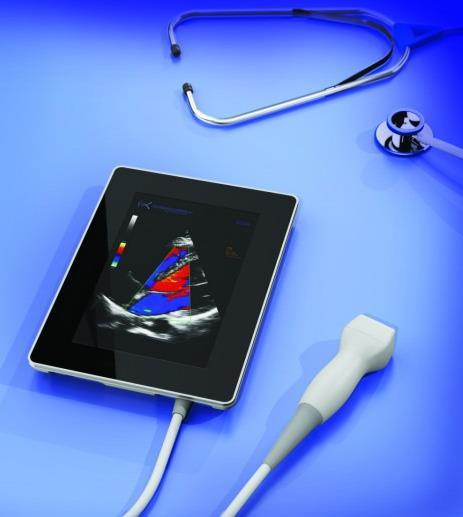
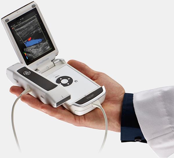
6 Ultrasound Ultrasound has advanced from a specialized imaging technique to a bedside test & clinical examination supplement Ultrasound is the principal imaging modality for visualization of the kidneys & urinary tract


7 In a patient with renal failure.

8 In a patient with AKI
9 Role of US Confirm normal anatomical position of kidneys Exclude structural anomalies. Assess size of the kidneys and collecting systems Exclude renal cortical scarring. Exclude renal or suprarenal masses (cystic or solid) Assess bladder filling and emptying
10 Common Neonatal & Pediatric Pathology Fusion Anomalies. (horseshoe, ectopia, cross-fusion) Hypoplasia or agenesis. Duplication anomalies. (supernumerary or variants of the collecting system and uterers) Congenital structural disease (Juveline PCKD, MCDK, dysplasia) Solid tumours
11 Limitations Co-operation is the biggest challenge with any pediatric study. If scanning a neonate, try to time the scan after a feed for best compliance. Full bladder if cooperative, bladder 1 st (before void) if not. Use WARM gel Ultrasound CANNOT exclude vesico-ureteric reflux.
12 EXAMINATION TECHNIQUE
13 Equipment & position 5+MHz curvilinear probe 3.5 MHz for larger adolescents High frequency (superficial) linear probe 8-12 MHz resolution Supine position Essential for bladder May use contra-lateral with caregiver support, posterolateral imaging, for kidneys Prone (if gases preclude visualization)
14 Scanning: Kidneys Confirm normal position Measure renal length Cortical thickness & echogenicity, CM differentiation, pyramids Cortical scars, cysts, NC Assess pelvis, calyces Scan entire kidney in LS & TS, may use higher freq. probe for detailed scan of cortex & med. Pyramids
15
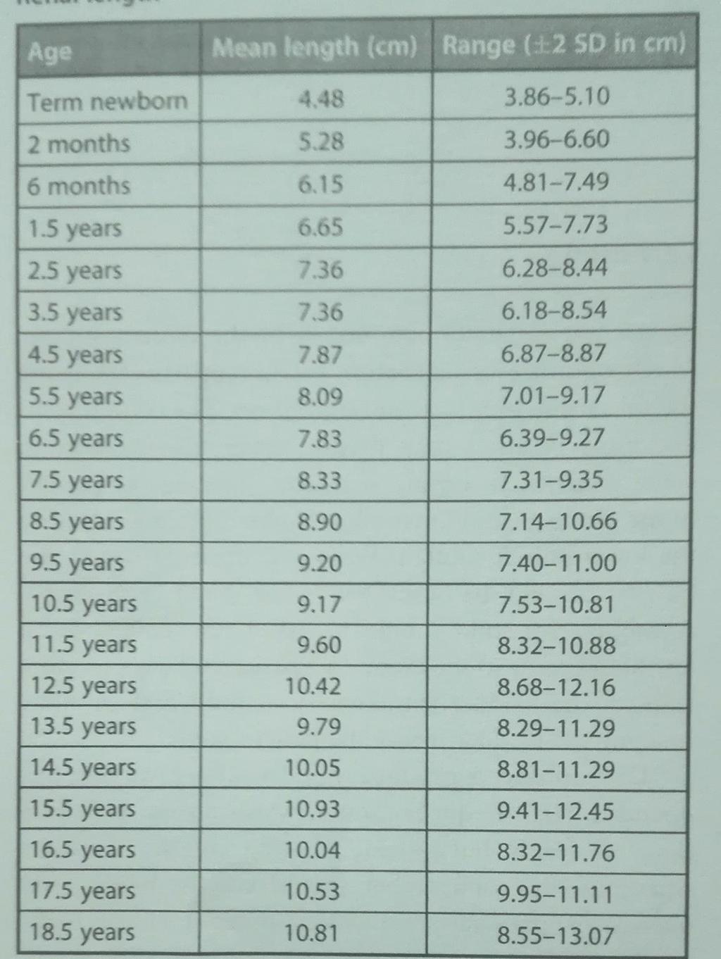
16 Renal length Supine measurement (prone underestimates) 2x Size correlates with pt height/ length more than age Lt kidney slightly larger (-5mm) in most cases
17
18
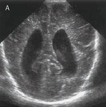
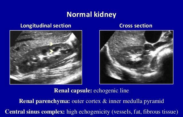
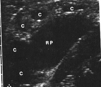
19 Features The renal cortex in patients older than 6 months of age is nearly always hypoechoic relative to the adjacent liver or spleen. The normal medullary pyramids are (minimally) hypoechoic. The identification of these pyramids is easier with hydration of the patient and diuresis. The renal sinus appears as a central echogenic area. (may be minimal-decreased vs adult) The renal pelvis, when visible, should be 10 mm or less in AP diameter.
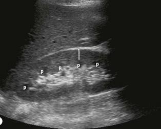
20 Neonatal kidney; 3 features 1. Echogenicity ( no of glomeruli ). 2. Prominent hypoechoic renal pyramids (larger medullary volumes, CM diff). Don t misinterpret as dilated collecting system 3. Renal sinus echogenicity (paucity of echogenic pelvic/ medullary fat)
21 Scanning: Kidneys Confirm normal position Measure renal length Cortical thickness & echogenicity, CM differentiation, pyramids Cortical scars, cysts, NC Assess pelvis, calyces Scan entire kidney in LS & TS, may use higher freq. probe for detailed scan of cortex & med. Pyramids
22 Echogenicity Renal parenchymal disease eg GN, Nephrotic present as echogenicity Cortical echogenicity Grade 0 Grade I Grade II Grade III Less than normal liver Equals normal liver Exceeds liver; less than renal sinus Exceeds liver; equals renal sinus echo Pyramids should be hypoechoic echogenicity suggests nephritis Echogenic lines throughout severe PN
23 Scanning: Kidneys Confirm normal position Measure renal length Cortical thickness & echogenicity, CM differentiation, pyramids Cortical scars, cysts, NC Assess pelvis, calyces urolithiasis Scan entire kidney in LS & TS, may use higher freq. probe for detailed scan of cortex & med. Pyramids
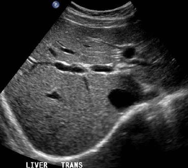
24 Collecting system
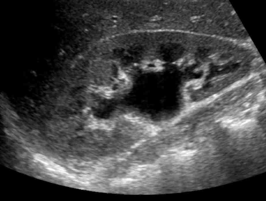
25 Pelvicalyceal without ureteric dilatation PUJO Intrauterine HN post-natal scan 4-5 d not before (dehydration & low GFR may give false ve early)
26 Grading of hydronephrosis Society of fetal urology grading system for hydronephrosis Grade U/S 0 No hydronephrosis 1 Only renal pelvis is seen 2 Renal pelvis & few calyces are seen 3 Virtually all calyces are seen 4 Virtually all calyces are seen + parenchymal thinning

27 Partial Duplex Kidney
28 Pyelonephritis Normal size & echogenicity Thickening of the wall of renal pelvis & calyceal distortion


29 Cortical scar KEYPOINTS: Scar- linear echogenic line from the cortical edge in towards a pyramid
30 In both the neonatal and paediatric kidney, the foetal cortical lobulations are pronounced and should span the pyramids If a lobulation dips into a pyramid, it is likely to be a cortical scar


31 Pyonephrosis (dilated system & echogenic content)

32 Stones highly echogenic + acoustic shadowing

33 Stones highly echogenic + acoustic shadowing Stone in pelvis Acoustic posterior shaddow


34 Nephrocalcinosis Calcium deposition in the renal parenchyma Medullary hyperechoic pyramids Diffuse cortical & medullary echog.
35 Nephrocalcinosis Causes: Idiopathic hypercalciuria Long term furosemide therapy in neonates esp. premature Hypervitaminosis D Hyperparathyroidism Renal tubular acidosis Hyperoxaluria Medullary sponge kidney Other causes of hyperechoic medullary pyramids: Tamm Horsfall proteins Vascular congestion Papillary necrosis. Transient in neonate with oliguria & perinatal anoxia.
36 Cystic renal diseases


37 Cystic renal diseases Small
 Turner syndrome Tuberous sclerosis. Cystic disease of renal medulla unilateral Simple Cyst. Multi Cystic Dysplasia.")
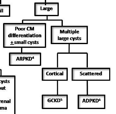
38 Renal Cystic disease Bilateral AR polycystic Kid AD polycystic Kid Glomerulo Cystic Disease Cysts associated with multiple malformation syndrome (e.g.) Turner syndrome Tuberous sclerosis. Cystic disease of renal medulla unilateral Simple Cyst. Multi Cystic Dysplasia. Multilocular Cystic nephroma
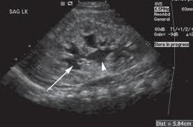
39 Polycystic Kidney Disease A.R Multiple small cysts1-2mm (dilated collecting tubules) Congenital hepatic fibrosis (presented later). By ultrasound Bilateral enlarged echogenic kidneys with poor delineation of the renal sinus, medulla and cortex. Presented in neonatal. Period with kidney function A.D. Large cysts are present in both kidneys. Congenital hepatic fibrosis is rare By ultrasound In neonates the same as A.R.polycystic kidney. In old child : multiple larg cysts in both kidneys Presented in 4 th or 5 th decade with hypertension or hematuria. Rarely in neonate presented with Abd. Mass.

40 ADPKD CRITERIA each 4+ each + EXCLUDES
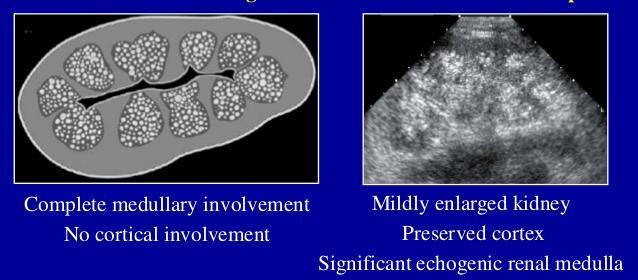
41 ARPKD
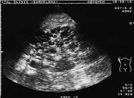
42 ARPKD


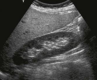
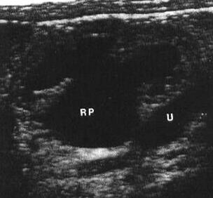
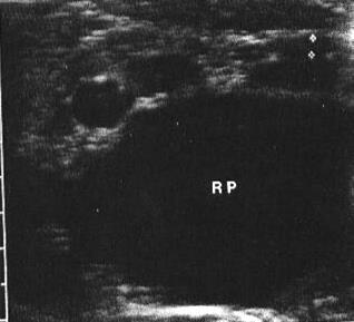
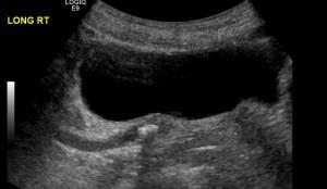
43 Multicystic-Dysplastic Kidney 2nd cause of neonatal abdominal mass after hydronephrosis. *kidney replaced by anechoic masses of variable size with: no communication between cysts no identifiable renal pelvis No normal (dysplastic) renal parenchyma *Scan No uptake
44 Bladder Urine Wall thickness (3mm full, empty: 5mm but may give false impression of ) Chronic pressure or infection Defects, stones, focal thickening, etc Lower ureter
45 Scanning: Bladder Begin in transverse with a slight caudal angle. Sweep trough the bladder for any structural defects or focal wall thickening. Distal ureteric dilatation, Ureteroceles, ureteric jets by Doppler. Post-voiding residual urine

46 Thick Bladder Wall

47 Bladder hematoma (post-biopsy)
48 Cystitis Diffuse thickening of bladder wall with extensive involvement, the inflammatory lesion can protrude into bladder lumen mimiking Rhabdomyo sarcoma of bladder Diff by cystoscopy or follow up after treatment of infection
49 PUV The most common cause of uretheral obst in boys. By ultrasound Bilateral hydronephrosis & Hydroureter Thick bladder wall
50

51 Ureterocele Congenital or infl. Obstruction ureter near trigone ballooning just proximal intravesical mass with thin sonolucent wall


52 Mega ureter Non obst Non refluxing Obst non refluxing Non obst. refluxing primary Secondary primary Secondary primary Secondary Idiopathic Ureteric dilation D.I Structure stenosis Calculi ureterocele Neurogenic bladder ureterocele Short or absent intranvesical ureter
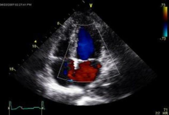
53 Special techniques Doppler imaging Renal vessels Ureteric jets Voiding urosonography (contrast/ Doppler) Post-voiding residual urine

54 Doppler imaging *Arterial *Venous *Perfusion

")

55 RVT: enlarged echog (oedema)

56 Ureteric jets by Doppler
57 Other roles of U/S Interventional Nephrology Biopsies, CVCs, etc Critical care nephrology
58 Therefore Ultrasound is the principal imaging modality for visualization of the kidneys & urinary tract Clinical examination & diagnostic tests are increasingly being integrated Remember ABG, CBG, dipstick, POC-testing, CXR,. A basic ultrasound examination can add a lot to a nephrology assessment And can guide further imaging/ investigation if needed


59
60 1. Multicystic dysplastic kidney is usually associated with A B C D multiple communicating renal cysts liver cysts polyhydramnios no uptake on isotopic scan
61 2. A neonate with unilateral hugely dilated renal pelvis without ureteric dilatation is most probably having A B C D vesicoureteric reflux pelviureteric obstruction posterior urethral valve ureterocele
62 3. Compared to older children, neonatal kidneys generally feature A B C D larger size increased echogenicity highly echogenic renal sinus absent renal pyramids
Obstetrics Content Outline Obstetrics - Fetal Abnormalities
 Obstetrics Content Outline Obstetrics - Fetal Abnormalities Effective February 2007 10 16% renal agenesis complete absence of the kidneys occurs when ureteric buds fail to develop Or degenerate before
Chapter 6: Genitourinary and Gastrointestinal Systems 93
 Chapter 6: Genitourinary and Gastrointestinal Systems 93 Chapter 6 Genitourinary and Gastrointestinal Systems Embryology Three sets of excretory organs or kidneys develop in human embryos: Pronephros:
RENAL SCINTIGRAPHY IN THE 21 st CENTURY
 RENAL SCINTIGRAPHY IN THE 21 st CENTURY 99m Tc- MAG 3 with zero time injection of Furosemide (MAG 3 -F 0 ) : A Fast and Easy Protocol, One for All Indications Clinical Experience Congenital Disorders PROTOCOL
GU Ultrasound in First Trimester
 Fetal Renal Malformations: The Role of Ultrasound in Diagnosis & Management Outline 1. Renal Anomalies Urinary Tract Dilation Aberrant Early Development Defects Terminal Maturation Alfred Abuhamad, M.D.
Acute flank pain in children: Imaging considerations
 Acute flank pain in children: Imaging considerations Carlos J. Sivit MD Rainbow Babies and Children s Hospital Case Western Reserve School of Medicine Flank pain Results from distention of ureter or renal
Fetal Renal Malformations: The Role of Ultrasound in Diagnosis & Management
 Fetal Renal Malformations: The Role of Ultrasound in Diagnosis & Management 12 weeks Alfred Abuhamad, M.D. Eastern Virginia Medical School 13 weeks 2nd trimester Medullary pyramids Renal Sinus Cortex 2nd
Excretory urography (EU) or IVP US CT & radionuclide imaging
 or IVP US CT & radionuclide imaging") Excretory urography (EU) or IVP US CT & radionuclide imaging MRI arteriography studies requiring catherization or direct puncture of collecting system EU & to a lesser extent CT provide both functional
Excretory urography (EU) or IVP US CT & radionuclide imaging MRI arteriography studies requiring catherization or direct puncture of collecting system EU & to a lesser extent CT provide both functional
Urinary system Ultrasound (Renal & Urinary bladder)
") Urinary system Ultrasound (Renal & Urinary bladder) Edited & Presented by ; Hussien A.B ALI DINAR. Msc.Phd ISRRT Associate Member Lecturer (National university) Reporting Sonographer (PHC) Objective By
Urinary system Ultrasound (Renal & Urinary bladder) Edited & Presented by ; Hussien A.B ALI DINAR. Msc.Phd ISRRT Associate Member Lecturer (National university) Reporting Sonographer (PHC) Objective By
Proceedings of the 34th World Small Animal Veterinary Congress WSAVA 2009
 www.ivis.org Proceedings of the 34th World Small Animal Veterinary Congress WSAVA 2009 São Paulo, Brazil - 2009 Next WSAVA Congress : Reprinted in IVIS with the permission of the Congress Organizers IMAGING
Abdominal Ultrasound : Aorta, Kidneys, Bladder
 Abdominal Ultrasound : Aorta, Kidneys, Bladder Nilam J. Soni, MD, MSc Associate Professor of Medicine Divisions of Hospital Medicine and Pulmonary/Critical Care Medicine Department of Medicine University
Urinary Tract Abnormalities
 Urinary Tract Abnormalities Dr Hennie Lombaard Senior Specialist Maternal and Fetal Medcine Department of Obstetrics and Gynecology Level 7 Pretoria Academic Hospital Pictures from The 18 to 23 weeks scan
US in non-traumatic acute abdomen. Lalita, M.D. Radiologist Department of radiology Faculty of Medicine ChiangMai university
 US in non-traumatic acute abdomen Lalita, M.D. Radiologist Department of radiology Faculty of Medicine ChiangMai university Sagittal Orientation Transverse (Axial) Orientation Coronal Orientation Intercostal
My Patient Has Abdominal Pain PoCUS of the Biliary Tract and the Urinary Tract
 My Patient Has Abdominal Pain PoCUS of the Biliary Tract and the Urinary Tract Objectives PoCUS for Biliary Disease PoCUS for Renal Colic PoCUS for Urinary Retention Biliary Disease A patient presents
Congenital Pediatric Anomalies: A Collection of Abdominal Scintigraphy Findings: An Imaging Atlas
 ISPUB.COM The Internet Journal of Nuclear Medicine Volume 5 Number 1 Congenital Pediatric Anomalies: A Collection of Abdominal Scintigraphy Findings: An Imaging Atlas V Vijayakumar, T Nishino Citation
CYSTIC DISEASES of THE KIDNEY. Dr. Nisreen Abu Shahin
 CYSTIC DISEASES of THE KIDNEY Dr. Nisreen Abu Shahin 1 Types of cysts 1-Simple Cysts 2-Dialysis-associated acquired cysts 3-Autosomal Dominant (Adult) Polycystic Kidney Disease 4-Autosomal Recessive (Childhood)
Functions of the kidney:
 Diseases of renal system : Normal anatomy of renal system : Each human adult kidney weighs about 150 gm, the ureter enters the kidney at the hilum, it dilates into a funnel-shaped cavity, the pelvis, from
Developmental Abnormalities of the Kidneys and GU System
 A5 Developmental Abnormalities of the Kidneys and GU System Erin Parilla, MD Neonatologist Pediatrix Medical Group, Tampa, FL The speaker has signed a disclosure form and indicated she has no significant
Kidneys and Urinary Tract Content Outline. Anatomy Coverings. Location. (Effective February 2007) (16%-24%)
 (16%-24%)") Kidneys and Urinary Tract Content Outline (Effective February 2007) (16%-24%) Anatomy Coverings true capsule perirenal fat surrounds capsule Gerota s fascia separates perirenal from extraperitoneal fat
Kidneys and Urinary Tract Content Outline (Effective February 2007) (16%-24%) Anatomy Coverings true capsule perirenal fat surrounds capsule Gerota s fascia separates perirenal from extraperitoneal fat
27-Apr-15 1 UAF ANOMALIES OF DEVELOPMENT RENAL SYSTEM - 1 DR. MUHAMMAD TARIQ JAVED UAF UAF
 RENAL SYSTEM - 1 DR. MUHAMMAD TARIQ JAVED Professor, Department of Pathology, Faculty of Veterinary Science, University of Agriculture, Faisalabad, Pakistan. Email: mtjaved@uaf.edu.pk RENAL AGENESIS Renal
ISUOG Basic Training. Distinguishing between Normal & Abnormal Appearances of the Urinary Tract. Seshadri Suresh, India
 ISUOG Basic Training Distinguishing between Normal & Abnormal Appearances of the Urinary Tract Seshadri Suresh, India Learning objectives 13 & 14 At the end of the lecture you will be able to: describe
Abdomen and Retroperitoneum Ultrasound Protocols
 Abdomen and Retroperitoneum Ultrasound Protocols Reviewed By: Anna Ellermeier, MD Last Reviewed: March 2018 Contact: (866) 761-4200, Option 1 **NOTE for all examinations: 1. If documenting possible flow
PROFESSIONAL SKILLS 1 3RD YEAR SEMESTER 6 RADIOGRAPHY. THE URINARY SYSTEM Uz. Fatema shmus aldeen Tel
 PROFESSIONAL SKILLS 1 3RD YEAR SEMESTER 6 RADIOGRAPHY THE URINARY SYSTEM Uz. Fatema shmus aldeen Tel. 0925111552 Professional skills-2 THE URINARY SYSTEM The urinary system (review anatomy and physiology)
Uroradiology For Medical Students
 Uroradiology For Medical Students Lesson 4: Cystography & Urethrography - Part 2 American Urological Association Review Cystography is useful in evaluating the bladder, the urethra and the competence of
Abdominal ultrasound:
 Abdominal ultrasound: Non-traumatic acute abdomen Wittanee Na-ChiangMai, MD Department of Radiology ChiangMai University 26/04/2017 Contents Technique of examination Normal anatomy Emergency conditions
Radiological and pathologic findings of fetal renal cystic diseases and associated fetal syndromes: A pictorial review
 Radiological and pathologic findings of fetal renal cystic diseases and associated fetal syndromes: A pictorial review Poster No.: C-2835 Congress: ECR 2010 Type: Educational Exhibit Topic: Pediatric Authors:
Renal Ultrasonography in Neonates. Abstract: Introduction:
 Renal Ultrasonography in Neonates. Mahmoud Adel Abdel-Moneim, Mahmoud Mohi-Eldin El-Kersh, Mohamed Alaa Thabet, Magdy Abdel-Fattah Ramadan and Hossam H. Zeid. From Department of Pediatrics, Faculty of
Guidelines, Policies and Statements D5 Statement on Abdominal Scanning
 Guidelines, Policies and Statements D5 Statement on Abdominal Scanning Disclaimer and Copyright The ASUM Standards of Practice Board have made every effort to ensure that this Guideline/Policy/Statement
Fetal Urologic Anomalies
 Fetal Urologic Anomalies Kathryn Drennan, MD Elizabeth McKinney, MD MultiCare Regional Maternal-Fetal Medicine What you should know They are common Account for 15%-20% of all congenital anomalies Associated
Hydronephrosis. Nephrosis. Refers to the kidney
 What is hydronephrosis? Hydro Nephrosis Refers to water or fluid Refers to the kidney A build-up of fluid (urine) in the kidney is the medical term for a build-up of urine in the kidney. As the urine builds
Pelvi-Ureteric Junction Obstruction Revisited
 Dr. Bimalendu Mukherjee was trained in Urology in the UK between 1956 to 1961. Upon return to India, he took up a teaching position in Calcutta National Medical College and ultimately retired as Professor
Summary and conclusions
 Summary and conclusions 7 Chapter 7 68 Summary and conclusions Chapter 1 provides a general introduction to this thesis focused on the use of ultrasound (US) in children with abdominal problems. The literature
PAEDIATRIC RENAL IMAGING. Dr A Brink
 PAEDIATRIC RENAL IMAGING Dr A Brink Causes of hydronephrosis includes: Pelvi-ureteric obstruction Vesico-ureteric reflux Vesico-ureteric obstruction Posterior uretral valves Duplex kidneys Radiopharmaceutical
Outline. Introduction to imaging modalities of the urinary system. Case base learning of common diseases in urinary tract
 Outline Introduction to imaging modalities of the urinary system Case base learning of common diseases in urinary tract Outline Introduction to imaging modalities of the urinary system Case base learning
Outline. Introduction to imaging modalities of the urinary system. Case base learning of common diseases in urinary tract
 Outline Introduction to imaging modalities of the urinary system Case base learning of common diseases in urinary tract Diagnostic Investigations in Urinary System PLAIN KUB EXCRETORY UROGRAPHY RETROGRADE
Basic of Ultrasound Physics E FAST & Renal Examination. Dr Muhammad Umer Ihsan MBBS,MD, DCH CCPU,DDU1,FACEM
 Basic of Ultrasound Physics E FAST & Renal Examination Dr Muhammad Umer Ihsan MBBS,MD, DCH CCPU,DDU1,FACEM What is Sound? Sound is Mechanical pressure waves What is Ultrasound? Ultrasounds are sound waves
1. Normal and pathological embryology of the urinary and genital tract 2. Nephrology 3. Infection
 1 1. Normal and pathological embryology of the urinary and genital tract 1.1. Development of the kidney and ureter 1.2. Development of the bladder and the urethra 1.3. Development of the female genital
Renal Cystic Disease. Dr H Bierman
 Renal Cystic Disease Dr H Bierman Objectives Be able to diagnose renal cystic disease Genetic / non-genetic Be able to describe patterns of various renal cystic disease on routine imaging studies Be able
Advanced Concept of Nursing- II UNIT-VI Advance Nursing Management of Genitourinary (GU) Diseases.
 Diseases.") In The Name of God (A PROJECT OF NEW LIFE COLLEGE OF NURSING KARACHI) Advanced Concept of Nursing- II UNIT-VI Advance Nursing Management of Genitourinary (GU) Diseases. Shahzad Bashir RN, BScN, DCHN,MScN
In The Name of God (A PROJECT OF NEW LIFE COLLEGE OF NURSING KARACHI) Advanced Concept of Nursing- II UNIT-VI Advance Nursing Management of Genitourinary (GU) Diseases. Shahzad Bashir RN, BScN, DCHN,MScN
URINARY SYSTEM I. Kidneys II. Nephron Unit and Urine Formation
 URINARY SYSTEM I. Kidneys A. Location and Structure 1. Retroperitoneal 2. Between T12 and L3 3. Rt. kidney slightly lower 4. Two bean shaped organs 5. Adrenal gland 6. Internal construction a. Renal cortex
Cystic Renal Disease, for USMLE Step One. Howard J. Sachs, MD
 Cystic Renal Disease, for USMLE Step One Howard J. Sachs, MD www.12daysinmarch.com The Major Players Medullary Sponge Kidney (MSK) Polycystic Kidney Disease (PKD) Autosomal Recessive: Childhood Autosomal
이학종분당서울대학교병원. Ultrasound in Urinary Colic
 이학종분당서울대학교병원 Ultrasound in Urinary Colic U l t r a s o u n d i n U r i n a US: Normal Kidney r y C o l i c Contents 1. 1. Definition and clinical consideration 2. 2. Pathophysiology 3. 3. US in in obstructive
Obstructive Uropathy. PATHOPHYSIOLOGIC CHANGES UUO vs BUO. Arry Rodjani Urology Department Ciptomangunkusumo Hospital Jakarta
 Obstructive Uropathy PATHOPHYSIOLOGIC CHANGES UUO vs BUO Arry Rodjani Urology Department Ciptomangunkusumo Hospital Jakarta INTRODUCTION Obstructive uropathy refers to the functional or anatomic obstruction
The functional anatomy of the urinary system. Human Anatomy Department Dr. Anastasia Bendelic
 The functional anatomy of the urinary system Human Anatomy Department Dr. Anastasia Bendelic Plan Development of the kidneys and their abnormalities Development of the urinary ways and their abnormalities
Prenatal Hydronephrosis
 Prenatal Hydronephrosis What is hydronephrosis? Hydronephrosis is dilation of the kidney, specifically the renal pelvis (place where urine is stored after its production). This can be the result of an
Case MDCT 3D reconstructed features of posterior urethral valve
 Case 12688 MDCT 3D reconstructed features of posterior urethral valve Hidayatullah Hamidi Third year Resident of Radiology French medical institute for children Radiology Department; Kabul, Afghanistan;
Imaging Ejaculatory Disorders and Hematospermia
 ATHENS 4-6 October 2018 European Society of Urogenital Radiology Imaging Ejaculatory Disorders and Hematospermia Parvati Ramchandani, MD Professor, Radiology and Surgery University of Pennsylvania Medical
Urinary tract infections, renal malformations and scarring
 Urinary tract infections, renal malformations and scarring Yaacov Frishberg, MD Division of Pediatric Nephrology Shaare Zedek Medical Center Jerusalem, ISRAEL UTI - definitions UTI = growth of bacteria
Role of imaging in RCC. Ultrasonography. Solid lesion. Cystic RCC. Solid RCC 31/08/60. From Diagnosis to Treatment: the Radiologist Perspective
 Role of imaging in RCC From Diagnosis to Treatment: the Radiologist Perspective Diagnosis Staging Follow up Imaging modalities Limitations and pitfalls Duangkamon Prapruttam, MD Department of Therapeutic
Clinical-Radiological management of congenital hydronephrosis.
 Clinical-Radiological management of congenital hydronephrosis. Poster No.: C-0983 Congress: ECR 2015 Type: Authors: Keywords: DOI: Educational Exhibit M. Vidal, D. Llanos, E. Pallares, I. de la Pedraja,
1. Hypogonadism is usually encountered in the following conditions, except
 1. Hypogonadism is usually encountered in the following conditions, except A. Congenital adrenal hyperplasia B. Noonan Syndrome C. Prader-Willi Syndrome D. Bardet-Biedl Syndrome 2. A 6 year old girl with
R adio logical investigations of urinary system
 R adio logical investigations of urinary system There are 4 main radiological Ix: 1 IVU: Intravenous urography. 2- U/S 3-CT scan 4-Radioisotope scan. Others (not frequently used): MRI, arteriography, antegrade
Renal Disease. Please refer to the assignment page Three online modules TBLs
 Renal Disease Please refer to the assignment page Three online modules TBLs 1 Renal Embryology 2 Lab Tests UA CBC Enzymes Creatinine Creatinine clearance Ammonia Abs C Bx 3 BUN Creatinine Creatinine Clearance
Genetics in Nephrology. Saeid Morovvati Associate Professor of BMSU Director of Biogene Laboratory
 Genetics in Nephrology Saeid Morovvati Associate Professor of BMSU Director of Biogene Laboratory Genetics in: A. Congenital Anomalies of the Kidney and Urinary Tract B. Cystic Diseases of the Kidney C.
Indications and effectiveness of the open surgery in vesicoureteral reflux
 Indications and effectiveness of the open surgery in vesicoureteral reflux Suzi DEMIRBAG, MD Department of Pediatric Surgery, Gulhane Military Medical Academy, Ankara, TURKEY Vesicoureteral reflux (VUR)
Development of the urinary system
 Development of the urinary system WSO School of Biomedical Sciences, University of Hong Kong. 3 sets of kidneys developing in succession (temporally and spatially) : Pronephros ] Mesonephros ]- Intermediate
Index. Note: Page numbers of article titles are in boldface type.
 Magn Reson Imaging Clin N Am 12 (2004) 587 591 Index Note: Page numbers of article titles are in boldface type. A Adenoma(s), adrenal, gadolinium-enhanced MR imaging in, 533 534 hyperfunctioning versus
Caveat sonologist Mistakes to avoid in Kidney Ultrasound
 Caveat sonologist Mistakes to avoid in Kidney Ultrasound Simon Freeman Derriford Hospital, Plymouth simonfreeman@nhs.net Bear trap 1 Report: There is a 4cm solid mass arising from the left kidney likely
Perineal Sonography in Diagnosis of an Ectopic Ureteric Opening Into the Urethra
 Case Series Perineal Sonography in Diagnosis of an Ectopic Ureteric Opening Into the Urethra S. Boopathy Vijayaraghavan, MD, DMRD Objective. To study the role of perineal sonography in the diagnosis of
IMAGING PROFILE OF CHILDREN BIRTH TO 12 YEARS PRESENTING WITH FIRST URINARY TRACT INFECTION (UTI) AT A TERTIARY CARE HOSPITAL
 AT A TERTIARY CARE HOSPITAL") IMAGING PROFILE OF CHILDREN BIRTH TO 12 YEARS PRESENTING WITH FIRST URINARY TRACT INFECTION (UTI) AT A TERTIARY CARE HOSPITAL Yengkhom Rameshwor Singh 1, Okram Pusparani Devi 2, Tonjam Hemchand Singh 3
IMAGING PROFILE OF CHILDREN BIRTH TO 12 YEARS PRESENTING WITH FIRST URINARY TRACT INFECTION (UTI) AT A TERTIARY CARE HOSPITAL Yengkhom Rameshwor Singh 1, Okram Pusparani Devi 2, Tonjam Hemchand Singh 3
Abdominal Ultrasound. Diane Hallinen, MD. Bloodroot
 Abdominal Ultrasound Diane Hallinen, MD Bloodroot Abdominal Ultrasound Vasculature Hepatobiliary Spleen Kidney Bladder Bowel Where to put the probe? Vasculature We are going to talk about Celiac Trunk
Pediatric Ure-Radiology*
 Pediatric Ure-Radiology* HERMAN GROSSMAN, M.D. Professor of Radiology and Pediatrics, Duke University Medical Center, Durham, North Carolina "Routine" radiologic studies do not, often enough, concentrate
Contents. Review anatomy of the urinary tract Imaging modalities
 Contents Review anatomy of the urinary tract Imaging modalities The Urinary Tract Kidneys ตาแหน งไต (position) อย ใน retroperitoneum ระด บ T12-L3 โดยไต ขวาจะม ระด บตากว าไตซ ายเล กน อย ร ปร าง (shape)
Hydronephrosis. What is hydronephrosis?
 What is hydronephrosis? Hydronephrosis Hydronephrosis describes the situation where the urine collecting system of the kidney is dilated. This may be a normal variant or it may be due to an underlying
Policies, Standards, and Guidelines. Guidelines for Abdominal Ultrasound Examination
 Policies, Standards, and Guidelines Guidelines for Abdominal Ultrasound Examination Approved by Council Feb 2018 Disclaimer and Copyright The ASUM Standards of Practice Board have made every effort to
16.1 Risk of UTI recurrence in children
 16. UTI prognosis 16.1 Risk of UTI recurrence in children Key question: What is the risk of recurrent UTI in children with no known structural or functional abnormalities of the urinary tract with a first
Controversies around antenatally detected PUJ syndrom. Amy Piepsz, CHU St Pierre, Brussels, Belgium
 Controversies around antenatally detected PUJ syndrom Amy Piepsz, CHU St Pierre, Brussels, Belgium Editors : Anthony Caldamone, USA Pierre Mouriquand, France Newborn boy History of prenatally diagnosed
Table of Contents: OVERVIEW AND INTRODUCTION. Imaging Approaches RETROPERITONEUM. Introduction to the Retroperitoneum
 Table of ontents: OVERVIEW AND INTRODUTION Imaging Approaches RETROPERITONEUM Introduction to the Retroperitoneum Duplications and Anomalies of IV Inflammati Retroperitoneal Fibrosis Deg Pelvic Lipomatosis
CONGENITAL ANOMALIES UROLOGY SUB DIVISION DEPARTMENT OF SURGERY MEDICAL SCHOOL UNIVERSITY OF SUMATERA UTARA
 CONGENITAL ANOMALIES UROLOGY SUB DIVISION DEPARTMENT OF SURGERY MEDICAL SCHOOL UNIVERSITY OF SUMATERA UTARA UPPER URINARY TRACT Abnormalities of the kidney position & number 1. Simple ectopia 2. Thoracic
Autosomal Dominant Polycystic Kidney Disease
 Case Studies [1] July 01, 2014 By Amar Udare, MBBS [2] Case History: 45-year-old female with vague pain in the abdomen. Case History: A 45-year-old female presented with vague pain in the abdomen. A USG
Vesicoureteral reflux and reflux nephropathy
 Vesicoureteral reflux and reflux nephropathy This infokid topic is for parents and carers about children s kidney conditions. Visit www.infokid.org.uk to find more topics about conditions, tests & diagnosis,
Zoltan Harkanyi M.D., Ph.D. Department of Radiology, Heim Pal Children s Hospital, Budapest, Hungary
 Zoltan Harkanyi M.D., Ph.D. Department of Radiology, Heim Pal Children s Hospital, Budapest, Hungary CEUS expereince 10 years Department of Radiology, Heim Pal Children s Hospital, Budapest US N o 1 study
Role of imaging in evaluation of genitourinary i trauma Spectrum of GU injuries Relevance of imaging findings in determining management Focus on MDCT
 Genitourinary Tract Injuries 6 th Nordic Course Scott D. Steenburg, MD Assistant Professor University of Maryland Department of Radiology Division of Trauma and Emergency Radiology R Adams Cowley Shock
Detection of Congenital Renal Anomalies in Children being Investigated by Tc99m-DMSA Renal Scan
 Detection of Congenital Renal Anomalies in Children being Investigated by Tc99m-DMSA Renal Scan Mais Halaseh MD*, Khaled Alkhawaldeh MD*, Akram Al-Ibraheem MD*, Hamza Al Adwan MD*, Hussam Al-Kaylani MD*
Uroradiology Tutorial For Medical Students
 Uroradiology Tutorial For Medical Students Lesson 3: Cystography & Urethrography Part 1 American Urological Association Introduction Conventional radiography of the urinary tract includes several diagnostic
IMAGING OF THE UROGENITAL TRACT
 IMAGING OF THE UROGENITAL TRACT 1 A) URINARY TRACT There are many methods of imaging the urinary tract but plain abdominal X-ray and ultrasound scan are usually done first in most cases, especially in
The Evolving Role of Antibiotic Prophylaxis for Vesicoureteral Reflux. Stephen Canon, MD Children s Urology
 The Evolving Role of Antibiotic Prophylaxis for Vesicoureteral Reflux Stephen Canon, MD Children s Urology www.childrensurology.com May 17, 2008 Objectives Review literature establishing the value of antibiotic
Cystic Renal Disease of Childhood
 Acta Radiológica Portuguesa, Vol.XIX, nº 74, pág. 90-107, Abr.-Jun., 2007 Cystic Renal Disease of Childhood Ellen Chung Armed Forces Institute of Pathology Terminology Cyst Polycystic kidney disease ARPKD
IT 의료융합 1 차임상세미나 복부질환초음파 이재영
 IT 의료융합 1 차임상세미나 2013-4-3 복부질환초음파 이재영 나는오늘누구를위하여 종을울리나? 전통적의료 의사 공학설계자 의사 최첨단진단장비들 USG, CT, MRI 환자 환자 현대의료 사용자중심의사고 US in the Abdomen Detection DDx Look Behavior Response by external stimuli Guiding Tool
Case Report Diagnosis of Rare Association of Orthotopic Multicystic Dysplasia with Crossed Fused Renal Ectopia
 Case Reports in Urology, Article ID 140850, 4 pages http://dx.doi.org/10.1155/2014/140850 Case Report Diagnosis of Rare Association of Orthotopic Multicystic Dysplasia with Crossed Fused Renal Ectopia
Radiological Assessment of the Kidney in Patients with Hematuria
 March 2005 Radiological Assessment of the Kidney in Patients with Hematuria Jeremy L. McKay, Harvard Medical School Year III Hematuria Signs and Symptoms Microscopic or gross hematuria Abdominal pain Fever
DISEASES AFFECTING TUBULES AND INTERSTITIUM
 DISEASES AFFECTING TUBULES AND INTERSTITIUM Acute tubular injury (ATI) Pyelonephritis Drug-induced tubulointerstitial nephritis (TIN) Myeloma cast NP Renal stones Urinary outflow obstruction: hydronephrosis
Nuclear medicine methods in the urogenital system
 Nuclear medicine methods in the urogenital system Anatomy of the kidneys I. Anatomy of the kidneys II. The types of examinations Static examinations (scintigraphy): 1) the radiopharmaceutical is administered
RADIO-NUCLIDE STUDIES FOR THE EVALUATION OF KIDNEY FUNCTIONS. DR RIANA NEL NUCLEAR MEDICINE DEPT 27 Sept 2010
 RADIO-NUCLIDE STUDIES FOR THE EVALUATION OF KIDNEY FUNCTIONS DR RIANA NEL NUCLEAR MEDICINE DEPT 27 Sept 2010 Alive patient RA + tracers Tc-99m(86%) + MAG 3 DMSA (DTPA) T½ = 6,5h Gamma camera ect. Radio-active
Genitourinary Radiology In-Training Test Questions for Diagnostic Radiology Residents
 Genitourinary Radiology In-Training Test Questions for Diagnostic Radiology Residents March, 2013 Sponsored by: Commission on Education Committee on Residency Training in Diagnostic Radiology 2013 by American
Alterations of Renal and Urinary Tract Function
 Alterations of Renal and Urinary Tract Function Chapter 29 Urinary Tract Obstruction Urinary tract obstruction is an interference with the flow of urine at any site along the urinary tract The obstruction
FHS Appendicitis US Protocol
 FHS Appendicitis US Protocol Reviewed By: Shireen Khan, MD; Sarah Farley, MD; Anna Ellermeier, MD Last Reviewed: May 2018 Contact: (866) 761-4200 **NOTE for all examinations: 1. If documenting possible
URINARY SYSTEM CHAPTER 28 I ANATOMY OF THE URINARY SYSTEM. Student Name
 Student Name CHAPTER 28 URINARY SYSTEM L iving produces wastes. Wherever people live or work or play, wastes accumulate. To keep these areas healthy, there must be a method of disposing of these wastes
Topic 5: Screening of the neonate/infant with prenatal hydronephrosis
 Topic 5: Screening of the neonate/infant with prenatal hydronephrosis Contents Index patient... 2 Introduction... 2 Methodology... 2 Outcomes Analysis... 3 Summary... 11 References... 12 Copyright 2010
Kristina M. Nowitzki, M.D., Ph.D. and Hao S. Lo, M.D. University of Massachusetts Medical School, Worcester, MA
 Kristina M. Nowitzki, M.D., Ph.D. and Hao S. Lo, M.D. University of Massachusetts Medical School, Worcester, MA Outline I. Introduction highlighting normal renal enhancement physiology including normal
Approach and Management of Obstructive Uropathy
 Approach and Management of Obstructive Uropathy INTRODUCTION One of the most urgent clinical entity need to be diagnosed 1. 10% of the causes of ARF and 4% of CRF 2. Symptoms and signs of obstruction are
Kidney Disease Profile of Syrian Refugee Children
 KIDNEY DISEASES Kidney Disease Profile of Syrian Refugee Children Mehtap Akbalık Kara, 1 Beltinge Demircioğlu Kılıç, 1 Nilgün Çöl, 2 Ayşe Aysima Özçelik, 3 Mithat Büyükçelik, 1 Ayşe Balat 1 1 Department
Lower Urinary Tract Obstruction LUTO or Bladder Outlet Obstruction BOO. Miss Harriet Corbett Consultant Paediatric Urologist
 Lower Urinary Tract Obstruction LUTO or Bladder Outlet Obstruction BOO Miss Harriet Corbett Consultant Paediatric Urologist Aims To give an overview of the anomalies we encounter presentation of LUTO how
Endoscopic Correction of Vesicoureteric Reflux in Children
 Endoscopic Correction of Vesicoureteric Reflux in Children Abstract Pages with reference to book, From 255 To 257 Khalid Rasheed ( Department of Surgery, The Aga Khan University Hospital, Karachi. ) Vesicoureteric
Unilateral renal agenesis does it matter?
 Clinical Research Facility Central Manchester University Hospitals NHS Foundation Trust Unilateral renal agenesis does it matter? Nicholas J A Webb BMedSci DM FRCP FRCPCH Honorary Professor of Paediatric
Multicystic dysplastic kidney: a review of eleven years ( )
") ORIGINAL ARTICLE Advance Access publication 14 February 2012 Multicystic dysplastic kidney: a review of eleven years (2000 2010) Helena Rios, Raquel Santos, Clara Gomes, António Jorge Correia Paediatric
ORIGINAL ARTICLE Advance Access publication 14 February 2012 Multicystic dysplastic kidney: a review of eleven years (2000 2010) Helena Rios, Raquel Santos, Clara Gomes, António Jorge Correia Paediatric
Renal survival analysis of CAKUT and outcomes in chronic kidney disease.
 Curr Pediatr Res 2017; 21 (4): 691-695 ISSN 0971-9032 www.currentpediatrics.com Renal survival analysis of CAKUT and outcomes in chronic kidney disease. Oke Rina Ramayani 1, Kiking Ritarwan 2, Putri Chairani
Scrotum Kacey Morrison Amanda Baxter Sabrina Tucker July 18, 2006 SCROTUM
 Scrotum Kacey Morrison Amanda Baxter Sabrina Tucker July 18, 2006 SCROTUM 1) Other Names: Scrotum None Testicles Testes (Curry Tempkin, p. 236, 2/3/2) Ductus deferens spermatic cord (Tempkin, p. 279, Anatomy
CHAPTER 2 NEW PATIENTS COMMENCING TREATMENT IN 2007
 CHAPTER 2 NEW PATIENTS COMMENCING TREATMENT IN 27 Stephen McDonald Leonie Excell Hannah Dent NEW PATIENTS ANZDATA Registry 28 Report Figure 2.1 Annual Intake of New Patients 23-27 (Number Per Million Population)
Urinary tract obstruction
 Urinary tract obstruction Common causes : stone, blood clot Radiographic findings depend on I. Level of obstruction II. Severity of obstruction : partial or complete III. Timing of obstruction Pathophysiology