Cognitive Neuroscience
|
|
|
- Dwayne Harper
- 6 years ago
- Views:
Transcription

1 Gazzaniga Ivry Mangun Cognitive Neuroscience FOURTH EDITION Chapter 4 Hemispheric Specialization


2 Science debate: Is it true?

3 Are the followings true? Left and right hemispheres perform different functions. Persons can be divided into left-brained and right-brained. Left-brained persons are analytic and right-brained persons are creative.


4 Different dominant functions in both hemispheres, but both are required to perform many functions


5


6
7 Split-brain research Split-brain research is a neuroscience subfield to investigate the cognitive and behavioral processes of the patients when their corpus callosum connecting the two hemispheres of the brain is severed to some degree by accidental damage or surgery. It is an association of symptoms produced by disruption of or interference with the connection between the hemispheres of the brain.
8 Because our speech center is in the left hemisphere, W.J. can name items presented to his right visual field Speech center
9 A picture was flashed to his left visual field, he responded not to see anything. He responded by pressing the key with his left hand (the right hemisphere controls the left hand), when a light was flashed to his left visual field.


10 The motor apparatus in the left hemisphere which controls the right hand is disconnected to left visual field, right hand do not respond appropriately.
11 The pattern in red on the right is the shape that the patient is trying to create with the blocks given to him. (a) With his right hand (left hemisphere), he is unable to duplicate the pattern. (b) With his left hand (right hemisphere), he is able to perform the task correctly.
12 The central nervous system is laterally specialized: Each of the two cerebral hemispheres performs processes that the other does not.


13 The Wada test, (intracarotid sodium amobarbital procedure, ISAP) This test is named after Japanese Canadian neurologist Juhn Atsushi Wada, is used to establish cerebral language and memory representation of each hemisphere. This test is performed to determine in which hemisphere speech center is located.
14 Wada test procedure The test is conducted with the patient awake. Essentially, a barbiturate (GABAA receptor agonist, which is usually sodium amobarbital) is introduced into one of the internal carotid arteries via a cannula or intra-arterial catheter from the femoral artery. The drug is injected into one hemisphere at a time. The effect is to shut down any language and/or memory function in that hemisphere to evaluate the other hemisphere ("half of the brain"). Then the patient is engaged in a series of language and memory related tests. The memory is evaluated by showing a series of items or pictures to the patient so that within a few minutes as soon as the effect of the medication is dissipated, the ability to recall can be tested.


15


16 Strong bias for language lateralization to the left hemisphere Regions of the right hemisphere are also engaged, especially for language tasks that require higher-level comprehension. Language and visuospatial activations in left-handers with typical and atypical language lateralization.
17 Lessons from Split-brain Most cognitive processes are redundant and each hemisphere is capable of carrying out those processes. Two separate, but coexisting brains do not result in split personalities, nor do they fight over control of the body. In short, the individual with the split brain does not feel conflicted. However, the two hemispheres do not represent information in an identical manner.
18 Anatomy of the hemispheres


19 The hemispheres of the brain are distinct yet connected. In the medial view are seen the commissures, the large white matter fiber tracts that connect the hemispheres.


20 The right protrudes in front, and the left protrudes in back. The right is chubbier in the front region, and the left is larger posteriorly in the occipital region.


21 Sylvian fissure or Lateral fissure


22 Sylvianfissure structure is different for both hemispheres.


23 Planum temporale The cortical area at the center of Wernicke s area (understanding of language) was larger in the left hemisphere.
24 Asymmetry in homotopic areas The left hemisphere has greater high-order dendritic branching than that of their homologs in the right hemisphere. Greater long-range connectivity in the language-associated regions of the left hemisphere. cf. homotopic: occurring at the same place upon the body.

 compared to the")
25 Visual examination reveals a subtle difference in the sizes of the largest subgroups of layer III pyramidal cells (stained here with acetylthiocholinesterase): in the left hemisphere they are larger (b) compared to the right (a).
26 How do two hemispheres interact each other?

27


28


29


30
31 The corpus callosum is the dense fiber tract (white matter) located below the folds of the cortex (250 million axonal fibers). The anterior portion is the genu, the middle portion is the body, and the posterior portion is the splenium.
 are located")

 located more")
32 Smaller fibers (~0.4micrometer) are located anteriorly (genu), fitfully grading to larger fibers (5 micrometer) located more posteriorly (splenium).
, premotor and supplementary motor areas (light blue), primary")
, occipital lobe (yellow), and temporal lobe")
33 Callosal fiber bundles projecting into the prefrontal lobe (coded in green), premotor and supplementary motor areas (light blue), primary motor cortex (dark blue), primary somatosensory cortex (red), parietal lobe (orange), occipital lobe (yellow), and temporal lobe (violet).
 connect corresponding sections of the two hemispheres via the corpus callosum; heterotopic connections (green) link different areas of the two hemispheres of the")
34 The caudal surface of a coronal section of brain roughly through the premotor cortical area. Homotopic callosal fibers (blue) connect corresponding sections of the two hemispheres via the corpus callosum; heterotopic connections (green) link different areas of the two hemispheres of the brain. In primates, both types of contralateral connections (blue and green), as well as ispilateral connections (red), start and finish at the same layer of neocortex.
35 Interhemispheric communication: cooperation or competition? Cooperation Perceptual categorization (right) vs. sementic categorization (left) Competition Primary mode of corpus callosum: inhibition Processing delays inherent in transcollosal communication (175mm, 6.5m/s = 30ms) may limit the extent to which the two hemispheres can cooperate.


36 Synchronization between cortical neurons in two hemispheres
37 Synchronization between cortical neurons in two hemispheres (a) When receptive fields (1 and 2) on either side of fixation are stimulated by two separate light bars moving in different directions (as indicated by the arrows), the firing rates of the two cells are not correlated. (b) In animals with an intact corpus callosum, cells with spatially separate receptive fields fire synchronously when they are stimulated by a common object, such as a long light bar spanning both fields. (c) In animals whose corpus callosum has been severed, synchrony is rarely observed.
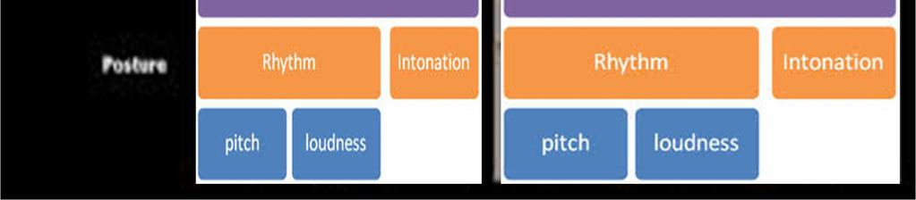
38 Split brain research for interhemispheric communications


39


40 Callosal section for epileptic seizure


41 The split-brain patient reports through the speaking hemisphere only the items flashed to the right half of the screen and denies seeing left-field stimuli or recognizing objects presented to the left hand. Nevertheless, the left hand correctly retrieves objects presented in the left visual field, about which the patient verbally denies knowing anything.


42 MRI scan showing that the splenium (arrow) was spared in the split-brain procedure performed on this patient. As a result, visual information can still be transferred between the cerebral hemispheres.


43 Schematic representation of split-brain patient J.W. s naming ability for objects in the left visual field at each operative stage.
44 Functional consequences of the split brain procedure Visual or tactile information presented to one half of the brain was not available to the other half. Verbal IQ and problem solving capacity is intact after callosotomy. Visual, tactile, and auditory sensory information is transferred by posterior half of callosum. The anterior part of callosum transfers semantic information about the stimulus.
.")
45 Letter priming: response latency between compatible (h-h) versus incompatible (t-h) trials The latencies for both types of trials are much longer for the left visual field (right hemisphere).
,")
 is")
46 The right hemisphere is capable of understanding language but not syntax. When presented with a horse stimulus in the left visual field (right hemisphere), the subject maintains through the left hemisphere that he saw nothing. When asked to draw what goes on the object, the left hand (right hemisphere) is able to draw a saddle.
 is simply required to point to a picture that best depicts what happens when the words are")
47 The capacity of the right hemisphere to make inferences is extremely limited. Two words are presented in serial order, and the right hemisphere (left hand) is simply required to point to a picture that best depicts what happens when the words are causally related. The left hemisphere finds these tasks trivial, but the right cannot perform the task.


48 Data from three patients show that the right hemisphere is more accurate than the left in recognizing unfamiliar faces.
49 Emotional prosody Emotional Prosody is characterized as an individual's tone of voice in speech that is conveyed through changes in pitch, loudness, timbre, speech rate, and pauses which is different from linguistic and semantic information. It can be isolated from linguistics and interacts with verbal content (i.e. sarcasm). It is perceived or decoded slightly worse than facial expressions but accuracy varies with emotions. Anger and sadness are perceived most easily followed by fear and happiness, with disgust being the most poorly perceive.


50
51 The brain in vocal emotion Language can be split into two components: the verbal and vocal channels. The verbal channel is the semantic content made by the speaker's chosen words. In the verbal channel, the semantic content of the speakers words determines the meaning of the sentence. The way a sentence is spoken however, can change its meaning which is the vocal channel. This channel of language conveys emotions felt by the speaker and gives us as listeners a better idea of the intended meaning. Nuances in this channel are expressed through intonation, intensity, a rhythm which combined for prosody. Usually these channels convey the same emotion, but sometimes they differ. Sarcasm and irony are two forms of humor based on this incongruent style.
52 Emotional prosody: The brain in vocal emotions Neurological processes integrating verbal and vocal (prosodic) components are relatively unclear. However, it is assumed that verbal content and vocal are processed in different hemispheres of the brain. Verbal content composed of syntactic and semantic information is processed the left hemisphere. Syntactic information is processed primarily in the frontal regions and a small part of the temporal lobe of the brain while semantic information is processed primarily in the temporal regions with a smaller part of the frontal lobes incorporated.
53 Emotional prosody: The brain in vocal emotions In contrast, prosody is processed primarily in the same pathway as verbal content, but in the right hemisphere. Neuroimaging studies using functional magnetic resonance imaging (fmri) machines provide further support for this hemisphere lateralization and temporo-frontal activation. Some studies however show evidence that prosody perception is not exclusively lateralized to the right hemisphere and may be more bilateral. There is some evidence that the basal ganglia may also play an important role in the perception of prosody.

54 Visuospatial task: self vs. familiar other The image on the far left contains 10% M.G. and 90% J.W. and changes in 10% increments from left to right, to 90% M.G. and 10% J.W. on the far right. The two original photographs of M.G. and J.W. pictured above and these nine morphed images were presented to each hemisphere randomly.


55 The proportion of yes responses to recognition judgments are plotted on the y-axis as a function of the percentage of the individual contained in the image and the cerebral hemisphere to which the image was presented.


56 Voluntary expressions that can signal intention have their own cortical networks in humans.

 The location of the section that has been")
57 The neural networks for spontaneous expressions involve older brain circuits and appear to be the same as those in chimpanzees. (inset) The location of the section that has been overlaid onto each face.
 The lesion did not interfere in spontaneous expression but (b) it did interfere with voluntary expression.")

58 The patient in the upper row suffered brain damage to the right hemisphere. (a) The lesion did not interfere in spontaneous expression but (b) it did interfere with voluntary expression. (c) This Parkinson s disease patient has a typical masked face. Because Parkinson s disease involves the part of the brain that controls spontaneous facial expression, the faces of these patients, when they are told to smile (d), light up because the other pathway is still intact. Spontaneous smile Voluntary smile


59


60


61


62

 pointed to the chicken (to go with the claw), and his")
63 Split brain patient P.S. His left hemisphere had seen a chicken claw and his right hemisphere had seen a snow scene. When asked to point to a picture associated with the image he had just seen, his right hand (guided by his left hemisphere) pointed to the chicken (to go with the claw), and his left hand pointed to the shovel ( to clean out the chicken shed ).
.")
.")

64 (a) Drawing of the LVF word Toad (ipsilateral to the drawing hand). (b) Drawing of the RVF Saw (contralateral to the drawing hand). (c) Drawing combining both words: Scraper and Sky (ipsilateral + contralateral).


 On between-field trials, the eye")
65 Cross-integration of spatial information. (a) On within-field trials, the eye moved to the stimulus that was surrounded by the probe. (b) On between-field trials, the eye moved to the corresponding stimulus in the other hemifield.
66
67 As more items are added to a set, for split brain patients the increase in reaction time for bilateral arrays is only half as fast as when all objects are confined to one side.


68 Hierarchical structure Global and local representations. We represent information at multiple scales. At its most global scale, this drawing is of a house. We can also recognize and focus on the component parts of the house.
69 Navon Letter task Navon task uses local and global stimuli to investigate hierarchical representation. Each stimulus is composed of a series of identical letters whose global arrangement forms a larger letter. The participants task is to indicate whether the stimulus contained an H or an L. When the stimulus set included competing targets at both levels (b), the participants were instructed to respond either to local targets only or to global targets only. Neither target is present in (e).


70 The left hemisphere is more adept at representing local information and the right hemisphere is better with global information.


71
72 Extreme failures of hierarchical processing following brain damage. Two patients were asked to draw the two figures shown in the left column of each panel. The patient with right-hemisphere damage was quite accurate in producing the local element the Z in (a) or the square in (b) but failed to arrange these elements into the correct global configuration. The patient with left-hemisphere damage drew the overall shapes but left out all of the local elements. Note that for each patient, the drawings were quite consistent for both the linguistic (a) and the nonlinguistic (b) stimuli, suggesting a task-independent representational deficit.
73 Theory of mind: Ann and Sally experiment Theory of mind is on the right hemisphere.


74 The attribution of beliefs is located in the right hemisphere s temporal parietal junction.


75 Competing messages are presented, one to the left ear and one to the right ear. Auditory information is projected bilaterally. Although most of the ascending fibers from the cochlear nucleus project to the contralateral thalamus, some fibers ascend on the ipsilateral side.


76 Participants are asked either to report the stimuli or to judge whether a probe stimulus was part of the dichotic message. Comparisons focus on whether they heard the reported information in the right or left ear, with the assumption that the predominant processing occurred in the contralateral hemisphere. With linguistic stimuli, participants are more accurate in reporting the information presented to the right ear.


77 Participants listened to a dichotic message in which each ear was presented with a series of letters sung to short melodies. When given a recognition memory test, participants were more accurate on the letters task for stimuli heard in the right ear. In contrast, a left-ear advantage was observed when the participants were tested on the melodies.
78 애국가 1. 동해물과백두산이마르고닳도록하느님이보우하사우리나라만세무궁화삼천리화려강산대한사람대한으로길이보전하세 3. 가을하늘공활한데높고구름없이밝은달은우리가슴일편단심일세무궁화삼천리화려강산대한사람대한으로길이보전하세 2. 남산위에저소나무철갑을두른듯바람서리불변함은우리기상일세무궁화삼천리화려강산대한사람대한으로길이보전하세 4. 이기상과이맘으로충성을다하여괴로우나즐거우나나라사랑하세무궁화삼천리화려강산대한사람대한으로길이보전하세
79 What determines hemispheric asymmetry or handedness?


80 This organization indicates that there is little overlap in the regions of space seen by each eye, and thus the visual input to the left hemisphere is independent of the visual input to the right hemisphere. This anatomical segregation would be expected to favor the emergence of hemispheric asymmetries.


, when a light")
81 A picture was flashed to his left visual field, he responded not to see anything. He responded by pressing the key with his left hand (the right hemisphere controls the left hand), when a light was flashed to his left visual field.


82 Functional asymmetries in manual coordination are sometimes attributed to the prenatal environment of the fetus. The position of the fetus in the uterus is thought to influence prenatal vestibular experience. Most fetuses are oriented with the right ear facing outward, resulting in different vestibular signals between two hemispheres. At birth, the left side of the body is more stable, freeing the right hand for exploration.
Cognitive Neuroscience Cortical Hemispheres Attention Language
 Cognitive Neuroscience Cortical Hemispheres Attention Language Based on: Chapter 18 and 19, Breedlove, Watson, Rosenzweig, 6e/7e. Cerebral Cortex Brain s most complex area with billions of neurons and
PSY 215 Lecture 17 (3/28/2010) (Lateralization in the Brain) Dr. Achtman PSY 215
 (Lateralization in the Brain) Dr. Achtman PSY 215") PSY 215 Lecture 17 Topic: Lateralization in the Brain Chapter 14.1, pages 403-414 Corrections: Lecture 16 (page 4) Broca s Area: trouble producing language, comprehension is okay. Announcements: Review
PSY 215 Lecture 17 Topic: Lateralization in the Brain Chapter 14.1, pages 403-414 Corrections: Lecture 16 (page 4) Broca s Area: trouble producing language, comprehension is okay. Announcements: Review
Higher Cortical Function
 Emilie O Neill, class of 2016 Higher Cortical Function Objectives Describe the association cortical areas processing sensory, motor, executive, language, and emotion/memory information (know general location
shows syntax in his language. has a large neocortex, which explains his language abilities. shows remarkable cognitive abilities. all of the above.
 Section: Chapter 14: Multiple Choice 1. Alex the parrot: pp.529-530 shows syntax in his language. has a large neocortex, which explains his language abilities. shows remarkable cognitive abilities. all
Lecture 35 Association Cortices and Hemispheric Asymmetries -- M. Goldberg
 Lecture 35 Association Cortices and Hemispheric Asymmetries -- M. Goldberg The concept that different parts of the brain did different things started with Spurzheim and Gall, whose phrenology became quite
P. Hitchcock, Ph.D. Department of Cell and Developmental Biology Kellogg Eye Center. Wednesday, 16 March 2009, 1:00p.m. 2:00p.m.
 Normal CNS, Special Senses, Head and Neck TOPIC: CEREBRAL HEMISPHERES FACULTY: LECTURE: READING: P. Hitchcock, Ph.D. Department of Cell and Developmental Biology Kellogg Eye Center Wednesday, 16 March
FAILURES OF OBJECT RECOGNITION. Dr. Walter S. Marcantoni
 FAILURES OF OBJECT RECOGNITION Dr. Walter S. Marcantoni VISUAL AGNOSIA -damage to the extrastriate visual regions (occipital, parietal and temporal lobes) disrupts recognition of complex visual stimuli
The origins of localization
 Association Cortex, Asymmetries, and Cortical Localization of Affective and Cognitive Functions Michael E. Goldberg, M.D. The origins of localization The concept that different parts of the brain did different
Homework Week 2. PreLab 2 HW #2 Synapses (Page 1 in the HW Section)
") Homework Week 2 Due in Lab PreLab 2 HW #2 Synapses (Page 1 in the HW Section) Reminders No class next Monday Quiz 1 is @ 5:30pm on Tuesday, 1/22/13 Study guide posted under Study Aids section of website
Homework Week 2 Due in Lab PreLab 2 HW #2 Synapses (Page 1 in the HW Section) Reminders No class next Monday Quiz 1 is @ 5:30pm on Tuesday, 1/22/13 Study guide posted under Study Aids section of website
Myers Psychology for AP*
 Myers Psychology for AP* David G. Myers PowerPoint Presentation Slides by Kent Korek Germantown High School Worth Publishers, 2010 *AP is a trademark registered and/or owned by the College Board, which
Association Cortex, Asymmetries, and Cortical Localization of Affective and Cognitive Functions. Michael E. Goldberg, M.D.
 Association Cortex, Asymmetries, and Cortical Localization of Affective and Cognitive Functions Michael E. Goldberg, M.D. The origins of localization The concept that different parts of the brain did different
Learning Objectives.
 Emilie O Neill, class of 2016 Learning Objectives 1. Describe the types of deficits that occur with lesions in association areas including: prosopagnosia, neglect, aphasias, agnosia, apraxia 2. Discuss
The human brain. of cognition need to make sense gives the structure of the brain (duh). ! What is the basic physiology of this organ?
. ! What is the basic physiology of this organ?") The human brain The human brain! What is the basic physiology of this organ?! Understanding the parts of this organ provides a hypothesis space for its function perhaps different parts perform different
The human brain The human brain! What is the basic physiology of this organ?! Understanding the parts of this organ provides a hypothesis space for its function perhaps different parts perform different
25/09/2012. Capgras Syndrome. Chapter 2. Capgras Syndrome - 2. The Neural Basis of Cognition
 Chapter 2 The Neural Basis of Cognition Capgras Syndrome Alzheimer s patients & others delusion that significant others are robots or impersonators - paranoia Two brain systems for facial recognition -
CEREBRUM Dr. Jamila Elmedany Dr. Essam Eldin Salama
 CEREBRUM Dr. Jamila Elmedany Dr. Essam Eldin Salama Objectives At the end of the lecture, the student should be able to: List the parts of the cerebral hemisphere (cortex, medulla, basal nuclei, lateral
"False tagging mechanism False Tagging Theory All idea initially believed Doubt occur when prefrontal cortex tags it as false Provides doubt and
 Ventromedial Notes Frontal lobe Prefrontal cortex 1. dorsolateral cortex Last to myelinate Sleep deprivation Executive functions Working memory Cognitive flexibility Planning 2. Orbitofrontal cortex Controls
fmri (functional MRI)
") Lesion fmri (functional MRI) Electroencephalogram (EEG) Brainstem CT (computed tomography) Scan Medulla PET (positron emission tomography) Scan Reticular Formation MRI (magnetic resonance imaging) Thalamus
Lesion fmri (functional MRI) Electroencephalogram (EEG) Brainstem CT (computed tomography) Scan Medulla PET (positron emission tomography) Scan Reticular Formation MRI (magnetic resonance imaging) Thalamus
Ch 8. Learning and Memory
 Ch 8. Learning and Memory Cognitive Neuroscience: The Biology of the Mind, 2 nd Ed., M. S. Gazzaniga, R. B. Ivry, and G. R. Mangun, Norton, 2002. Summarized by H.-S. Seok, K. Kim, and B.-T. Zhang Biointelligence
CEREBRUM. Dr. Jamila EL Medany
 CEREBRUM Dr. Jamila EL Medany Objectives At the end of the lecture, the student should be able to: List the parts of the cerebral hemisphere (cortex, medulla, basal nuclei, lateral ventricle). Describe
Exam 1 PSYC Fall 1998
 Exam 1 PSYC 2022 Fall 1998 (2 points) Briefly describe the difference between a dualistic and a materialistic explanation of brain-mind relationships. (1 point) True or False. George Berkely was a monist.
Layered organization of cortex: Paleocortex 3 layers hippocampal formation / ventral & medial cortex closest to brainstem
 Layered organization of cortex: Paleocortex 3 layers hippocampal formation / ventral & medial cortex closest to brainstem Archicortex 3-4 layers hippocampal formation / amygdala Neocortex 6 layers more
Ch 8. Learning and Memory
 Ch 8. Learning and Memory Cognitive Neuroscience: The Biology of the Mind, 2 nd Ed., M. S. Gazzaniga,, R. B. Ivry,, and G. R. Mangun,, Norton, 2002. Summarized by H.-S. Seok, K. Kim, and B.-T. Zhang Biointelligence
PsychoBrain. 31 st January Dr Christos Pliatsikas. Lecturer in Psycholinguistics in Bi-/Multilinguals University of Reading
 PsychoBrain 31 st January 2018 Dr Christos Pliatsikas Lecturer in Psycholinguistics in Bi-/Multilinguals University of Reading By the end of today s lecture you will understand Structure and function of
Cerebral Cortex 1. Sarah Heilbronner
 Cerebral Cortex 1 Sarah Heilbronner heilb028@umn.edu Want to meet? Coffee hour 10-11am Tuesday 11/27 Surdyk s Overview and organization of the cerebral cortex What is the cerebral cortex? Where is each
Chapter 6 Section 1. The Nervous System: The Basic Structure
 Chapter 6 Section 1 The Nervous System: The Basic Structure Essential Question: How does studying the biology of the brain give us an understanding of our behavior? Draw or type 2 things you already know
Cerebrum-Cerebral Hemispheres. Cuneyt Mirzanli Istanbul Gelisim University
 Cerebrum-Cerebral Hemispheres Cuneyt Mirzanli Istanbul Gelisim University The largest part of the brain. Ovoid shape. Two incompletely separated cerebral hemispheres. The outer surface of the cerebral
Neocortex. Hemispheres 9/22/2010. Psychology 472 Pharmacology of Psychoactive Drugs. Structures are divided into several section or lobes.
 Neocortex Psychology 472 Pharmacology of Psychoactive Drugs 1 Is the most developed in Humans Has many folds and fissures The folds of tissue are called gyri or a gyrus (single) The fissures or valleys
Sensorimotor Functioning. Sensory and Motor Systems. Functional Anatomy of Brain- Behavioral Relationships
 Sensorimotor Functioning Sensory and Motor Systems Understanding brain-behavior relationships requires knowledge of sensory and motor systems. Sensory System = Input Neural Processing Motor System = Output
Motor Functions of Cerebral Cortex
 Motor Functions of Cerebral Cortex I: To list the functions of different cortical laminae II: To describe the four motor areas of the cerebral cortex. III: To discuss the functions and dysfunctions of
Wetware: The Biological Basis of Intellectual Giftedness
 Wetware: The Biological Basis of Intellectual Giftedness Why is "giftedness" such a puzzle for parents? Why is there so much confusion? The most common plea heard on TAGFAM is "my child is different; please
synapse neurotransmitters Extension of a neuron, ending in branching terminal fibers, through which messages pass to other neurons, muscles, or glands
 neuron synapse The junction between the axon tip of a sending neuron and the dendrite of a receiving neuron Building block of the nervous system; nerve cell Chemical messengers that cross the synaptic
1--One Brain...or Two?--2
 Página 1 de 5 1--One Brain...or Two?--2 Left How many brains do you have - one or two? Actually, this is quite easy to answer...you have only one brain. However, the cerebral hemispheres are divided right
Psych 56L/ Ling 51: Acquisition of Language
 Psych 56L/ Ling 51: Acquisition of Language Lecture 4 Biological Bases of Language II Announcements Be working on HW1 (due 1/26/12) Be working on bio bases review questions Check out the reference material
MULTI-CHANNEL COMMUNICATION
 INTRODUCTION Research on the Deaf Brain is beginning to provide a new evidence base for policy and practice in relation to intervention with deaf children. This talk outlines the multi-channel nature of
Cerebral Cortex Structure, Function, Dysfunction Reading Ch 10 Waxman Dental Neuroanatomy Lecture. Suzanne Stensaas, Ph.D.
 Cerebral Cortex Structure, Function, Dysfunction Reading Ch 10 Waxman Dental Neuroanatomy Lecture Suzanne Stensaas, Ph.D. March 7, 2012 Anatomy Review Lobes and layers Brodmann s areas Vascular Supply
The Nervous System. Divisions of the Nervous System. Branches of the Autonomic Nervous System. Central versus Peripheral
 The Nervous System Divisions of the Nervous System Central versus Peripheral Central Brain and spinal cord Peripheral Everything else Somatic versus Autonomic Somatic Nerves serving conscious sensations
Theories of memory. Memory & brain Cellular bases of learning & memory. Epileptic patient Temporal lobectomy Amnesia
 Cognitive Neuroscience: The Biology of the Mind, 2 nd Ed., M. S. Gazzaniga, R. B. Ivry, and G. R. Mangun, Norton, 2002. Theories of Sensory, short-term & long-term memories Memory & brain Cellular bases
Does Wernicke's Aphasia necessitate pure word deafness? Or the other way around? Or can they be independent? Or is that completely uncertain yet?
 Does Wernicke's Aphasia necessitate pure word deafness? Or the other way around? Or can they be independent? Or is that completely uncertain yet? Two types of AVA: 1. Deficit at the prephonemic level and
Organization of the nervous system. The withdrawal reflex. The central nervous system. Structure of a neuron. Overview
 Overview The nervous system- central and peripheral The brain: The source of mind and self Neurons Neuron Communication Chemical messengers Inside the brain Parts of the brain Split Brain Patients Organization
Introduction to Physiological Psychology Review
 Introduction to Physiological Psychology Review ksweeney@cogsci.ucsd.edu www.cogsci.ucsd.edu/~ksweeney/psy260.html n Learning and Memory n Human Communication n Emotion 1 What is memory? n Working Memory:
REFERENCES. Hemispheric Processing Asymmetries: Implications for Memory
 TENNET XI 135 Results Allocentric WM. There was no difference between lesioned and control rats; i.e., there was an equal proportion of rats from the two groups in the target quadrant (saline, 100%; lesioned,
Language Speech. Speech is the preferred modality for language.
 Language Speech Speech is the preferred modality for language. Outer ear Collects sound waves. The configuration of the outer ear serves to amplify sound, particularly at 2000-5000 Hz, a frequency range
Brain-Behavior Network. Central Nervous System. Cerebral Cortex Gyrus and Sulcus. Nervous System
 Brain-Behavior Network Nervous System Sensory information comes into and decisions come out of the central nervous system (CNS) Central Nervous System The nerves outside the CNS are called the peripheral
Chapter 14, Part 2! Chapter 14 Part 2 Brain/Cranial Nerves! The Cerebrum and Cranial Nerves! pp !
 Chapter 14, Part 2! The Cerebrum and Cranial pp. 482 505! SECTION 14-9! The cerebrum, the largest region of the brain, contains motor, sensory, and association areas! 2! White Matter of the Cerebrum! 1.
The Central Nervous System I. Chapter 12
 The Central Nervous System I Chapter 12 The Central Nervous System The Brain and Spinal Cord Contained within the Axial Skeleton Brain Regions and Organization Medical Scheme (4 regions) 1. Cerebral Hemispheres
Cortical Organization. Functionally, cortex is classically divided into 3 general types: 1. Primary cortex:. - receptive field:.
 Cortical Organization Functionally, cortex is classically divided into 3 general types: 1. Primary cortex:. - receptive field:. 2. Secondary cortex: located immediately adjacent to primary cortical areas,
Chapter 14, Part 2! The Cerebrum and Cranial Nerves! pp !
 Chapter 14, Part 2! The Cerebrum and Cranial pp. 482 505! SECTION 14-9! The cerebrum, the largest region of the brain, contains motor, sensory, and association areas! 2! 1! ! Chapter 14 Part 2 Brain/Cranial
Leah Militello, class of 2018
 Leah Militello, class of 2018 Objectives 1. Describe the general organization of cerebral hemispheres. 2. Describe the locations and features of the different functional areas of cortex. 3. Understand
Title:Atypical language organization in temporal lobe epilepsy revealed by a passive semantic paradigm
 Author's response to reviews Title:Atypical language organization in temporal lobe epilepsy revealed by a passive semantic paradigm Authors: Julia Miro (juliamirollado@gmail.com) Pablo Ripollès (pablo.ripolles.vidal@gmail.com)
Okami Study Guide: Chapter 2 1
 Okami Study Guide: Chapter 2 1 Chapter Test 1. A cell that receives information and transmits it to other cells via an electrochemical process is called a(n) a. neuron b. hormone c. glia d. endorphin Answer:
Advances in Clinical Neuroimaging
 Advances in Clinical Neuroimaging Joseph I. Tracy 1, PhD, ABPP/CN; Gaelle Doucet 2, PhD; Xaiosong He 2, PhD; Dorian Pustina 2, PhD; Karol Osipowicz 2, PhD 1 Department of Radiology, Thomas Jefferson University,
Essentials of Human Anatomy & Physiology. Seventh Edition. The Nervous System. Copyright 2003 Pearson Education, Inc. publishing as Benjamin Cummings
 Essentials of Human Anatomy & Physiology Seventh Edition The Nervous System Copyright 2003 Pearson Education, Inc. publishing as Benjamin Cummings Functions of the Nervous System 1. Sensory input gathering
The Nervous System and the Endocrine System
 The Nervous System and the Endocrine System Neurons: The Building Blocks of the Nervous System Nervous System The electrochemical communication system of the body Sends messages from the brain to the
CNS composed of: Grey matter Unmyelinated axons Dendrites and cell bodies White matter Myelinated axon tracts
 CNS composed of: Grey matter Unmyelinated axons Dendrites and cell bodies White matter Myelinated axon tracts The Brain: A Quick Tour Frontal Lobe Control of skeletal muscles Personality Concentration
Computational Explorations in Cognitive Neuroscience Chapter 7: Large-Scale Brain Area Functional Organization
 Computational Explorations in Cognitive Neuroscience Chapter 7: Large-Scale Brain Area Functional Organization 1 7.1 Overview This chapter aims to provide a framework for modeling cognitive phenomena based
Physiology Unit 2 CONSCIOUSNESS, THE BRAIN AND BEHAVIOR
 Physiology Unit 2 CONSCIOUSNESS, THE BRAIN AND BEHAVIOR In Physiology Today What the Brain Does The nervous system determines states of consciousness and produces complex behaviors Any given neuron may
For more information about how to cite these materials visit
 Author(s): Peter Hitchcock, PH.D., 2009 License: Unless otherwise noted, this material is made available under the terms of the Creative Commons Attribution Non-commercial Share Alike 3.0 License: http://creativecommons.org/licenses/by-nc-sa/3.0/
Neurophysiology of systems
 Neurophysiology of systems Motor cortex (voluntary movements) Dana Cohen, Room 410, tel: 7138 danacoh@gmail.com Voluntary movements vs. reflexes Same stimulus yields a different movement depending on context
Name: Period: Test Review: Chapter 2
 Name: Period: Test Review: Chapter 2 1. The function of dendrites is to A) receive incoming signals from other neurons. B) release neurotransmitters into the spatial junctions between neurons. C) coordinate
Chapter 6. Attention. Attention
 Chapter 6 Attention Attention William James, in 1890, wrote Everyone knows what attention is. Attention is the taking possession of the mind, in clear and vivid form, of one out of what seem several simultaneously
Ways we Study the Brain. Accidents Lesions CAT Scan PET Scan MRI Functional MRI
 The Brain Ways we Study the Brain Accidents Lesions CAT Scan PET Scan MRI Functional MRI Accidents Phineas Gage Story Personality changed after the accident. What this this tell us? That different part
Psychology Formative Assessment #2 Answer Key
 Psychology Formative Assessment #2 Answer Key 1) C 2) B 3) B 4) C 5) D AP Objective: Discuss the influence of drugs on neurotransmitters 6) E AP Objective: Discuss the influence of drugs on neurotransmitters
Human Paleoneurology and the Evolution of the Parietal Cortex
 PARIETAL LOBE The Parietal Lobes develop at about the age of 5 years. They function to give the individual perspective and to help them understand space, touch, and volume. The location of the parietal
Inside Your Patient s Brain Michelle Peterson, APRN, CNP Centracare Stroke and Vascular Neurology
 Inside Your Patient s Brain Michelle Peterson, APRN, CNP Centracare Stroke and Vascular Neurology Activity Everyone stand up, raise your right hand, tell your neighbors your name 1 What part of the brain
The Central Nervous System
 The Central Nervous System Cellular Basis. Neural Communication. Major Structures. Principles & Methods. Principles of Neural Organization Big Question #1: Representation. How is the external world coded
Basic Brain Structure
 The Human Brain Basic Brain Structure Composed of 100 billion cells Makes up 2% of bodies weight Contains 15% of bodies blood supply Uses 20% of bodies oxygen and glucose Brain Protection Surrounded by
Chapter 2 Test. 1. Evolutionary structures within the are the most primitive. *a. hindbrain b. thalamus c. forebrain d. midbrain e.
 Cognitive Psychology In and Out of the Laboratory 5th Edition Galotti TEST BANK Full clear download (no formatting errors) at: https://testbankreal.com/download/cognitive-psychology-laboratory-5thedition-galotti-test-bank/
Prof. Greg Francis 5/23/08
 Brain parts The brain IIE 269: Cognitive Psychology Greg Francis Lecture 02 The source of cognition (consider transplant!) Weighs about 3 pounds Damage to some parts result in immediate death or disability
Vision II. Steven McLoon Department of Neuroscience University of Minnesota
 Vision II Steven McLoon Department of Neuroscience University of Minnesota 1 Ganglion Cells The axons of the retinal ganglion cells form the optic nerve and carry visual information into the brain. 2 Optic
Physiology Unit 2 CONSCIOUSNESS, THE BRAIN AND BEHAVIOR
 Physiology Unit 2 CONSCIOUSNESS, THE BRAIN AND BEHAVIOR What the Brain Does The nervous system determines states of consciousness and produces complex behaviors Any given neuron may have as many as 200,000
XIXth Century: Localization of Functions to Different Parts of the Brain
 XIXth Century: Localization of Functions to Different Parts of the Brain Studies by Bell and Magendie initiated an extremely important scientific procedure,, where a specific part of the nervous system
THE CENTRAL NERVOUS SYSTEM. The Brain & Spinal Cord
 THE CENTRAL NERVOUS SYSTEM The Brain & Spinal Cord Review: Nervous System Parallel Distributed Processing Composition of the CNS Nuclei: Clusters of neurons in the CNS ( neighborhoods ) Fiber Tracts/Pathways:
Psy /16 Human Communication. By Joseline
 Psy-302 11/16 Human Communication By Joseline Lateralization Left Hemisphere dominance in speech production in 95% of right handed and 70% of left handed people Left -> Timing, Sequence of events Right
Outline of the next three lectures
 Outline of the next three lectures Lecture 35 Anatomy of the human cerebral cortex gross and microscopic cell types connections Vascular supply of the cerebral cortex Disorders involving the cerebral cortex
Brain Structures. Some scientists divide the brain up into three parts. Hindbrain Midbrain Forebrain
 The Brain Phineas Gage Play The Frontal Lobes and Behavior: The Story of Phineas Gage (12:03) Module #25 from The Brain: Teaching Modules (2 nd edition). http://www.learner.org/resources/series1 42.html
Retinotopy & Phase Mapping
 Retinotopy & Phase Mapping Fani Deligianni B. A. Wandell, et al. Visual Field Maps in Human Cortex, Neuron, 56(2):366-383, 2007 Retinotopy Visual Cortex organised in visual field maps: Nearby neurons have
Neural Basis of Motor Control
 Neural Basis of Motor Control Central Nervous System Skeletal muscles are controlled by the CNS which consists of the brain and spinal cord. Determines which muscles will contract When How fast To what
Announcement. Danny to schedule a time if you are interested.
 Announcement If you need more experiments to participate in, contact Danny Sanchez (dsanchez@ucsd.edu) make sure to tell him that you are from LIGN171, so he will let me know about your credit (1 point).
Disorders affecting region: depression anxiety
 Amygdala Involved in learning, and the processing of emotional memories. Measures sensory input for potential threat level, then hypothalamus Regulates volatile emotions like fear and anger. Disorders
Cognitive Neuroscience Attention
 Cognitive Neuroscience Attention There are many aspects to attention. It can be controlled. It can be focused on a particular sensory modality or item. It can be divided. It can set a perceptual system.
BRAIN: CONTROL CENTER
 BRAIN: CONTROL CENTER ORCHESTRA Scientists now believe the brain functions much like an orchestra, where different instruments each play a different part. Scans show that the brain divides different aspects
CEREBRUM & CEREBRAL CORTEX
 CEREBRUM & CEREBRAL CORTEX Seonghan Kim Dept. of Anatomy Inje University, College of Medicine THE BRAIN ANATOMICAL REGIONS A. Cerebrum B. Diencephalon Thalamus Hypothalamus C. Brain Stem Midbrain Pons
Neuroscience Tutorial
 Neuroscience Tutorial Brain Organization : cortex, basal ganglia, limbic lobe : thalamus, hypothal., pituitary gland : medulla oblongata, midbrain, pons, cerebellum Cortical Organization Cortical Organization
CISC 3250 Systems Neuroscience
 CISC 3250 Systems Neuroscience Levels of organization Central Nervous System 1m 10 11 neurons Neural systems and neuroanatomy Systems 10cm Networks 1mm Neurons 100μm 10 8 neurons Professor Daniel Leeds
Introduction to the Nervous System. Code: HMP 100/ UPC 103/ VNP 100. Course: Medical Physiology. Level 1 MBChB/BDS/BPharm
 Introduction to the Nervous System. Code: HMP 100/ UPC 103/ VNP 100. Course: Medical Physiology Level 1 MBChB/BDS/BPharm Lecture 2. Functional Organisation of the Nervous System Lecture Outline 1.1 Introduction
1. Processes nutrients and provides energy for the neuron to function; contains the cell's nucleus; also called the soma.
 1. Base of brainstem; controls heartbeat and breathing 2. tissue destruction; a brain lesion is a naturally or experimentally caused destruction of brain tissue 3. A thick band of axons that connects the
The Frontal Lobes. Anatomy of the Frontal Lobes. Anatomy of the Frontal Lobes 3/2/2011. Portrait: Losing Frontal-Lobe Functions. Readings: KW Ch.
 The Frontal Lobes Readings: KW Ch. 16 Portrait: Losing Frontal-Lobe Functions E.L. Highly organized college professor Became disorganized, showed little emotion, and began to miss deadlines Scores on intelligence
Biocomputer Wired for Action MWABBYH CTBIR LOBES
 Biocomputer Wired for Action MWABBYH CTBIR LOBES 100 100 100 100 100 200 200 200 200 200 300 300 300 300 300 400 400 400 400 400 500 500 500 500 500 Biocomputer Wired for Action MWABBYH CTBIR LOBES 100
Contents. Boxes xii Preface xiii Acknowledgments. Background and Methods
 Contents Boxes xii Preface xiii Acknowledgments xv PARTI Background and Methods 1 A Brief History of Cognitive Neuroscience 2 A Historical Perspective 4 The Brain Story 5 The Psychological Story 10 The
FRONTAL LOBE. Central Sulcus. Ascending ramus of the Cingulate Sulcus. Cingulate Sulcus. Lateral Sulcus
 FRONTAL LOBE Central Ascending ramus of the Cingulate Cingulate Lateral Lateral View Medial View Motor execution and higher cognitive functions (e.g., language production, impulse inhibition, reasoning
PHY3111 Mid-Semester Test Study. Lecture 2: The hierarchical organisation of vision
 PHY3111 Mid-Semester Test Study Lecture 2: The hierarchical organisation of vision 1. Explain what a hierarchically organised neural system is, in terms of physiological response properties of its neurones.
Ch 5. Perception and Encoding
 Ch 5. Perception and Encoding Cognitive Neuroscience: The Biology of the Mind, 2 nd Ed., M. S. Gazzaniga, R. B. Ivry, and G. R. Mangun, Norton, 2002. Summarized by Y.-J. Park, M.-H. Kim, and B.-T. Zhang
CYTOARCHITECTURE OF CEREBRAL CORTEX
 BASICS OF NEUROBIOLOGY CYTOARCHITECTURE OF CEREBRAL CORTEX ZSOLT LIPOSITS 1 CELLULAR COMPOSITION OF THE CEREBRAL CORTEX THE CEREBRAL CORTEX CONSISTS OF THE ARCHICORTEX (HIPPOCAMPAL FORMA- TION), PALEOCORTEX
Forebrain Brain Structures Limbic System. Brain Stem Midbrain Basil Ganglia. Cerebellum Reticular Formation Medulla oblongata
 Brain structures (1) Cut out the following cards (2) Identify the three major divisions of the brain (as defined by your book). Initially, try this without any form of aid such as your textbook. (3) Organize
Human Brain. Lateralization of Function. An extension of the spinal cord. Dr. Coulson Cognitive Science Department UCSD
 Lateralization of Function Human Brain An extension of the spinal cord Dr. Coulson Cognitive Science Department UCSD Cerebral Hemispheres Corpus Callosum Cerebral Lobes Neurons Brain composed of neurons
Okami Study Guide: Chapter 2 1
 Okami Study Guide: Chapter 2 1 Chapter in Review 1. The human nervous system is a complex biological system designed for nearly instantaneous communication among billions of neurons throughout the body.
Notes: Organization. Anatomy of the Nervous System. Cerebral cortex. Cortical layers. PSYC 2: Biological Foundations - Fall Professor Claffey
 PSYC 2: Biological Foundations - Fall 2012 - Professor Claffey Notes: Organization Version: 10/30/12 - original version Anatomy of the Nervous System Content covered in Hans's lecture: CNS & PNS Directions/Planes
2Lesson. Outline 1.5. Lesson Plan. The OVERVIEW. Lesson 1.5: How do the parts of my brain work together? LESSON. Unit1.2
 Outline 2Lesson Unit1.2 OVERVIEW Rationale: This lesson is intended to emphasize the need of the structures of the brain to work together to create and control behaviors, like the creation and comprehension
Anatomy Lab (1) Theoretical Part. Page (2 A) Page (2B)
 Theoretical Part. Page (2 A) Page (2B)") Anatomy Lab (1) This sheet only includes the extra notes for the lab handout regarding the theoretical part, as for the practical part it includes everything the doctor mentioned. Theoretical Part Page
Anatomy Lab (1) This sheet only includes the extra notes for the lab handout regarding the theoretical part, as for the practical part it includes everything the doctor mentioned. Theoretical Part Page
EVIDENCE FOR PARACALLOSAL VERBAL TRANSFER AFTER CALLOSAL SECTION
 Brain (1982), 105,53-63 EVIDENCE FOR PARACALLOSAL VERBAL TRANSFER AFTER CALLOSAL SECTION A POSSIBLE CONSEQUENCE OF BILATERAL LANGUAGE ORGANIZATION by MICHAEL S. GAZZANIGA, JOHN J. SIDTIS, BRUCE T. VOLPE,