FDG-PET e parkinsonismi
|
|
|
- Garry Fleming
- 5 years ago
- Views:
Transcription

1 Parkinsonismi FDG-PET e parkinsonismi Valentina Berti Dipartimento di Scienze Biomediche, Sperimentali e Cliniche Sez. Medicina Nucleare Università degli Studi di Firenze

2 History 140 PubMed: FDG AND parkinsonism

3 Clinical differentiation of parkisonian syndromes can be challenging Neuroimaging contributes to the correct diagnosis of parkinsonism Dopaminergic system imaging is recommended by international guidelines, but no/low accuracy in differential diagnosis FDG-PET Good accuracy in the differential diagnosis of parkinsonisms Correlation with clinical symptoms Correlation with pathological alterations

4 Parkinsonisms - Pathology α-synucleinopathies α-synuclein structural protein localized primarily in synaptic terminals and involved in synaptic plasticity Parkinson s disease - PD Parkinson s disease dementia PDD Dementia with Lewy bodies - DLB Multisystemic atrophy Tauopathies Tau protein microtubule-associated protein involved in axonal transport Progressive Supranuclear Palsy Corticobasal Degeneration/Syndrome
5 Parkinson s disease - PD Parkinson s disease dementia PDD Dementia with Lewy bodies - DLB Multisystemic Atrophy Progressive Supranuclear Palsy Cortico-basal syndrome / Cortico-basal Degeneration - CBS
 II.")
 IV.Mesocortex (transentorhinal region) V.")
6 PD Brain pathology α-synuclein-immunopositive Lewy neurites and Lewy bodies Braak staging of Lewy body pathology I. Medulla oblongata (Dorsal IX/X motor and anterior olfactory nucleus) II. Medulla oblongata and pontine tegmentum (caudal raphe nuclei, gigantocellular reticular nucleus, and coeruleus subcoeruleus complex) III.Midbrain (pars compacta substantia nigra) IV.Mesocortex (transentorhinal region) V.Neocortex (high order sensory association and prefrontal areas) VI.Neocortex (first order sensory association areas and premotor areas)


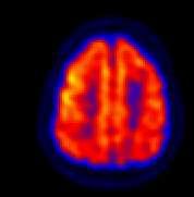
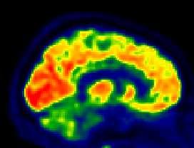
7 PD - FDG-PET pattern NL PD PD-early Widespread mild to severe cortical hypometabolism Parieto-temporo-occipital regions Frontal lobes Relative sparing of striata, thalamus, cerebellum PD-advanced


8 PD - FDG-PET pattern Key point Relative sparing of striata PD - early R


9 PD motor symptoms UPDRS III CMRGlu bilateral premotor cortex Putaminal dopaminergic deficit CMRGlu dorso-lateral prefrontal cortex premotor cortex anterior prefrontal cortex orbitofrontal cortex


10 PD cognitive symptoms Executive dysfunction: anterior cingulate cortex dorso-lateral prefrontal cortex orbito-frontal cortex Executive dysfunction: Dorso-lateral prefrontal cortex PCC Caudate nucleus dopaminergic deficit: anterior cingulate cortex dorso-lateral prefrontal cortex orbito-frontal cortex

11 PD cognitive pattern

12 From PD to PDD Hypometabolic cortical areas frequently observed in nondemented PD correspond to those in PDD and DLB FDG-PET findings in patients with PD without dementia span a spectrum from normality to pronounced posterior cortical and sometimes frontal hypometabolism as observed in patients with PDD Meyer et al. Current Opinion in Neurology 2014

13 PDD vs controls PD vs controls

14 From PD to PDD PD-MCI PD-MCI vs PD-nMCI PD-MCI vs controls Hypometabolism Prefrontal Cortex Superior and Inferior Parietal Cortex Associative Occipital Cortex


15 From PD to PDD PD-MCI Overall pattern of hypometabolism is similar in PDD and PD-MCI However, metabolic deficits were significantly more severe in PDD
 than PD-MCI patients, and also, albeit to a lesser extent, in the right frontal lobe PD-MCI: vs PDCN: mild")
16 From PD to PDD PD-MCI PDD: vs PDCN: extensive bilateral areas of reduced FDG uptake in the frontal, parietal, occipital and temporal lobes and in the posterior cingulate cortex; vs PDMCI: lower metabolism mainly in posterior brain areas (parietal, occipital and temporal lobes) than PD-MCI patients, and also, albeit to a lesser extent, in the right frontal lobe PD-MCI: vs PDCN: mild hypometabolism in the left frontal lobe and to a lesser extent in the left parietal lobe Garcia-Garcia et al. Eur J Nucl Med Mol Imaging 2012
 uptake; Language: anterior areas mainly the frontal lobe Garcia-Garcia et al.")
17 PD-MCI, PDD cognitive symptoms regional glucose metabolism correlated with executive, memory, language and visuospatial functions Executive function: parietal, frontal and occipitotemporal junction; Memory: temporal and parietal regions; Visuospatial function: posterior areas (occipitoparietal and temporal) uptake; Language: anterior areas mainly the frontal lobe Garcia-Garcia et al. Eur J Nucl Med Mol Imaging 2012


18 From PDD to DLB Both PDD and DLB: widespread lateral frontal, temporoparietal and occipital hypometabolism PDD vs DLB: metabolic differences appear to be minor, if any

19 DLB metabolic pattern Congruence with Cortical cholinergic dysfunction Cognitive impairment Meyer et al. Current Opinion in Neurology 2014

20 DLB metabolic correlations Correlation between MMSE and regional FDG uptake in DLB lateral frontal, temporal and parietal association cortices cingulate gyrus and precuneus caudate nucleus no significant correlation is found in occipital cortex, in line with the finding that occipital hypometabolism may be observed well before and unrelated to cognitive impairment Meyer et al. Current Opinion in Neurology 2014

21 Conversion from PD to PDD Converters to PDD Baseline: significant hypometabolism in posterior cingulate, occipital cortex and caudate nucleus 2-year follow-up: widespread metabolic decline in association cortices, posterior cingulate, hippocampus and thalamus Primary visual cortex hypometabolism was also observed in cognitively stable PD patients.


22 Conversion to DLB Converters to DLB: hypometabolism in the parietal and the lateral occipital cortex compared to non-converters.
23 Parkinson s disease - PD Parkinson s disease dementia PDD Dementia with Lewy bodies - DLB Multisystemic Atrophy Progressive Supranuclear Palsy Cortico-basal syndrome / Cortico-basal Degeneration - CBS


24 MSA - Brain pathology MSA-C Predominant cerebellar ataxia MSA-P Parkinsonian clinical phenotype OPCA inferior olivary nucleus, pontine nuclei, cerebellar hemispheres and vermis SND substantia nigra (SbN), putamen, caudate nucleus and globus pallidus abnormal α-synuclein positive glial cytoplasmic inclusions in oligodendrocytes
 cerebellum (cerebellar Purkinje cells) brainstem (substantia nigra, locus ceruleus, dorsal vagal nuclei, vestibular nuclei, inferior")
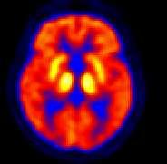
25 MSA FDG-PET pattern NL MSA consistent with MSA neuropathological features Degenerative changes in: Striata (putamen < caudate) Cerebellum Brainstem More advenced stages: Frontal lobes Parietal lobes Occipital lobes basal ganglia (putamen and globus pallidus) cerebellum (cerebellar Purkinje cells) brainstem (substantia nigra, locus ceruleus, dorsal vagal nuclei, vestibular nuclei, inferior olives, and pontine nuclei) Hypometabolic pattern - reflects the loss of neurons and synaptic connections in these sites


26 MSA FDG-PET pattern R L

27 MSA motor symptoms ATAXIA negative CMRGlu CEREBELLUM
28 MSA Clinicometabolic correlation PARKINSONISM negative CMRglu STRIATUM AUTONOMIC DYSFUNCTION negative CMRglu THALAMUS
29 Tauopathies Neurodegenerative parkinsonism with disturbances in tau protein handling PSP and CBD similarities with frontotemporal dementia overlap in pathology with Alzheimer s disease
30 Parkinson s disease - PD Parkinson s disease dementia PDD Dementia with Lewy bodies - DLB Multisystemic Atrophy Progressive Supranuclear Palsy Cortico-basal syndrome / Cortico-basal Degeneration - CBS

31 PSP - Brain pathology Tau-positive star-shaped astrocytic tufts and neurofibrillary tangles

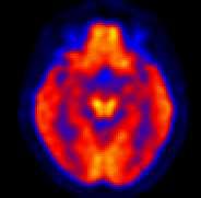
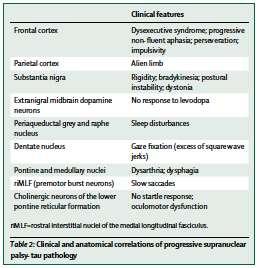
32 PSP FDG-PET pattern NL PSP consistent with Richardson's syndrome neuropathological features Subcortical Striata (caudate<putamen) Thalamus Brainstem Hypometabolism reflects the loss of neurons and synaptic connections in these sites Cortical Frontal lobe ACC Medial frontal cortex Neuropathologic alterations and neuronal loss in frontal lobes Functional deprivation of the frontal lobe due to pallidal degeneration
33 PSP FDG-PET pattern R L Statistical maps of hypometabolism from Teune et al. Mov Disord 2010

, low sensitivity (29%) possible or definite pimple sign : sensitivity of")
34 PSP FDG-PET pattern Midbrain hypometabolism Pimple sign focal area of midbrain hypometabolism on statistical maps of FDG-PET scans in some patients with PSP-Richardson s syndrome associated with midbrain atrophy helpful in differentiating PSP-RS from CBS and MSA definite pimple sign : high specificity (100%), low sensitivity (29%) possible or definite pimple sign : sensitivity of 79%


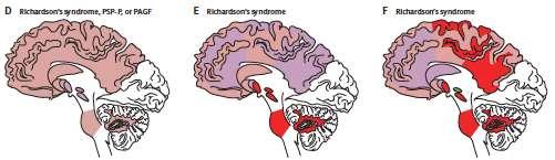
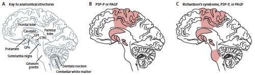
35 Atypical PSP FDG-PET pattern PSP-P Striata Thalamus Frontal lobe RS PSP-P


36 Atypical PSP FDG-PET pattern PSP-PAGF Midbrain Frontal lobe RS PAGF
37 PSP Clinicometabolic correlation COGNITIVE DEFICIT DISEASE DURATION positive negative CMRglu FRONTAL cortex MOTOR SYMPTOMS negative CMRglu CAUDATE NUCLEUS THALAMUS
38 Parkinson s disease - PD Parkinson s disease dementia PDD Dementia with Lewy bodies - DLB Multisystemic Atrophy Progressive Supranuclear Palsy Cortico-basal syndrome / Cortico-basal Degeneration - CBS


39 CBS - Brain pathology ballooned neurons and characteristic glial pathology, including tau-positive astrocytic plaques Same pathology Different clinical syndromes Same clinical syndrome Different pathologies
")
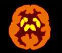
40 CBS FDG-PET pattern NL CBS ASYMMETRICAL DISTRIBUTION Hypometabolism contralateral to the clinically most affected side CORTICAL Frontal lobe Parieto-temporal regions SUBCORTICAL Striata (putamen=caudate) Thalamus
")
41 CBS FDG-PET pattern ASYMMETRICAL DISTRIBUTION Hypometabolism contralateral to the clinically most affected side CORTICAL Frontal lobe Parieto-temporal regions SUBCORTICAL Striata (putamen=caudate) Thalamus

42 Ad Interim Summary NL Disease-specific metabolic pattern in FDG-PET PD PDD DLB MSA Sparing (relative hypermetabolism) of putamen/pallidum and, possibly, thalamus and cerebellum Hypometabolism of occipital, temporoparietal and frontal cortices Hypometabolism of putamen, pons and/or cerebellum PSP CBS Hypometabolism of midline frontal, upper brainstem, thalamus and caudate nucleus Asymmetric hypometabolism of cortical and subcortical regions (striatum and thalamus), including parietal, primary sensorimotor, midline frontal and premotor cortices


43 PD vs atypical parkinsonisms Accuracy 95% MSA diagnosis Sensitivity 76.9% Specificity 96.9% PSP diagnosis Sensitivity 73.9% Specificity 95.2% CBD diagnosis Sensitivity 75.0% Specificity 91.7%
44 Juh et al.eur J Radiology 2004 Kwon et al. Eur J Neurol 2008 Baudrexel et al. Mov Disord 2014 FDG-PET in parkinsonism Differential diagnosis PD Sparing of the striata vs MSA Hypometabolism Occipital lobe and visual cortex Discriminating regions Putamen, pons vs PSP Occipital lobe Midbrain, midline frontal, thalamus
45 Juh et al.eur J Radiology 2004 Kwon et al. Eur J Neurol 2008 Baudrexel et al. Mov Disord 2014 FDG-PET in parkinsonism Differential diagnosis MSA Accuracy: 82-96% vs PD Hypometabolism Putamen (posterior>anterior), pons, cerebellum Discriminating regions Putamen, pons vs PSP Pons Midbrain, cingulated gyrus, pons
46 Juh et al.eur J Radiology 2004 Kwon et al. Eur J Neurol 2008 Baudrexel et al. Mov Disord 2014 FDG-PET in parkinsonism Differential diagnosis PSP vs PD vs MSA Hypometabolism Cingulated gyrus, caudate nucleus, thalamus and midbrain Cingulated gyrus, thalamus and midbrain Discriminating regions Midbrain, cingulated gyrus, thalamus Midbrain, cingulated gyrus, pons
47 Conclusion FDG-PET: Differential diagnosis Clinical variants Clinico-pathological correlation Thank you
Parkinson e decadimento cognitivo. Stelvio Sestini
 Parkinson e decadimento cognitivo Stelvio Sestini Patients with PD can develop a spectrum of cognitive symptoms Heterogeneity of cognitive deficits The cognitive symptoms can evolve to dementia (Mov Disorder
Neuropathology of Neurodegenerative Disorders Prof. Jillian Kril
 Neurodegenerative disorders to be discussed Alzheimer s disease Lewy body diseases Frontotemporal dementia and other tauopathies Huntington s disease Motor Neuron Disease 2 Neuropathology of neurodegeneration
Diagnosis before NIA AA The impact of FDG PET in. Diagnosis after NIA AA Neuropathology and PET image 2015/10/16
 The impact of FDG PET in degenerative dementia diagnosis Jung Lung, Hsu MD, Ph.D (Utrecht) Section of dementia and cognitive impairment Department of Neurology Chang Gung Memorial Hospital, Linkou, Taipei
Update on functional brain imaging in Movement Disorders
 Update on functional brain imaging in Movement Disorders Mario Masellis, MSc, MD, FRCPC, PhD Assistant Professor & Clinician-Scientist Sunnybrook Health Sciences Centre University of Toronto 53 rd CNSF
Imaging biomarkers for Parkinson s disease
 3 rd Congress of the European Academy of Neurology Amsterdam, The Netherlands, June 24 27, 2017 Teaching Course 6 MDS-ES/EAN: Neuroimaging in movement disorders - Level 2 Imaging biomarkers for Parkinson
Dementia and Healthy Ageing : is the pathology any different?
 Dementia and Healthy Ageing : is the pathology any different? Professor David Mann, Professor of Neuropathology, University of Manchester, Hope Hospital, Salford DEMENTIA Loss of connectivity within association
Lecture 42: Final Review. Martin Wessendorf, Ph.D.
 Lecture 42: Final Review Martin Wessendorf, Ph.D. Lecture 33 cortex Heilbronner 5 lobes of the cortex Lateral view (left side) Mid-saggital view (right side) Cellular organization of cortex White matter
Neuro degenerative PET image from FDG, amyloid to Tau
 Neuro degenerative PET image from FDG, amyloid to Tau Kun Ju Lin ( ) MD, Ph.D Department of Nuclear Medicine and Molecular Imaging Center, Chang Gung Memorial Hospital ( ) Department of Medical Imaging
Anatomy and Physiology (Bio 220) The Brain Chapter 14 and select portions of Chapter 16
 The Brain Chapter 14 and select portions of Chapter 16") Anatomy and Physiology (Bio 220) The Brain Chapter 14 and select portions of Chapter 16 I. Introduction A. Appearance 1. physical 2. weight 3. relative weight B. Major parts of the brain 1. cerebrum 2.
Anatomy and Physiology (Bio 220) The Brain Chapter 14 and select portions of Chapter 16 I. Introduction A. Appearance 1. physical 2. weight 3. relative weight B. Major parts of the brain 1. cerebrum 2.
Supplementary Material S3 Further Seed Regions
 Supplementary Material S3 Further Seed Regions Figure I. Changes in connectivity with the right anterior insular cortex. (A) wake > mild sedation, showing a reduction in connectivity between the anterior
Chapter 3. Structure and Function of the Nervous System. Copyright (c) Allyn and Bacon 2004
 Allyn and Bacon 2004") Chapter 3 Structure and Function of the Nervous System 1 Basic Features of the Nervous System Neuraxis: An imaginary line drawn through the center of the length of the central nervous system, from the
Chapter 3 Structure and Function of the Nervous System 1 Basic Features of the Nervous System Neuraxis: An imaginary line drawn through the center of the length of the central nervous system, from the
Neurodegenerative Disease. April 12, Cunningham. Department of Neurosciences
 Neurodegenerative Disease April 12, 2017 Cunningham Department of Neurosciences NEURODEGENERATIVE DISEASE Any of a group of hereditary and sporadic conditions characterized by progressive dysfunction,
review of existing studies on ASL in dementia Marion Smits, MD PhD
 review of existing studies on ASL in dementia Marion Smits, MD PhD Associate Professor of Neuroradiology Department of Radiology, Erasmus MC, Rotterdam (NL) Alzheimer Centre South-West Netherlands, Rotterdam
Detailed protocol Only dissected human brain samples are stored. The microdissection is performed on frozen brains and the samples are kept on -70 C.
 2008 Detailed protocol Only dissected human brain samples are stored. The microdissection is performed on frozen brains and the samples are kept on -70 C. BrainNet Europe II Project Co-ordinator: Prof.
212 Index C-SB-13,
 Index A Acetylcholinesterase inhibitor, treatment, 15 Age-associated memory impairment (AAMI), 5 Alzheimer s disease (AD), 40, 95 96 apolipoprotein E genotype and risk for, 58 cellular neurodegeneration
PET ligands and metabolic brain imaging Prof. Karl Herholz
 PET ligands Karl Herholz, University of Manchester PET images in this lecture, unless indicated otherwise, are from Max-Planck-Institute for Neurological Research, Cologne, Germany 1 Positron-Emission-Tomography
Brainstem. Steven McLoon Department of Neuroscience University of Minnesota
 Brainstem Steven McLoon Department of Neuroscience University of Minnesota 1 Course News Change in Lab Sequence Week of Oct 2 Lab 5 Week of Oct 9 Lab 4 2 Goal Today Know the regions of the brainstem. Know
Pathogenesis of Degenerative Diseases and Dementias. D r. Ali Eltayb ( U. of Omdurman. I ). M. Path (U. of Alexandria)
. M. Path (U. of Alexandria)") Pathogenesis of Degenerative Diseases and Dementias D r. Ali Eltayb ( U. of Omdurman. I ). M. Path (U. of Alexandria) Dementias Defined: as the development of memory impairment and other cognitive deficits
Pathogenesis of Degenerative Diseases and Dementias D r. Ali Eltayb ( U. of Omdurman. I ). M. Path (U. of Alexandria) Dementias Defined: as the development of memory impairment and other cognitive deficits
III./3.1. Movement disorders with akinetic rigid symptoms
 III./3.1. Movement disorders with akinetic rigid symptoms III./3.1.1. Parkinson s disease Parkinson s disease (PD) is the second most common neurodegenerative disorder worldwide after Alzheimer s disease.
Dementia spectrum disorders: lessons learnt from decades with PET research
 Journal of Neural Transmission (2019) 126:233 251 https://doi.org/10.1007/s00702-019-01975-4 NEUROLOGY AND PRECLINICAL NEUROLOGICAL STUDIES - REVIEW ARTICLE Dementia spectrum disorders: lessons learnt
10/3/2016. T1 Anatomical structures are clearly identified, white matter (which has a high fat content) appears bright.
 appears bright.") H2O -2 atoms of Hydrogen, 1 of Oxygen Hydrogen just has one single proton and orbited by one single electron Proton has a magnetic moment similar to the earths magnetic pole Also similar to earth in that
H2O -2 atoms of Hydrogen, 1 of Oxygen Hydrogen just has one single proton and orbited by one single electron Proton has a magnetic moment similar to the earths magnetic pole Also similar to earth in that
Imaging biomarkers in Parkinson s disease and Parkinsonian syndromes: current and emerging concepts
 Saeed et al. Translational Neurodegeneration (2017) 6:8 DOI 10.1186/s40035-017-0076-6 REVIEW Imaging biomarkers in Parkinson s disease and Parkinsonian syndromes: current and emerging concepts Usman Saeed
The neurvous system senses, interprets, and responds to changes in the environment. Two types of cells makes this possible:
 NERVOUS SYSTEM The neurvous system senses, interprets, and responds to changes in the environment. Two types of cells makes this possible: the neuron and the supporting cells ("glial cells"). Neuron Neurons
PSY 302: CHAPTER 3 NOTES THE BRAIN (PART II) - 9/5/17. By: Joseline
 - 9/5/17. By: Joseline") PSY 302: CHAPTER 3 NOTES THE BRAIN (PART II) - 9/5/17 By: Joseline Left 3 MAJOR FISSURES : 2HEMISPHERES Right Lateral Ventricle Central Fissure Third Ventricle Sulcus Lateral Fissure Gyros Fissure- Fissures
PSY 302: CHAPTER 3 NOTES THE BRAIN (PART II) - 9/5/17 By: Joseline Left 3 MAJOR FISSURES : 2HEMISPHERES Right Lateral Ventricle Central Fissure Third Ventricle Sulcus Lateral Fissure Gyros Fissure- Fissures
Kurt A. Jellinger. 2 nd Int. Conference BrainNet Europe, Munich, Dec , 2008 NAC A30P A53T ALPHA HELICAL HYDROPHOBIC ACID (GLU-PRO) COOH
 COOH") 2 nd Int. Conference BrainNet Europe, Munich, Dec. 10-12, 2008 NH 3 1 NAC 125 133 136 A30P A53T 125 140 ALPHA HELICAL HYDROPHOBIC ACID (GLU-PRO) COOH 29 71 82 125 129 (Src) (GRK5, CK-1 & CK-2) Kurt A.
2 nd Int. Conference BrainNet Europe, Munich, Dec. 10-12, 2008 NH 3 1 NAC 125 133 136 A30P A53T 125 140 ALPHA HELICAL HYDROPHOBIC ACID (GLU-PRO) COOH 29 71 82 125 129 (Src) (GRK5, CK-1 & CK-2) Kurt A.
COGNITIVE SCIENCE 107A. Motor Systems: Basal Ganglia. Jaime A. Pineda, Ph.D.
 COGNITIVE SCIENCE 107A Motor Systems: Basal Ganglia Jaime A. Pineda, Ph.D. Two major descending s Pyramidal vs. extrapyramidal Motor cortex Pyramidal system Pathway for voluntary movement Most fibers originate
Round table: Moderator; Fereshteh Sedaghat, MD, PhD Brain Mapping in Dementias and Non-invasive Neurostimulation
 Round table: Moderator; Fereshteh Sedaghat, MD, PhD Brain Mapping in Dementias and Non-invasive Neurostimulation 1. Reflection of Mild Cognitive Impairment (MCI) and Dementias by Molecular Imaging, PET
FRONTOTEMPORAL DEGENERATION: OVERVIEW, TRENDS AND DEVELOPMENTS
 FRONTOTEMPORAL DEGENERATION: OVERVIEW, TRENDS AND DEVELOPMENTS Norman L. Foster, M.D. Director, Center for Alzheimer s Care, Imaging and Research Chief, Division of Cognitive Neurology, Department of Neurology
Role of Neuropsychiatric Assessment in Diagnosis and Research
 Role of Neuropsychiatric Assessment 163 11 Role of Neuropsychiatric Assessment in Diagnosis and Research Dag Aarsland, Uwe Ehrt, and Clive Ballard INTRODUCTION Although the basal ganglia have traditionally
Nsci 2100: Human Neuroanatomy 2017 Examination 3
 Name KEY Lab Section Nsci 2100: Human Neuroanatomy 2017 Examination 3 On this page, write your name and lab section. On your bubble answer sheet, enter your name (last name, space, first name), internet
The Neuroscience of Music in Therapy
 Course Objectives The Neuroscience of Music in Therapy Unit I. Learn Basic Brain Information Unit II. Music in the Brain; Why Music Works Unit III. Considerations for Populations a. Rehabilitation b. Habilitation
Biological Bases of Behavior. 3: Structure of the Nervous System
 Biological Bases of Behavior 3: Structure of the Nervous System Neuroanatomy Terms The neuraxis is an imaginary line drawn through the spinal cord up to the front of the brain Anatomical directions are
International Journal of Health Sciences and Research ISSN:
 International Journal of Health Sciences and Research www.ijhsr.org ISSN: 2249-9571 Case Report Multiple System Atrophy-Cerebellar Type (MSA-C): A Case Report Mohd Abbas Ilyas, Pramod Shaha, Kulamani Sahoo,
Announcement. Danny to schedule a time if you are interested.
 Announcement If you need more experiments to participate in, contact Danny Sanchez (dsanchez@ucsd.edu) make sure to tell him that you are from LIGN171, so he will let me know about your credit (1 point).
Anatomy of the basal ganglia. Dana Cohen Gonda Brain Research Center, room 410
 Anatomy of the basal ganglia Dana Cohen Gonda Brain Research Center, room 410 danacoh@gmail.com The basal ganglia The nuclei form a small minority of the brain s neuronal population. Little is known about
Dementia Update. October 1, 2013 Dylan Wint, M.D. Cleveland Clinic Lou Ruvo Center for Brain Health Las Vegas, Nevada
 Dementia Update October 1, 2013 Dylan Wint, M.D. Cleveland Clinic Lou Ruvo Center for Brain Health Las Vegas, Nevada Outline New concepts in Alzheimer disease Biomarkers and in vivo diagnosis Future trends
The Central Nervous System I. Chapter 12
 The Central Nervous System I Chapter 12 The Central Nervous System The Brain and Spinal Cord Contained within the Axial Skeleton Brain Regions and Organization Medical Scheme (4 regions) 1. Cerebral Hemispheres
COGNITIVE IMPAIRMENT IN PARKINSON S DISEASE
 1 GENERAL INTRODUCTION GENERAL INTRODUCTION PARKINSON S DISEASE Parkinson s disease (PD) is a neurodegenerative movement disorder, named after James Parkinson who described some of its characteristic
DISCLOSURES. Objectives. THE EPIDEMIC of 21 st Century. Clinical Assessment of Cognition: New & Emerging Tools for Diagnosing Dementia NONE TO REPORT
 Clinical Assessment of Cognition: New & Emerging Tools for Diagnosing Dementia DISCLOSURES NONE TO REPORT Freddi Segal Gidan, PA, PhD USC Keck School of Medicine Rancho/USC California Alzheimers Disease
Making Things Happen 2: Motor Disorders
 Making Things Happen 2: Motor Disorders How Your Brain Works Prof. Jan Schnupp wschnupp@cityu.edu.hk HowYourBrainWorks.net On the Menu in This Lecture In the previous lecture we saw how motor cortex and
Introduction to the Central Nervous System: Internal Structure
 Introduction to the Central Nervous System: Internal Structure Objective To understand, in general terms, the internal organization of the brain and spinal cord. To understand the 3-dimensional organization
Clinicopathologic and genetic aspects of hippocampal sclerosis. Dennis W. Dickson, MD Mayo Clinic, Jacksonville, Florida USA
 Clinicopathologic and genetic aspects of hippocampal sclerosis Dennis W. Dickson, MD Mayo Clinic, Jacksonville, Florida USA The hippocampus in health & disease A major structure of the medial temporal
Exam 2 PSYC Fall (2 points) Match a brain structure that is located closest to the following portions of the ventricular system
 Match a brain structure that is located closest to the following portions of the ventricular system") Exam 2 PSYC 2022 Fall 1998 (2 points) What 2 nuclei are collectively called the striatum? (2 points) Match a brain structure that is located closest to the following portions of the ventricular system
Exam 2 PSYC 2022 Fall 1998 (2 points) What 2 nuclei are collectively called the striatum? (2 points) Match a brain structure that is located closest to the following portions of the ventricular system
Fluorodeoxyglucose Positron Emission Tomography in Richardson s Syndrome and Progressive Supranuclear Palsy-Parkinsonism
 BRIEF REPORT Fluorodeoxyglucose Positron Emission Tomography in Richardson s Syndrome and Progressive Supranuclear Palsy-Parkinsonism Karin Srulijes, MD, 1,2 Matthias Reimold, MD, 3 Rajka M. Liscic, MD,
By Dr. Saeed Vohra & Dr. Sanaa Alshaarawy
 By Dr. Saeed Vohra & Dr. Sanaa Alshaarawy 1 By the end of the lecture, students will be able to : Distinguish the internal structure of the components of the brain stem in different levels and the specific
Pietro Cortelli. IRCCS Istituto delle Scienze Neurologiche di Bologna DIBINEM, Alma Mater Studiorum - Università di Bologna
 Pietro Cortelli IRCCS Istituto delle Scienze Neurologiche di Bologna DIBINEM, Alma Mater Studiorum - Università di Bologna HYSTORY 1900 description of OPCA (Dejerine, Thomas) 1960 description of Shy-Drager
Course Calendar
 Clinical Neuroscience BMS 6706C Charles, Ph.D., Course Director charles.ouimet@med.fsu.edu (850) 644-2271 2004 2005 Course Calendar Click here to return to the syllabus Meeting Hours for entire semester:
La neurosonologia. Ecografia cerebrale e nuove applicazioni nelle malattie neurodegenerative. Nelle patologie degenerative e vascolari cerebrali
 La neurosonologia Nelle patologie degenerative e vascolari cerebrali Andrea Pilotto Ecografia cerebrale e nuove applicazioni nelle malattie neurodegenerative Prof. Daniela Berg Department of Neurodegeneration
UNIT 5 REVIEW GUIDE - NERVOUS SYSTEM 1) State the 3 functions of the nervous system. 1) 2) 3)
 State the 3 functions of the nervous system. 1) 2) 3)") UNIT 5 REVIEW GUIDE - NERVOUS SYSTEM State the 3 functions of the nervous system. Briefly describe the general function(s) of each of the following neuron types: a) SENSORY NEURONS: b) INTERNEURONS: c)
UNIT 5 REVIEW GUIDE - NERVOUS SYSTEM State the 3 functions of the nervous system. Briefly describe the general function(s) of each of the following neuron types: a) SENSORY NEURONS: b) INTERNEURONS: c)
Form D1: Clinician Diagnosis
 Initial Visit Packet Form D: Clinician Diagnosis NACC Uniform Data Set (UDS) ADC name: Subject ID: Form date: / / Visit #: Examiner s initials: INSTRUCTIONS: This form is to be completed by the clinician.
b. The groove between the two crests is called 2. The neural folds move toward each other & the fuse to create a
 Chapter 13: Brain and Cranial Nerves I. Development of the CNS A. The CNS begins as a flat plate called the B. The process proceeds as: 1. The lateral sides of the become elevated as waves called a. The
Course Calendar - Neuroscience
 2006-2007 Course Calendar - Neuroscience Meeting Hours for entire semester: Monday - Friday 1:00-2:20 p.m. Room 1200, COM August 28 August 29 August 30 August 31 September 1 Course introduction, Neurocytology:
Supplementary Online Material Supplementary Table S1 to S5 Supplementary Figure S1 to S4
 Supplementary Online Material Supplementary Table S1 to S5 Supplementary Figure S1 to S4 Table S1: Brain regions involved in the adapted classification learning task Brain Regions x y z Z Anterior Cingulate
DEMENTIA 101: WHAT IS HAPPENING IN THE BRAIN? Philip L. Rambo, PhD
 DEMENTIA 101: WHAT IS HAPPENING IN THE BRAIN? Philip L. Rambo, PhD OBJECTIVES Terminology/Dementia Basics Most Common Types Defining features Neuro-anatomical/pathological underpinnings Neuro-cognitive
CEREBRUM & CEREBRAL CORTEX
 CEREBRUM & CEREBRAL CORTEX Seonghan Kim Dept. of Anatomy Inje University, College of Medicine THE BRAIN ANATOMICAL REGIONS A. Cerebrum B. Diencephalon Thalamus Hypothalamus C. Brain Stem Midbrain Pons
Systems Neuroscience Dan Kiper. Today: Wolfger von der Behrens
 Systems Neuroscience Dan Kiper Today: Wolfger von der Behrens wolfger@ini.ethz.ch 18.9.2018 Neurons Pyramidal neuron by Santiago Ramón y Cajal (1852-1934, Nobel prize with Camillo Golgi in 1906) Neurons
Evaluating the Patterns of Aging-Related Tau Astrogliopathy Unravels Novel Insights Into Brain Aging and Neurodegenerative Diseases
 J Neuropathol Exp Neurol Vol. 76, No. 4, April 2017, pp. 270 288 doi: 10.1093/jnen/nlx007 ORIGINAL ARTICLE Evaluating the Patterns of Aging-Related Tau Astrogliopathy Unravels Novel Insights Into Brain
Brain anatomy and artificial intelligence. L. Andrew Coward Australian National University, Canberra, ACT 0200, Australia
 Brain anatomy and artificial intelligence L. Andrew Coward Australian National University, Canberra, ACT 0200, Australia The Fourth Conference on Artificial General Intelligence August 2011 Architectures
Clinical Features and Treatment of Parkinson s Disease
 Clinical Features and Treatment of Parkinson s Disease Richard Camicioli, MD, FRCPC Cognitive and Movement Disorders Department of Medicine University of Alberta 1 Objectives To review the diagnosis and
Synaptic changes in dementia: links to cognition and behaviour
 Synaptic changes in dementia: links to cognition and behaviour Paul T Francis, PhD Professor of Neurochemistry Director, Brains for Dementia Research Agenda Discuss synaptic changes in various dementias
Motor Functions of Cerebral Cortex
 Motor Functions of Cerebral Cortex I: To list the functions of different cortical laminae II: To describe the four motor areas of the cerebral cortex. III: To discuss the functions and dysfunctions of
Human Paleoneurology and the Evolution of the Parietal Cortex
 PARIETAL LOBE The Parietal Lobes develop at about the age of 5 years. They function to give the individual perspective and to help them understand space, touch, and volume. The location of the parietal
Stanley Pruisinger 1980's
 Neuroanatomy Prion disease cerebellum chapter b/c cerebellar ataxia here as a warning for obvious reasons. Creutzfeldt - Jakob Disease (CJD) "Spongiform" (brain turns to sponge) Jews in Lybia who ate
DOWNLOAD PDF DOPAMINERGIC IMAGING IN PARKINSONS DISEASE : SPECT CHRISTOPH SCHERFLER AND WERNER POEWE
 Chapter 1 : Imaging Approaches to Parkinson Disease The diagnosis of idiopathic Parkinson's disease (PD) can often be made on clinical grounds with a high degree of accuracy particularly in cases with
A. General features of the basal ganglia, one of our 3 major motor control centers:
 Reading: Waxman pp. 141-146 are not very helpful! Computer Resources: HyperBrain, Chapter 12 Dental Neuroanatomy Suzanne S. Stensaas, Ph.D. March 1, 2012 THE BASAL GANGLIA Objectives: 1. What are the main
NIH Public Access Author Manuscript Semin Neurol. Author manuscript; available in PMC 2014 November 14.
 NIH Public Access Author Manuscript Published in final edited form as: Semin Neurol. 2013 September ; 33(4): 386 416. doi:10.1055/s-0033-1359312. Neuroimaging Biomarkers of Neurodegenerative Diseases and
SWI including phase and magnitude images
 On-line Table: MRI imaging recommendation and summary of key features Sequence Pathologies Visible Key Features T1 volumetric high-resolution whole-brain reformatted in axial, coronal, and sagittal planes
Chapter 8. Control of movement
 Chapter 8 Control of movement 1st Type: Skeletal Muscle Skeletal Muscle: Ones that moves us Muscles contract, limb flex Flexion: a movement of a limb that tends to bend its joints, contraction of a flexor
A. General features of the basal ganglia, one of our 3 major motor control centers:
 Reading: Waxman pp. 141-146 are not very helpful! Computer Resources: HyperBrain, Chapter 12 Dental Neuroanatomy Suzanne S. Stensaas, Ph.D. April 22, 2010 THE BASAL GANGLIA Objectives: 1. What are the
Course Booklet. We have felt the pain that Neuroscience is giving you.
 Exams Stressing You Out? Take Action! Course Booklet NEUR 1202 Carleton University* *TranscendFinals is not affiliated with the university We have felt the pain that Neuroscience is giving you. Our mission
Biomedical Technology Research Center 2011 Workshop San Francisco, CA
 Diffusion Tensor Imaging: Parkinson s Disease and Atypical Parkinsonism David E. Vaillancourt court1@uic.edu Associate Professor at UIC Departments t of Kinesiology i and Nutrition, Bioengineering, and
CEREBRUM Dr. Jamila Elmedany Dr. Essam Eldin Salama
 CEREBRUM Dr. Jamila Elmedany Dr. Essam Eldin Salama Objectives At the end of the lecture, the student should be able to: List the parts of the cerebral hemisphere (cortex, medulla, basal nuclei, lateral
NEUROPATHOLOGY BRAIN CUTTING MANUAL LAST UPDATED ON 6/22/2015
 NEUROPATHOLOGY BRAIN CUTTING MANUAL LAST UPDATED ON 6/22/2015 Neuropathology Faculty involved in Brain Cutting: Dr. Sandra Camelo-Piragua Dr. Andrew Lieberman (Chief of the Division) Dr. Kathryn A. McFadden
I: To describe the pyramidal and extrapyramidal tracts. II: To discuss the functions of the descending tracts.
 Descending Tracts I: To describe the pyramidal and extrapyramidal tracts. II: To discuss the functions of the descending tracts. III: To define the upper and the lower motor neurons. 1. The corticonuclear
Neural plasticity in infants - relevance to baby swimming. Morten Overgaard
 Neural plasticity in infants - relevance to baby swimming Morten Overgaard Programme What is neuroscience? Totally superficial neuroanatomy Paradoxes of functional localization Mechanisms of neural plasticity
Overview. Overview. Parkinson s disease. Secondary Parkinsonism. Parkinsonism: Motor symptoms associated with impairment in basal ganglia circuits
 Overview Overview Parkinsonism: Motor symptoms associated with impairment in basal ganglia circuits The differential diagnosis of Parkinson s disease Primary vs. Secondary Parkinsonism Proteinopathies:
Cerebral correlates of psychotic syndromes in neurodegenerative diseases
 J. Cell. Mol. Med. Vol 16, No 5, 2012 pp. 995-1012 Cerebral correlates of psychotic syndromes in neurodegenerative diseases Kurt A. Jellinger Institute of Clinical Neurobiology, Vienna, Austria Received:
Basal nuclei, cerebellum and movement
 Basal nuclei, cerebellum and movement MSTN121 - Neurophysiology Session 9 Department of Myotherapy Basal Nuclei (Ganglia) Basal Nuclei (Ganglia) Role: Predict the effects of various actions, then make
FTD basics! Etienne de Villers-Sidani, MD!
 FTD basics! Etienne de Villers-Sidani, MD! Frontotemporal lobar degeneration (FTLD) comprises 3 clinical syndromes! Frontotemporal dementia (behavioral variant FTD)! Semantic dementia (temporal variant
! slow, progressive, permanent loss of neurologic function.
 UBC ! slow, progressive, permanent loss of neurologic function.! cause unknown.! sporadic, familial or inherited.! degeneration of specific brain region! clinical syndrome.! pathology: abnormal accumulation
PETER PAZMANY CATHOLIC UNIVERSITY Consortium members SEMMELWEIS UNIVERSITY, DIALOG CAMPUS PUBLISHER
 PETER PAZMANY CATHOLIC UNIVERSITY SEMMELWEIS UNIVERSITY Development of Complex Curricula for Molecular Bionics and Infobionics Programs within a consortial* framework** Consortium leader PETER PAZMANY
Transcranial sonography in movement disorders
 Transcranial sonography in movement disorders Uwe Walter 1st Residential Training of the European Society of Neurosonology and Cerebral Hemodynamics September 7-12, 2008 Bertinoro, Italy Department of
Nervous System. 1. What N.S. division controls skeletal muscles? 3. What kind of neuroglia myelinates axons in the PNS?
 . What N.S. division controls skeletal muscles? Nervous System SRS Review %. Central nervous system %. Peripheral nervous system %. Afferent division %. Somatic division %. Autonomic division %. Sympathetic
Cerebellum. Steven McLoon Department of Neuroscience University of Minnesota
 Cerebellum Steven McLoon Department of Neuroscience University of Minnesota 1 Anatomy of the Cerebellum The cerebellum has approximately half of all the neurons in the central nervous system. The cerebellum
1.1. Parkinson disease
 1.TREMOR 1.1. Parkinson disease Parkinson Disease Progressive disorder: tremor, rigidity, and slowness of movements Neuronal loss of the substantia nigra Non motor features (dementia and dysautonomia),
Dementia. Stephen S. Flitman, MD Medical Director 21st Century Neurology
 Dementia Stephen S. Flitman, MD Medical Director 21st Century Neurology www.neurozone.org Dementia is a syndrome Progressive memory loss, plus Progressive loss of one or more cognitive functions: Language
Cheyenne 11/28 Neurological Disorders II. Transmissible Spongiform Encephalopathy
 Cheyenne 11/28 Neurological Disorders II Transmissible Spongiform Encephalopathy -E.g Bovine4 Spongiform Encephalopathy (BSE= mad cow disease), Creutzfeldt-Jakob disease, scrapie (animal only) -Sporadic:
CN V! touch! pain! Touch! P/T!
 CN V! touch! pain! Touch! P/T! Visual Pathways! L! R! B! A! C! D! LT! E! F! RT! G! hypothalamospinal! and! ALS! Vestibular Pathways! 1. Posture/Balance!!falling! 2. Head Position! 3. Eye-Head Movements
Functional Distinctions
 Functional Distinctions FUNCTION COMPONENT DEFICITS Start Basal Ganglia Spontaneous Movements Move UMN/LMN Cerebral Cortex Brainstem, Spinal cord Roots/peripheral nerves Plan Cerebellum Ataxia Adjust Cerebellum
Dementia: A Comprehensive Update Neuroimaging, CSF, and genetic biomarkers in dementia
 Dementia: A Comprehensive Update 2016 Neuroimaging, CSF, and genetic biomarkers in dementia Bradford C. Dickerson, M.D. Associate Professor of Neurology, Harvard Medical School Departments of Neurology
Biological Bases of Behavior. 8: Control of Movement
 Biological Bases of Behavior 8: Control of Movement m d Skeletal Muscle Movements of our body are accomplished by contraction of the skeletal muscles Flexion: contraction of a flexor muscle draws in a
Teach-SHEET Basal Ganglia
 Teach-SHEET Basal Ganglia Purves D, et al. Neuroscience, 5 th Ed., Sinauer Associates, 2012 Common organizational principles Basic Circuits or Loops: Motor loop concerned with learned movements (scaling
Altered proteins in the aging brain
 Digital Comprehensive Summaries of Uppsala Dissertations from the Faculty of Medicine 1182 Altered proteins in the aging brain ADILA ELOBEID ACTA UNIVERSITATIS UPSALIENSIS UPPSALA 2016 ISSN 1651-6206 ISBN
Study Guide Unit 2 Psych 2022, Fall 2003
 Study Guide Unit 2 Psych 2022, Fall 2003 Subcortical Anatomy 1. Be able to locate the following structures and be able to indicate whether they are located in the forebrain, diencephalon, midbrain, pons,
Chronic Traumatic Encephalopathy Provider and Parent Essentials
 Chronic Traumatic Encephalopathy Provider and Parent Essentials Concussion Global Cast July 30, 2014 John Lockhart, MD Seattle Children s Hospital Chronic Traumatic Encephaly (CTE) Working Definition Chronic
Treatment of Neurological Disorders. David Stamler, MD Chief Medical Officer and SVP, Clinical Development January, 2018
 Treatment of Neurological Disorders David Stamler, MD Chief Medical Officer and SVP, Clinical Development January, 2018 1 Corporate Overview Developing first-in-class therapies to treat orphan and non-orphan
The Frontal Lobes. Anatomy of the Frontal Lobes. Anatomy of the Frontal Lobes 3/2/2011. Portrait: Losing Frontal-Lobe Functions. Readings: KW Ch.
 The Frontal Lobes Readings: KW Ch. 16 Portrait: Losing Frontal-Lobe Functions E.L. Highly organized college professor Became disorganized, showed little emotion, and began to miss deadlines Scores on intelligence
For more information about how to cite these materials visit
 Author(s): Peter Hitchcock, PH.D., 2009 License: Unless otherwise noted, this material is made available under the terms of the Creative Commons Attribution Non-commercial Share Alike 3.0 License: http://creativecommons.org/licenses/by-nc-sa/3.0/
Non Alzheimer Dementias
 Non Alzheimer Dementias Randolph B Schiffer Department of Neuropsychiatry and Behavioral Science Texas Tech University Health Sciences Center 9/11/2007 Statement of Financial Disclosure Randolph B Schiffer,,
神經解剖學 NEUROANATOMY BASAL NUCLEI 盧家鋒助理教授臺北醫學大學醫學系解剖學暨細胞生物學科臺北醫學大學醫學院轉譯影像研究中心.
 神經解剖學 NEUROANATOMY BASAL NUCLEI 盧家鋒助理教授臺北醫學大學醫學系解剖學暨細胞生物學科臺北醫學大學醫學院轉譯影像研究中心 http://www.ym.edu.tw/~cflu OUTLINE Components and Pathways of the Basal Nuclei Functions and Related Disorders of the Basal Nuclei