Simple linear measurements of the normal liver: Interobserver agreement and correlation with hepatic volume on MRI
|
|
|
- Sherman Wright
- 6 years ago
- Views:
Transcription

1 Thomas Jefferson University Jefferson Digital Commons Department of Radiology Faculty Papers Department of Radiology Spring Simple linear measurements of the normal liver: Interobserver agreement and correlation with hepatic volume on MRI Sachit K. Verma Thomas Jefferson University, Kristen McClure Thomas Jefferson University, Laurence Parker Thomas Jefferson University, Donald G. Mitchell Thomas Jefferson University, Manisha Verma Thomas Jefferson University, See next page for additional authors Let us know how access to this document benefits you Follow this and additional works at: Part of the Medical Sciences Commons, and the Radiology Commons Recommended Citation Verma, Sachit K.; McClure, Kristen; Parker, Laurence; Mitchell, Donald G.; Verma, Manisha; and Bergin, Diane, "Simple linear measurements of the normal liver: Interobserver agreement and correlation with hepatic volume on MRI" (2010). Department of Radiology Faculty Papers. Paper 8. This Article is brought to you for free and open access by the Jefferson Digital Commons. The Jefferson Digital Commons is a service of Thomas Jefferson University's Center for Teaching and Learning (CTL). The Commons is a showcase for Jefferson books and journals, peer-reviewed scholarly publications, unique historical collections from the University archives, and teaching tools. The Jefferson Digital Commons allows researchers and interested readers anywhere in the world to learn about and keep up to date with Jefferson scholarship. This article has been accepted for inclusion in Department of Radiology Faculty Papers by an authorized administrator of the Jefferson Digital Commons. For more information, please contact: JeffersonDigitalCommons@jefferson.edu.
2 Authors Sachit K. Verma, Kristen McClure, Laurence Parker, Donald G. Mitchell, Manisha Verma, and Diane Bergin This article is available at Jefferson Digital Commons:
3 Simple linear measurements of the normal liver: interobserver agreement and correlation with hepatic volume on MRI Sachit K. Verma a, Kristen McClure a, Donald G. Mitchell a, Laurence Parker a, Manisha Verma a and Diane Bergin a,b a Department of Radiology, Thomas Jefferson University Hospital, Philadelphia, PA, USA b Current address: Department of Radiology, Galway University Hospital, Galway, Ireland. Introduction Estimation of liver size can be used as an index to monitor various aspects of liver disease and response to treatment [1, 2] Serial magnetic resonance imaging (MRI) may be used to monitor patient treatment and determine management [3]. Midclavicular (MCL), craniocaudad (CC), or midhepatic (MHP) CC measurements have been used in ultrasound (US) to estimate liver size [4, 5]. These methods have been extrapolated to advanced imaging modalities, including computed tomography (CT) and MRI [6-9]. There are no studies correlating simple linear hepatic measurements on MRI and hepatic volume. The aim of the present study was to determine interobserver agreement for the following linear hepatic measurements: MHP CC, maximum CC to liver tip (Max CC), maximum transverse, and MHP anteroposterior (AP) dimensions. Individual liver measurements and their products (Max CC and MHP CC by AP) were correlated with hepatic volume. Materials and methods Patients This retrospective study was compliant with the Health Insurance Portability and Accountability Act and was approved by our institutional review board, who waived the requirement for informed patient consent. The hospital radiology information system was retrospectively searched to select patients who had undergone contrast enhanced abdominal MRI for conditions unrelated to the hepatobiliary system between December 2006 and September All patients had normal liver function.the final study group consisted of 116 patients (40 men, 76 women; age range 16-89, mean; 55.5 years). MRI technique All MR exams were performed during suspended respiration with a 1.5-T system and a phasedarray coil (Signa; GE Medical Systems, Milwaukee, Wis). The sequences included twodimensional coronal and transverse single-shot fast spin-echo T2-weighted MR imaging (echo time, msec), transverse fat-suppressed fast spin-echo T2-weighted MR imaging (repetition time msec/echo time msec, /70 90), and spoiled dual gradient-echo T1- weighted in- and out-of-phase MR imaging ( /2.3 and 4.6, 90 flip angle). Parameters for two-dimensional images included 5 8-mm-thick sections with a 1-mm intersection gap, 256 x
4 matrix, cm 2 field of view. Three-dimensional (3D) dynamic contrast-enhanced spoiled dual gradient-echo dynamic fat-suppressed MR images were obtained at 2.5-mm increments by using zerofill interpolation, 6.1/2.1, and flip angle. Twenty milliliters of intravenous contrast (Magnevist; Berlex Laboratories, Wayne, NJ) was administered with a power injector (Optistar LE; Mallinckrodt, Hazelwood, Mo) at 2 ml/sec. This was followed by a 20-mL saline solution flush. First-pass arterial enhancement was optimized by using a timing bolus sequence or by observing enhancement on images reconstructed in real time. Dynamic imaging was performed during breath holding before the injection (unenhanced), immediately after the injection (hepatic arterial phase), seconds after the injection (portal venous phase). Additional images of the entire liver were acquired in the delayed phase with a twodimensional single-section spoiled dual gradient-echo technique with 19 20/ and a flip angle of 40. Hepatic linear measurements may have been performed on any sequence, but for standardization, portal venous enhanced 3D gradient echo images were utilized. Linear hepatic measurements MRI data sets were transferred to a dedicated 3D workstation (Vitrea 2, Vital Images, Minnetonka, Minnesota, USA). Two experienced radiologists (S.K.V., D.B) independently evaluated the MRI images. The following measurements of the liver were performed independently by readers (1). Midhepatic point craniocaudad (MHP CC); (2). Maximum CC to liver tip (Max CC); (3). Maximum transverse dimension and (4). MHP anteroposterior (AP) dimension of the liver. The plane of the horizontal component of the main portal vein was identified and used as a reference point for measurements. The MHP was defined as half way between the mid vertebra and right lateral margin of the liver at the level of main portal vein on a transverse section (Fig. 1). MHP CC was defined as a perpendicular measurement on the coronal images from the hepatic dome to the inferior margin of the liver passing through the midhepatic point (Fig. 2). The Max CC was defined as the greatest obtainable craniocaudad dimension of the liver from the hepatic dome to the liver tip on coronal or sagittal reconstructed images (Fig. 2). Maximum transverse dimension was the maximum measurement from the right to left margins of the liver at the level of the portal vein (Fig. 3). MHP AP measurement was taken at the level of the midhepatic point from anterior to posterior margin of the liver (Fig. 3). Hepatic volume measurement Hepatic volume measurements were performed by a radiologist (S.K.V) experienced in volume measurements and blinded to linear hepatic measurements. Hepatic volume measurements were performed by tracing the contours of the liver on sequential 5 mm axial images. For consistency and because of best demonstration of the portal vein, volumetric measurements were also acquired on the portal venous phase images. Manual tracing was performed on all transverse images starting from the superior margin of the liver to the inferior margin in a series similar to algorithm proposed by Heymsfield et al [10]. Extraparenchymal blood vessels such as the inferior vena cava, portal vein and fissure for ligamentous teres were excluded. The gallbladder was also excluded. Intrahepatic veins and intraparenchymal inferior vena cava were included. A 3D volume dataset of the liver with calculated hepatic volume was generated from compilation of traced hepatic contour on sequential transverse images.
5 Statistical Analysis Single linear measurements and hepatic volume measurements were recorded. Products of linear measurements MHP CC x MHP AP and Max CC x MHP AP were determined Statistical analysis were conducted using SAS V for Windows (2006; SAS Institute, Gary, NC, USA). Descriptive statistics (mean, SD) were provided where appropriate. Linear hepatic measurements: MHP CC, Max CC, maximum transverse, MHP AP dimensions and products of both CC measurements with MHP AP dimension were compared with hepatic volumes by using Pearson product moment correlation coefficient (Pearson s r). Student s t test was used to calculate statistical significance. A p value of less than.05 was statistically significant. The intraclass correlation coefficient (ICC) of the two reader s measurements was determined by using the Shrout-Fliess Random test [11] Results Linear hepatic and volume measurements Linear hepatic dimensions (expressed as means ± standard deviations), as determined by two observers are summarized in Table 1. Product of MHP CC with MHP AP dimensions ranged from to cm² (mean cm² ± cm²) and of Max CC with MHP AP dimensions ranged from to cm² (mean ± cm²). 113 (96%) patients had MHP CC dimension of 16 cm or less (mean 12.2 cm; range, cm). 33 (28%) patients had Max CC dimension of 16 cm or less (mean 14.8 cm; range, cm). The volume of liver ranged from 533 to 2417 cm 3 (mean 1106 ± 392 cm 3 ). Relationship between linear hepatic measurements and volume There was a positive correlation between hepatic volume and linear hepatic measurements; MHP CC (r = 0.44, p<0.0001), Max CC (r = 0.51, p<0.0001) and MHP AP (r = 0.53, p<0.0001). There was no correlation between hepatic volume and maximum transverse measurement (r=0.15, p=0.09). There was a positive correlation between hepatic volume and product of MHP CC with MHP AP dimension (r=0.78, p<0.0001) (Fig. 5) and product of Max CC with MHP AP dimension (r= 0.68, p<0.0001) (Fig. 6). Interobserver agreement assessment There was an excellent interobserver agreement (ICC range 0.89 to 0.95) between readers for all linear hepatic measurements. Discussion Comprehensive US and radionuclide studies has been done to assess liver size, limited by poor resolution, body habitus, respiratory movements, field of view, poor acoustic window and observer dependency [5, 12-15] Cadaver studies have determined the reliability of sonographic
6 measurement of the liver in the right MCL plane as an indicator of liver size [16, 17], but we are not aware of any criteria to define the liver size by linear measurements from CT or MRI. Measuring hepatic volume may be time consuming and software dependent. Therefore, for a busy clinical radiology practice, it is useful to have a simplified measuring method by which one can reliably compare liver size on comparative studies. In this study four separate linear liver measurements were assessed as indicators of liver size. MHP was described by Gosink and Leymaster [16] as an index hepatic point on US to determine CC hepatic measurement. The results of the present study show a good correlation between single linear measurements (MHP CC, Max CC and MHP AP) of the liver and hepatic volume (Pearson r, 0.44, 0.51 and 0.53). It has been well documented that the product of the two longest diameter for tumor volume even though non perpendicular reduces the risk of potential bias [18]. In the present study there was excellent correlation between hepatic volume and products of the single measurements (MHP CC x MHP AP) or (Max CC x MHP AP) are used (Pearson r; 0.68 and 0.78). The present study validates the use of single linear hepatic measurements or their product as reliable and fast methods for estimating liver size. High interobserver agreement between our readers for linear measurements indicated reproducibility of these measurements. On longitudinal sections, we evaluated two alternative liver dimensions; MHP CC and Max CC. Max CC is easier to measure, but this measurement may potentially be less reliable in the presence of tongue like projection known as Reidel s lobe arising from the right lobe of the liver. Max CC was found to be at least as good an indicator of hepatic size as MHP CC (Pearson r; Max CC 0.51 and MHP 0.44) in the present study. Kratzer et al [19] suggested liver length greater than 16 cm as criteria of defining hepatomegaly on US. 11.5% of patients in their study group exceeded 16 cm. In the present study, only 4% (5/116) patients had MHP CC greater than 16 cm, confirming the present cohort of patients with healthy livers. We are not aware of any consensus for defining normal hepatic volume by cross-sectional imaging. A recent study reported hepatic volumes (mean 1186 cm 3, range; cm 3 ) similar to ours study (mean 1106 cm 3, range; cm 3 ) of normal healthy livers [20]. Performing individual orthogonal liver measurements to assess liver size is a practical and simple indicator of liver size in a busy clinical radiology practice. Hepatic linear measurements may be obtained on any picture archiving and communication systems (PACS) without the need of additional image processing or a 3D workstation, and are readily comparable between serial studies of the same patient. The present method of utilizing individual liver linear measurements in normal liver patients is validated by high correlation with the hepatic volume parameters and excellent interobserver agreement. Future studies will be necessary to validate these measurements in diseased livers. Although MRI images were used for measurement, it is anticipated that similar results would be obtained if CT rather than MRI are used to measure liver size using the same measurement techniques. In conclusion, the present study validates single hepatic measurements; MHP CC, Max CC and MHP AP dimensions and their products as good indicators of hepatic size and a reliable method of comparing liver size on serial studies. Both CC measurements had similar correlation with hepatic volume. Max CC measurement of liver size to liver tip and MHP CC hepatic dimensions are easy and practical measurement methods for routine use. Thus linear hepatic measurements and their products can be reliably used to indicate hepatic size on MRI
7 References 1. Gao L, Heath DG, Kuszyk BS, et al. Automatic liver segmentation technique for threedimensional visualization of CT data. Radiology 1996;201: Strunk H, Stuckmann G, Textor J, et al. Limitations and pitfalls of couinaud s segmentation of the liver in transaxial imaging. Eur Radiol 2003;13: Ito K, Mitchell DG, Hann HW et al. Progressive viral-induced cirrhosis: serial MR imaging findings and clinical correlation. Radiology 207: Naylor CD, McCormack DG, Sullivan SN. The midclavicular line: the midclavicular line: a wandering landmark. Can Med Assoc J 1987;136: Leung NW, Farrant P, Peters TJ. Liver volume measurement by ultrasonography in normal subjects and alcoholic patients. J Hepatol 1986;2: Emirzeoglu M, Sahin B, Selcuk M, et al. The effects of section thickness on the estimation of liver volume by the cavalieri principle using computed tomography images. Eur J Radiol 2005;56: Cheng YF, Chen CL, Huang TL, et al. Single imaging modality evaluation of living donors in liver transplantation: magnetic resonance imaging. Transplantation 2001;72: Breiman RS, Beck JW, Korobkin M, et al. Volume determinations using computed tomography. AJR Am J Roentgenol 1982;138: McNeal GR, Maynard WH, Branch R, et al. Liver volume measurements and threedimensional display from MR images. Radiology 1988;169: Heymsfield SB, Fulenwider T, Nordlinger B, et al. Accurate measurement of liver, kidney, and spleen volume and mass by computerized axial tomography. Ann Intern Med 1979;90: Shrout PE, Fleiss JL. Intraclass correlations: uses in assessing rater reliability. Psychol Bull 1979;86: Hermoye L, Laamari-Azjal I, Cao Z, et al. Liver segmentation in living liver transplant donors: comparison of semiautomatic and manual methods. Radiology 2005;234: Nakayama Y, Li Q, Katsuragawa S, et al. Automated hepatic volumetry for living related liver transplantation at multisection CT. Radiology 2006;240: Rosenfield AT, Schneider PB. Rapid evaluation of hepatic size on radioisotope scan. J Nucl Med 1970;11:
8 15. Rollo FD, DeLand FH. The determination of liver mass by radionuclide images. Radiology 1968;91: Gosink BB, Leymaster CE. Ultrasonic determination of hepatomegaly. J Clin Ultrasound 1981;9: Raeth U, Johnson PJ, Williams R. Ultrasound determination of liver size and assessment of patients with malignant liver disease. Liver 1984;4: Monsky WL, Raptopoulos V, Keogan MT, et al. Reproducibility of linear tumor measurements using PACS: comparison of caliper method with edge-tracing method. Eur Radiol 2004;14: Kratzer W, Fritz V, Mason RA, et al. Roemerstein study Group. Factors affecting liver size: a sonographic survey of 2080 subjects. J Ultrasound Med 2003;22: Chandramohan A, Eapen A, Govil S, et al. Determining standard liver volume: assessment of existing formulae in Indian population. Indian J Gastroenterol 2007;26:22 5.

9 Figures Figure 1 Figure 2
10 Figure 3
11 Table 1: Linear Dimensions of the Liver Dimension Measurement a (cm) MHP CC (12.4±2.3) Max CC (17.8±2.3) Maximum transverse (18.4±2.6) MHP AP (14.8±2.7) MHP, mid hepatic point; Max, maximum; CC, craniocaudad; AP, anteroposterior. a Data are range intervals and its mean diameter ± standard deviations in the parenthesis.
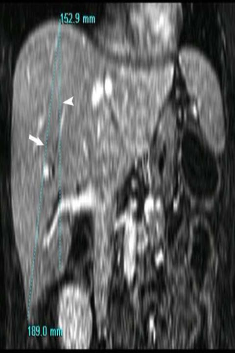
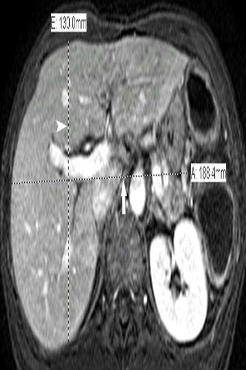
12 Figure Legends Figure 1 Axial, T1-weighted, 3D, gradient-echo MRI (TR/TE, 3.9/1.9 ms) in a 47-year-old woman shows mid-hepatic point (black arrow) half way between the mid vertebra and right lateral margin of the liver (white lines). Figure 2 Coronal, T1-weighted, 3D, gradient-echo MRI (TR/TE, 3.9/1.9 ms) in a 47- year-old woman illustrates the method of depicting the midhepatic point craniocaudad (MHP CC) and maximum CC (Max CC) lengths of the liver. MHP CC is measured as the greatest overall dimension (arrowhead) and Max CC as the longest distance between superior to inferior-most tip of the liver (arrow). Figure 3 Axial T1-weighted, 3D, gradient-echo MRI (TR/TE, 3.9/1.9 ms) in a 47-year-old woman illustrates the maximum transverse and midhepatic point anteroposterior (MHP AP) lengths of the liver. The maximum transverse dimension is measured as maximum distance between left and right lateral liver margins (arrow) and the MHP AP as the maximum AP dimension of the liver (arrowhead).
Dilated cisternae chyli. A sign of uncompensated cirrhosis at MR imaging
 Thomas Jefferson University Jefferson Digital Commons Department of Radiology Faculty Papers Department of Radiology January 2008 Dilated cisternae chyli. A sign of uncompensated cirrhosis at MR imaging
Index words : Bile duct radiography, technology Bile ducts MR Bile ducts surgery Liver, transplantation
 Hilar Branching Anatomy of Living Adult Liver Donors: Comparison of T2-MR Cholangiography and Contrast Enhanced T1-MR Cholangiography in Terms of Diagnostic Utility 1 Joon Seok Lim, M.D. 1, Myeong-Jin
Renal vascular evaluation with 64 Multislice Computerized Tomography Daniela Stoisa, Fabrizzio E. Galiano, Andrés Quaranta, Roberto L.
 Renal vascular evaluation with 64 Multislice Computerized Tomography Daniela Stoisa, Fabrizzio E. Galiano, Andrés Quaranta, Roberto L. Villavicencio Footnote Diagnóstico Médico Oroño. Bv. Oroño 1515. 2000.
US and MR imaging features of benign cystic mesothelioma of the liver: A diagnostic dilemma
 Thomas Jefferson University Jefferson Digital Commons Department of Radiology Faculty Papers Department of Radiology 5-2009 US and MR imaging features of benign cystic mesothelioma of the liver: A diagnostic
MR Advance Techniques. Vascular Imaging. Class II
 MR Advance Techniques Vascular Imaging Class II 1 Vascular Imaging There are several methods that can be used to evaluate the cardiovascular systems with the use of MRI. MRI will aloud to evaluate morphology
Background: Bedside ultrasound is emerging as a useful tool in the assessment of
 Abstract: Background: Bedside ultrasound is emerging as a useful tool in the assessment of intravascular volume status by examining measurements of the inferior vena cava (IVC). Many previous studies do
Aims and objectives. Page 2 of 10
 Diagnostic performance of automated breast volume scanner (ABVS) versus hand-held ultrasound (HHUS) as second look for breast lesions detected only on magnetic resonance imaging. Poster No.: C-1701 Congress:
Non Contrast MRA. Mayil Krishnam. Director, Cardiovascular and Thoracic Imaging University of California, Irvine
 Non Contrast MRA Mayil Krishnam Director, Cardiovascular and Thoracic Imaging University of California, Irvine No disclosures Non contrast MRA-Why? Limitations of CTA Radiation exposure Iodinated contrast
Sensitivity and Specificity in Detection of Labral Tears with 3.0-T MRI of the Shoulder
 Magee and Williams MRI for Detection of Labral Tears Musculoskeletal Imaging Clinical Observations C M E D E N T U R I C L I M G I N G JR 2006; 187:1448 1452 0361 803X/06/1876 1448 merican Roentgen Ray
ASSESSING THE PLAIN ABDOMINAL RADIOGRAPH M A A M E F O S U A A M P O F O
 ASSESSING THE PLAIN ABDOMINAL RADIOGRAPH M A A M E F O S U A A M P O F O Introduction The abdomen (less formally called the belly, stomach, is that part of the body between the thorax (chest) and pelvis,
Normal Sonographic Anatomy
 hapter 2:The Liver DUNSTAN ABRAHAM Normal Sonographic Anatomy Homogeneous, echogenic texture (Figure 2-1) Measures approximately 15 cm in length and 10 12.5 cm anterior to posterior; measurement taken
Biliary Ultrasonography Kathleen O Brien MD MPH RDMS Kaiser Permanente South Sacramento
 Biliary Ultrasonography Kathleen O Brien MD MPH RDMS Kaiser Permanente South Sacramento https://www.google.com/search?sa=g&hl=en&q=public+disclosure&tbm=isch&tbs=simg:caqsigeahwelekju2aqaaawlelcmpwgaygpgcamskpib_1qnza7ai
Methods. Yahya Paksoy, Bülent Oğuz Genç, and Emine Genç. AJNR Am J Neuroradiol 24: , August 2003
 AJNR Am J Neuroradiol 24:1364 1368, August 2003 Retrograde Flow in the Left Inferior Petrosal Sinus and Blood Steal of the Cavernous Sinus Associated with Central Vein Stenosis: MR Angiographic Findings
Magnetic Resonance Angiography
 Magnetic Resonance Angiography 1 Magnetic Resonance Angiography exploits flow enhancement of GR sequences saturation of venous flow allows arterial visualization saturation of arterial flow allows venous
Manifestations of rheumatoid arthritis: epidural pannus and atlantoaxial subluxation resulting in basilar invagination.
 Thomas Jefferson University Jefferson Digital Commons Department of Rehabilitation Medicine Faculty Papers Department of Rehabilitation Medicine 1-1-2012 Manifestations of rheumatoid arthritis: epidural
Liver metastases: treatment planning. PJ Valette
 Liver metastases: treatment planning PJ Valette Liver metastases removal December 2010 April 2011 : after chemotherapy June 2011 : after resection of left lobe mets & portal embol. Sept 2011 : 1 year after
B-Flow, Power Doppler and Color Doppler Ultrasound in the Assessment of Carotid Stenosis: Comparison with 64-MD-CT Angiography
 Med. J. Cairo Univ., Vol. 85, No. 2, March: 805-809, 2017 www.medicaljournalofcairouniversity.net B-Flow, Power Doppler and Color Doppler Ultrasound in the Assessment of Carotid Stenosis: Comparison with
My Patient Has Abdominal Pain PoCUS of the Biliary Tract and the Urinary Tract
 My Patient Has Abdominal Pain PoCUS of the Biliary Tract and the Urinary Tract Objectives PoCUS for Biliary Disease PoCUS for Renal Colic PoCUS for Urinary Retention Biliary Disease A patient presents
JlntSocPlastination, Vol4:16-22,
 JlntSocPlastination, Vol4:16-22, 1990 16 SECTIONAL ANATOMY: STANDARDIZED METHODOLOGY Alexander Lane, Coordinator of Anatomy and Physiology, Triton College, Visiting Associate Professor, University of Illinois
Modified Oblique Sagittal Magnetic Resonance Imaging of Rotator Cuff Tears: Comparison with Standard Oblique Sagittal Images
 Journal of Magnetics 22(3), 519-524 (2017) ISSN (Print) 1226-1750 ISSN (Online) 2233-6656 https://doi.org/10.4283/jmag.2017.22.3.519 Modified Oblique Sagittal Magnetic Resonance Imaging of Rotator Cuff
Anatomical and Functional MRI of the Pancreas
 Anatomical and Functional MRI of the Pancreas MA Bali, MD, T Metens, PhD Erasme Hospital Free University of Brussels Belgium mbali@ulb.ac.be Introduction The use of MRI to investigate the pancreas has
Added value of MR myelography using 3D COSMIC sequence in the diagnosis of lumbar canal stenosis: comparison with routine MR imaging
 Added value of MR myelography using 3D COSMIC sequence in the diagnosis of lumbar canal stenosis: comparison with routine MR imaging Poster No.: C-1099 Congress: ECR 2012 Type: Authors: Scientific Exhibit
Alice Fung, MD Oregon Health and Science University
 Alice Fung, MD Oregon Health and Science University Disclosure Comments The speaker Alice Fung, MD Has relevant financial relationships to disclose. Received honorarium from (Guerbet). This individual
Posterior longitudinal ligament status in cervical spine bilateral facet dislocations
 Thomas Jefferson University Jefferson Digital Commons Department of Orthopaedic Surgery Faculty Papers Department of Orthopaedic Surgery November 2005 Posterior longitudinal ligament status in cervical
Lung Perfusion Analysis New Pathways in Lung Imaging. Case Study Brochure PLA 309 Hospital
 Lung Perfusion Analysis New Pathways in Lung Imaging Case Study Brochure PLA 309 Hospital http://www.toshibamedicalsystems.com Toshiba Medical Systems Corporation 2012 all rights reserved. Design and specifications
MRI Abdomen Protocol Pancreas/MRCP with Contrast
 MRI Abdomen Protocol Pancreas/MRCP with Contrast Reviewed By: Brett Mollard, MD; Anna Ellermeier, MD Last Reviewed: July 2018 Contact: (866) 761-4200 Standard uses: 1. Characterization of cystic and solid
Essentials of Clinical MR, 2 nd edition. 99. MRA Principles and Carotid MRA
 99. MRA Principles and Carotid MRA As described in Chapter 12, time of flight (TOF) magnetic resonance angiography (MRA) is commonly utilized in the evaluation of the circle of Willis. TOF MRA allows depiction
Evolution of Clinical Anatomy with Ultrasonography Past Present and Future
 College of Medicine Evolution of Clinical Anatomy with Ultrasonography Past Present and Future Andrew F. Payer, Ph.D. Human Body Structure and Function Module Director Christine Bellew, M.D., Caridad Hernandez,
Hepatic Imaging: What Every Practitioner Should Know
 Hepatic Imaging: What Every Practitioner Should Know Shuchi K. Rodgers, MD Section Chief, Abdominal Imaging Director of Ultrasound Department of Radiology Einstein Medical Center rodgerss@einstein.edu
Paraumbilical collateral veins on MRI as possible protection against portal venous thrombosis in candidates for liver transplantation
 Thomas Jefferson University Jefferson Digital Commons Department of Radiology Faculty Papers Department of Radiology October 2007 Paraumbilical collateral veins on MRI as possible protection against portal
Ultrasound anatomy of the normal liver
 Clinical Ultrasound in Hepatology: Training for Hepatologists UCL Institute for Liver and Digestive Health, Royal Free Hospital, London, UK 29-30 April 2017 Ultrasound anatomy of the normal liver IVICA
Anatomy Jessica Ferguson Ashley Dobos May 31, 2006 LIVER
 Anatomy Jessica Ferguson Ashley Dobos May 31, 2006 LIVER 1) Other Names: Reidel s Lobe normal anatomic variant; projection of the right lobe that can extend as far as the iliac crest (Tempkin, p.54, Anatomy).
MRI of the Pancreas UNIT A18.1 A18.1.1
 MRI of the UNIT A18.1 MRI provides comprehensive information on the full range of pancreatic diseases. We employ a set protocol incorporating various types of sequences including transverse and coronal
Vascular abnormalities of the breast include a wide spectrum of arterial and
 Clinical Medicine & Research Volume 11, Number 4: 237-241 2013 Marshfield Clinic clinmedres.org Case Report Coexistence of a Congenital Arteriovenous Fistula of the Left Breast with a True Aneurysm of
Abdomen and Retroperitoneum Ultrasound Protocols
 Abdomen and Retroperitoneum Ultrasound Protocols Reviewed By: Anna Ellermeier, MD Last Reviewed: March 2018 Contact: (866) 761-4200, Option 1 **NOTE for all examinations: 1. If documenting possible flow
Role of Imaging in Medical Schools of Tomorrow
 PERSPECTIVE STUDY Role of Imaging in Medical Schools of Tomorrow Sanja Plavsic Kupesic DSJUOG Role of Imaging in Medical Schools of Tomorrow Professor and Clinical Professor of Obstetrics and Gynecology
Small Pulmonary Nodules: Our Preliminary Experience in Volumetric Analysis of Doubling Times
 Small Pulmonary Nodules: Our Preliminary Experience in Volumetric Analysis of Doubling Times Andrea Borghesi, MD Davide Farina, MD Roberto Maroldi, MD Department of Radiology University of Brescia Brescia,
A quality control program for MR-guided focused ultrasound ablation therapy
 JOURNAL OF APPLIED CLINICAL MEDICAL PHYSICS, VOLUME 3, NUMBER 2, SPRING 2002 A quality control program for MR-guided focused ultrasound ablation therapy Tao Wu* and Joel P. Felmlee Department of Radiology,
Prevalence of Meniscal Radial Tears of the Knee Revealed by MRI After Surgery
 Downloaded from www.ajronline.org by 46.3.207.114 on 12/22/17 from IP address 46.3.207.114. Copyright RRS. For personal use only; all rights reserved Thomas Magee 1 Marc Shapiro David Williams Received
CAN SOFT TISSUES STRUCTURES DIFFERENTIATE BETWEEN DYSPLASIA AND CAM-FAI OF THE HIP?
 CAN SOFT TISSUES STRUCTURES DIFFERENTIATE BETWEEN DYSPLASIA AND CAM-FAI OF THE HIP? A Le Bouthillier, KS Rakhra 1, PE Beaulé 2, RCB Foster 1 1 Department of Medical Imaging 2 Division of Orthopaedic Surgery
Hepatobiliary Ultrasound Rimon Bengiamin, MD, RDMS Assistant Clinical Professor Director of Emergency Ultrasound UCSF Fresno. Objectives. Why?
 Hepatobiliary Ultrasound Rimon Bengiamin, MD, RDMS Assistant Clinical Professor Director of Emergency Ultrasound UCSF Fresno Objectives Discuss the goals of point-of-care biliary ultrasound Review the
Sonographic Findings of Adductor Insertion Avulsion Syndrome With Magnetic Resonance Imaging Correlation
 Case Report Sonographic Findings of Adductor Insertion Avulsion Syndrome With Magnetic Resonance Imaging Correlation Jennifer S. Weaver, MD, Jon A. Jacobson, MD, David A. Jamadar, MBBS, Curtis W. Hayes,
Value of Volumetric and Morphological Parameters on Computed Tomography for Assessing Severity of Viral-Induced Liver Cirrhosis
 Med. J. Cairo Univ., Vol. 80, No. 2, June: 189-194, 2012 www.medicaljournalofcairouniversity.com Value of Volumetric and Morphological Parameters on Computed Tomography for Assessing Severity of Viral-Induced
Evaluation of Canal Diameter by MRI in Sudanese Population
 American Scientific Research Journal for Engineering, Technology, and Sciences (ASRJETS) ISSN (Print) 2313-4410, ISSN (Online) 2313-4402 Global Society of Scientific Research and Researchers http://asrjetsjournal.org/
Lab Monitor Images Dissection of the Abdominal Vasculature + Lower Digestive System
 Lab Monitor Images Dissection of the Abdominal Vasculature + Lower Digestive System Stomach & Duodenum Frontal (AP) View Nasogastric tube 2 1 3 4 Stomach Pylorus Duodenum 1 Duodenum 2 Duodenum 3 Duodenum
Abdominal Ultrasound. Diane Hallinen, MD. Bloodroot
 Abdominal Ultrasound Diane Hallinen, MD Bloodroot Abdominal Ultrasound Vasculature Hepatobiliary Spleen Kidney Bladder Bowel Where to put the probe? Vasculature We are going to talk about Celiac Trunk
CARDIAC MRI. Cardiovascular Disease. Cardiovascular Disease. Cardiovascular Disease. Overview
 CARDIAC MRI Dr Yang Faridah A. Aziz Department of Biomedical Imaging University of Malaya Medical Centre Cardiovascular Disease Diseases of the circulatory system, also called cardiovascular disease (CVD),
Use of IV-contrast versus IV-and oral-contrast in the evaluation of abdominal pain on CT in the emergency department
 Use of IV-contrast versus IV-and oral-contrast in the evaluation of abdominal pain on CT in the emergency department Poster No.: B-0693 Congress: ECR 2016 Type: Authors: Scientific Paper M. Wasserman 1,
Diagnostic Imaging
 www.fisiokinesiterapia.biz Diagnostic Imaging Diagnostic Imaging is no longer limited to radiography. Major technological advancements have lead to the use of new and improved imaging technologies. The
Effect of intravenous contrast medium administration on prostate diffusion-weighted imaging
 Effect of intravenous contrast medium administration on prostate diffusion-weighted imaging Poster No.: C-1766 Congress: ECR 2015 Type: Authors: Keywords: DOI: Scientific Exhibit J. Bae, C. K. Kim, S.
Pediatric Lung Ultrasound (PLUS) In Diagnosis of Community Acquired Pneumonia (CAP)
 In Diagnosis of Community Acquired Pneumonia (CAP)") Pediatric Lung Ultrasound (PLUS) In Diagnosis of Community Acquired Pneumonia (CAP) Dr Neetu Talwar Senior Consultant, Pediatric Pulmonology Fortis Memorial Research Institute, Gurugram Study To compare
Pediatric Lung Ultrasound (PLUS) In Diagnosis of Community Acquired Pneumonia (CAP) Dr Neetu Talwar Senior Consultant, Pediatric Pulmonology Fortis Memorial Research Institute, Gurugram Study To compare
Fracture risk in unicameral bone cyst. Is magnetic resonance imaging a better predictor than plain radiography?
 Acta Orthop. Belg., 2011, 77, 230-238 ORIGINAL STUDY Fracture risk in unicameral bone cyst. Is magnetic resonance imaging a better predictor than plain radiography? Nathalie PiREAU, Antoine DE GHELDERE,
Compute-aided Differentiation of Focal Liver Disease in MR Imaging
 1063 Compute-aided Differentiation of Focal Liver Disease in MR Imaging Xuejun Zhang a, Masayuki Kanematsu b, Hiroshi Fujita c, Takeshi Hara c, Hiroshi Kondo b, Xiangrong Zhou c, Wenguang Li a and Hiroaki
Surgical Correction of Severe Bilateral Thumb Pincer-Nail Deformity
 Thomas Jefferson University Jefferson Digital Commons Department of Orthopaedic Surgery Faculty Papers Department of Orthopaedic Surgery September 2006 Surgical Correction of Severe Bilateral Thumb Pincer-Nail
Comparison of T2-weighted MRCP before and after injection of Gd-EOB-DTPA in patients with primary sclerosing cholangitis (PSC)
") Comparison of T2-weighted MRCP before and after injection of Gd-EOB-DTPA in patients with primary sclerosing cholangitis (PSC) Poster No.: C-0051 Congress: ECR 2010 Type: Scientific Exhibit Topic: Abdominal
Comparison of T2-weighted MRCP before and after injection of Gd-EOB-DTPA in patients with primary sclerosing cholangitis (PSC) Poster No.: C-0051 Congress: ECR 2010 Type: Scientific Exhibit Topic: Abdominal
Using lesion washout volume fraction as a biomarker to improve suspicious breast lesion characterization
 JOURNAL OF APPLIED CLINICAL MEDICAL PHYSICS, VOLUME 16, NUMBER 5, 2015 Using lesion washout volume fraction as a biomarker to improve suspicious breast lesion characterization Jie Huang, a Sarah M. Schafer,
Accessory Glands of Digestive System
 Accessory Glands of Digestive System The liver The liver is soft and pliable and occupies the upper part of the abdominal cavity just beneath the diaphragm. The greater part of the liver is situated under
US LI-RADS v2017 CORE
 US LI-RADS v2017 CORE Screening or surveillance US in patient at high risk for HCC US category US-1 US-2 US-3 Negative Subthreshold Positive Category Concept Definition US-1 Negative US-2 Subthreshold
Common Occurrence of Benign Liver Lesions in Patients With Newly Diagnosed Breast Cancer Investigated by MRI for Suspected Liver Metastases
 JOURNAL OF MAGNETIC RESONANCE IMAGING 10:165 169 (1999) Original Research Common Occurrence of Benign Liver Lesions in Patients With Newly Diagnosed Breast Cancer Investigated by MRI for Suspected Liver
Vascular Technology Examination Content Outline
 Vascular Technology Examination Content Outline (Outline Summary) # Domain Subdomain Percentage 1 Normal Anatomy, Perfusion, and Function Evaluate normal anatomy, perfusion, function 2 Pathology, Perfusion,
3D ultrasound applied to abdominal aortic aneurysm: preliminary evaluation of diameter measurement accuracy
 3D ultrasound applied to abdominal aortic aneurysm: preliminary evaluation of diameter measurement accuracy Poster No.: C-0493 Congress: ECR 2011 Type: Authors: Keywords: DOI: Scientific Paper A. LONG
Liver Perfusion Analysis New Frontiers in Dynamic Volume Imaging. Case Study Brochure Chang Gung Memorial Hospital.
 New Frontiers in Dynamic Volume Imaging dynamic volume CT Case Study Brochure Chang Gung Memorial Hospital http://www.toshibamedicalsystems.com Toshiba Medical Systems Corporation 2010-2011. All rights
MR imaging of the knee in marathon runners before and after competition
 Skeletal Radiol (2001) 30:72 76 International Skeletal Society 2001 ARTICLE W. Krampla R. Mayrhofer J. Malcher K.H. Kristen M. Urban W. Hruby MR imaging of the knee in marathon runners before and after
Customizing Contrast Injection for Body MDCT: Algorithmic Approach
 Customizing Contrast Injection for Body MDCT: Algorithmic Approach Lincoln L. Berland, M.D., F.A.C.R. University of Alabama at Birmingham Before Contrast Prep and Hydration Hydration single most important
Guidelines, Policies and Statements D5 Statement on Abdominal Scanning
 Guidelines, Policies and Statements D5 Statement on Abdominal Scanning Disclaimer and Copyright The ASUM Standards of Practice Board have made every effort to ensure that this Guideline/Policy/Statement
Research Article Comparison of Colour Duplex Ultrasound with Computed Tomography to Measure the Maximum Abdominal Aortic Aneurysmal Diameter
 International Vascular Medicine, Article ID 574762, 4 pages http://dx.doi.org/10.1155/2014/574762 Research Article Comparison of Colour Duplex Ultrasound with Computed Tomography to Measure the Maximum
CLINICAL PRESENTATION AND RADIOLOGY QUIZ QUESTION
 Donald L. Renfrew, MD Radiology Associates of the Fox Valley, 333 N. Commercial Street, Suite 100, Neenah, WI 54956 6/23/2012 Radiology Quiz of the Week # 78 Page 1 CLINICAL PRESENTATION AND RADIOLOGY
NEW SUBTRACTION ALGORITHMS FOR EVALUATION OF BREAST LESIONS ON DYNAMIC CONTRAST ENHANCED MR MAMMOGRAPHY
 A-056 NEW SUBTRACTION ALGORITHMS FOR EVALUATION OF BREAST LESIONS ON DYNAMIC CONTRAST ENHANCED MR MAMMOGRAPHY So Hee Cho, M.D., Byung Gil Choi, M.D., Hak Hee Kim, M.D., Euy Neyng Kim, M.D., Bum-soo Kim,
Recommendations for cross-sectional imaging in cancer management, Second edition
 www.rcr.ac.uk Recommendations for cross-sectional imaging in cancer management, Second edition Renal and adrenal tumours Faculty of Clinical Radiology www.rcr.ac.uk Contents Renal cell carcinoma 3 Clinical
Objectives. Hepatobiliary Ultrasound: Anatomy, Technique, Pathology. RUQ: Normal Anatomy. Emergency Ultrasound: Gallbladder Location
 Hepatobiliary Ultrasound: Anatomy, Technique, Pathology Laleh Gharahbaghian, MD FAAEM Associate Director, EM Ultrasound Co-Director, EM Ultrasound Fellowship Stanford University Medical Center Seric Cusick,
RECTAL CARCINOMA: A DISTANCE APPROACH. Stephanie Nougaret
 RECTAL CARCINOMA: A DISTANCE APPROACH Stephanie Nougaret stephanienougaret@free.fr Despite the major improvements that have been made due to total mesorectal excision (TME) management of rectal cancer
MRI of Bucket-Handle Te a rs of the Meniscus of the Knee 1
 MRI of ucket-handle Te a rs of the Meniscus of the Knee 1 Joon Yong Park, M.D., Young-uk Lee M.D., Eun-Chul Chung M.D., Hae-Won Park M.D., E u n - Kyung Youn M.D., Shin Ho Kook, M.D., Young Rae Lee, M.D.
Simplifying liver assessment in internal medicine
 Ultrasound Customer story Simplifying liver assessment in internal medicine Philips Affiniti ultrasound for elastography and contrast-enhanced ultrasound (CEUS) Where Sonography Institute, Uster, Switzerland
KNEE ALIGNMENT SYSTEM (KAS) MRI Protocol
 MRI Protocol") KNEE ALIGNMENT SYSTEM (KAS) MRI Protocol Sample referral sticker Referral Sticker Insert here Corin 17 Bridge Street Pymble NSW Australia 2073 P: +61 (0)2 9497 7400 F: +61 (0)2 9497 7498 E: KAS.customerservice@coringroup.com
KNEE ALIGNMENT SYSTEM (KAS) MRI Protocol Sample referral sticker Referral Sticker Insert here Corin 17 Bridge Street Pymble NSW Australia 2073 P: +61 (0)2 9497 7400 F: +61 (0)2 9497 7498 E: KAS.customerservice@coringroup.com
Basic Abdominal Sonography
 24S Basic Abdominal Sonography Procedural Overview JOHN FATCHETT II, RDMS is provided. Patient preparation (i.e., fasting) scanning techniques, spleen, transducer. evaluation of abdominal anatomy in the
Accuracy of SPECT bone scintigraphy in diagnosis of meniscal tears ABSTRACT
 1 Iran J Nucl Med 2005; 23 Accuracy of SPECT bone scintigraphy in diagnosis of meniscal tears M. Saghari 1, M. Moslehi 1, J. Esmaeili 2, M.N. Tahmasebi 3, A. Radmehr 4, M. Eftekhari 1,2, A. Fard-Esfahani
Supplementary Online Content
 Supplementary Online Content Hooshmand B, Magialasche F, Kalpouzos G, et al. Association of vitamin B, folate, and sulfur amino acids with brain magnetic resonance imaging measures in older adults: a longitudinal
Infraclavicular brachial plexus blocks have been designed
 The Supraclavicular Lateral Paravascular Approach for Brachial Plexus Regional Anesthesia: A Simulation Study Using Magnetic Resonance Imaging Øivind Klaastad, MD* and Örjan Smedby, Dr Med Sci *Department
Pituitary Gland Assessment by MR Volumetry in the Normal Indian Adolescent Population
 International Journal of Medical Imaging 2015; 3(6): 105-109 Published online October 31, 2015 (http://www.sciencepublishinggroup.com/j/ijmi) doi: 10.11648/j.ijmi.20150306.11 ISSN: 2330-8303 (Print); ISSN:
Look differently. Invenia ABUS. Automated Breast Ultrasound
 Look differently. Invenia ABUS Automated Breast Ultrasound InveniaTM ABUS from GE Healthcare offers a view beyond mammography, with breast screening technology that looks differently. 40 % The unseen risk.
2D and 3D MR imaging in the assessment of Fallopian tube features
 2D and 3D MR imaging in the assessment of Fallopian tube features Poster No.: C-1292 Congress: ECR 2010 Type: Topic: Scientific Exhibit Genitourinary Authors: J. Takahama, S. Kitano, N. Marugami, A. Takahashi,
Childhood obesity and blood pressure: back to the future?
 Thomas Jefferson University Jefferson Digital Commons Department of Pediatrics Faculty Papers Department of Pediatrics 11-1-2011 Childhood obesity and blood pressure: back to the future? Bonita Falkner
Half-Fourier Acquisition Single-Shot Turbo Spin-Echo (HASTE) MR: Comparison with Fast Spin-Echo MR in Diseases of the Brain
 MR: Comparison with Fast Spin-Echo MR in Diseases of the Brain") Half-Fourier Acquisition Single-Shot Turbo Spin-Echo (HASTE) MR: Comparison with Fast Spin-Echo MR in Diseases of the Brain Mahesh R. Patel, Roman A. Klufas, Ronald A. Alberico, and Robert R. Edelman PURPOSE:
Half-Fourier Acquisition Single-Shot Turbo Spin-Echo (HASTE) MR: Comparison with Fast Spin-Echo MR in Diseases of the Brain Mahesh R. Patel, Roman A. Klufas, Ronald A. Alberico, and Robert R. Edelman PURPOSE:
Spontaneous portosystemic venous shunts in liver cirrhosis: Anatomy, pathophysiology, hemodynamic changes and imaging findings
 Spontaneous portosystemic venous shunts in liver cirrhosis: Anatomy, pathophysiology, hemodynamic changes and imaging findings Poster No.: C-3193 Congress: ECR 2010 Type: Educational Exhibit Topic: Vascular
Acute abdominal venous thromboses- the hyperdense noncontrast CT sign
 Acute abdominal venous thromboses- the hyperdense noncontrast CT sign Poster No.: C-1095 Congress: ECR 2011 Type: Educational Exhibit Authors: M. Goldstein, K. Jhaveri; Toronto, ON/CA Keywords: Abdomen,
ACUTE PANCREATITIS: NEW CLASSIFICATION OF AN OLD FOE. T Barrow, A Nasrullah, S Liong, V Rudralingam, S A Sukumar
 ACUTE PANCREATITIS: NEW CLASSIFICATION OF AN OLD FOE T Barrow, A Nasrullah, S Liong, V Rudralingam, S A Sukumar LEARNING OBJECTIVES q Through a series of cases illustrate the updated Atlanta symposium
PULMONARY EMBOLISM ANGIOCT (CTA) ASSESSMENT OF VASCULAR OCCLUSION EXTENT AND LOCALIZATION OF EMBOLI 1. BACKGROUND
 ASSESSMENT OF VASCULAR OCCLUSION EXTENT AND LOCALIZATION OF EMBOLI 1. BACKGROUND") JOURNAL OF MEDICAL INFORMATICS & TECHNOLOGIES Vol. 11/2007, ISSN 1642-6037 Damian PTAK * pulmonary embolism, AngioCT, postprocessing techniques, Mastora score PULMONARY EMBOLISM ANGIOCT (CTA) ASSESSMENT
JOURNAL OF MEDICAL INFORMATICS & TECHNOLOGIES Vol. 11/2007, ISSN 1642-6037 Damian PTAK * pulmonary embolism, AngioCT, postprocessing techniques, Mastora score PULMONARY EMBOLISM ANGIOCT (CTA) ASSESSMENT
Laminar Thickness: Correlation Against Symptoms of Spinal Stenosis and Changes with Age
 Laminar Thickness: Correlation Against Symptoms of Spinal Stenosis and Changes with Age Hamed Shalikar, MD 1, Sean L. Borkowski, MS 2, Juan Pablo Villablanca, MD 1, Sophia N. Sangiorgio, PhD 2, Edward
Depiction of Lateral Ligament Complex of the Ankle using 3D MRI in Healthy Subjects and Patients with Chronic Ankle Instability.
 Depiction of Lateral Ligament Complex of the Ankle using 3D MRI in Healthy Subjects and Patients with Chronic Ankle Instability. Satoshi Yamaguchi, M.D. 1, Hiroshi Matsumoto, R.T. 2, Atsuya Watanabe, M.D.
The latest developments - Automated Breast Volume Scanning. Dr. med. M. Golatta
 The latest developments - Automated Breast Volume Scanning Dr. med. M. Golatta Automated Breast Volume US: Why? o Mammography is limited in dense breasts: high false negative rate o Many of these tumors
Improvement of Image Quality with ß-Blocker Premedication on ECG-Gated 16-MDCT Coronary Angiography
 16-MDCT Coronary Angiography Shim et al. 16-MDCT Coronary Angiography Sung Shine Shim 1 Yookyung Kim Soo Mee Lim Received December 1, 2003; accepted after revision June 1, 2004. 1 All authors: Department
Coronary angiography is the standard way of visualizing
 Clinical Investigation and Reports Coronary Artery Fly-Through Using Electron Beam Computed Tomography Peter M.A. van Ooijen, MSc; Matthijs Oudkerk, MD, PhD; Robert J.M. van Geuns, MD; Benno J. Rensing,
Hepatic pseudolesion and pseudotumor due to third inflow: Prevalence and correlation with liver fibrosis on multi-phasic MDCT
 Hepatic pseudolesion and pseudotumor due to third inflow: Prevalence and correlation with liver fibrosis on multi-phasic MDCT Poster No.: C-1940 Congress: ECR 2015 Type: Scientific Exhibit Authors: K.
Computed Tomography of Normal Adrenal Glands in Indian Population
 IOSR Journal of Dental and Medical Sciences (IOSR-JDMS) e-issn: 2279-0853, p-issn: 2279-0861.Volume 17, Issue 01 Ver. V January. (2018), PP 26-30 www.iosrjournals.org Computed Tomography of Normal Adrenal
Innovations in HCC Imaging: MDCT/MRI
 Innovations in HCC Imaging: MDCT/MRI Anthony E. Cheng, M.D. Cardinal MRI Center Cardinal Santos Medical Center, Wilson Street, San Juan Innovations in HCC Imaging: Goals/Objectives MDCT/MRI Learn the diagnostic
Gemstone Spectral Imaging quantifies lesion characteristics for a confident diagnosis
 GE Healthcare Gemstone Spectral Imaging quantifies lesion characteristics for a confident diagnosis CT clinical case study lesion characterization Desiree Morgan, MD Vice Chair of Clinical Research Professor
Usefulness of the Navigator-echo triggering Technique for Free-Breathing 3D MRCP
 Usefulness of the Navigator-echo triggering Technique for Free-Breathing 3D MRCP Poster No.: C-1257 Congress: ECR 2012 Type: Scientific Exhibit Authors: K. Matsunaga, G. Ogasawara, K. Fujii, T. Irie, T.
Oak foundation for donating the 3T Siemens Verio scanner. Board of directors BBH and Frh Hospitals for supporting the
 Knee pain and inflammation in the infrapatellar fat pad estimated by conventional and dynamic contrast-enhanced magnetic resonance imaging in obese patients with osteoarthritis: a crosssectional study