Metabolic liver disease
|
|
|
- Dina Dean
- 5 years ago
- Views:
Transcription

1
2 Metabolic liver disease
3 Metabolic liver disease Group of diseases due to disorders of metabolism Acquired Inherited A- Acquired Non alcoholic fatty liver disease B- Inherited Hemochromatosis Wilson disease Α 1 antitrypsin deficiency
4 Non alcoholic fatty liver disease (NAFLD) Non alcoholic fatty liver disease (NAFLD) group of conditions : Include; 1. Simple hepatic steatosis (fatty liver) 2. Steatosis accompanied by non specific inflam ( stable conditions with out significant clinical problems) 3. Non alcoholic steatohepatitis (NASH) Individuals with No H/o alcohol consumption / take very small quantity - < 20 gm of ethanol / week
5 Non Alcoholic Steatohepatitis- NASH NASH : A condition with hepatocyte injury may progress to cirrhosis - in % cases. Main components are ; Hepatocyte ballooning Lobular inflammation Steatosis Progression of disease fibrosis Men = women
6 Non Alcoholic Steatohepatitis- NASH Ass. with ; obesity & other components of metabolic synd. as. Dyslipidemia, Hyperinsulinemia Insulin resistance 70% of obese individuals - NAFLD;- Most common cause of cryptogenic cirrhosis (cirrhosis of unknown origin) contribute to progression of other liver diseases HCV infectopn, & HCC.
7 Pathogenesis; Pathogenesis; Has two sequential events: (1) Hepatic fat accumulation & (2) Hepatic oxidative stress. Oxidative stress - acts upon accumulated hepatic lipids, - leads to Lipid peroxidation & Release of lipid peroxides, - produce reactive oxygen species.

8
 In less sever disease Only -Elevated liver enzymes No inflam, hepatocyte death /")
9 Fatty liver - Morphology Steatosis; Involves > 5% of hepatocytes ( rarely - > 90 % ) Micro & macro vesicular (droplets of fat in hepatocytes) In less sever disease Only -Elevated liver enzymes No inflam, hepatocyte death / scaring


10 Two patterns of hepatic steatosis (1) Micro vesicular steatosis: Cytoplasm - replaced by bubbles of fat - do not displace the nucleus; & (2) Macrovesicular steatosis: Cytoplasm - replaced by a large bubble of fat that displaces the nucleus to edge of cell.
11 Morphology Steatohepatitis (NASH); Steatosis, Parenchymal inflam- Multi focal, mainly neutrophils Mallory bodies Hepatocyte death (ballooning degen & apoptosis), Fibrosis; Sinusoidal, In portal tract & Around terminal hepatic V Cirrhosis - may develop after years

12 Steatohepatitis Micro ; Macro vesicular steatosis, Cytologic ballooning, Mallory bodies, and Scattered lobular inflam
 Morphologic hallmarks of alcoholic and nonalcoholic steatohepatitis.")
13 Mallory's hyaline bodies; Pink filamentous structures, - cytoplasmic inclusions in hepatocytes (ab. keratin, hyaline, & other pr). Mallory's hyaline bodies Consequence of cellular injury. Found in ballooned hepatocytes (black arrow) Morphologic hallmarks of alcoholic and nonalcoholic steatohepatitis. Hepatocytes, - macro vesicular fat globules (white arrow), displacing nucleus (white arrowhead) to periphery

14 Steatohepatitis with cirrhosis. A nodule of liver tissue is circumscribed by scar tissue.

15 NASH Elevations in liver tests, ALT / AST No apparent reason for liver disease (medications, viral hepatitis, / alcohol) x rays or imaging studies - show fat, - suspect - NASH. Tissue shows fat without inflam / damage - simple fatty liver / NAFLD.
16 Clinical features Simple steatosis asymptomatic Symptoms - Related to other metabolic derangements (Obesity, insulin resistance and diabetes) General symptoms- Fatigue Right abd. Discomfort - due to hepatomegaly NASH with ass - dyslipidemia, hyperinsulinemia and insulin resistance Cardiovascular disease frequent cause of death
17 Clinical features Diagnosis; Elevated - serum AST & ALT in 90% - pts with NASH Imaging studies fat accumulation - in liver Liver biopsy diagnostic tool for NASH. Determine the; Extent of - steatosis, Presence of - steatohepatitis and Degree of - fibrosis Goal of treatment to; Reverse steatosis and Prevent cirrhosis Liver transplantation - only treat for advanced cirrhosis with liver failure,
18 Hemochromatosis
19 Hemochromatosis Hemochromatosis can be; Primary An inherited disorder- Caused by excessive iron absorption Secondary - acquired H./ hemosiderosis Accumulation of iron in tissues - due to parental administration of iron as transfusion / other causes
20 Ch by : Hemochromatosis Excessive accumulation of body iron, - in parenchymal organs liver & pancreas. / heart, joints, endocrine organs Normally : - Total body iron pool -2-6 gm in adults. 0.5gm in liver, 98 % of this in hepatocytes In primary hemochromatosis; Total body iron - > 50 gm, 1/3 rd of which in liver
21 Hemochromatosis Primary hemochromatosis : - Fully developed cases show Micronodular cirrhosis in all pts Diabetes mellitus 75 80% of pts Skin pigmentation % pts Iron accumulation Is life long, but Injury by excessive iron - slow & progressive so Symptom - in 5 th 6 th decades of life Men > women
22 I. Hereditary Hemochromatosis Mutations in genes II. Hemosiderosis (Secondary Hemochromatosis) Classification of Iron Overload A. Parenteral iron overload Transfusions Long-term hemodialysis Aplastic anemia Sickle cell disease Myelodysplastic syndromes Leukemias Iron-dextran injections Sideroblastic anemia Pyruvate kinase deficiency C. Increased oral intake of iron African iron overload (Bantu siderosis) D. Congenital atransferrinemia E. Chronic liver disease Chronic alcoholic liver disease Porphyria cutanea tarda B. Ineffective erythropoiesis with increased erythroid activity β-thalassemia F. Neonatal hemochromatosis
23 Hemochromatosis Pathogenesis. Total body iron - regulated by intestinal absorption In hemochromatosis- ; Regulation of intestinal absorption of dietary iron is - lost Net iron accum to 1.0 gm / year mainly in liver. - After 20 gm of storage iron - Disease.
24 Pathogenesis Excessive iron - directly toxic to host tissues by; (1)Lipid per oxidation via iron-catalyzed free radical reactions (2) Stimulation of collagen formation (3) Interactions of reactive oxygen species & iron with DNA,- lethal injury / hepatocellular carcinoma Process - reversible in cells - not fatally injured, Removal of excess iron by therapy - promotes recovery of tissue function
25 Hepcidin level is regulated by; Hemojuvelin ( Pr expressed by liver, heart & sk muscle) Transferrin Receptor 2 - on hepatocytes HFE - product of hemochromatosis gene Pathogenesis Main regulators of iron absorption is hepcidin pr. encoded by HAMP gene, produced by hepatocytes. Its production Increased by - inflam cytokines & iron & Decreased by - iron def, hypoxia & ineffective erythropoiesis. Hepcidin - prevents - release of iron from intestinal cells & macrophages lowers plasma iron level
26 Pathogenesis Mutation of ; HAMP gene & hemojuvelin Sever form of - hereditary - juvenile hemochromatosis. HFE &, transferrin receptor 2 Classic form of - mild hereditary adult hemochromatosis.
27 Hereditary Hemochromatosis Morphologic Changes - ch. by Deposition of hemosiderin - in dec. order of frequency in ; liver, Pancreas, Myocardium, Pituitary gland, Adrenal gland, Thyroid, parathyroid, Joints & Skin Cirrhosis Pancreatic fibrosis "Hemosiderosis" - relatively benign accum. of iron. Hemochromatosis" - when organ dysfunction occurs.

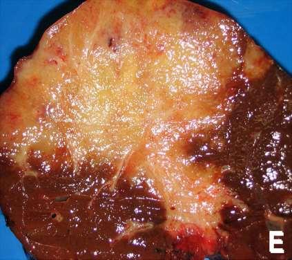
28 Morphology Golden yellow hemosiderin granules - of periportal hepatocytes (cytoplasm) Stain blue with Prussian blue stain With increasing iron load Progressive involvement of - rest of lobule, Bile duct epith & Kupffer cell - pigmentation, At this stage; liver slightly enlarged, & chocolate brown. Fibrous septa - slow Micro nodular pattern of cirrhosis in intensely pigmented liver Prussian blue iron stain -Blue granules of hemosiderin in; hepatocytes & Kupffer cells.

 on sectioning - is due to extensive iron")
29 Hemochromatosis A cross-section of a normal, healthy liver Dark brown color of ; Liver, Pancreas (bottom center) & Lymph nodes (bottom right) on sectioning - is due to extensive iron deposition
30 Morphology Pancreas Intensely pigmented, Diffuse interstitial fibrosis, Parenchymal atrophy. Hemosiderin present both in acinar & islet cells &fibrous stroma Heart Enlarged, Hemosiderin granules in myocardial fibers Skin - pigmentation Pigmentation of synovial lining, and testes


31 Hemochromatosis Extensive fibrosis with small nodules of hepatocytes without fatty change. Iron stain of liver - presence of hemosiderin granules.

32 Hemochromatosis Hepatocytes & Kupffer cells Granular brown deposits of hemosiderin Due to - accumulation of excess iron in the liver.
33 > in male, Rare - < 40 y Presents as; Hepatomegaly- Abdominal pain, Skin pigmentations, Diabetes mellitus - due to destruction of pancreatic islands, Clinical features Cardiac dysfunction- (arrhythmias, cardiomyopathy), Arthritis Classic triad- late in the course; 1)- Pigment cirrhosis with hepatomegaly, 2)- Skin pigmentation, 3)- Diabetes mellitus
34 Clinical features Death - may occur due to; Cirrhosis, Cardiac disease / Hepatocellular Ca Diagnosis: High serum iron and ferritin levels, Exclusion of secondary causes of iron overload Liver biopsy Prusssian blue histological reaction Treatment : Pre - cirrhotic stage - treated by regular phlebotomy
35 Wilson's disease
36 Wilson's disease Wilson's disease / hepatolenticular degeneration An autosomal recessive disorder caused by mutations in (ATP 7B) gene. Resulting in; Impaired - copper excretion into bile & Failure to incorporate copper - in ceruloplasmin Toxic levels of copper accumulates in tissues & organs, esp Liver, Brain & Eyes;
37 Normally; Wilson's disease % of ingested copper (2-5 mg /day)- absorbed in duodenum & proximal small intestine To portal circulation - with albumin & histadine Free copper dissociates - & taken up by hepatocytes Incorporates in to enzymes & binds to apoceruloplasmin to form ceruloplasmin secreted in blood. Excess copper transported into bile, % of plasma copper ceruloplasmin - eventually - degraded by liver cells Released copper is excreted into bile. Total body copper mg
38 Wilson's disease ATP 7B gene located on ch 13, Encodes - a trans memb. Copper transporting ATPase expressed on hepatocyte canalicular memb. Mutations def of - ATP7B pr. decrease copper transport in bile Impairs - incorporation in cerulopl Dec cerulopl sec in blood Copper accum in liver toxic liver inj

39 Liver - mainly effected, Morphology Hepatic changes; - minor to massive damage Fatty change - mild to moderate with ; vacuolated nuclei and occasional focal hepatocyte necrosis Acute hepatitis feature of - acute viral hepatitis with fatty change A bluish-coppery colored liver,
40 Morphology Chronic hepatitis of Wilson disease Moderate to severe inflammation Hepatocyte necrosis & Macro vesicular steatosis. Vacuolated hepatocellular nuclei and Mallory bodies Progress to cirrhosis Hepatic copper content - > 250 µg / gm dry weight- most diagnostic In the brain - toxic injury to basal ganglia - atrophy / cavitation


41 Eyes: Kayser-Fleischer rings (KF rings) visible around the iris. Wilson's disease Due to copper deposition in descemet's memb of cornea. Golden Brown / copper-colored rings - Kayser-Fleischer rings around - corneas
42 Clinical features Symptoms usually appear between years Most common presentation acute / chronic liver disease Neuropsychiatric manifestations including; Mild behavioral changes, Frank psychosis or Parkinson disease like syndrome e.g. tremors
43 Clinical features Biochemical diagnosis of Wilson disease is based on; Decreased - serum ceruloplasmin, Increased - hepatic copper content & - (most sensitive test) Increased - urinary excretion of copper- (most sp. Screening test) Serum copper level low / normal / elevated depending on the stage of disease - not diagnostic Pts with hepatitis / cirrhosis- liver transplantation
44 Alpha 1- antitrypsin deficiency
45 Alpha antitrypsin-1 deficiency Autosomal Recessive Disorder Very low levels of Alpha 1 antitrypsin - a plasma glycoprotein - synthesized by hepatocytes Major function of this protein Inhibition of proteases - esp neutrophil elastase, cathepsin G & protinase 3 normally released from neutrophils at sites of inflam.
46 Alpha antitrypsin-1 deficiency Alpha antitrypsin-1 deficiency cause pulmonary emphysema due to unopposed destructive proteases Also causes; Liver disease due to accum of this pr in hepatocytes Cutaneous paniculitis, Arterial aneurysm, Bronchiectasis and Wegener s granulomatosis. Defective secretion of Alpha-1 antitrypsin by hepatocytes Chronic active hepatitis, Cirrhosis, HCC in child


47 Alpha-1 antitrypsin deficiency
48 Alpha 1-antitrypsin- deficiency - Morphology Hepatic pathology Range from; Neonatal hepatitis with / without cholestasis & fibrosis to childhood cirrhosis / Chronic active Hep / Cirrhosis - appears late in life Distinctive feature of hepatic diseases; PAS positive globules, - ( Mostly only distinctive feature) Fatty change & Mallory bodies

49 Morphology ; Alpha-1-antitrypsin deficiency Round - oval cytoplasmic globular inclusions Mostly, in hepatocytes, surrounding the portal tracts Acidophilic in H&E, PAS positive.
 On the right - normal liver")
50 Alpha-1-antitrypsin deficiency Large globules in the liver cells ( PAS positive granules) On the right - normal liver cells
51 Alpha 1-antitrypsin- deficiency C/F ;- Neonatal - hepatitis & jaundice 10 20% of newborns with antitrypsin deficiency In adolescence symptoms of hepatitis / cirrhosis Hepatitis may subside with recovery or chronic H - cirrhosis in middle to latter life 2-3 % of adults develop HCC Treatment Liver transplantation
52 Intrahepatic billiary disease
53 Intrahepatic billiary disease Disorders of intrahepatic bile ducts 1. Secondary billiary cirrhosis 2. Primary billiary cirrhosis 3. Primary sclerosing cholangitis Secondary billiary cirrhosis- uncorrected obst. of extra hepatic billiary tree Primary billiary cirrhosis destructive disorder of intrahepatic billiary tree Primary sclerosing cholangitis- involves both extra hepatic & intra hepatic billiary tree
54 Primary biliary cirrhosis / Secondary billiary cirrhosis Primary billiary cirrhosis Secondary billiary cirrhosis Etiology autoimmune extra hepatic bile duct obstruction; biliary atresia, gall stones, stricture, ca head of pancreas Sex predilection ( F : M) 6 : 01 None Symptoms / signs Lab findings Important pathological findings- before cirrhosis develops pruritus, jaundice, malaise, dark urine, light stools, hepatosplenomegaly, - insidious onset Conjugated hyperbilirubinemia, Increased serum alk. Phosphatase, bile acids, cholesterol, IgM auto antibodies Dense lymphocytic infiltrate in portal Tracts with Granulomatous destruction of bile ducts same as primary biliary cirrhosis Conjugated hyperbilirubinemia Increased serum alk. Phosphatase, bile acids, cholesterol, Prominent bile stasis in bile ducts, Bile ductular proliferation with surrounding neutrophils, Portal tract edema


55 Primary billiary cirrhosis Ch by; Destruction of bile ductules within triads of liver. Dense chronic inflam. Infilt. With loss of bile ductules in portal tract. Periductal epithelioid granulomas. Micro nodular cirrhosis.

56 Characterized by: Secondary billiary cirrhosis Regeneration nodules, surrounded by coarse fibrous septa. regenerating hepatocytes are disorderly disposed. Fibrous septa - inflam. infiltrate (lymph, macrophages)
. Bile retention - in the parenchyma, as \"bile lakes\".")
57 Embedded in septa are; Secondary billiary cirrhosis Small & large bile ducts ( prolif / distended) Bile stasis - as bile thrombi (brown-green, amorphous). Bile retention - in the parenchyma, as "bile lakes".
58 Tumors of Liver
59 Tumors of Liver Non-Neoplastic Neoplastic 1. Cystic masses Abscess (pyogenic / amebic) Hydatid cyst Polycystic liver disease Caroli Disease 2. Solid Nodular hyperplasia 1. Benign Adenoma Hemangioma 2. Malignant Hepatoblastoma Hepatocellular ca. Fibro lamellar ca. Cholangio ca. Angio sarcoma 3. Metastatic
60 Hepatocellular carcinoma Usually after 5 th decade, M > F (2.4 : 1) More common in Africa & South east Asia (with high rates of chronic HBV).
61 Pathogenesis Four major etiologic factors ass. with HCC ; 1. Chronic viral infection (HBV, HCV) 2. Chronic alcoholism 3. Non alcoholic steatohepatitis (NASH) 4. Food contaminants - Aflatoxins Aflatoxin: a toxin produced by fungus (aspergillus flavus) - contaminate peanuts & grains (aflatoxins bind with cellular DNA cause specific mutation in p53)
62 Pathogenesis Other predisposing conditions :- 1. Tyrosinemia - rare 40% pts develop HCC despite dietary control 2. Glycogen storage disease 3. Hereditary hemochromatosis 4. Non alcoholic fatty liver disease 5. α 1 anti trypsin deficiency 6. Other contributing factors include; Genetic factors, Age, gender, Chemicals, hormones
63 Pathogenesis 1. In countries with high prevalence regions of HBV infection main cause Begins in infancy from infected mother 200 times increased risk for HCC - by adulthood Cirrhosis - absent in 50 % pts Cancer occurs at yrs of age 2. In western world (where HBV not prevalent) Ass with other Ch. Liver diseases e.g. Alcoholism Non alcoholic steatohepatitis Ch. HCV infection Hemochromatosis Cirrhosis present in 75 90% 3. In china &southern Africa (HBV endemic) aflatoxins
64 Events in pathogenesis; Pathogenesis Ch. Hepatitis - from any cause Repeated cycles of cell death & regeneration imp in pathogenesis of HCC. Resulting accum. of mutations may Damage - DNA repair mechanisms, Transform hepatocytes. Preneoplastic - dysplasia - Progression to HCC - may occur from point mutations of different genes
65 In HBV infected individuals- Pathogenesis Viral integration - precedes / accompanies transforming event disrupt cell genome Depending on integration sites, HBV may; Activate - proto oncogenes tumorigenic or Transform- HBV X protein,- a transcriptional activator of many genes, HCV - RNA virus Does not disrupt DNA / produce oncogenic proteins, May participate via -HCV core & NS5A- protein of hepatitis C virus - to develop HCC

66 Grossly; Hepatocellular carcinoma Liver enlarged tumor; Unifocal usually large mass, Multifocal, widely distributed nodules of variable size Diffusely infiltrative ca.- may involve entire liver in cirrhotic background Paler than surrounding liver, Green hue - when composed of well diff. hepatocytes - secreting bile Hepatocellular carcinoma with a greenish yellow hue.


67 Hepatocellular carcinoma Multinodular. Unifocal


68 Hepatocellular carcinoma Diffuse Hepatocellular ca in cirrhosis
69 Morphology All patterns may invade; Vascular structures extensive intrahepatic metastasis, Portal vein / inferior venacava - may extend to right heart Metastasis outside the liver - Via; Hematogenous to lung, Lymphatics - lymph node (perihilar, peripancreatic & para - aortic nodes) above & below the diaphragm

70 Hepatocellular carcinoma May spread extensively within the liver by; Contiguous growth & By development of satellite nodules - driven from parent tumor Satellite nodules represent either; Intrahepatic spread of tumor or Multicentric origin of tumor.

71 Microscopically ; Morphology HCCs range well diff - highly anaplastic / undiff lesions. Well diff & moderately diff tumors ; Recognizable hepatocytes > than 2 cell thick plates forming Trabeculae, Solid / tubular structures Acinar / pseudo glandular pat. Well diff T - composed of liver cords wider than two cells thick Loss of lobular architecture, Vascular structures - present.
72 Morphology Important clues are Atypical mitotic figures, Cluster of enlarged hyper chromatic nuclei Blood vessel with thrombi. Intra nuclear / cytoplasmic inclusion may be seen. Cytoplasm may show Mallory s hyaline bodies, / bile pigment. Kupffer cells - scant and irregularly scattered. CEA -negative / focally positive AFP - specific but insensitive,

")
73 Morphology Well differentiated ca. Tumor cells Resemble hepatocytes, Form cords & nests, May contain bile pig. in cytoplasm Malignant cells (mostly on right) interdigitate with Normal, larger hepatocytes (mostly at left).
, developed on liver cirrhosis. Discohesive, pleomorphic, anaplastic, giant cells.")
74 Hepatocelular carcinoma Poorly diff T- Tumor cells; Pleomorphic with numerous anaplastic giant cells, Or Small & completely undiff, - May resemble spindle cell sarcoma Poorly diff ( upper right), developed on liver cirrhosis. Discohesive, pleomorphic, anaplastic, giant cells. Scant stroma and central necrosis because of the poor vascularization.

75 Morphology Poorly diff - hepatocellular carcinoma.


76 Hepatocellular carcinoma Cytoplasm showing --- bile pigment. Hepatocellular Ca. in lymphatic vessels

77 Fibro lamellar carcinoma- Fibro lamellar Carcinoma A variant of HCC,-(5% of HCC) Young males & adult females Equal incidence No underlying ch. Hepatic disease Etiology unknown Presents as ; Single, large, hard scirrhous tumor with fibrous bands coursing through it Prognosis better than HCC

78 Microscopically composed of; Fibro lamellar Carcinoma Well diff polygonal cells - in nests / cords Separated by lamellae of dense collagen bundles Tumor cells have Abundant eosinophilic cytoplasm & Prominent nucleoli
79 Clinical Features of HCC Often masked by that of cirrhosis / ch. Hepatitis Mostly pts. have ill defined upper abdominal pain, Malaise Fatigue Wt. loss Abdominal mass / fullness Jaundice Fever GI / esophageal variceal bleeding
80 Clinical Features Lab findings - helpful but rarely conclusive Serum α feto pr elevated - 50% cases GLYPICAN-3 (GPC-3) is a heparin sulphate proteoglycan & an oncofetal protein. Useful serum marker in pts with hepatocellular ca GPC3 immunohistochemistry (IHC) - used to distinguish early HCC from dysplastic nodule
81 Clinical Features Diagnosis- ultrasound, hepatic angiography, MRI, Natural course:- progressive enlargement of primary mass disturbs hepatic function / metastasizes first to lungs, than - other sites Death may occurs from Cachexia, GI / esophageal variceal bleeding, Liver failure with hepatic coma, 5 yr survival rate; With large tumor - < 2 yrs Detection of Tumor < 2cm excision good prognosis
82 Cholangiocarcinoma
83 Cholangiocarcinoma Malignancy of biliary tree, arising from bile ducts within & out side the liver Risk factors include; Primary sclerosing cholangitis, Congenital fibro polycystic disease of biliary system (esp choledochal cysts & Caroli disease - rare inherited disorder, ch by dilatation of intrahepatic bile ducts) HCV infection & Previous exposure to Thorotrast (for radiography of biliary disease).
84 Acc to location are classified in Intrahepatic & Extra hepatic CCA Cholangiocarcinoma Extra hepatic % - including; Perihilar (Klatskin tumors) at the jn of Rt & Lt hepatic ducts & Distal bile duct tumors, Subgroup - tumors near ampulla of Vater. Periampullary ca - also include ; Adeno. ca of duodenal mucosa and pancreatic ca
85 Cholangiocarcinoma Survival only 15 % - at 2 yrs after diagnosis Intrahepatic CCAs - diagnosed late - after obstruction of bile flow or As liver mass Hilar and distal tumors - present with symptoms of; biliary obst, cholangitis and right upper quadrant pain
86 Morphology Extrahepetic CCAs Small lesions at the time of diagnosis Firm, gray nodules within bile duct wall Diffusely infiltrating, / papillary /polypoid lesions Most Adeno ca - with / without mucin secretion Squamous features, present - rare Epith prolif. with abundant fibrous stroma Klatskin tumors ( hilar cholangiocarcinoma); Have slower growth than other CCAs Show prominent fibrosis & Have distant metastasis

87 Cholangiocarcinoma Ca of extrahepatic bile ducts. tumor extends from the junction of the cystic and hepatic ducts to the ampulla of Vater.

88 Cholangiocarcinoma Intrahepatic CCAs Occur in non cirrhotic liver, Track along intrahepatic portal tract system create tree like mass or Massive tumor nodule develops Vascular invasion & propagation along portal lymphatics prominent Extensive intrahepatic metastasis

89 Cholangiocarcinoma

90 Microscopy; Cholangiocarcinoma Cholangio ca - resemble adeno ca of other sites Mostly well to moderately diff sclerosing adeno ca with Glandular and tubular structures lined by cuboidal to low col epith Markedly desmoplastic dense collagenous stroma separating the glandular element tumor extremely firm & gritty
91 Cholangiocarcinoma Metastasis; Lymphatic lymph nodes, Hematogenous - lungs and bones, adrenals and brain Mixed variants occur in which elements of both HCC & CCA are present 3- forms recognized; 1. Separate tumor masses of HCC & CCA within same liver 2. Collision tumors : tumorous masses of HCC & CCA comingle at, identifiable interface 3. Tumors in which elements of HCC & CCA - mixed at microscopic level

92 Cholangiocarcinoma


93 Cholangiocarcinoma Cholangio ca, well diff intrahepatic CCA- adeno ca showing; Tubular and / or papillary structures Lined by anaplastic cuboidal to low columnar epithelial cells. Variable fibrous stroma. Cholangio ca, moderately diff
94 Metastatic Carcinoma

95 Metastases to the liver Numerous mass lesions of variable size. Some of the larger ones demonstrate central necrosis..

96 Metastatic carcinoma Cross section of liver, showing; Multiple tumor deposits of Adeno ca - body of the pancreas.

97 Metastatic carcinoma Metastatic infiltrating ductal carcinoma from breast, with Normal liver parenchyma.

98
Hemosiderin. Livia Vida 2018
 Hemosiderin Livia Vida 2018 Questions Histochemical caracteristics of the different pigments. Exogenous pigments. Hemoglobinogenic pigments. Causes and forms of jaundice. Hemoglobinogenic pigments. Pathological
Pathological Classification of Hepatocellular Carcinoma
 3 rd APASL Single Topic Conference: HCC in 3D Pathological Classification of Hepatocellular Carcinoma Glenda Lyn Y. Pua, M.D. HCC Primary liver cancer is the 2 nd most common cancer in Asia HCC is the
-sheet 3. -Waseem Alhaj. Maha Shomaf
 -sheet 3 -Basheer egbaria -Waseem Alhaj Maha Shomaf 1 P a g e Viral hepatitis have many types each type is associated with different outcomes complication, some can result in acute one,others result in
Liver Tumors. Prof. Dr. Ahmed El - Samongy
 Liver Tumors Prof. Dr. Ahmed El - Samongy Objective 1. Identify the most important features of common benign liver tumors 2. Know the risk factors, diagnosis, and management of hepatocellular carcinoma
Slide 7 demonstrates acute pericholangitisis with neutrophils around proliferating bile ducts.
 Many of the histologic images and the tables are from MacSween s Pathology of the Liver (5 th Edition). Other images were used from an online source called PathPedia.com. A few images from other sources
Approach to the Patient with Liver Disease
 Approach to the Patient with Liver Disease Diagnosis of liver disease Careful history taking Physical examination Laboratory tests Radiologic examination and imaging studies Liver biopsy Liver diseases
Dhanpat Jain Yale University School of Medicine, New Haven, CT
 Dhanpat Jain Yale University School of Medicine, New Haven, CT Case history 15 years old female presented with fatigue. Found to have features suggestive of cirrhosis with esophageal varices, splenomegaly
A Review of Liver Function Tests. James Gray Gastroenterology Vancouver
 A Review of Liver Function Tests James Gray Gastroenterology Vancouver Copyright 2017 by Sea Courses Inc. All rights reserved. No part of this document may be reproduced, copied, stored, or transmitted
Liver, Pancreas and Gall Bladder Pathology
 Liver, Pancreas and Gall Bladder Pathology SCBM342 Systemic Pathology Witchuda Payuhakrit, Ph.D. (Pathobiology) Email: witchuda.pay@mahidol.ac.th Objectives 1. Understand etiology and pathogenesis of common
Anatomy of the biliary tract
 Harvard-MIT Division of Health Sciences and Technology HST.121: Gastroenterology, Fall 2005 Instructors: Dr. Jonathan Glickman Anatomy of the biliary tract Figure removed due to copyright reasons. Biliary
Cell injury, adaptation and death. Unite one Second Lab.
 Cell injury, adaptation and death Unite one Second Lab. The two lung abscesses seen here are examples of liquefactive necrosis in which there is a liquid center in an area of tissue injury. One abscess
Digestive system L 4. Lecturer Dr. Firdous M. Jaafar Department of Anatomy/Histology section
 Digestive system L 4 Lecturer Dr. Firdous M. Jaafar Department of Anatomy/Histology section objectives 1-Describe the structure of liver. 2-Define liver lobule, and identify its zones. 3-Define portal
PITFALLS IN THE DIAGNOSIS OF MEDICAL LIVER DISEASE WITH TWO CONCURRENT ETIOLOGIES I HAVE NOTHING TO DISCLOSE CURRENT ISSUES IN ANATOMIC PATHOLOGY 2017
 CURRENT ISSUES IN ANATOMIC PATHOLOGY 2017 I HAVE NOTHING TO DISCLOSE Linda Ferrell PITFALLS IN THE DIAGNOSIS OF MEDICAL LIVER DISEASE WITH TWO CONCURRENT ETIOLOGIES Linda Ferrell, MD, UCSF THE PROBLEM
Histology. The pathology of the. bile ducts. pancreas. liver. The lecture in summary. Vt-2006
 Vt-2006 The pathology of the liver, bile ducts and pancreas Richard Palmqvist Docent, ST-läkare, Klin Pat Lab, Labcentrum The lecture in summary Introduction, histology & physiology in brief General phenomenon
What is Liver Cancer? About the Liver
 Your liver is important and it has many functions. The top three are that it cleans your blood of toxins, gives you energy and produces bile for digestion. What is Liver Cancer? Cancer starts when cells
BREAST PATHOLOGY. Fibrocystic Changes
 BREAST PATHOLOGY Lesions of the breast are very common, and they present as palpable, sometimes painful, nodules or masses. Most of these lesions are benign. Breast cancer is the 2 nd most common cause
Metabolic Liver Disease
 Metabolic Liver Disease Peter Eichenseer, MD No relationships to disclose. Outline Overview Alpha-1 antitrypsin deficiency Wilson s disease Hereditary hemochromatosis Pathophysiology Clinical features
Disorders of the Liver and Pancreas
 Disorders of the Liver and Pancreas Liver Lobule Hexagonal plates Sinusoids Triads Bile duct branch Arteriole Venuole Blood flows from periphery to Central vein Space of Dissé Lobular Microanatomy Hepatocytes
Malignant Focal Liver Lesions
 Malignant Focal Liver Lesions Other Than HCC Pablo R. Ros, MD, MPH, PhD Departments of Radiology and Pathology University Hospitals Cleveland Medical Center Case Western Reserve University Pablo.Ros@UHhospitals.org
PATHOLOGY OF LIVER TUMORS
 PATHOLOGY OF LIVER TUMORS Pathobasic, 31.05.2016 WHO Classification Approach to a Liver Mass Lesion in a patient with chronic liver disease? Lesion in a patient without chronic liver disease? Malignant
Basic patterns of liver damage what information can a liver biopsy provide and what clinical information does the pathologist need?
 Basic patterns of liver damage what information can a liver biopsy provide and what clinical information does the pathologist need? Rob Goldin r.goldin@imperial.ac.uk Fatty liver disease Is there fatty
Steatosis/Steatohepatitis
 Prepared by Kurt Schaberg Introduction to Medical Liver Steatosis/Steatohepatitis Alcoholic Hepatitis Hepatocyte injury and inflammation resulting from chronic alcohol consumption AST/ALT ratio typically
Diseases of liver. Dr. Mohamed. A. Mahdi 4/2/2019. Mob:
 Diseases of liver Dr. Mohamed. A. Mahdi Mob: 0123002800 4/2/2019 Cirrhosis Cirrhosis is a complication of many liver disease. Permanent scarring of the liver. A late-stage liver disease. The inflammation
Biliary tract tumors
 Short Course 2010 Annual Fall Meeting of the Korean Society for Pathologists Biliary tract tumors Joon Hyuk Choi, M.D., Ph.D. Professor, Department of Pathology, Yeungnam Univ. College of Medicine, Daegu,
Note: The cause of testicular neoplasms remains unknown
 - In the 15- to 34-year-old age group, they are the most common tumors of men. - Tumors of the testis are a heterogeneous group of neoplasms that include: I. Germ cell tumors : 95%; all are malignant.
HEPATO-BILIARY IMAGING
 HEPATO-BILIARY IMAGING BY MAMDOUH MAHFOUZ MD PROF.OF RADIOLOGY CAIRO UNIVERSITY mamdouh.m5@gmail.com www.ssregypt.com CT ABDOMEN Indications Patient preparation Patient position Scanogram Fasting 4-6 hours
Special stains in liver pathology
 Current Issues in Surgical Pathology 2014 Special stains in liver pathology Which, why, how Really? Sanjay Kakar, MD University of California, San Francisco Outline Which stains Why the stain is done How
Diseases of the breast (1 of 2)
") Diseases of the breast (1 of 2) Introduction A histology introduction Normal ducts and lobules of the breast are lined by two layers of cells a layer of luminal cells overlying a second layer of myoepithelial
Diseases of the breast (1 of 2) Introduction A histology introduction Normal ducts and lobules of the breast are lined by two layers of cells a layer of luminal cells overlying a second layer of myoepithelial
Neoplasms of the Canine, Feline and Lemur Liver:
 Neoplasms of the Canine, Feline and Lemur Liver: Classification and Prognosis Annual Seminar of the French Society of Veterinary Pathology John M. Cullen VMD PhD DACVP North Carolina State University Primary
Evaluation of Liver Mass Lesions. American College of Gastroenterology 2013 Regional Postgraduate Course
 Evaluation of Liver Mass Lesions American College of Gastroenterology 2013 Regional Postgraduate Course Lewis R. Roberts, MB ChB, PhD Division of Gastroenterology and Hepatology Mayo Clinic College of
Hereditary Haemochromatosis (A pint too many: discussing haemochromatosis) John Lee
 John Lee") Hereditary Haemochromatosis (A pint too many: discussing haemochromatosis) John Lee Hereditary Haemochromatosis A disorder of iron metabolism Inherited disorder Iron Essential micro-nutrient Toxicity when
Hereditary Haemochromatosis (A pint too many: discussing haemochromatosis) John Lee Hereditary Haemochromatosis A disorder of iron metabolism Inherited disorder Iron Essential micro-nutrient Toxicity when
Hepatitis. Causes: - infectious Hepatitis: viral, Bacterial, Parasitic, and Helminthic - Autoimmune Hepatitis - Drug- and Toxin (Alcohol) - Metabolic
 - Metabolic") Liver diseases II Hepatitis Causes: - infectious Hepatitis: viral, Bacterial, Parasitic, and Helminthic - Autoimmune Hepatitis - Drug- and Toxin (Alcohol) - Metabolic infectious Hepatitis Several clinical
Liver diseases II Hepatitis Causes: - infectious Hepatitis: viral, Bacterial, Parasitic, and Helminthic - Autoimmune Hepatitis - Drug- and Toxin (Alcohol) - Metabolic infectious Hepatitis Several clinical
Metabolic Liver Diseases
 Metabolic Liver Diseases Howard J. Worman, M. D. Department of Medicine Columbia University College of Physicians and Surgeons Three Classical Inherited Disorders of Metabolism Affecting the Liver Hereditary
LIVER DISEASES. Pathology Department, Zhejiang University School of Medicine,
 LIVER DISEASES Pathology Department, Zhejiang University School of Medicine, 马丽琴,maliqin198@zju.edu.cn Viral Hepatitis Cirrhosis of liver Liver cancer Viral Hepatitis DEFINITION Primary hepatic infections
Gross appearance of nodular hyperplasia in material obtained from suprapubic prostatectomy. Note the multinodular appearance and the admixture of
 Tiền liệt tuyến Tiền liệt tuyến Gross appearance of nodular hyperplasia in material obtained from suprapubic prostatectomy. Note the multinodular appearance and the admixture of solid and microcystic areas.
CHAPTER 1. Alcoholic Liver Disease
 CHAPTER 1 Alcoholic Liver Disease Major Lesions of Alcoholic Liver Disease Alcoholic fatty liver - >90% of binge and chronic drinkers Alcoholic hepatitis precursor of cirrhosis Alcoholic cirrhosis end
What Is Cirrhosis? CIRRHOSIS. Cirrhosis occurs when the liver is. by chronic conditions and diseases. permanently scarred or injured
 What Is Cirrhosis? Cirrhosis occurs when the liver is permanently scarred or injured by chronic conditions and diseases. Common causes of cirrhosis include: Long-term alcohol abuse. Chronic viral hepatitis
Hepatocytes produce. Proteins Clotting factors Hormones. Bile Flow
 R.J.Bailey MD Hepatocytes produce Proteins Clotting factors Hormones Bile Flow Trouble.. for the liver! Trouble for the Liver Liver Gall Bladder Common Alcohol Hep C Fatty Liver Cancer Drugs Viruses Uncommon
Normal thyroid tissue
 Thyroid Pathology Overview Normal thyroid tissue Normal thyroid tissue with follicles filled with colloid. Thyroid cells form follicles, spheres of epithelial cells (always single layered in health, usually
XIII. Tumours of the liver and biliary system
 XIII. Tumours of the liver and biliary system V. PONOMARKOV 1 & L. J. MACKEY 2 In this histological classification of liver and gall bladder tumours the tumour types largely correspond to those found in
Jaundice. Agnieszka Dobrowolska- Zachwieja, MD, PhD
 Jaundice Agnieszka Dobrowolska- Zachwieja, MD, PhD Jaundice definition Jaundice, as in the French jaune, refers to the yellow discoloration of the skin. It arises from the abnormal accumulation of bilirubin
Liver Diseases. Yasmine Lashine MD, PhD
 Liver Diseases Yasmine Lashine MD, PhD ILOs Recognize different causes of Live failure Recall and understand clinical picture and complication of Liver failure Discuss causes and clinical picture of hepatic
Extrahepatic Biliary Obstruction. Ductal Diseases: Stones Tumors. Acute Injury: Viral Hepatitis Toxin (APAP/Etoh) Reye s Shock.
 Reye s Shock.") Extrahepatic Biliary Obstruction Stones Tumors Ductal Diseases: Ductal Diseases: Primary Biliary Primary Biliary Cirrhosis Cirrhosis Sclerosing Cholangitis Sclerosing Cholangitis Acute Injury: Viral Hepatitis
Extrahepatic Biliary Obstruction Stones Tumors Ductal Diseases: Ductal Diseases: Primary Biliary Primary Biliary Cirrhosis Cirrhosis Sclerosing Cholangitis Sclerosing Cholangitis Acute Injury: Viral Hepatitis
Liver Cancer Causes, Risk Factors, and Prevention
 Liver Cancer Causes, Risk Factors, and Prevention Risk Factors A risk factor is anything that affects your chance of getting a disease such as cancer. Learn more about the risk factors for liver cancer.
Pitfalls in the diagnosis of well-differentiated hepatocellular lesions
 2013 Colorado Society of Pathology Pitfalls in the diagnosis of well-differentiated hepatocellular lesions Sanjay Kakar, MD University of California, San Francisco Outline Hepatocellular adenoma: new WHO
Radiology of hepatobiliary diseases
 GI cycle - Lecture 14 436 Teams Radiology of hepatobiliary diseases Objectives 1. To Interpret plan x-ray radiograph of abdomen with common pathologies. 2. To know the common pathologies presentation.
Linda Ferrell, MD Distinguished Professor Vice Chair Director of Surgical Pathology Dept of Pathology
 Linda Ferrell, MD Distinguished Professor Vice Chair Director of Surgical Pathology Dept of Pathology Nonalcoholic steatohepatitis and Fatty Liver Disease Liver manifestations of the obesity epidemic Changes
Chapter 18 LIVER BILIARY TRACT
 Chapter 18 LIVER & BILIARY TRACT DUCT SYSTEM N O FIBROUS TISSUE PORTAL TRIAD CENTRAL VEIN PATTERNS OF HEPATIC INJURY Degeneration: Balooning, feathery degeneration, fat, pigment Inflammation:
Maram Abdaljaleel, MD Dermatopathologist and Neuropathologist University of Jordan, School of Medicine
 Maram Abdaljaleel, MD Dermatopathologist and Neuropathologist University of Jordan, School of Medicine The most common non-skin malignancy of women 2 nd most common cause of cancer deaths in women, following
2014 CURRENT ISSUES IN PATHOLOGY
 2014 CURRENT ISSUES IN PATHOLOGY SPECIAL STAINS IN LIVER BIOPSY PATHOLOGY Sanjay Kakar, MD University of California, San Francisco Trichrome stain : (1) Assess degree of fibrosis. H&E stain is not reliable
Section 8 Liver and Gallbladder
 General and Systemic Histopathology C601 and C602 Section 8 As we will see in this unit, the liver is subject to many types of injury. Additionally, many systemic diseases have a liver component and sometimes
Liver Cancer And Tumours
 Liver Cancer And Tumours What causes liver cancer? Many factors may play a role in the development of cancer. Because the liver filters blood from all parts of the body, cancer cells from elsewhere can
SESSION 1: GENERAL (BASIC) PATHOLOGY CONCEPTS Thursday, October 16, :30am - 11:30am FACULTY COPY
 PATHOLOGY CONCEPTS Thursday, October 16, :30am - 11:30am FACULTY COPY") SESSION 1: GENERAL (BASIC) PATHOLOGY CONCEPTS Thursday, October 16, 2008 9:30am - 11:30am FACULTY COPY GOAL: Describe the basic morphologic (structural) changes which occur in various pathologic conditions.
SESSION 1: GENERAL (BASIC) PATHOLOGY CONCEPTS Thursday, October 16, 2008 9:30am - 11:30am FACULTY COPY GOAL: Describe the basic morphologic (structural) changes which occur in various pathologic conditions.
GASTROINTESTINAL IMAGING STUDY GUIDE
 GASTROINTESTINAL IMAGING STUDY GUIDE Pharynx Diverticula Foreign bodies Trauma o Motility Disorders Esophagus Diverticula Trauma Esophagitis Barrett esophagus Rings, webs, and strictures Varices Benign
Salivary Gland Cytology
 Salivary Gland Cytology Diagnostic challenges and potential pitfalls Tarik M. Elsheikh, MD Professor and Medical Director Anatomic Pathology Cleveland Clinic FNA Salivary Gland Lesions Indications Distinguish
Neoplasia 2018 Lecture 2. Dr Heyam Awad MD, FRCPath
 Neoplasia 2018 Lecture 2 Dr Heyam Awad MD, FRCPath ILOS 1. List the differences between benign and malignant tumors. 2. Recognize the histological features of malignancy. 3. Define dysplasia and understand
Navigating the Biliary Tract with CT & MR: An Imaging Approach to Bile Duct Obstruction
 Navigating the Biliary Tract with CT & MR: An Imaging Approach to Bile Duct Obstruction Ann S. Fulcher, MD Medical College of Virginia Virginia Commonwealth University Richmond, Virginia Objectives To
Causes of acquired hemosidrosis : 1-multiple transfusions 2-ineffective erythropoiesis (β-thalassemia ) 3-increased iron intake (Bantu sidrosis )
 3-increased iron intake (Bantu sidrosis )") Hemochromatosis Excessive accumalation of body iron (liver & pancreas) 1ry or 2ry (genetic or acquired ) Genetic hemochromatosis ( 4 variants) The most common form is aut.recessive disease of adult onset
Hemochromatosis Excessive accumalation of body iron (liver & pancreas) 1ry or 2ry (genetic or acquired ) Genetic hemochromatosis ( 4 variants) The most common form is aut.recessive disease of adult onset
Workup of a Solid Liver Lesion
 Workup of a Solid Liver Lesion Joseph B. Cofer MD FACS Chief Quality Officer Erlanger Health System Affiliate Professor of Surgery UTHSC-Chattanooga I have no financial or other relationships with any
Autoimmune Liver Diseases
 2nd Pannonia Congress of pathology Hepato-biliary pathology Autoimmune Liver Diseases Vera Ferlan Marolt Institute of pathology, Medical faculty, University of Ljubljana Slovenia Siofok, Hungary, May 2012
British Liver Transplant Group Pathology meeting September Leeds cases
 British Liver Transplant Group Pathology meeting September 2014 Leeds cases Leeds Case 1 Male 61 years Liver transplant for HCV cirrhosis with HCC in January 2014. Now raised ALT and bilirubin,? acute
NEOPLASMS AND TUMOR-LIKE CONDITIONS OF LIVER
 NEOPLASMS AND TUMOR-LIKE CONDITIONS OF LIVER Epithelial Tumors Focal nodular hyperplasia Focal nodular hyperplasia is a localized hyperplasic overgrowth of hepatocytes around a vascular anomaly, particularly
Liver Specialty Evening Conference. Matthew M. Yeh, MD, PhD Professor of Pathology Adjunct Professor of Medicine University of Washington, Seattle
 Liver Specialty Evening Conference Matthew M. Yeh, MD, PhD Professor of Pathology Adjunct Professor of Medicine University of Washington, Seattle Case History A 65 year-old man presents with abdominal
Case year female. Routine Pap smear
 Case 1 57 year female Routine Pap smear Diagnosis? 1. Atypical glandular cells of unknown significance (AGUS) 2. Endocervical AIS 3. Endocervical adenocarcinoma 4. Endometrial adenocarcinoma 5. Adenocarcinoma
WSC , Conference 9, Case 1. Tissue from a nyala.
 WSC 2009-2010, Conference 9, Case 1. Tissue from a nyala. MICROSCOPIC DESCRIPTION: Heart, atrium (1 pt.): Approximately 40% of the atrial myocardium is replaced by areas of fibrous connective tissue (1
Interesting Cases from Liver Tumor Board. Jeffrey C. Weinreb, M.D.,FACR Yale University School of Medicine
 Interesting Cases from Liver Tumor Board Jeffrey C. Weinreb, M.D.,FACR Yale University School of Medicine jeffrey.weinreb@yale.edu Common Liver Diseases Hemangioma Cyst FNH Focal Fat/Sparing THID Non-Cirrhotic
DIGESTIVE SYSTEM II ACCESSORY DIGESTIVE ORGANS
 DIGESTIVE SYSTEM II ACCESSORY DIGESTIVE ORGANS Dr. Larry Johnson Texas A& M University Objectives Distinguish between the parotid and submandibular salivary glands. Understand and identify the structural
Video Microscopy Tutorial 8
 Video Microscopy Tutorial 8 Common and Uncommon Lesions of the Liver Gladwyn Leiman, MD There are no disclosures necessary. Common and Uncommon Lesions in Liver FNA Gladwyn Leiman University of Vermont
Non alcoholic fatty liver and Non alcoholic Steatohepatitis. By Dr. Seham Seif
 Non alcoholic fatty liver and Non alcoholic Steatohepatitis By Dr. Seham Seif Definition NAFL describe a common clinicopathological conditions characterized by significant lipid deposition in the hepatocytes
PATHOLOGY MCQs. The Pancreas
 PATHOLOGY MCQs The Pancreas A patient with cystic fibrosis is characteristically: A. more than 45 years of age B. subject to recurring pulmonary infections C. obese D. subject to spontaneous fractures
Metabolic Liver Disease: What s New in Diagnosis and Therapy?
 Metabolic Liver Disease: What s New in Diagnosis and Therapy? Bruce R. Bacon, M.D. James F. King M.D. Endowed Chair in Gastroenterology Professor of Internal Medicine Division of Gastroenterology and Hepatology
Case n 1 ( B 92 / 4208 ) Case n 2 ( B 00 / 8249 ) Case n 3 ( B 98 / 8352 )
 Case n 2 ( B 00 / 8249 ) Case n 3 ( B 98 / 8352 )") Slide Seminar Case n 1 ( B 92 / 4208 ) 16 month-old girl. HBV serology +. Clinic in favour of chronic hepatitis. 4 portal triads! classification limited Viral B chronic hepatitis Mild activity (Fig. 1
Slide Seminar Case n 1 ( B 92 / 4208 ) 16 month-old girl. HBV serology +. Clinic in favour of chronic hepatitis. 4 portal triads! classification limited Viral B chronic hepatitis Mild activity (Fig. 1
Biliary Atresia. Karen F. Murray, MD Professor of Pediatrics Director, Hepatobiliary Program Seattle Children s
 Biliary Atresia Karen F. Murray, MD Professor of Pediatrics Director, Hepatobiliary Program Seattle Children s Biliary Atresia Incidence: 1/8,000-15,000 live births Girls > boys 1.5:1 The most common cause
Kidney Case 1 SURGICAL PATHOLOGY REPORT
 Kidney Case 1 Surgical Pathology Report February 9, 2007 Clinical History: This 45 year old woman was found to have a left renal mass. CT urography with reconstruction revealed a 2 cm medial mass which
Chronic Biliary Disease. Dr Susan Davies & Dr Bill Griffiths
 Chronic Biliary Disease Dr Susan Davies & Dr Bill Griffiths Chronic Biliary Disease Terminology is confusing with pathologists and hepatologists using the same language BUT with different meanings. Chronic
New insights into fatty liver disease. Rob Goldin Centre for Pathology, Imperial College
 New insights into fatty liver disease Rob Goldin Centre for Pathology, Imperial College r.goldin@imperial.ac.uk Prevalence of NASH Global prevalence of NAFLD is 25% with highest prevalence in the Middle
Steatotic liver disease
 Steatotic liver disease Fatty liver disease Prof. Dr. ANNE HOORENS Non-Neoplastic Liver Pathology December 8th 2018 Working Group of Digestive Pathology Belgian Society of Pathology OUTLINE NAFLD = Non-Alcoholic
Neoplasia literally means "new growth.
 NEOPLASIA Neoplasia literally means "new growth. A neoplasm, defined as "an abnormal mass of tissue the growth of which exceeds and is uncoordinated with that of the normal tissues and persists in the
Autoimmune Hepatitis Chronic hepatitis with
 Autoimmune Hepatitis -Chronic hepatitis with immunologic abnormalities -Histologic features are similar to chronic viral hepatitis -Indolent or severe course -Dramatic response to immunosuppressive therapy
Biliary tract diseases of the liver
 Biliary tract diseases of the liver Digestive Diseases Course Bucharest 2016 Rob Goldin r.goldin@imperial.ac.uk How important are biliary tract diseases? Hepatology 2011 53(5):1608-17 Approximately 16%
Gastrointes*nal and Liver Pathology. Kris*ne Kra5s, M.D.
 Gastrointes*nal and Liver Pathology Kris*ne Kra5s, M.D. GI Pathology Outline Esophagus Stomach Intes*ne Liver Gallbladder Pancreas GI Pathology Outline Esophagus Stomach Intes*ne Liver Hepa**s Alcoholic
Intrahepatic cholangiocarcinoma Histologic spectrum, novel markers and molecular assays
 2018 Current Issues in Surgical Pathology Summary (not actual lecture) Intrahepatic cholangiocarcinoma Histologic spectrum, novel markers and molecular assays Sanjay Kakar, MD University of California,
Imaging Guided Biopsy. Edited & Presented by ; Hussien A.B ALI DINAR. Msc Lecturer,Reporting Sonographer
 Imaging Guided Biopsy Edited & Presented by ; Hussien A.B ALI DINAR. Msc Lecturer,Reporting Sonographer Objective By the End of this lessons you should : Define what biopsy Justify Aim to perform biopsy
Basic patterns of liver damage what information can a liver biopsy provide and what clinical information does the pathologist need?
 Basic patterns of liver damage what information can a liver biopsy provide and what clinical information does the pathologist need? Rob Goldin r.goldin@imperial.ac.uk @robdgol FATTY LIVER DISEASE Brunt
Pathology lab 4 DONE BY : MORAD ABU QAMAR
 Pathology lab 4 DONE BY : MORAD ABU QAMAR Chronic interstitial inflammation, lung Certain etiologic agents such as viruses are more likely to lead to chronic inflammation, as seen here in the lung of a
!! 2 to 3% of All New Visceral Cancers.!! Peak Incidence is 6th Decade!! M:F = 2:1
 !! Kathleen M. O Toole, M.D.!! 2 to 3% of All New Visceral Cancers!! Peak Incidence is 6th Decade!! M:F = 2:1!! Grossly is a Bright Yellow, Necrotic Mass with a Pseudocapsule 1 !!Conventional RCC! Clear
Neoplasias Quisticas del Páncreas
 SEAP -Aproximación Práctica a la Patología Gastrointestinal- Madrid, 26 de mayo, 2006 Neoplasias Quisticas del Páncreas Gregory Y. Lauwers, M.D. Director, Service Massachusetts General Hospital Harvard
2 to 3% of All New Visceral Cancers Peak Incidence is 6th Decade M:F = 2:1 Grossly is a Bright Yellow, Necrotic Mass with a Pseudocapsule
 GENITOURINARY PATHOLOGY Kathleen M. O Toole, M.D. Renal Cell Carcinoma 2 to 3% of All New Visceral Cancers Peak Incidence is 6th Decade M:F = 2:1 Grossly is a Bright Yellow Necrotic Mass Grossly is a Bright
Noncalculous Biliary Disease Dean Abramson, M.D. Gastroenterologists, P.C. Cedar Rapids. Cholestasis
 Noncalculous Biliary Disease Dean Abramson, M.D. Gastroenterologists, P.C. Cedar Rapids Cholestasis Biochemical hallmark Impaired bile flow from liver to small intestine Alkaline phosphatase is primary
Alpha-1 Antitrypsin Deficiency: Liver Disease
 Alpha-1 Antitrypsin Deficiency: Liver Disease Who is at risk to develop Alpha-1 liver disease? Alpha-1 liver disease may affect children and adults who have abnormal Alpha-1 antitrypsin genes. Keys to
End Stage Liver Disease & Disease Specific Indications for Liver Transplant. Susan Kang, RN, MSN, ANP-BC
 End Stage Liver Disease & Disease Specific Indications for Liver Transplant Susan Kang, RN, MSN, ANP-BC Introduction (https://www.srtr.org) What does the liver do? STORAGE METABOLIC DETOXIFICATION SYNTHETIC
End Stage Liver Disease & Disease Specific Indications for Liver Transplant Susan Kang, RN, MSN, ANP BC
 End Stage Liver Disease & Disease Specific Indications for Liver Transplant Susan Kang, RN, MSN, ANP BC Introduction (https://www.srtr.org) 1 What does the liver do? STORAGE METABOLIC DETOXIFICATION SYNTHETIC
HEALTH SERVICES POLICY & PROCEDURE MANUAL
 PAGE 1 of 6 PURPOSE To establish basic understanding of indications and contraindications for transplantation of various organs. POLICY The N.C. Department of Correction, Division of Prisons, Health Services
number Done by Corrected by Doctor Maha Shomaf
 number 16 Done by Waseem Abo-Obeida Corrected by Zeina Assaf Doctor Maha Shomaf MALIGNANT NEOPLASMS The four fundamental features by which benign and malignant tumors can be distinguished are: 1- differentiation
Mody. AIS vs. Invasive Adenocarcinoma of the Cervix
 Common Problems in Gynecologic Pathology Michael T. Deavers, M.D. Houston Methodist Hospital, Houston, Texas Common Problems in Gynecologic Pathology Adenocarcinoma in-situ (AIS) of the Cervix vs. Invasive
B. Environmental Factors. a. The major risk factor to papillary thyroid cancer is exposure to ionizing radiation, during the first 2 decades of life.
 B. Environmental Factors. a. The major risk factor to papillary thyroid cancer is exposure to ionizing radiation, during the first 2 decades of life. b. Deficiency of dietary iodine: - Is linked with a
the urinary system pathology Dr. Fairoz A Eltorgman
 the urinary system pathology Dr. Fairoz A Eltorgman Tumors of the renal pelvis & kidney Benign tumors of the renal pelvis: Hemangioma Leiomyoma Malignant tumors: Transitional cell carcinoma Squamous cell
Omar Sami. Maha Shomaf. 1 P a g e
 aa 4 Omar Sami - Maha Shomaf 1 P a g e In the last lecture we have talked about inherited metabolic liver diseases, and we started with Hemochromatosis, throughout this sheet we will talk about Wilson
Histopathology: Cell necrosis and cytoplasmic accumulations
 Histopathology: Cell necrosis and cytoplasmic accumulations These presentations are to help you identify basic histopathological features. They do not contain the additional factual information that you
Salivary Glands 3/7/2017
 Salivary Glands 3/7/2017 Goals and objectives Focus on the entities unique to H&N Common board type facts Information for your future practice Salivary Glands Salivary Glands Major gland. Paratid. Submandibular.