Neck lumps in children
|
|
|
- Walter Sims
- 5 years ago
- Views:
Transcription

1 Neck lumps in children Midline Lateral


2 Midline neck lumps Thyroglossal cyst - 80% Dermoid cyst Submental lymph node Ectopic thyroid Some rare lesions
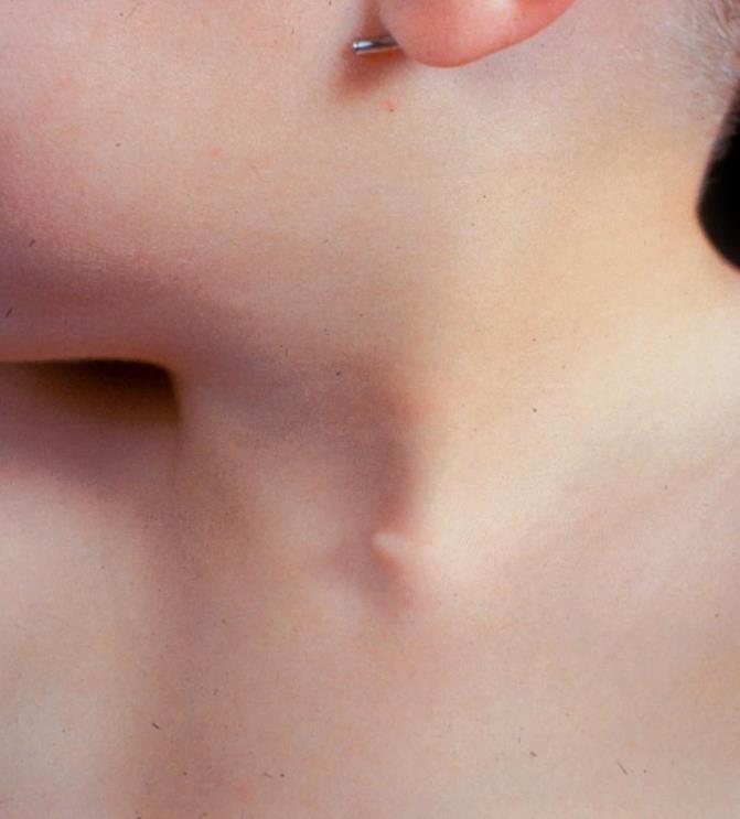
3 Thyroglossal cyst Diagnosis: midline, usually overlying hyoid bone demonstrate attachment to hyoid Investigations only if diagnostic uncertainty


4 Thyroglossal cyst Most common midline lesion Adherent to the hyoid bone Usually midline or just to left Moves on protrusion of the tongue Can present acutely as an abscess

5 Submental lymphadenopathy More anterior than thyroglossal cyst May be multiple nodes Look for source of infection eg dental caries, mouth ulcer Little movement with tongue protrusion or swallowing

6 Lateral neck lumps in children Lymph nodes reactive hyperplasia lymphadenitis/abscess MAIS Malignancy Rarely: branchial remnants, parotitis, cystic hygroma

7 Reactive hyperplasia Normal response to infection Vary in size, never disappear May be mildly tender No overlying skin changes History usually gives diagnosis FNA not indicated

8 Cervical abscess Common in first years of life Poorly localised swelling Starts as acute lymphadenitis Fails to respond to antibiotics Redness usually indicates suppuration Central softening is the signal for I & D Fluctuance often not demonstrable


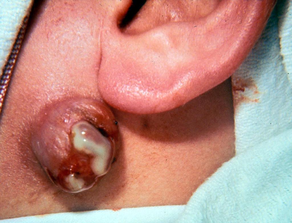
9 Endemic MAIS = Mycobacterium avium, intracellulare, & scrofulaceum Painless enlargement of lymph nodes over 3-6 weeks Common in pre-school kids (2-6) Starts as a non-tender lump Suppuration penetrates fascia to give collar stud abscess Skin discolouration means abscess Chronic discharging sinus if untreated

10 Hodgkin s disease Adolescent Painless enlargement of cervical lymph nodes Fever Night sweats Rubbery contiguous nodes
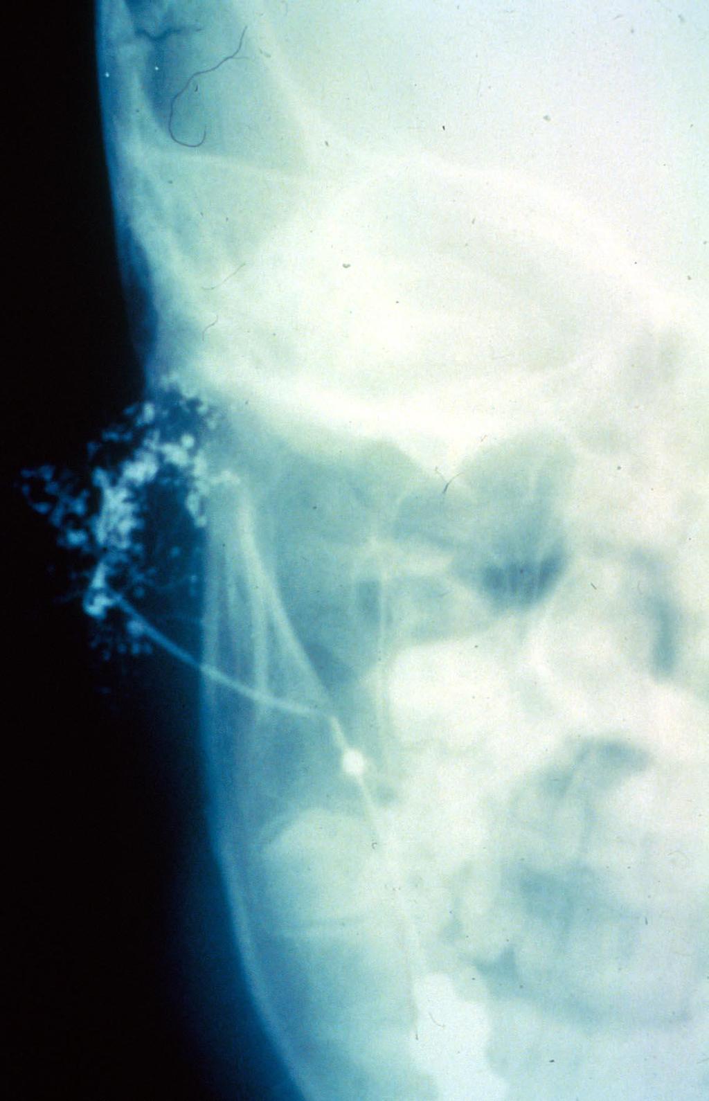
11 Remnants of 2 nd Branchial Cleft Sinuses are common, fistulae less so Internal opening of fistulae in tonsillar fossa Fistulae penetrate platysma and cervical fascia, ascend along the carotid sheath to level of the hyoid then pass between carotids. Remnants can be found anywhere along this course

12 Branchial sinus Inconspicuous opening at junction of middle and lower thirds of sternomastoid muscle Commonly discharge clear saliva from ectopic salivary glands May pass down or up

13 Branchial cyst - 2 nd cleft Treatment is excision Better cold i.e. not infected If inflamed Increased risk of nerve injury Higher incidence of recurrence Technically more difficult Malignant transformation later can occur
14
15


16 Pre-auricular Pits & Sinuses Ectodermal inclusions Stratified squamous epithelial lining Sinuses usually short and end blindly Attached to or penetrate cartilage of crus Become branched after infection Familial and often bilateral Common in Chinese


17 Cystic hygroma Is a hamartoma of the jugular lymph sacs Often associated with venous malformation More common in boys May be simple or multicystic Sudden swelling (bleed or infection) can compromise airway Otherwise, treatment is for cosmetic reasons Sclerosant injection now preferred

18 Torticollis Sternomastoid muscle fibrosis Postural torticollis Cervical hemivertebrae Squint Posterior fossa tumours Retropharyngeal abscess/cervical lymphadenitis Atlanto-occipital subluxation

19 Sternomastoid tumour Shortening by fibrosis of the sternomastoid muscle Appears in third week of life History of breech and forceps common 90% resolve in 9-12 months Role of physiotherapy controversial
20

21 External angular dermoid Common anomaly of cosmetic significance Fusion between the frontonasal and maxillary processes trap ectoderm. Often beneath the pericranium Excision curative Beware internal angular & midline dermoids -?deep extension, nasal glioma

22 Mucous retention cyst Usually inside lower lip May be traumatised by biting Often resolve over 3-6 months Excision curative if needed Diathermy excision, no sutures, is preferred

23 Tongue tie Does not interfere with swallowing May affect pronunciation of some consonants No other effects on speech May interfere with latch on Can improve spontaneously Usually divided under GA at 2-4 years

24
PEDIATRICS WK 3 HEAD AND NECK ALISON WALLACE MD, PHD
 PEDIATRICS WK 3 HEAD AND NECK ALISON WALLACE MD, PHD Topics 1. Cervical lymphadenopathy 2. Lymphatic malformation 3. Thyroglossal duct cysts 4. Branchial cleft cysts 5. Thyroid masses CASE 1 Case 1 A 2
Congenital Neck Masses C. Stefan Kénel-Pierre, MD
 Congenital Neck Masses C. Stefan Kénel-Pierre, MD SUNY-LICH Medical Center Department of Surgery Case Presentation xx year old male presents with sudden onset left lower neck swelling x 1 week Denies pain,
Thyroglossal cyst our experience
 Volume 3 Issue 1 2013 ISSN: 2250-0359 Thyroglossal cyst our experience Balasubramanian Thiagarajan 1 Ulaganathan Venkatesan 2 Geetha Ramamoorthy 1 1 Stanley Medical College 2 Meenakshi Medical College
HEAD & NECK SWELLINGS
 HEAD & NECK SWELLINGS EXCLUDING GOITRE FAISAL GHANI SIDDIQUI MBBS; FCPS; MCPS-HPE; PGDIP-BIOETHICS PROFESSOR OF SURGERY J I N N A H S I N D H M E D I C A L U N I V E R S I T Y MIDLINE SWELLINGS NECK SWELLINGS
LUMPS AND BUMPS: EVALUATION AND MANAGEMENT OF SOFT TISSUE MASSES IN PEDIATRICS. By Elizabeth A. Paton, MSN, RN-BC, PPCNP-BC, FAEN
 LUMPS AND BUMPS: EVALUATION AND MANAGEMENT OF SOFT TISSUE MASSES IN PEDIATRICS By Elizabeth A. Paton, MSN, RN-BC, PPCNP-BC, FAEN I. Objectives II. By the end of this presentation, the learner will be able
Chapter 13: Mass in the Neck. Raymond P. Wood II:
 Chapter 13: Mass in the Neck Raymond P. Wood II: In approaching the problem of a mass in the neck, one immediately encounters the fact that there are normally palpable masses in the neck (eg, almost all
Neckmasses in infancy and childhood: Clinical and radiological classification and imaging approaches M. Mearadji
 Neckmasses in infancy and childhood: Clinical and radiological classification and imaging approaches M. Mearadji International Foundation for Pediatric Imaging Aid Introduction Neck masses are a frequent
Dr Nick McIvor. Dr John Chaplin. Head & Neck Surgeon Auckland City Hospital Auckland. Auckland Head & Neck Surgeon Gillies Hospital Auckland
 Dr Nick McIvor Head & Neck Surgeon Auckland City Hospital Auckland Dr John Chaplin Auckland Head & Neck Surgeon Gillies Hospital Auckland 14:00-14:55 WS #148: Case Studies of Lumps in the Neck 15:05-16:00
THYROID & PARATHYROID. By Prof. Saeed Abuel Makarem & Dr. Sanaa Al-Sharawy
 THYROID & PARATHYROID By Prof. Saeed Abuel Makarem & Dr. Sanaa Al-Sharawy 1 OBJECTIVES By the end of the lecture, the student should be able to: Describe the shape, position, relations and structure of
Evaluation of Neck Mass. Disclosure. Learning Objectives 3/24/2014. Karen T. Pitman MD, FACS Banner MDACC, Gilbert AZ. Nothing to disclose
 Evaluation of Neck Mass Karen T. Pitman MD, FACS Banner MDACC, Gilbert AZ Nothing to disclose Disclosure Learning Objectives 1. Describe a systematic method to evaluate a patient with a neck mass 2. Select
Thyroid gland. importance. relations and connections. external laryngeal nerves. malformations.
 Thyroid gland 1. Recognize and understand the coverings of the thyroid gland and their clinical importance. 2. Recognize and understand the main parts of the thyroid gland and their locations, relations
Peripheral mycobacterial lymphadenitis (TB, NTM and BCG)
") Peripheral mycobacterial lymphadenitis (TB, NTM and BCG) H Simon Schaaf Desmond Tutu TB Centre, Department of Paediatrics and Child Health, Stellenbosch University, Cape Town, South Africa Questions Peripheral
Peripheral mycobacterial lymphadenitis (TB, NTM and BCG) H Simon Schaaf Desmond Tutu TB Centre, Department of Paediatrics and Child Health, Stellenbosch University, Cape Town, South Africa Questions Peripheral
 Drawings illustrating the human pharyngeal apparatus. Drawings illustrating the human pharyngeal apparatus. Drawings illustrating the human pharyngeal apparatus. Drawings illustrating the human pharyngeal
REVIEW OF CLINICAL EMBRYOLOGY OF HEAD AND NECK
 REVIEW OF CLINICAL EMBRYOLOGY OF HEAD AND NECK OUTLINE - EMBRYOLOGY UNDERLYING CLINICAL CONDITIONS I. EARLY DEVELOPMENT OF FACE: CLEFT LIP, CLEFT PALATE, OBSTRUCTED NASOLACRIMAL DUCT II. BRANCHIAL ARCHES
Management of thyroglossal duct cysts in children
 Pediatrics International (2004) 46, 77 80 Original Article Management of thyroglossal duct cysts in children ZAFER TÜRKYILMAZ, 1 KAAN SÖNMEZ, 1 RAMAZAN KARABULUT, 1 BILLUR DEMIR}OULLARI, 1 CEM SEZER, 2
PAPILLARY THYROID CARCINOMA PRESENTING AS A LATERAL NECK MASS MASS. Dr. Pamela Hanson DO PGY3
 PAPILLARY THYROID CARCINOMA PRESENTING AS A LATERAL NECK MASS MASS Dr. Pamela Hanson DO PGY3 MK CASE PRESENTATION 28 yo Female presented to the ENT Clinic in October 2016, with the complaint of chronic
Neoplasms that present as a swelling in the neck may be either
 Problems in otolaryngology Inflammatory swellings Viral and bacterial infection are frequent causes of swellings in the neck. Enlargement of the cervical lymph nodes is most likely but a dormant branchial
PART COMMON PAEDIATRIC SURGICAL PROBLEMS
 PART 9 COMMON PAEDIATRIC SURGICAL PROBLEMS Ch009-F10280.indd 261 7/27/2007 5:35:23 PM Ch009-F10280.indd 262 7/27/2007 5:35:23 PM Common surgical conditions in children S. W. Beasley 9.1 The penis and foreskin
Chapter 20: Branchial cleft anomalies, thyroglossal cysts and fistulae. P. D. M. Ellis. Branchial cleft anomalies. Embryology
 Chapter 20: Branchial cleft anomalies, thyroglossal cysts and fistulae P. D. M. Ellis Branchial cleft anomalies and thyroglossal cysts and fistulae are the end result of defects in development in the neck
"Mummy what's this on my neck? - A pictorial review of paediatric neck masses"
 "Mummy what's this on my neck? - A pictorial review of paediatric neck masses" Poster No.: C-0405 Congress: ECR 2014 Type: Educational Exhibit Authors: A. Farrugia, A. S. Gatt; Msida/MT Keywords: Education,
Cystic Head and Neck Lesions
 Cystic Head and Neck Lesions Disclosures None Brad Wright, MD 19 March 2018 Key points Huge variety of cystic lesions in H&N May be cystic, necrotic, or solid but cystic-appearing Patient age, clinical
Carcinoma of Unknown Primary site (CUP) in HEAD & NECK SURGERY
 in HEAD & NECK SURGERY") Carcinoma of Unknown Primary site (CUP) in HEAD & NECK SURGERY SEARCHING FOR THE PRIMARY? P r o f J P P r e t o r i u s H e a d : C l i n i c a l U n i t C r i t i c a l C a r e U n i v e r s i t y O f
Carcinoma of Unknown Primary site (CUP) in HEAD & NECK SURGERY SEARCHING FOR THE PRIMARY? P r o f J P P r e t o r i u s H e a d : C l i n i c a l U n i t C r i t i c a l C a r e U n i v e r s i t y O f
Veins of the Face and the Neck
 Veins of the Face and the Neck Facial Vein The facial vein is formed at the medial angle of the eye by the union of the supraorbital and supratrochlear veins. connected through the ophthalmic veins with
Subdivided into Vestibule & Oral cavity proper
 Extends from the lips to the oropharyngeal isthmus The oropharyngeal isthmus: Is the junction of mouth and pharynx. Is bounded: Above by the soft palate and the palatoglossal folds Below by the dorsum
The Neck the lower margin of the mandible above the suprasternal notch and the upper border of the clavicle
 The Neck is the region of the body that lies between the lower margin of the mandible above and the suprasternal notch and the upper border of the clavicle below Nerves of the neck Cervical Plexus Is formed
Remember from the first year embryology Trilaminar disc has 3 layers: ectoderm, mesoderm, and endoderm
 Development of face Remember from the first year embryology Trilaminar disc has 3 layers: ectoderm, mesoderm, and endoderm The ectoderm forms the neural groove, then tube The neural tube lies in the mesoderm
Tikrit University College of Dentistry Dr.Ban I.S. head & neck anatomy 2 nd y.
 Lec [3]/The scalp The scalp extends from the supraorbital margins anteriorly to the nuchal lines at the back of the skull and down to the temporal lines at the sides. The forehead, from eyebrows to hairline,
MALIGNANT TUMOURS OF THE JAWS
 MALIGNANT TUMOURS OF THE JAWS MALIGNANT TUMOURS OF THE JAWS Squamous cell carcinoma Osteogenic sarcoma Chondrosarcoma Fibrosarcoma Malignant lymphomas (incl. Burkitt s) Multiple myeloma Ameloblastoma Secondary
Branchial Cleft and Pouch Anomalies
 Branchial Cleft and Pouch Anomalies Prof.Mohamed Hesham Alexandria Faculty of Medicine Alexandria, Egypt Emberyological Basis Branchial Clefts 1st 2nd Pinna EAC 3rd 4th 4th 6th Cervical sinus Branchial
Collar stud abscess an interesting case report
 Volume 2 issue 2 2012 ISSN 2250-0359 Collar stud abscess an interesting case report Kameshwaran Kannappan Punniyakodi * Balasubramanian Thiagarajan* *Stanley Medical College Chennai, Tamilnadu Abstract
ENT in Primary Care. Learning Objectives. Eustachian Tube (ET) Dysfunction. Eustachian Tube (ET) Dysfunction. Middle Ear Effusion
 Dysfunction. Eustachian Tube (ET) Dysfunction. Middle Ear Effusion") Learning Objectives ENT in Primary Care Paul A. Kedeshian, MD Associate Clinical Professor David Geffen School of Medicine at UCLA Department of Head and Neck Surgery Identifying common ENT problems and
Learning Objectives ENT in Primary Care Paul A. Kedeshian, MD Associate Clinical Professor David Geffen School of Medicine at UCLA Department of Head and Neck Surgery Identifying common ENT problems and
Alexander C Vlantis. Selective Neck Dissection 33
 05 Modified Radical Neck Dissection Type II Alexander C Vlantis Selective Neck Dissection 33 Modified Radical Neck Dissection Type II INCISION Various incisions can be used for a neck dissection. The incision
Clinical Anatomy of the Thyroid and Adrenal Glands
 Clinical Anatomy of the Thyroid and Adrenal Glands Handout download: http://www.oucom.ohiou.edu/dbms-witmer/gs-rpac.htm 28 October 2003 Lawrence M. Witmer, PhD Department of Biomedical Sciences College
Pharyngeal Apparatus. Pouches Endoderm Grooves Ectoderm Arch Neural Crest Somitomeres Aortic Arch - Vessel
 Pharyngeal Apparatus Pouches Endoderm Grooves Ectoderm Arch Neural Crest Somitomeres Aortic Arch - Vessel Segmental Organization Humans: Arch 1-4 prominent Arch 5 absent Arch 6 - transient First Arch Face
1/13/2009. Classification:
 SUPPURATIONS OF SPACES RELATED TO THE PHARYNX Assistant Professor, Department of Otolaryngology Head & Neck Surgery Faculty of Medicine, Alexandria University Classification: I. Intratonsillar abscess.
Case Presentation and Discussion on Posterior Neck Mass. Martin Joseph S. Cabahug
 Case Presentation and Discussion on Posterior Neck Mass Martin Joseph S. Cabahug General Data: C.A, 60 y/o male Sta. Ana, Mla Chief Complaint: Posterior Neck Mass History and Physical Exam 2 wks PTA mass,
Infection of the Pharyngeal Spaces
 Lecture (4) pharynx د.سنمار Infection of the Pharyngeal Spaces Parapharyngeal Abscess Definition: Collection of pus in the parapharyngeal space which is a connective tissue space lies on the lateral side
Neck-2. Dr. Heba Kalbouneh Associate Professor of Anatomy and Histology
 Neck-2 ` Dr. Heba Kalbouneh Associate Professor of Anatomy and Histology Triangles of the neck Side of the neck Midline Lower border of mandible Line between angle of mandible and mastoid Superior nuchal
DEVELOPMENT & STRUCTURE OF THYROID GLAND DR TATHEER ZAHRA ASSISTANT PROFESSOR ANATOMY
 DEVELOPMENT & STRUCTURE OF THYROID GLAND DR TATHEER ZAHRA ASSISTANT PROFESSOR ANATOMY DEVELOPMENT OF THYROID Concept of pharyngeal arch 3 rd week 4 th week Adults 7 th week HISTOGENESIS OF THYROID GLAND
Asian Journal of Pharmacy and Life Science ISSN Vol.3 (2), April-June, 2013
, April-June, 2013") RECURRENT NECK INFECTION ASSOCIATED WITH FOURTH BRANCHIAL POUCH SINUS: A RARE CLINICAL ENTITY WITH DELAYED DIAGNOSIS Bhawana Pant* 1, Sanjay Gaur 2, Shahzad Ahmad 1 1. Department of E.N.T, Government Medical
RECURRENT NECK INFECTION ASSOCIATED WITH FOURTH BRANCHIAL POUCH SINUS: A RARE CLINICAL ENTITY WITH DELAYED DIAGNOSIS Bhawana Pant* 1, Sanjay Gaur 2, Shahzad Ahmad 1 1. Department of E.N.T, Government Medical
04 Development of the Face and Neck. Development of the Face Development of the neck
 04 Development of the Face and Neck Development of the Face Development of the neck Development of the face Overview of facial development The fourth week ~ the twelfth week of prenatal development Between
Dr. Muhammad Shamim. Assistant Professor, Dept. of Surgery College of Medicine, Prince Sattam bin Abdulaziz University
 Dr. Muhammad Shamim FCPS (Pak), FACS (USA), FICS (USA). JMHPE (Nl & Eg) Assistant Professor, Dept. of Surgery College of Medicine, Prince Sattam bin Abdulaziz University Email: surgeon.shamim@gmail.com
A clinical study on branchial arch anomalies
 IOSR Journal of Dental and Medical Sciences (IOSR-JDMS) e-issn: 2279-0853, p-issn: 2279-0861.Volume 18, Issue 1 Ver. 5 (January. 2019), PP 05-10 www.iosrjournals.org Ashim Sarkar 1, Ritam Ray 2 1 (Clinical
Anatomy: head and Neck (6 questions) 1. Prevertebral Flexor Musculature (lying in front of the vertebrae) include all, EXCEPT: Longus Colli.
 1. Prevertebral Flexor Musculature (lying in front of the vertebrae) include all, EXCEPT: Longus Colli.") Anatomy: head and Neck (6 questions) 1. Prevertebral Flexor Musculature (lying in front of the vertebrae) include all, EXCEPT: Longus Colli. Rectus Capitis Anterior. Rectus Capitis Lateralis. Rectus Capitis
Anatomy: head and Neck (6 questions) 1. Prevertebral Flexor Musculature (lying in front of the vertebrae) include all, EXCEPT: Longus Colli. Rectus Capitis Anterior. Rectus Capitis Lateralis. Rectus Capitis
The Neck. BY: Lina Abdullah & Rahaf Jreisat
 The Neck BY: Lina Abdullah & Rahaf Jreisat Boundaries of the Neck: generally from base of the skull to root of the neck Superior margin :From superior nuchal line of occipital bone up to mastoid process
Case Presentation. x year old African American male seen in Pediatric Surgery Clinic. History: NKDA
 Case Presentation ALIREZA SADEGHI MD Kings County Hospital Center University Hospital of Brooklyn Downstate Medical Center Division of Pediatric Surgery July 7 th 2006 Case Presentation x year old African
INFECTION. HIV Infection DWI
 HIV Infection INFECTION DWI Fig Axial CT and MRI images show multiple enlarged lymph nodes in the neck as well as in the parotid gland bilaterally. These nodes were suppurative with positive diffusion.
Ultrasound Interpretation of Non-Thyroid Neck Pathology
 Ultrasound Interpretation of Non-Thyroid Neck Pathology Kevin T. Brumund, M.D., F.A.C.S. Associate Professor of Surgery Head and Neck Surgery University of California, San Diego Health Sciences VA Medical
Chronic Tonsillitis 1
 Chronic Tonsillitis 1 Aetiology Complication of acute tonsillitis Subclinical infections of tonsils Children and young adults Chronic infection in sinuses or teeth 2 Types Chronic follicular tonsillitis
Head and Neck Case Studies
 Head and Neck Case Studies John Chaplin & Nick McIvor www.headneck.co.nz Head and Neck lumps every lump must have a diagnosis Working diagnosis» +/- investigations Review» +/- investigations auckland head
Rafal Zielinski*, Anna Zakrzewska Submental epidermoid cysts in children. 2.1 Case 1
 Open Med. 2015; 10: 77-81 Case Report Open Access Rafal Zielinski*, Anna Zakrzewska Submental epidermoid cysts in children Abstract: Epidermoid cysts are lesions, which form as a result of implantation
Contents. Basic Ultrasound Principles and Terminology. Ultrasound Nodule Characteristics
 Contents Basic Ultrasound Principles and Terminology Basic Ultrasound Principles... 1 Ultrasound System... 2 Linear Transducer for Superficial Images and Ultrasound-Guided FNA... 3 Scanning Planes... 4
CERVICAL LYMPH NODES
 CERVICAL LYMPH NODES (ANATOMY & EXAMINATION) Hemant (DTCD 1 st YEAR) 1. Lymphatic Tissues: A Type of connective tissue that contains large numbers of lymphocytes. 2. Lymphatic Vessels: Are Tubes that assist
"The Space Between Us:" A Radiographic Review of Common and Uncommon Pathologic Findings within the Deep Spaces of the Neck
 "The Space Between Us:" A Radiographic Review of Common and Uncommon Pathologic Findings within the Deep Spaces of the Neck Poster No.: C-2457 Congress: ECR 2015 Type: Educational Exhibit Authors: A. K.
Evaluation of Head and Neck Masses in Adults
 Evaluation of Head and Neck Masses in Adults Kristi Chang, MD Associate Professor Department of Otolaryngology-Head and Neck Surgery University of Iowa Hospitals and Clinics Annual Refresher Course for
Deepak M. Sampathu MD, PhD Assistant Professor of Clinical Radiology University of Pennsylvania
 Deepak M. Sampathu MD, PhD Assistant Professor of Clinical Radiology University of Pennsylvania Objectives Recognize benign masses and masslike lesions of the neck and skull base Understand the imaging
ANTERIOR CERVICAL TRIANGLE (Fig. 2.1 )
") 2 Neck Anatomy ANTERIOR CERVICAL TRIANGLE (Fig. 2.1 ) The boundaries are: Lateral: sternocleidomastoid muscle Superior: inferior border of the mandible Medial: anterior midline of the neck This large triangle
2 Neck Anatomy ANTERIOR CERVICAL TRIANGLE (Fig. 2.1 ) The boundaries are: Lateral: sternocleidomastoid muscle Superior: inferior border of the mandible Medial: anterior midline of the neck This large triangle
The following images were all acquired using a CTI Biograph
 Positron Emission Tomography/ Computed Tomography Imaging of Head and Neck Tumors: An Atlas Michael M. Graham, MD, PhD, and Yusuf Menda, MD Department of Radiology, University of Iowa, Iowa City, IA. Address
Salivary Glands. The glands are found in and around your mouth and throat. We call the major
 Salivary Glands Where Are Your Salivary Glands? The glands are found in and around your mouth and throat. We call the major salivary glands the parotid, submandibular, and sublingual glands. They all secrete
SYLLABUS OF ORAL AND MAXILLOFACIAL SURGERY
 MEDICAL UNIVERSITY OF VARNA FACULTY OF DENTAL MEDICINE DEPARTMENT OF ORAL AND MAXILLOFACIAL SURGERY AND SID SYLLABUS OF ORAL AND MAXILLOFACIAL SURGERY (State examination) ACADEMIC YEAR 2015 2016 1. Asepsis
Abscess. A abscess is a localized collection of pus in the skin and may occur on any skin surface and be formed in any part of body.
 Abscess A abscess is a localized collection of pus in the skin and may occur on any skin surface and be formed in any part of body. Ethyology Bacteria causing cutaneous abscesses are typically indigenous
Swelling in the head and neck. Bernhard Schuknecht. Order of business. Choice of diagnostic technique
 Swelling in the head and neck next speaker: Bernhard Schuknecht Bernhard Schuknecht MRI Medical Radiological Institute Zurich Switzerland ESHNR Sept 24-26 2015 Krakow Depends on Choice of diagnostic technique
Face. Definition: The area between the two ears and from the chin to the eye brows. The muscles of the face
 Face Definition: The area between the two ears and from the chin to the eye brows. The muscles of the face The muscle of facial expression (include the muscle of the face and the scalp). All are derived
Shadow because the air
 Thyroid Ultrasound Thyroid US examination needs: 1. high frequency transducer 2. extended patient's neck 3. check all the neck area because the swelling could be in areas other than the thyroid such as
Chapter 10: Salivary Gland Disorders. Raymond P. Wood. History
 Chapter 10: Salivary Gland Disorders Raymond P. Wood Dysfunction of the salivary glands is usually manifested in one of two ways: swelling of the gland, either diffuse or discrete, or by dry mouth (xerostomia).
Development of the Pharyngeal Arches
 Development of the Pharyngeal Arches Thomas A. Marino, Ph.D. Temple University School of Medicine Competencies: Upon completion of this section of the course, the student must be able to: 1. Recall the
Index. Infect Dis Clin N Am 21 (2007) Note: Page numbers of article titles are in boldface type.
 Note: Page numbers of article titles are in boldface type.") Infect Dis Clin N Am 21 (2007) 591 599 Index Note: Page numbers of article titles are in boldface type. A Abscess(es) epidural, subdural empyema and, 584 586 periotonsillar, microbiologic investigations
Infect Dis Clin N Am 21 (2007) 591 599 Index Note: Page numbers of article titles are in boldface type. A Abscess(es) epidural, subdural empyema and, 584 586 periotonsillar, microbiologic investigations
Lecture 07. Lymphatic's of Head & Neck. By: Dr Farooq Amanullah Khan PMC
 Lecture 07 Lymphatic's of Head & Neck By: Dr Farooq Amanullah Khan PMC Dated: 28.11.2017 Lymphatic Vessels Of the 800 lymph nodes in the human body, 300 are in the Head & neck region. The lymphatic vessels
Thyroid and Parathyroid Glands
 Thyroid and Parathyroid Glands Please view our Editing File before studying this lecture to check for any changes. Color Code Important Doctors Notes Notes/ explanation Objectives: By the end of the lecture,
REVIEW/PREVIEW OF HEAD AND NECK ANATOMY FOR ENT EXAM
 REVIEW/PREVIEW OF HEAD AND NECK ANATOMY FOR ENT EXAM - 2017 PALPATE CAROTID ARTERY: AT LEVEL OF CAROTID BIFURCATION VERTEBRAL LEVEL C4 Sternocleidomastoid Muscle INTERNAL CAROTID EXTERNAL CAROTID COMMON
SALIVARY GLAND DISEASES. Omar alnoubani MD,MRCS
 SALIVARY GLAND DISEASES Omar alnoubani MD,MRCS Salivary Glands Overview Parotid gland Sublingual gland Submandibular gland Salivary glands - Types 3 Major Salivary Glands Parotid Submandibular Sublingual
Evaluation and Management of Pediatric Neck masses
 Evaluation and Management of Pediatric Neck masses Steven T. Wright, M.D. Faculty Advisor: Ronald Deskin, M.D. The University of Texas Medical Branch Department of Otolaryngology Grand Rounds Presentation
Anatomy of Oral Cavity DR. MAAN AL-ABBASI
 Anatomy of Oral Cavity DR. MAAN AL-ABBASI By the end of this lecture you should be able to: 1. Differentiate different parts of the oral cavity 2. Describe the blood and nerve supply of mucosa and muscles
Hemangioma of Tongue with Phlebolith: A Rare presentation
 Journal of Government Dental College and Hospital, October 2017, Vol.-04, Issue- 01, P. 20-25 Original article: Hemangioma of Tongue with Phlebolith: A Rare presentation 1 Dr. Jigna S Shah (MDS) 1, 2 Dr.
Lecture 01. The Thyroid & Parathyroid Glands. By: Dr Farooq Khan PMC Date: 12 th March. 2018
 Lecture 01 The Thyroid & Parathyroid Glands By: Dr Farooq Khan PMC Date: 12 th March. 2018 INTRODUCTION LAYERS OF THE NECK The neck has four major compartments or layer which are enclosed by an outer musculofascial
Module 1, Stomatology
 Module 1, Stomatology Department of operative surgery and topographic anatomy Test questions for self training for passing Module 1, writing part Paractical questions with images The patient has been scheduled
42 yr old male with h/o Graves disease and prior I 131 treatment presents with hyperthyroidism and undetectable TSH. 2 hr uptake 20%, 24 hr uptake 50%
 Pinhole images of the neck are acquired in multiple projections, 24hrs after the oral administration of approximately 200 µci of I123. Usually, 24hr uptake value if also calculated (normal 24 hr uptake
Dupuytren's Contracture Assessment
 Dupuytren's Contracture Assessment Link to guidance: http://www.enhertsccg.nhs.uk/ bedfordshire-and-hertfordshire-priorities-forum Dupuytren's contracture - clinical presentation for patients History Examination
The Scalp and Face Protocol. Julie Goodwin, BA, LMT
 The Scalp and Face Protocol Julie Goodwin, BA, LMT The Scalp and Face Protocol Julie Goodwin, BA, LMT Julie Goodwin 2014 2 Agenda Pertinent Anatomy and Physiology Treatment Planning Strokes, Techniques
DEVELOPMENTAL ANATOMY OF THE FACE, JAW AND NECK. O.M. Oluwatosin Department of Surgery
 DEVELOPMENTAL ANATOMY OF THE FACE, JAW AND NECK O.M. Oluwatosin Department of Surgery 1 2 By the end of this lecture, you should be able to: Discuss the embryology of the face Relate congenital anomalies
OTOLARYNGOLOGY ONLINE. Preauricular sinus. Management. Dr. T. Balasubramanian 6/17/2010
 1 OTOLARYNGOLOGY ONLINE Preauricular sinus Management Dr. T. Balasubramanian 6/17/2010 This e book discusses the Etiopathogenesis of preauricular sinus. Even though it is commonly encountered surgical
*in general the blood supply of the nose comes from branches of the internal and external carotid arteries.
 In the previous lecture we talked about the anatomy of the nasal cavity, today we will talk about its blood supply, venous drainage, innervations, and finally about the paranasal sinuses. When we describe
Case Presentation 主治醫師 : 宋文鑫日期 :
 Case Presentation 主治醫師 : 宋文鑫日期 : 2015-2-28 General Data Name:OOO Chart Number:OOOOOOO Date of Admission:2014 年 08 月 04 日 Age: 33 y/o Sex:female Occupation : 會計 Chief Complaint Palpable soft tissue mass
Case Scenario. 7/13/12 Anterior floor of mouth biopsy: Infiltrating squamous cell carcinoma, not completely excised.
 Case Scenario 7/5/12 History A 51 year old white female presents with a sore area on the floor of her mouth. She claims the area has been sore for several months. She is a current smoker and user of alcohol.
Head & Neck Contouring
 Head & Neck Contouring Presented by James Wheeler, MD Center for Cancer Care Goshen, IN 46526 September 12, 2014 Special Thanks to: Spencer Boulter, Director of Operations (AAMD) Adam Moore, RT(T), CMD
Distribution of branchial anomalies in a paediatric Asian population
 Singapore Med J 2015; 56(4): 203-207 doi: 10.11622/smedj.2015060 Distribution of branchial anomalies in a paediatric Asian population Neville Wei Yang Teo 1, MBBS, MRCS, Shahrul Izham Ibrahim 2, MBBCh,
Pediatric Otolaryngology Disorders for Primary Care ASHOK N. REDDY, MD CONCORD OTOLARYNGOLOGY HEAD AND NECK SURGERY CONCORD, NH
 Pediatric Otolaryngology Disorders for Primary Care ASHOK N. REDDY, MD CONCORD OTOLARYNGOLOGY HEAD AND NECK SURGERY CONCORD, NH DISCLOSURES None of the planners or presenters of this session have disclosed
Case Scenario 1. 7/13/12 Anterior floor of mouth biopsy: Infiltrating squamous cell carcinoma, not completely excised.
 Case Scenario 1 7/5/12 History A 51 year old white female presents with a sore area on the floor of her mouth. She claims the area has been sore for several months. She is a current smoker and user of
Management of unknown primary with neck node metastasis: Current evidence
 Management of unknown primary with neck node metastasis: Current evidence Dr. Pooja Nandwani Patel Associate Professor Dept. of Radiation Oncology GCRI, Ahmedabad Introduction- Approach to Topic What is
Prevertebral Region, Pharynx and Soft Palate
 Unit 20: Prevertebral Region, Pharynx and Soft Palate Dissection Instructions: Step1 Step 2 Step 1: Insert your fingers posterior to the sternocleidomastoid muscle, vagus nerve, internal jugular vein,
The SCALP. Prof. Dr. Muhammad Imran Qureshi
 The SCALP By Prof. Dr. Muhammad Imran Qureshi The SCALP includes FIVE layers external to the Calvaria. These are: S: Skin & Superficial Fascia C: Connective Tissue A: Aponeurosis (Epicranial) L: Loose
THE INTERIOR OF THE PHARYNX. By Dr. Muhammad Imran Qureshi
 THE INTERIOR OF THE PHARYNX By Dr. Muhammad Imran Qureshi The Cavity The cavity of the pharynx is divided into: 1. The Nasal part (called Nasopharynx) 2. The Oral part (called the Oropharynx), 3. And the
3-Deep fascia: is absent (except over the parotid gland & buccopharngeal fascia covering the buccinator muscle)
") The Face 1-Skin of the Face The skin of the face is: Elastic Vascular (bleed profusely however heal rapidly) Rich in sweat and sebaceous glands (can cause acne in adults) It is connected to the underlying
The Face 1-Skin of the Face The skin of the face is: Elastic Vascular (bleed profusely however heal rapidly) Rich in sweat and sebaceous glands (can cause acne in adults) It is connected to the underlying
Anterior triangle of neck
 Anterior triangle of neck Dept. of Anatomy Zhou Hong Ying Outline boundary and subdivisions of ant. triangle contents of the triangle Muscles: suprahyoid muscles, infrahyoid muscles Nerves: CNⅩ, CNⅪ, CNⅫ,
2015 Otolaryngology Survival Guide
 2015 Otolaryngology Survival Guide Chapter 12: Scope Procedures Know All Your New Sinus Endoscopy Procedures Choices You have three codes in the CPT manual for sinus endoscopy procedures that are meant
NECK MASS. Clinical history and examination: Document detail history of mass. Imaging: US or CT of neck
 ENT ENT Referral Referral Guidelines Guidelines Austin Health ENT Clinic holds fortnightly multidisciplinary meetings with Plastics/ Maxillary Facial and Oncology units to discuss and plan the treatment